clinical study computed tomography angiography for
TRANSCRIPT

Clinical StudyComputed Tomography Angiography for Detection ofMiddle Meningeal Artery Lesions Associated with AcuteEpidural Hematomas
Wellingson Silva Paiva,1 Almir Ferreira Andrade,1 Robson Luis Oliveira De Amorim,1
Edson Bor-Seng-Shu,1 Gabriel Gattas,2 Iuri Santana Neville,1 Jose Guilherme Caldas,2
Eberval Gadelha Figueiredo,1 and Manoel Jacobsen Teixeira1
1 Division of Neurological Surgery, University of Sao Paulo Medical School, 255 Eneas Aguiar Street, Office 4080,05403010 Sao Paulo, SP, Brazil
2 Institute of Radiology, University of Sao Paulo Medical School, 470 Alves Guimaraes Street,05410-000 Sao Paulo, SP, Brazil
Correspondence should be addressed to Wellingson Silva Paiva; [email protected]
Received 25 November 2013; Accepted 11 February 2014; Published 13 March 2014
Academic Editor: David Maintz
Copyright © 2014 Wellingson Silva Paiva et al. This is an open access article distributed under the Creative Commons AttributionLicense, which permits unrestricted use, distribution, and reproduction in any medium, provided the original work is properlycited.
Background. The natural history of traumatic aneurysms of the middle meningeal artery (MMA) is not well known, but patientswith these lesions aremore likely to have delayed bleeds. In this paper, we described a series of patients with epidural hematomawhounderwent angiotomography (CTA) for MMA vascular lesion diagnosis.Methods. Eleven patients admitted to our emergency unitwith small acute epidural hematoma were prospectively studied. All patients with temporal acute epidural hematomas underwentCTA and cerebral angiogram at our institution for diagnosis of posttraumatic lesions of middle meningeal artery. The findings ofangiotomography and digital angiography were reviewed by radiologist and angiographers, respectively, to ensure that the lesionswere readily diagnosed without knowing the results of angiotomography and to compare CTA findings with standard angiogram.Results. The causes of head injury were traffic accidents, falls, and aggression. Three of these patients presented traumatic MMApseudoaneurysm. CT angiography was able to diagnose all of them, with dimensions ranging from 1.5 to 2.8mm. Conventionalangiography confirmed the findings of CT angiography, and the lesions presented with similar dimensions at both methods.Conclusions. We believe that angiotomography can be a useful technique for diagnosis of vascular lesion associated with smallepidural hematoma.
1. Introduction
Acute epidural hematomas (AEDHs) are common traumaticlesions [1]. It is well known that small AEDHs withoutsignificant mass effect may be treated conservatively, andtheir ideal management has not been clearly established thusfar [2–4].
Late enlargement of previously small hematomas is awell-recognized clinical occurrence [2, 5]. Patients with cra-nial fractures crossing over dural arteries or veins are proneto experience rebleedingwith consequent hematoma enlarge-ment [2, 6]. In patients who present with fractures crossingthe middle meningeal artery (MMA), the possibility of false
aneurysm should be kept in mind [2, 7]. Patients withtraumatic pseudoaneurysms are more likely to have delayedbleeds, which account for typical prolonged lucid interval[8, 9]. It is important to diagnose and treat these aneurysmsat the earliest to prevent catastrophic events. Recent studyhas suggested that the incidence of posttraumatic pseudoa-neurysms is higher than previously thought [2].
Intracranial vascular lesions related to cranial fracturesand small AEDHs have not been adequately studied thusfar, and their incidence, natural history, clinical relevance,and ideal management have not been well established.Angiograms have been used to identify pseudoaneurysmsassociated with small epidural hematomas [2]. However, it
Hindawi Publishing CorporationBioMed Research InternationalVolume 2014, Article ID 413916, 5 pageshttp://dx.doi.org/10.1155/2014/413916

2 BioMed Research International
Table 1: Summary of the patients, causes, and radiological characteristics.
Patient Sex Age Trauma mechanism Hematoma location Angio-CT Angiography GCS GOS1 M 19 Traffic accident Temporal Pseudoaneurysm Pseudoaneurysm 15 52 M 18 Aggression Temporal Normal Normal 15 53 F 19 Traffic accident Temporal Normal Normal 15 54 M 29 Traffic accident Temporal Pseudoaneurysm Pseudoaneurysm 14 55 M 32 Fall Temporal Normal Normal 15 56 M 27 Aggression Temporal Normal Normal 14 57 M 25 Traffic accident Temporal Normal Normal 15 58 F 39 Aggression Temporoparietal Normal Normal 14 59 M 30 Traffic accident Temporal Pseudoaneurysm Pseudoaneurysm 15 510 F 44 Fall Temporoparietal Normal Normal 15 511 M 32 Traffic accident Temporal Normal Normal 14 5
presents several drawbacks and the associated morbiditycannot be neglected. The role of “less invasive” methods,such as CT angiography, to diagnose these lesions has notbeen determined thus far. The aim of the current study is todescribe the radiological findings in 11 patients who harborsmall AEDHs associatedwith linear cranial fractures crossingover the MMA trajectory and to compare CT angiography(CTA) versus conventional angiography.
2. Methods
Eleven consecutive patients admitted to the emergency unitin the Division of Neurological Surgery at University of SaoPaulo were prospectively studied. All patients with temporalor parietal AEDHs underwent CTA and cerebral angiogramat our institution for the diagnosis of posttraumatic lesionsof middle meningeal artery. Eight patients were male andthree were female with mean age of 25.3 years old (rangingfrom 18 to 44). All of the patients presented small AEDHsin regions corresponding to bleeding from branches of theMMA. Patients having traumatic lesions with mass effect,midline shift, or other associated intracranial injuries werenot included in this study. Patients with moderate or largesized lesions underwent surgical evacuation and were notincluded as well. All patients had previous transient loss ofconsciousness following the trauma but had a score of 14 or 15on the Glasgow Coma Scale on admission. Headache was themain complaint and no neurological deficits were observed.
The mean time from trauma to the patients’ admissionto our emergency center was 102 minutes (range: 25–260minutes). Table 1 provides a summary of the causes and othercharacteristics of the AEDHs.
Patients were treated according to ATLS protocol;patients with mild TBI underwent head CT scan. Thesepatients with small epidural hematoma of middle fossawith temporal fracture were evaluated with noncontrastCT examination followed immediately by 3D CTA using aGeneral Electric Light-Speed Advantage CT scanner withAdvantage Windows 3D workstation (General Electric, Mil-waukee, Wis). Noncontrast CT scans were performed with5mm contiguous axial sections through the posterior fossafollowed by 10mmcontiguous axial sections to the vertex. CT
angiography was performed using an injection of nonioniccontrast at a rate of 3mL/s for 80mL initiated with a 15-second prescan delay. A 1mm collimated helical scan with a1 : 1 pitch was obtained from the cavernous carotid cephalicfor at least 3.5 cm. On average, a total dose of 24 g of iodinewas administered. Patients were kept under clinical observa-tion in ICU. All patients underwent ipsilateral digital externalcarotid artery angiography which was performed within 12hours of the initial study. The findings of angiotomographyand digital angiography were reviewed by radiologists andangiographers, respectively, to ensure that the lesions werereadily diagnosed without knowing the results of angioto-mography. If any vascular lesion was found, embolization ofthe MMA and branches was performed after superselectiveinjection with a microguidewire up to an area just before thearterial lesion had been reached.The ethics committee of ourinstitution approved this study and informed consent for thisstudy was obtained in all cases.
3. Results
The causes of head injury were traffic accidents (𝑛 = 6patients), falls (𝑛 = 3 patients), and aggression (𝑛 = 2patients). The largest hematoma had a thickness of 10mm.Three of these patients presented traumatic MMA pseudoa-neurysm. CT angiography was able to diagnose all of them,with dimensions ranging from 1.5 to 2.8mm. Conventionalangiography confirmed the findings of CT angiography, andthe lesions presented with similar dimensions at both meth-ods. No additional lesion was demonstrated by angiogram.
Scan duration was 35 to 42 seconds in all cases. AverageCTA reconstruction time was 15 minutes. The acquisitiontime did not exceed 30 minutes including 3D reconstruction.The average period until discharge was 6.9 days (range:5–9 days) for all patients and 2.7 days (range: 2–4 days)for the 3 patients with AEDHs treated with embolization.Embolization of the pseudoaneurysm itself or of the parentvessel was successfully performed in 3 patients. The postop-erative course was uneventful and no complications relatedto the procedure were noted. All of the lesions were followed
BioMed Research International 3
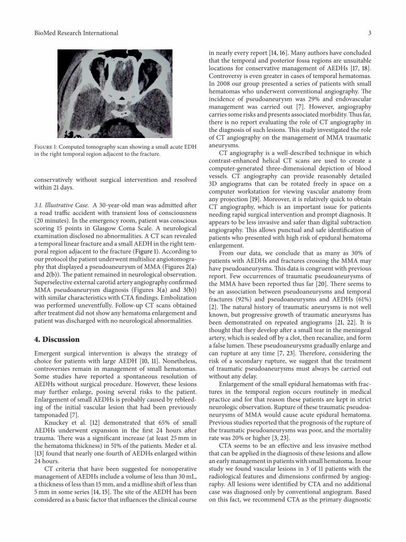
Figure 1: Computed tomography scan showing a small acute EDHin the right temporal region adjacent to the fracture.
conservatively without surgical intervention and resolvedwithin 21 days.
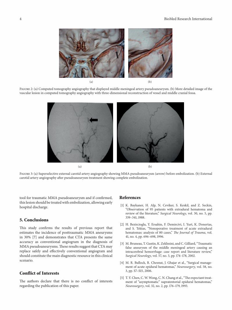
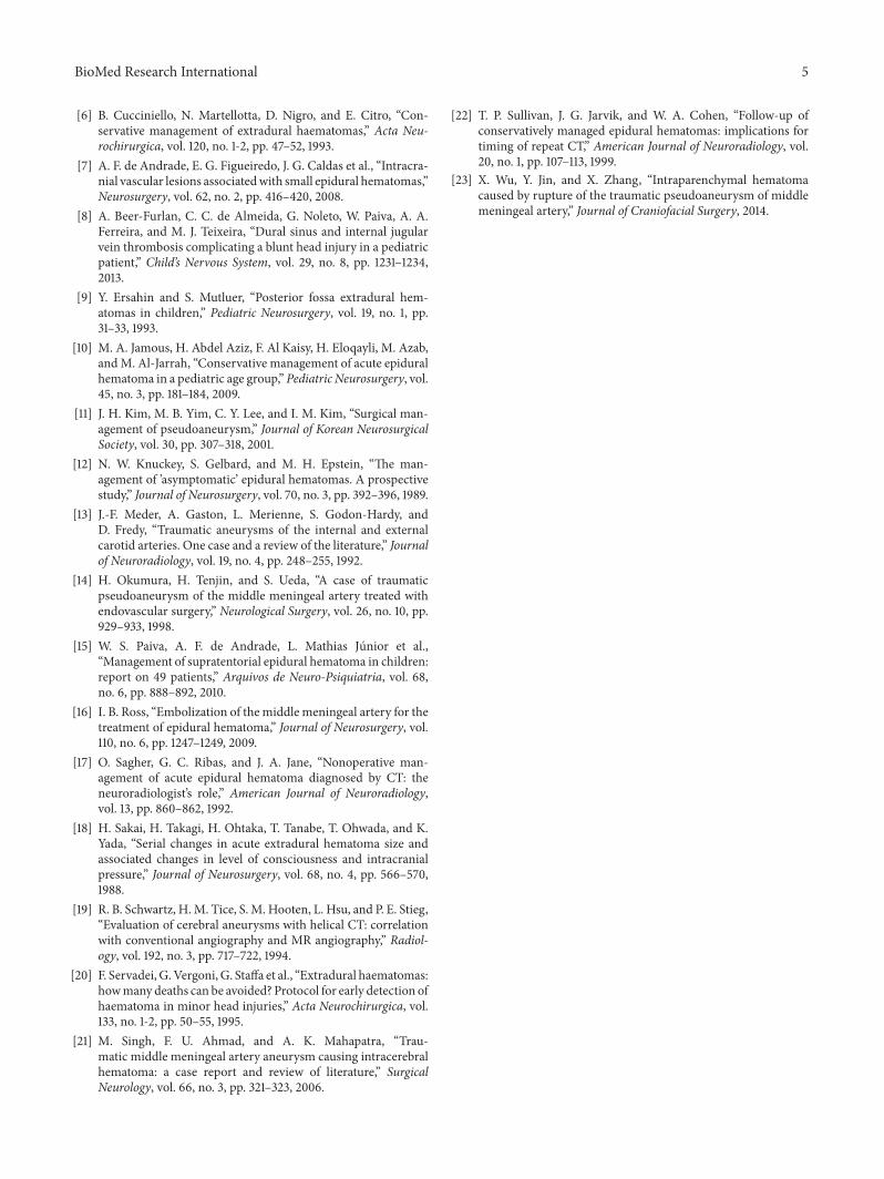
3.1. Illustrative Case. A 30-year-old man was admitted aftera road traffic accident with transient loss of consciousness(20 minutes). In the emergency room, patient was consciousscoring 15 points in Glasgow Coma Scale. A neurologicalexamination disclosed no abnormalities. A CT scan revealeda temporal linear fracture and a small AEDH in the right tem-poral region adjacent to the fracture (Figure 1). According toour protocol the patient underwentmultislice angiotomogra-phy that displayed a pseudoaneurysm of MMA (Figures 2(a)and 2(b)). The patient remained in neurological observation.Superselective external carotid artery angiography confirmedMMA pseudoaneurysm diagnosis (Figures 3(a) and 3(b))with similar characteristics with CTA findings. Embolizationwas performed uneventfully. Follow-up CT scans obtainedafter treatment did not show any hematoma enlargement andpatient was discharged with no neurological abnormalities.
4. Discussion
Emergent surgical intervention is always the strategy ofchoice for patients with large AEDH [10, 11]. Nonetheless,controversies remain in management of small hematomas.Some studies have reported a spontaneous resolution ofAEDHs without surgical procedure. However, these lesionsmay further enlarge, posing several risks to the patient.Enlargement of small AEDHs is probably caused by rebleed-ing of the initial vascular lesion that had been previouslytamponaded [7].
Knuckey et al. [12] demonstrated that 65% of smallAEDHs underwent expansion in the first 24 hours aftertrauma. There was a significant increase (at least 25mm inthe hematoma thickness) in 51% of the patients. Meder et al.[13] found that nearly one-fourth of AEDHs enlarged within24 hours.
CT criteria that have been suggested for nonoperativemanagement of AEDHs include a volume of less than 30mL,a thickness of less than 15mm, and amidline shift of less than5mm in some series [14, 15]. The site of the AEDH has beenconsidered as a basic factor that influences the clinical course
in nearly every report [14, 16]. Many authors have concludedthat the temporal and posterior fossa regions are unsuitablelocations for conservative management of AEDHs [17, 18].Controversy is even greater in cases of temporal hematomas.In 2008 our group presented a series of patients with smallhematomas who underwent conventional angiography. Theincidence of pseudoaneurysm was 29% and endovascularmanagement was carried out [7]. However, angiographycarries some risks andpresents associatedmorbidity.Thus far,there is no report evaluating the role of CT angiography inthe diagnosis of such lesions. This study investigated the roleof CT angiography on the management of MMA traumaticaneurysms.
CT angiography is a well-described technique in whichcontrast-enhanced helical CT scans are used to create acomputer-generated three-dimensional depiction of bloodvessels. CT angiography can provide reasonably detailed3D angiograms that can be rotated freely in space on acomputer workstation for viewing vascular anatomy fromany projection [19]. Moreover, it is relatively quick to obtainCT angiography, which is an important issue for patientsneeding rapid surgical intervention and prompt diagnosis. Itappears to be less invasive and safer than digital subtractionangiography. This allows punctual and safe identification ofpatients who presented with high risk of epidural hematomaenlargement.
From our data, we conclude that as many as 30% ofpatients with AEDHs and fractures crossing the MMA mayhave pseudoaneurysms.This data is congruent with previousreport. Few occurrences of traumatic pseudoaneurysms ofthe MMA have been reported thus far [20]. There seems tobe an association between pseudoaneurysms and temporalfractures (92%) and pseudoaneurysms and AEDHs (61%)[2]. The natural history of traumatic aneurysms is not wellknown, but progressive growth of traumatic aneurysms hasbeen demonstrated on repeated angiograms [21, 22]. It isthought that they develop after a small tear in the meningealartery, which is sealed off by a clot, then recanalize, and forma false lumen.These pseudoaneurysms gradually enlarge andcan rupture at any time [7, 23]. Therefore, considering therisk of a secondary rupture, we suggest that the treatmentof traumatic pseudoaneurysms must always be carried outwithout any delay.
Enlargement of the small epidural hematomas with frac-tures in the temporal region occurs routinely in medicalpractice and for that reason these patients are kept in strictneurologic observation. Rupture of these traumatic pseudoa-neurysms of MMA would cause acute epidural hematoma.Previous studies reported that the prognosis of the rupture ofthe traumatic pseudoaneurysms was poor, and the mortalityrate was 20% or higher [3, 23].
CTA seems to be an effective and less invasive methodthat can be applied in the diagnosis of these lesions and allowan earlymanagement in patientswith small hematoma. In ourstudy we found vascular lesions in 3 of 11 patients with theradiological features and dimensions confirmed by angiog-raphy. All lesions were identified by CTA and no additionalcase was diagnosed only by conventional angiogram. Basedon this fact, we recommend CTA as the primary diagnostic
4 BioMed Research International
(a) (b)
Figure 2: (a) Computed tomography angiography that displayed middle meningeal artery pseudoaneurysm. (b) More detailed image of thevascular lesion in computed tomography angiography with three-dimensional reconstruction of vessel and middle cranial fossa.
(a) (b)
Figure 3: (a) Superselective external carotid artery angiography showing MMA pseudoaneurysm (arrow) before embolization. (b) Externalcarotid artery angiography after pseudoaneurysm treatment showing complete embolization.
tool for traumatic MMA pseudoaneurysm and if confirmed,this lesion should be treatedwith embolization, allowing earlyhospital discharge.
5. Conclusions
This study confirms the results of previous report thatestimates the incidence of posttraumatic MMA aneurysmsin 30% [7] and demonstrates that CTA presents the sameaccuracy as conventional angiogram in the diagnosis ofMMApseudoaneurysms.These results suggest that CTAmayreplace safely and effectively conventional angiogram andshould constitute themain diagnostic resource in this clinicalscenario.
Conflict of Interests
The authors declare that there is no conflict of interestsregarding the publication of this paper.
References
[1] K. Baykaner, H. Alp, N. Ceviker, S. Keskil, and Z. Seckin,“Observation of 95 patients with extradural hematoma andreview of the literature,” Surgical Neurology, vol. 30, no. 5, pp.339–341, 1988.
[2] H. Bezircioglu, Y. Ersahin, F. Demircivi, I. Yurt, K. Donertas,and S. Tektas, “Nonoperative treatment of acute extraduralhematomas: analysis of 80 cases,” The Journal of Trauma, vol.41, no. 4, pp. 696–698, 1996.
[3] M. Bruneau, T. Gustin, K. Zekhnini, andC.Gilliard, “Traumaticfalse aneurysm of the middle meningeal artery causing anintracerebral hemorrhage: case report and literature review,”Surgical Neurology, vol. 57, no. 3, pp. 174–178, 2002.
[4] M. R. Bullock, R. Chesnut, J. Ghajar et al., “Surgical manage-ment of acute epidural hematomas,” Neurosurgery, vol. 58, no.3, pp. S7–S15, 2006.
[5] T. Y. Chen, C.W.Wong, C. N. Chang et al., “The expectant treat-ment of “asymptomatic” supratentorial epidural hematomas,”Neurosurgery, vol. 32, no. 2, pp. 176–179, 1993.
BioMed Research International 5
[6] B. Cucciniello, N. Martellotta, D. Nigro, and E. Citro, “Con-servative management of extradural haematomas,” Acta Neu-rochirurgica, vol. 120, no. 1-2, pp. 47–52, 1993.
[7] A. F. de Andrade, E. G. Figueiredo, J. G. Caldas et al., “Intracra-nial vascular lesions associatedwith small epidural hematomas,”Neurosurgery, vol. 62, no. 2, pp. 416–420, 2008.
[8] A. Beer-Furlan, C. C. de Almeida, G. Noleto, W. Paiva, A. A.Ferreira, and M. J. Teixeira, “Dural sinus and internal jugularvein thrombosis complicating a blunt head injury in a pediatricpatient,” Child’s Nervous System, vol. 29, no. 8, pp. 1231–1234,2013.
[9] Y. Ersahin and S. Mutluer, “Posterior fossa extradural hem-atomas in children,” Pediatric Neurosurgery, vol. 19, no. 1, pp.31–33, 1993.
[10] M. A. Jamous, H. Abdel Aziz, F. Al Kaisy, H. Eloqayli, M. Azab,andM. Al-Jarrah, “Conservative management of acute epiduralhematoma in a pediatric age group,” Pediatric Neurosurgery, vol.45, no. 3, pp. 181–184, 2009.
[11] J. H. Kim, M. B. Yim, C. Y. Lee, and I. M. Kim, “Surgical man-agement of pseudoaneurysm,” Journal of Korean NeurosurgicalSociety, vol. 30, pp. 307–318, 2001.
[12] N. W. Knuckey, S. Gelbard, and M. H. Epstein, “The man-agement of ’asymptomatic’ epidural hematomas. A prospectivestudy,” Journal of Neurosurgery, vol. 70, no. 3, pp. 392–396, 1989.
[13] J.-F. Meder, A. Gaston, L. Merienne, S. Godon-Hardy, andD. Fredy, “Traumatic aneurysms of the internal and externalcarotid arteries. One case and a review of the literature,” Journalof Neuroradiology, vol. 19, no. 4, pp. 248–255, 1992.
[14] H. Okumura, H. Tenjin, and S. Ueda, “A case of traumaticpseudoaneurysm of the middle meningeal artery treated withendovascular surgery,” Neurological Surgery, vol. 26, no. 10, pp.929–933, 1998.
[15] W. S. Paiva, A. F. de Andrade, L. Mathias Junior et al.,“Management of supratentorial epidural hematoma in children:report on 49 patients,” Arquivos de Neuro-Psiquiatria, vol. 68,no. 6, pp. 888–892, 2010.
[16] I. B. Ross, “Embolization of the middle meningeal artery for thetreatment of epidural hematoma,” Journal of Neurosurgery, vol.110, no. 6, pp. 1247–1249, 2009.
[17] O. Sagher, G. C. Ribas, and J. A. Jane, “Nonoperative man-agement of acute epidural hematoma diagnosed by CT: theneuroradiologist’s role,” American Journal of Neuroradiology,vol. 13, pp. 860–862, 1992.
[18] H. Sakai, H. Takagi, H. Ohtaka, T. Tanabe, T. Ohwada, and K.Yada, “Serial changes in acute extradural hematoma size andassociated changes in level of consciousness and intracranialpressure,” Journal of Neurosurgery, vol. 68, no. 4, pp. 566–570,1988.
[19] R. B. Schwartz, H.M. Tice, S. M. Hooten, L. Hsu, and P. E. Stieg,“Evaluation of cerebral aneurysms with helical CT: correlationwith conventional angiography and MR angiography,” Radiol-ogy, vol. 192, no. 3, pp. 717–722, 1994.
[20] F. Servadei, G.Vergoni, G. Staffa et al., “Extradural haematomas:howmany deaths can be avoided? Protocol for early detection ofhaematoma in minor head injuries,” Acta Neurochirurgica, vol.133, no. 1-2, pp. 50–55, 1995.
[21] M. Singh, F. U. Ahmad, and A. K. Mahapatra, “Trau-matic middle meningeal artery aneurysm causing intracerebralhematoma: a case report and review of literature,” SurgicalNeurology, vol. 66, no. 3, pp. 321–323, 2006.
[22] T. P. Sullivan, J. G. Jarvik, and W. A. Cohen, “Follow-up ofconservatively managed epidural hematomas: implications fortiming of repeat CT,” American Journal of Neuroradiology, vol.20, no. 1, pp. 107–113, 1999.
[23] X. Wu, Y. Jin, and X. Zhang, “Intraparenchymal hematomacaused by rupture of the traumatic pseudoaneurysm of middlemeningeal artery,” Journal of Craniofacial Surgery, 2014.

Submit your manuscripts athttp://www.hindawi.com
Stem CellsInternational
Hindawi Publishing Corporationhttp://www.hindawi.com Volume 2014
Hindawi Publishing Corporationhttp://www.hindawi.com Volume 2014
MEDIATORSINFLAMMATION
of
Hindawi Publishing Corporationhttp://www.hindawi.com Volume 2014
Behavioural Neurology
EndocrinologyInternational Journal of
Hindawi Publishing Corporationhttp://www.hindawi.com Volume 2014
Hindawi Publishing Corporationhttp://www.hindawi.com Volume 2014
Disease Markers
Hindawi Publishing Corporationhttp://www.hindawi.com Volume 2014
BioMed Research International
OncologyJournal of
Hindawi Publishing Corporationhttp://www.hindawi.com Volume 2014
Hindawi Publishing Corporationhttp://www.hindawi.com Volume 2014
Oxidative Medicine and Cellular Longevity
Hindawi Publishing Corporationhttp://www.hindawi.com Volume 2014
PPAR Research
The Scientific World JournalHindawi Publishing Corporation http://www.hindawi.com Volume 2014
Immunology ResearchHindawi Publishing Corporationhttp://www.hindawi.com Volume 2014
Journal of
ObesityJournal of
Hindawi Publishing Corporationhttp://www.hindawi.com Volume 2014
Hindawi Publishing Corporationhttp://www.hindawi.com Volume 2014
Computational and Mathematical Methods in Medicine
OphthalmologyJournal of
Hindawi Publishing Corporationhttp://www.hindawi.com Volume 2014
Diabetes ResearchJournal of
Hindawi Publishing Corporationhttp://www.hindawi.com Volume 2014
Hindawi Publishing Corporationhttp://www.hindawi.com Volume 2014
Research and TreatmentAIDS
Hindawi Publishing Corporationhttp://www.hindawi.com Volume 2014
Gastroenterology Research and Practice
Hindawi Publishing Corporationhttp://www.hindawi.com Volume 2014
Parkinson’s Disease
Evidence-Based Complementary and Alternative Medicine
Volume 2014Hindawi Publishing Corporationhttp://www.hindawi.com