clonal chromosome abnormalities in 54 cases of ovarian
TRANSCRIPT
Clonal Chromosome Abnormalities in 54 Cases of Ovarian Carcinoma
F. H. Thompson, J. Emerson, D. Alberts, Y. Liu, X-Y. Guan, A. Burgess, S. Fox, R. Taetle, R. Weinstein, R. Makar, D. Powell, and J. Trent
ABSTRACT: As a prelude to assessing the relationship of chromosome alterations to clinical outcome in ovarian carcinoma, we report on the cytogenetic analysis on short-term cultures from 54 patients. All patients had histopathalogically confirmed malignancy, with the majority of cases demonstrating serous ovarian adenocarcinomas. Structural alterations were evident in 52 cases, whereas numeric changes were identified in 13 cases. The most notable numeric abnormalities were loss of the X-chromosome (9/13 total cases) and +7 (3/9 diploid cases). Structural alterations most frequently involved chromosomes 1, 3, 6, 7, 11, and 12. Chromosomal breakpoints were shown to cluster in several chromosomal banding regions, including lp36, lp11-q21, 3p23-plO, 7p (especially 7p22), llp, 11q, 12p13-q12, and 12q24. The frequency of structural alterations involving the following chromosome arms was found to be significantly increased: lp (p <~ 0.01), 7p (p < 0.01), 11p (p < 0.01), 11q (p < 0.05), and 12p (p < 0.05). An analysis of the net gain or loss of chromosome segments was also performed, with the most consistent tendency observed being over-representation of lq and chromosome 7, deletion of lp, and loss of the X chromosome.
INTRODUCTION
Ovarian cancer continues as the leading cause of death from gynecologic cancer in the United States, with an estimated 20,700 new cases and 12,500 deaths in 1991 [1]. This cancer is most common in Western countries, with the incidence per 100,000 population being highest in Sweden (21.0) but nearly as high in white females in the United States (15.6) [2]. In contrast, the incidence is as low as 3.1 per 100,000 in Japan [2].
Previously described prognostic factors from prospective, randomized studies of patients with stages III and IV dis- ease include: patient age <50 years; performance status 0-1 {SWOG); stage III disease; absence of macroscopic tumor at the end of initial exploratory laparotomy; and well-differ- entiated (i.e., grade 1] serous cystadenocarcinoma histo- pathology [3-8]. Commonly, the most important predictor of survival in patients with stage III c~ncers is the degree of residual disease remaining following initial exploratory
From the Arizona Cancer Center (E H. T., ]. E., Y. L., S. F., B. T.), Section of Hematology and Oncology, Department of Medi- cine (D. A.), and Department of Pathology (B. W., Ft. M.), Univer- sity o/Arizona, Tucson, Arizona; Department of Pathology and Lab- oratory Medicine (D. P.), University of Kentucky, College of Medicine, Lexington, Kentucky; and the University of Michigan Medical Cen- ter (X-Y. G., A. B.), Departments ofBadiation Oncology and Haman Genetics (J. T.), Ann Arbor. Michigan.
Address requests to: Floyd H. Thompson, Cytogenetic Oncology Lab, Arizona Cancer Center, 1515 N. Campbell Ave. Tucson, AZ, 85724,
Beceived June 21, 1993; accepted August 31, 1993.
O 1994 Elsevier Science Inc. 655 Avenue of the Americas, New York, NY 10010
laparotomy [3-5]. Additionally, the serum CA-125 concen- t_ration after chemotherapy may prove an independent prog- nostic factor with respect to survival duration in patients with advanced ovarian cancer [9-13]. However, there is also an ongoing search for a pretreatment predictor of prognosis. A potentially important area is the identification of specific cytogenetic abnormalities in the ovarian tumors.
Relatively little is known of specific cytogenetic altera- tions in ovarian cancer, particularly for tumor karyotypas from the primary specimens and regional metastatic sites at initial laparotomy. Metastatic specimens {including ascites fluids) show frequent rearrangements {or allelic loss of het- erozygosity) involving 3p, 6q, 1113, 17q, and 17p13 [14-16]. However, the only karyetypic alteration that has been shown to be associated with ovarian cancer as a primary change is + 12, also found in benign tumors (fibromas, thecomas, and
adenomas of the ovary) [15]. Clearly, given the importance of this tumor, further knowledge regarding the cytogenetics of this disorder would be useful. In this report, we describe cytogenetic findings in 54 cases of previously unpublished ovarian carcinoma.
MATERIALS AND METHODS
Patient Population Solid tumor samples for cytogenetic analysis were obtained from patient with clinical ovarian cancer at initial laparot- amy. During the period from January 1987 to April 1991, 480 ovarian tumor samples were received; 317 of these samples
33 Cancer Genet Cytogenet 73:33-45 (1994) 01654608/94/$07.00
34 F . H . Thompson et al.
(66%) were successful ly cultured. From this group 169 sam- pies {from 165 patients) met e l ig ibi l i ty criteria for s tudy in- clusion, specifically: or igin from a solid tumor sample from a newly d iagnosed pat ient previous ly untreated for epi the- l ial ovarian cancer. Of these 165 patients, c lonal abnormali- t ies were detected in 84 pat ients (~ 50%). The present se- ries descr ibes the first 54 pat ients wi th c lonal abnormal i t ies on w h o m systematic h is topathologic review has been com- pleted.
His topathologic review of tumor t issue conf i rmed the di- agnosis of pr imary ovarian epi thel ia l malignancy. Histelogic classif icat ion based on s tandard WHO criteria [17] was per- formed independen t of karyotype date. Tumors were graded his tological ly as well , moderately, or poor ly differentiated according to a grading system ut i l iz ing a combina t ion of ar- chitectural, cytoplasmic, and nuclear features as descr ibed by Russell and BAnnatyne [18]. Clinical staging was based on FIGO classif icat ion cri teria [19].
Cytosene~c Ana lys i s
Cytogenetic nnalysis was performed as previously descr ibed [20] after short-term cul ture (1-32 days, mean = 8.2 days) in ei ther McCoy's, RPMI-1640, or modif ica t ions of L15 med ium [21]. Typically, 25 or more metaphases were counted and examined, and a m in imum of five ~ s [and gener- a l ly 10 or more) of the modal popu la t ion ware prepared . Descript ion of karyotypic abnormal i t ies followed the recom- mendat ions of the ISCN [22, 23]. Structural abnormal i t ies were ident i f ied as c lonal if found in two or more cells. Nu- meric changes (two or more cells for gain, three or more cells for loss) were descr ibed relative to p lo idy of the abnormal modal popula t ion , as pe r the recommenda t ions of the Can- cer Cytogenetics Supp lemen t [1991], and these were deter- mined only if a mode or moda l range represented at least 20% of the cel ls counted.
Stati lfdcal Ana lys i s of ~ e o m o s o m a l Al te ra t ions
The frequency of occurrence of structural abnormali t ies was analyzed us ing the statistical approach of Brodeur et al. [24]. This method allows assessment of statistically significant in- volvemant of part icular chromosome arms. With this method, the expected probabi l i ty of structural abnormal i t ies is as- sumed to be propor t iona l to chromosome arm length and chromosomal abnormal i t ies are assumed to occur as inde- penden t events.
Segment Representation Profiles A n analysis to assess the recurrent gains or losses of spe- cific chromosomal segments was performed. We have termed this approach a chromosomal segment representa t ion pro- file (CSRP) [25]. This analysis takes into account both gain and loss of normal chromosomes and the presence of struc- turally altered homologs, with the combined results producing a visual representation of the net gain or loss of chromosomes or chromosomal segments. CSRP analysis was performed only in cases in wh ich the abnormal cell popula t ions had def ined modes {~20% of cel ls examined} and karyotypes. Addit ional ly, CSRPs ware al l descr ibed relative to d ip lo id , as we wished to s tudy the effects of karyotype alterations ac- quired by the normal progenitors.
Table 1 Cl inicopathologic characterist ics of ovar ian cancer cases
Clinical Histologic Case Age Histologic type Stage Differentiation
T87-112 58 Serous III Poor T87-134 64 Serous IV Poor T88-173 57 Serous IV Moderate T88-178 62 Undifferentiated I Poor T88-183 71 Serous III Poor T88-207 50 Serous III Poor T88-267 65 Endometroid III Poor T88-286 76 Undifferentiated HI Poor T88-297 38 Serous III Poor T88-301 51 Serous Ill Poor T88-304 65 Serous III Moderate T88-320 40 Serous IV Poor T88-339 65 Serous III Moderate T88-372 69 Serous III Poor T88-411 66 Serous III Well T89-003 34 Undifferentiated III Poor T89-007 81 Endometroid III Poor T89-026 43 Undifferentiated III Poor T89-032 40 Serous III Poor T89-052 45 Serous Ill Poor T89-060 54 Serous III Moderate T89-070 46 Undifferentiated III Poor T89-097 63 Serous III Moderate T89-107 61 Clear cell III Moderate T89-126 44 Endometroid IV Moderate T89-133 69 Serous III Poor T89-134 59 Serous III Poor T89-143 72 Serous HI Moderate T89-148 57 Endometroid III Moderate T89-154 61 Serous Ill Well T89-232 30 Serous III Well T89-243 61 Undifferentiated III Poor T89-244 85 Serous II Moderate T89-321 38 Endometroid I Well T89-348 62 Serous HI Poor T90-023 71 Serous III Poor T90-031 62 Mucinous HI Poor T90-062 71 Serous III Moderate T90-063 65 Serous IV Poor T90-064 52 Serous III Poor T90-067 48 Serous HI Moderate T90-068 58 Serous IV Poor T90-073 53 Serous III Moderate T90-116 69 Endometroid III Moderate T90-123 73 Serous HI Poor T90-146 64 Serous III Well T90-216 55 Serous II Poor T90-245 69 Endometroid IV Moderate T90-248 58 Serous III Moderate T90-257 38 Serous III Moderate T91-007 60 Endometroid IV Moderate T91-010 70 Serous llI Poor T91-015 61 Serous ill Poor T91-094 66 Serous llI Moderate
RESULTS
Clinicopathologic characteristics of the 54 patients are sum- mar ized in Table 1. A majori ty (70%) had serous ovarian
54 Cases of Ovarian Cancer 35
adenocarcinomas. Most {93%) had Stage HI or IV disease, and more than half (56%] had Grade HI tumors. The average age of patients at the time of diagnosis was 59 years, with the range being 30-85 years. Tumors from 52 of the 54 pa- tients in this series (96%) had clonal structural abnormali- ties, while 13 of 54 patient tumors {24%) had defined abnor- mal model populations and clonal numeric abnormalities.
A majority of cases had modes that were near diploid, with one case being pseudo-diploid (1"88-411). In six cases, the mode was normal diploid, but the abnormal cell com- ponent {sideline) did not have a modal number as defined by 920% of cells examined. Seven cases were bimodal in chromosome numbers, with one mode of 46 in each case.
Eleven cases were so heterogeneous or complex that no mo- dal numbers could be determined.
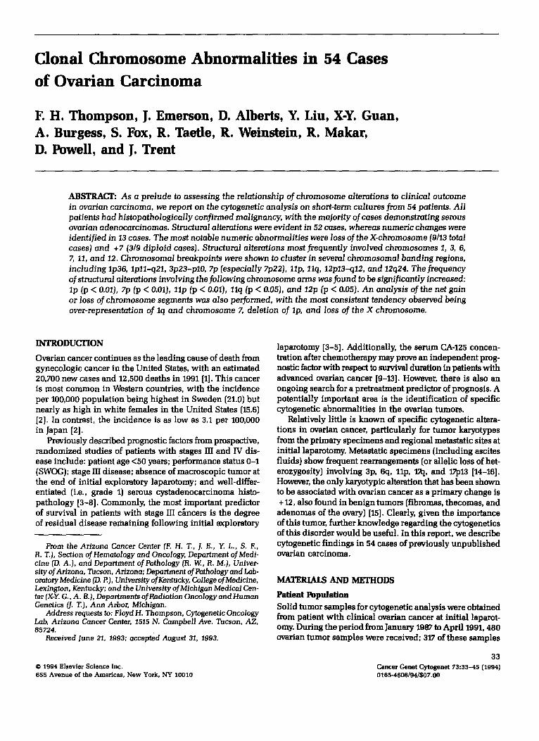
Normal metaphases were found in 12 cases, presumably representing mixtures of tumor and nontumor cells. In 22 cases extremely complex abnormal chromosome prepara- tions were obtained. These cells typically contained frag- mented chromosomes, quadriradials and/or triradials, and varying complex structural rearrangements such that com- plete karyotype descriptions were not possible. This find- ing is characteristic of ovarian carcinomas but rare in other tumors [26]. An example of such a cell is shown in Figure 1. The small proportion of cases with clonal numeric abnor- malities is no doubt due in part to the presence of these corn-
A
Figure I (A) Example of a highly abnormal, unanalyzable chromosome spread f~om case T90-06~. The presence of this kind of cell helped define the AX karyotype profile assigned to this and similar cases. (B) Examples of clonal structural abnormalities identified in case T90-248 {another case with an AX profile), See Table I for ,~X definition.
W
: . . . . i~ !i i ¸ i ̧ ~ • ;~:~•-~ ~i•:i ~i ~: ~ : i ~ i::i ~ •• / i i i~ i~ i :! I '̧•
q
. . . . i k:il il iiii !
i ! i ii! ii ! i!ii i i~i!i~!!~ i !~!iJ :̧ ~i:̧ ¸̧ ~
i: ! i i iil
iiii!iiiii:~i~

3 6
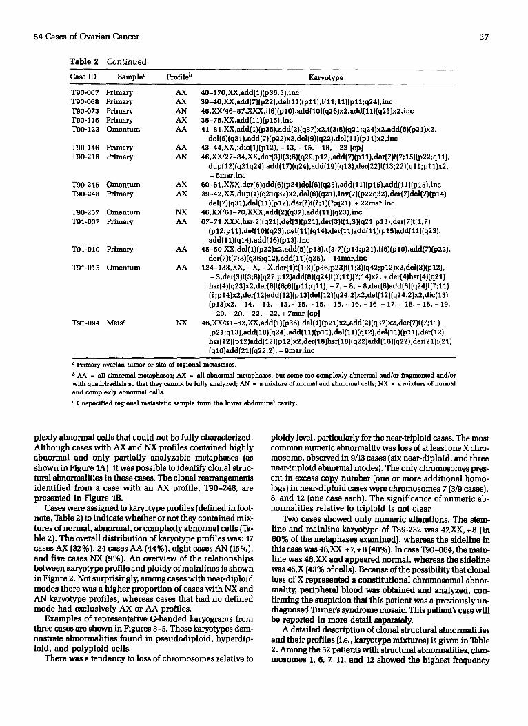
T a b l e 2 K a r y o t y p e s f r o m 54 c a s e s o f o v a r i a n c a r c i n o m a
Case ID Sample a Profile b Karyotype
T87-112 O m e n t u m T87-134 Primary T88-I 73 Pr imary T88-178 Primary
T88-183 O m a n t u m
T88-207 Primary T88-267 O m e n t u m T88-286 Primary
T88-297 Mets c
T88-301 O m e n t u m T88-304 O m a n t u m T88-320 O m e n t u m
T88-339 Primary
T88-372 O m e n t u m
T88-411 Primary T89-003 Abdomina l wall T69-007 Primary T89-026 Pr imary T89-032 Primary T69-052 O m e n t u m
T89-060 Primary T89-070 Omentum
T80-097 Mets c
T89-I07 O m e n t u m T89-126 Primary
T89-133 O m e n t u m T89-134 Intraperi tonea]
T89-143 Mets c T89-148 Primary T89-154 Je junum T89-232 Primary T89-243 Primary T89-244 Primary T89-321 Adnexa T89-348 Adnexa T90-023 PrimAry
T90-031 Primary
I"90-062 Primary
T90-063 Primary T90-064 Primary
AN 46,XX/56-57,XX,del(1]{q10),dal(1)(p10),add(11){p15),add(12](pll), inc NX 200,XXXXXXXXX,deI(10)(q25)x4,der(12)t( 1; 12)(q21 ;q24),inc/46,XX AA 55-170,XX,del[1)(q21),add(6)(q25),der(12)t(12;14)[p13;q13),inc AX 37-120,XX,add(6)(q13),del(11)(p13),hsr( l l )(q22),der(11)add(l l)(p14)
add(11)(q23),add(12)(p12),inc AA 56-63,XX, + X,dup(12)(q24q21),der(12)dup(12)(q24q11)dup(12)
(q l lq21) , inc AX 43-44,XX,del(1)(q25),add(5)(p 15),inc AN 46,XX/39-105,XX,dal(1)(p21),del(1)(q25),inc
AA 45, - X, - X,add(1J(p36.5J,i(1)(q10~,add(5){p15),t(6;13){q25;q34), - 15, - 16, t(18;22)(p11.1;q11.2), - 19, + 3mar [cp]
AA 32-75,X,del(X)(q23),add(1)(pll),del(7)(q32),der(13)t(11;13)(q12;q22), add(16)(q13],add(17)(q25),inc
AX 41-45,XX,add(1)(q44),add(7)(q36),inc AA 41-147,XX,dell(2)(p15),hsr(7](p21),add(11)(p15), inc AA 35-67,XX,del(1)(p34),add(3)(p25),add(6)(q27),hsr(6)(p22),add(7)(q36),
del(7)(p15],del(7)(q31],add(llJ(q25],ider(12)(q10]del(12)(qZZ},inc AA 53-58,XX,add(2)(q34],add(3)(pl l) ,add(12)(pl l) ,add(16)(q24),add(19)
(q13.5),inc AA 60-86,XX, - X, - X,add(1)(pll) ,add(1)(p33),del(1)(q23), - 2, - 2, - 3, - 3,
add(3)(q27}, - 4, - 4, - 5, - 5, - 6, - 6,del(7)(p14), + del(7)(q34), - 8, - 8, - 9 , - 9 , - 1 0 , - 1 1 , - 1 3 , - 1 4 , - 1 5 , - 1 5 , - 1 6 , - 1 7 , - 1 8 , - 1 8 , - 1 9 , - 19, - 20, - 21, - 22, - 22, - 2, + 15mar [cp]
AA 46,XX/44-92,XX,hsr(17) (q25),inc AX 56-143,XX,add(1)(p31),add(11)(q24),inc AX 46-100,XXXX,del(3)(p13),inc AX 41 ,XX,del{6){ql 6),der{7)add{7){p22)add{7){q34),inc AX 36,XX,add(12)(q24},inc AX 67-72,XXX,add(1}{p34),add{1)(q32),del(3){q13),add{6)(p22)x2,
add(11)(q23),inc AA 45,X, - X, - 1,der(9)t(1;9)(q11;p13)x2, + mar [cp] AN 46,XX/44,XX,add(1)(p36),add(6)(q22),add(7)(p22),der(11)t(ll ; 11)(p15;q13)
dup( l l ) (q13q25) , + 12mar, inc AA 64,XX, - X,del(1)(p35)x2, + hsr(1)(p33),add(2)(q37), - 4,der(6)t{5;6)
(q l l ;q24) , + add(7)(ql l ) ,der(TJt (7;11)(ql l ;q13)x2, - 8 , - 11,del(11)(q21), del(12)(p12), - 13, - 14, - 14,add(15)(p11), - 17, - 18, - 18, - 19, - 26, [cp]
NX 46,XX/36-37,XX,add(11)(pl4) ,del(12)(pl2.1) , inc AA 38,X, - X,add(1)(p33), - 3, - 4, - 4, - 5, - 5, - 6, - 6,del(7)(pl5),der(9)t(6;9)
( p l l ; p l l ) , + add(9](q34) ,der( l l ) t ( l l ;14) , (p14;q23) ,der(12)hsr(12)(pl l ) add(12J(ql5}, - 13,add(13}(pll] , - 14, + add(16)(p11], - 17, - 18, + 4mar[cp]
A N 46,XX/50-54,XX,del(7)(p12), inc AA 43,XX,add[l){p36),add(3){q29),add[3)[q29),hsr[6)(p21),add(7)(q31),add(11)
(q13),del{12)(p12),der(14)t(13;14)(q11;p13), + 7mar, inc AN 46,XX/45-66,XX,add(S)(q35),add(9)(p24),inc AA 39,X,add(X)(q28],i(1] (q10),der(1)t(1;11)(p36;q13),inc AA 46,X, - X, + i(1)(ql0), + 7, - 22/45,X, - X,i(1)(ql0), + 7, - 22 AA 47,XX, + 8/48,XX, + 7, + 6 AX 69-72,XXX,add(1)(p32),del(6)(q23),del(7)(p12),add(11)(p15),inc AX 49-76,XXX,add(3)(p26),add(12)[q24),inc AA 49,XX, + del(3}(q21}, + 7, + 7 AX 56-57,XX,del(1)(p12),add(1)(p11),del(6){q15),hsr(ll)(p14),add(12)(q24),inc AA 43,X, - X, - 1,del(1)(q24q32), + 12, - 13, - 14, - 15,dar(16)t(1;16)(q21;q24),
- 1 8 , - 1 9 , a d d ( 2 2 ) ( q 1 3 ) , + 3 m A r [ c p ]
AA 68-70,XXX, + X, + 1, + 3, - 4,der(7)t(7;9)(p22;q12)x2, + 6, - 12, - 13, - 14, - 1 6 , - 1 7 , - 1 9 , + 2 0 , + 4 m a r [ c p ]
A X 65,XXX,i(1)(qlO),der(7)t(3;7)(q13;p21),hsrf11)(pll),der(17)t[8;17) (ql 1;p13),i(21)(qlO),inc
NX 46,XX/68-72,XXX,add(1)(p36.5),add[12){p13),inc AN 46,XXc/45,X, - Xc
cont inued
54 Cases of Ovarian Cancer
Table 2 Continued
37
Case ID S ampl e a Profile b Karyo type
T90-067 P r imary T90-068 P r imary T90-073 Pr imary T90-116 Pr imary T90-123 O m e n t u m
T90-146 P r imary T90-216 Pr imary
T90-245 O m e n t u m T90-248 P r imary
T90-257 O m e n t u m T91-007 Pr imary
T91-010 P r imary
T91-015 O m e n t u m
T91-094 Mets c
AX 40-170,XX,add(1}(p36.5) , inc AX 39-40,XX,add(7}(p22) ,de l (11)(p l 1),t(11; 11)(p11;q24), inc AN 46,XX/46-87,XXX,i(6}(p10),add{10)(q26)x2,add{11)(q23)x2,inc AX 36-75,XX,add(11} [p15), inc A A 41-81,XX,add(1}(p36},add(2)(q37}x2,t(3;8}(q21;q24}x2,add{6)(p21}x2,
del[6)(q21},add(7)Co22)x2,del(9)(q22},del(11}(pll}x2,inc A A 43-44,XX,idic(1}(p12}, - 13, - 15, - 18, - 22 [cp] AN 46,XX/27-84,XX,derC3}t(3;6)(q29;p12},add(7)(pll),der(7)t(7;15}(p22;q11},
dup(12}(q21q24),add(17}(q24},add(19){q13},der(22)t(13;22)(q11;p11)x2, ÷ 6mar , inc
AX 60-61,XXX,derI6)add(6)(p24)del(6)(q23),add(11) Cpl5),add(11)(pl5),inc AX 39-42,XX,dup(1}Cq21q32)x2,del(6}(q21},inv(7}{p22q32),der{7}del(7)lp14}
dellT){q31),del(ll)(pl2},derC?)t(?;1)(?;q21), + 22mar,inc NX 46,XX/61-70,XXX,add(2)(q37},add(ll)(q23),inc AA 67-71,XXX,hsr(2)(q21),del(3)(p21},der(3)tC1;3)(q21;p13),der(7}t(1;7}
(p12;p11},delC10}(q23},del(11)(q14),der(ll}add(11}(p15}add(11}(q23}, add[11)(q14},add(16)(p13},inc
AA 45-50,XX,del{1}(p22)x2,add(5)(p13},t{3;7}(p14;p21),t(6}(p10},add(7)(p22), derC7)t(7;8){q36;q12),add(ll){q25}, + 14mar,inc
APt 124-133,XX, - X, - X,der(1)t(1;3)(p36;p23)tC1;3)(q42;p12}x2,del(3){p12}, - 3,der(3)tC3;8)(q27;p12}add(8)(q24}t(?;11)(?;14}x2, + der(4)hsr(4}(q21) hsr(4)(q23}x2,der(6}t(6;6}(p11;qll}, - 7, - 8, - 8,der{8)add(8}lq24)t(?;11} (?;p14)x2,der{12}addC12}(p13}delI12)(q24.2)x2,del(12}(q24.2)x2,dic(13} (p13)x2, - 14, - 14, - 15, - 15, - 15, - 15, - 16, - 16, - 17, - 18, - 18, - 19, - 20, - 20, - 22, - 22, + 7mar [cp]
NX 46,XX/31-62,XX,add(1}(p36),del(1}(p21}x2,add(2)(q37}x2,der(7}t{7;11) (p21;q13),addC10}(q24),add(11)(pl l},del(11)(ql2) ,del{11}(pl 1},der(12) hsr(12)(p12)add(12)(p12}x2 ,der(18}hsr(18}(q22}add(18)[q22),der(21)i(21) (q10)add(21}(q22.2). + 9mar , inc
a Primary ovarian tumor or site of regional metastases.
b AA ffi all abnormal metaphases; AX = all abnormal metaphases, but some too complexly abnormal and/or fragmented and/or with quadriradials so that they cannot be fully analyzed; AN ffi a mixture of normal and abnormal cells; NX ffi a mixture of normal and complexly abnormal cells.
e Unspecified regional metastatic sample from the lower abdominal cavity.
plexly abnormal cells that could not be fully characterized. Although cases with AX and NX profiles contained highly abnormal and only partially analyzable metaphases {as shown in Figure 1A), it was possible to identify clonal struc- tural abnormalities in these cases. The clonal rearrangements identified from a case with an AX profile, T90-248, are presented in Figure lB.
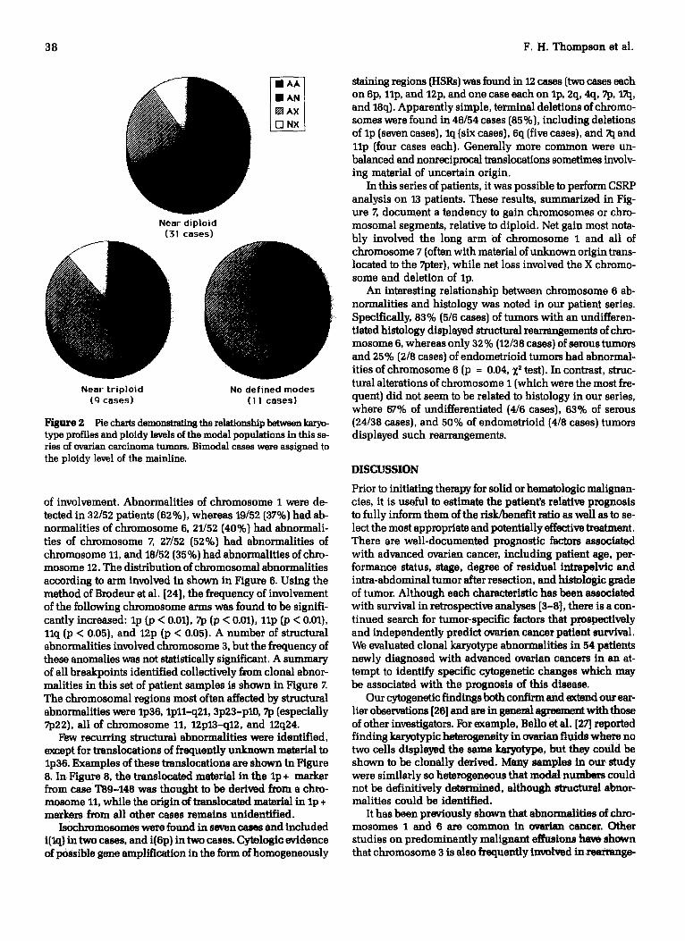
Cases were assigned to karyotype profiles {defined in foot- note, Table 2} to indicate whether or not they contained mix- tures of normal, abnormal, or complexly abnormal cells {Ta- ble 2). The overall distribution of karyotype profiles was: 17 cases AX {32%}, 24 cases AA {44%), eight cases AN {15%}, and five cases NX {9%). An overview of the relationships between karyotype profile and ploidy of mainlines is shown in Hgure 2. Not surprisingly, among cases with near-diploid modes there was a higher proport ion of cases with NX and AN karyotype profiles, whereas cases that had no defined mode had exclusively AX or AA profiles.
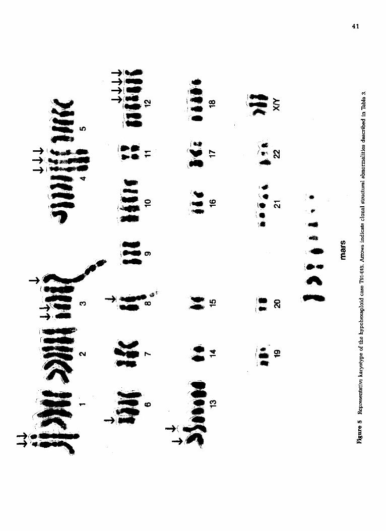
Examples of representative G-banded karyograms from three cases are shown in Figures 3-5. These karyotypes dem- onstrate abnormalities found in pseudodiploid, hyperdip- loid, and polyploid cells.
There was a tendency to loss of chromosomes relative to
ploidy level, particularly for the near-triploid cases. The most common numeric abnormality was loss of at least one X chro- mosome, observed in 9/13 cases (six near-diploid, and three near-triploid abnormal modes). The only chromosomes pres- ent in excess copy number (one or more additional homo- logs) in near-diploid cases were chromosomes 7 (3/9 cases), 8, and 12 (one case each). The significance of numeric ab- normalities relative to triploid is not clear.
Two cases showed only numeric alterations. The stem- line and mainline karyotype of T89-232 was 4ZXX, + 8 (in 60% of the metaphases examined), whereas the sideline in this case was 48,XX, + Z + 8 (40%). In case T90-064, the main- line was 46,XX and appeared normal, whereas the sideline was 45,X (43% of cells). Because of the possibility that clonal loss of X represented a constitutional chromosomal abnor- mality, peripheral blood was obtained and analyzed, con- firming the suspicion that this patient was a previously un- diagnosed Turner's syndrome mosaic. This patient's case will be reported in more detail separately.
A detailed description of clonal structural abnormalities and their profiles {i.e., karyotype mixtures) is given in Table 2. Among the 52 patients with structural abnormalities, chro- mosomes 1, 6, Z 11, and 12 showed the highest frequency
38 F .H. Thompson et al.
Near d ip lo id (31 cases)
Near t r i p lo id No defined modes (9 cases) (I I cases)
Figure 2 Pie charts demonstrating the relationship between karyo- type profiles and ploidy levels of the modal populations in this se- ries of ovarian carcinoma tumors. Bimodal cases were assigned to the ploidy level of the mainline.
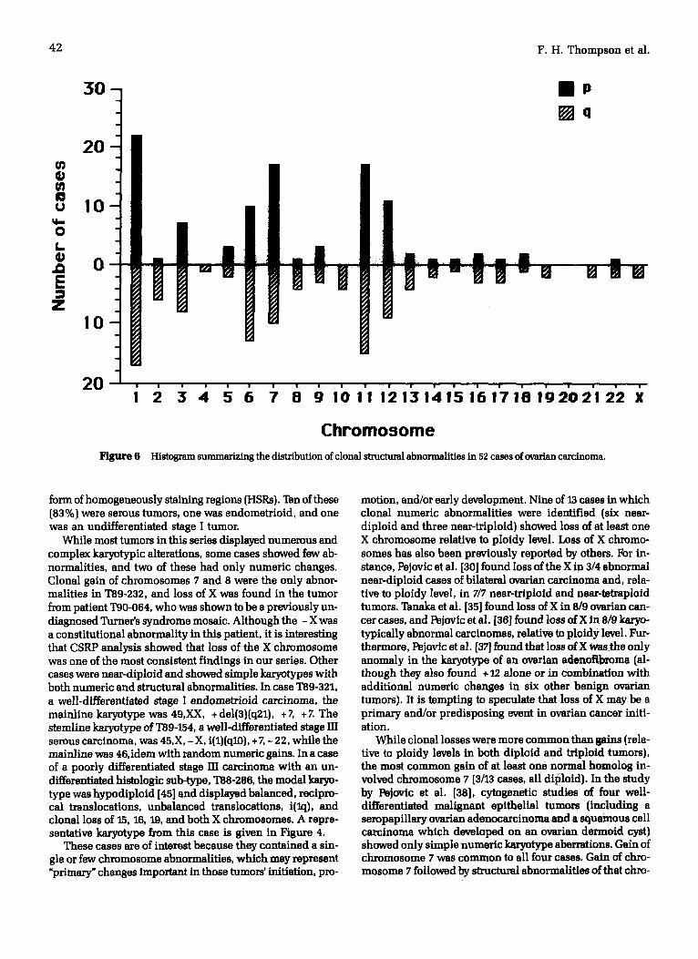
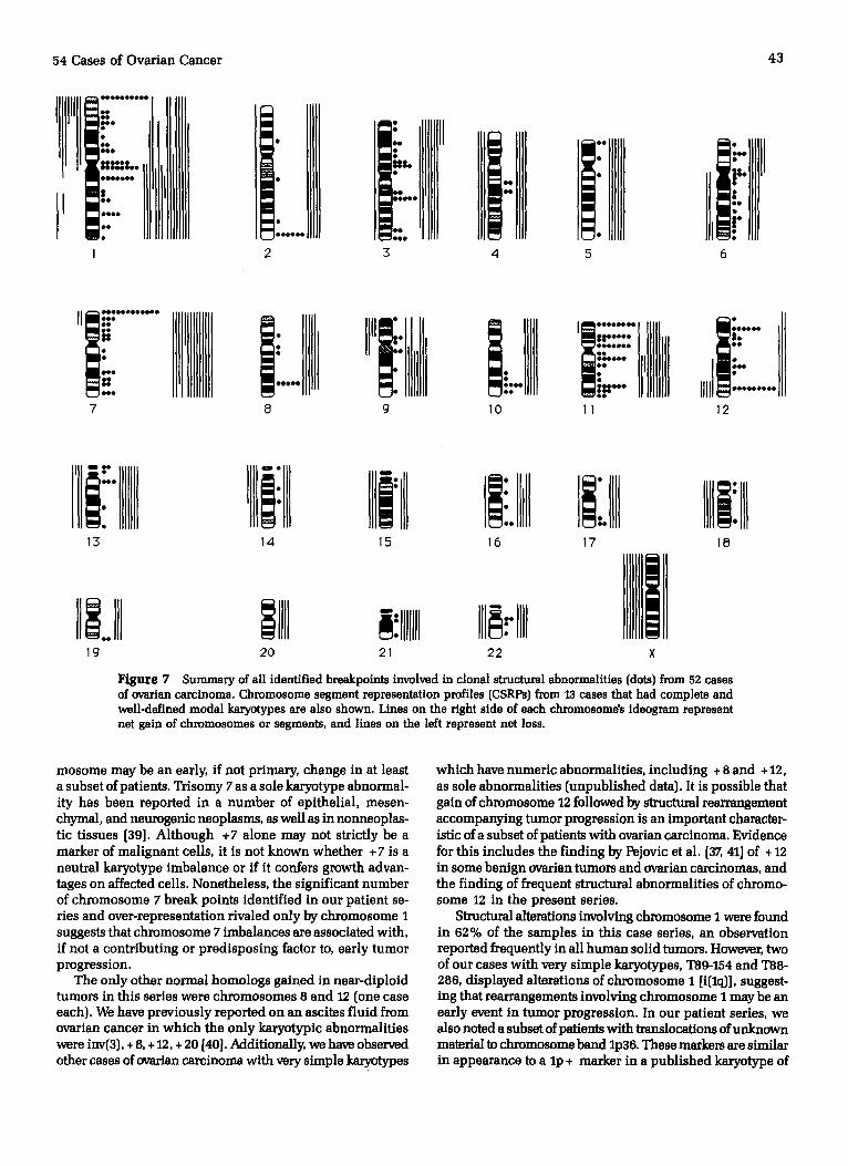
of involvement. Abnormalities of chromosome 1 were de- tected in 32/52 patients [62%}, whereas 19/52 (37%) had ab- normalities of chromosome 6, 21/52 {40%) had abnormali- ties of chromosome 7, 27/52 (52%) had abnormalities of chromosome 11, and 18/52 [35%) had abnormalities of chro- mosome 12. The distribution of chromosomal abnormalities according to arm involved in shown in Figure 6. Using the method of Brodeur et al. [24], the frequency of involvement of the following chromosome arms was found to be signifi- cantly increased: lp [p < 0.01), 7p {p < 0.01), l lp {p < 0.01), l lq (p < 0.05), and 12p {p < 0.05). A number of structural abnormalities involved chromosome 3, but the frequency of these anomalies was not statistically significant. A summary of all breakpoints identified collectively f ~ m clonal abnor- malities in this set of patient samples is shown in Figure 7. The chromosomal regions most often affected by structural abnormalities were lp36, 1p11-q21, 3p23-p10, 7p {especially 7p22), all of chromosome 11, 12p13-q12, and 12q24.
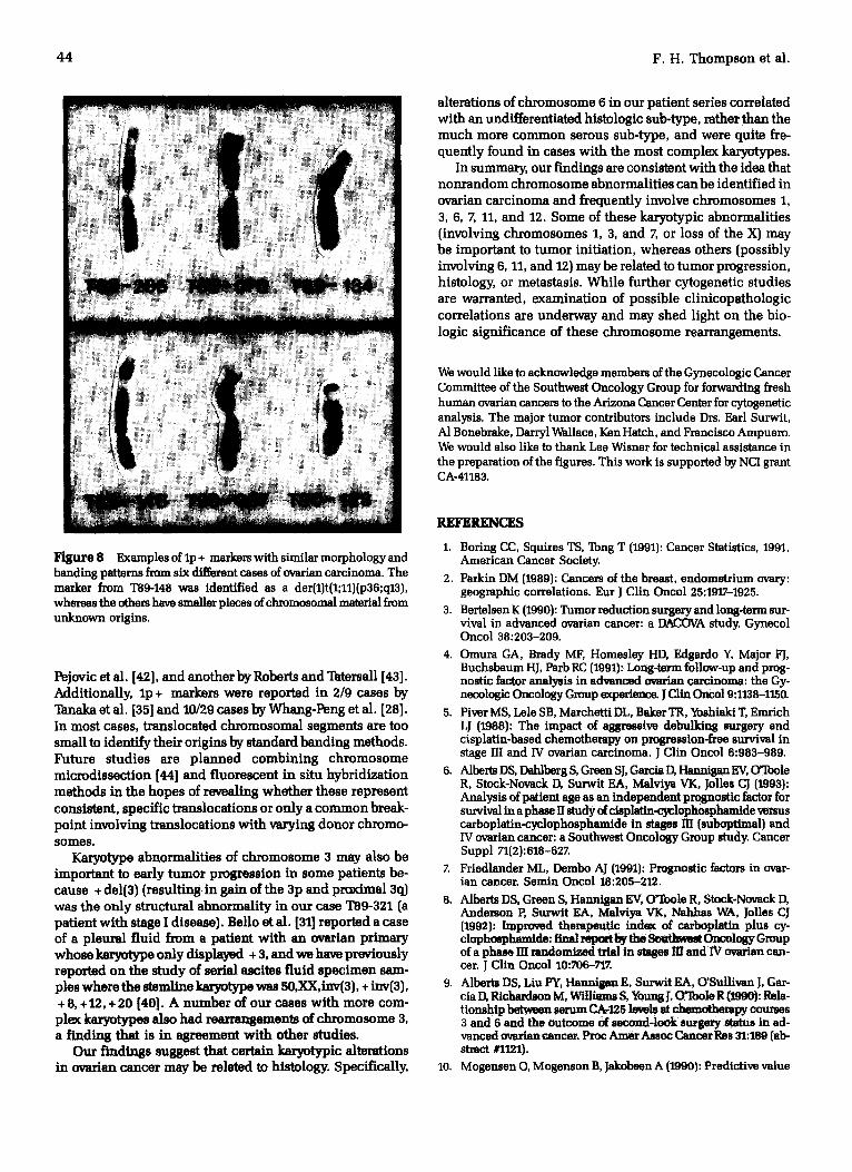
Few recurring structural abnormalities were identified, except for translocations of frequently unknown material to lp36. Examples of these translocations are shown in Figure 8. In Figure 8, the translocated material in the lp + marker from case T89-148 was thought to be derived from a chro- mosome 11, while the origin of translocated material in lp + markers from all other cases remains unidentified.
Isochromosomes were found in seven cases and included i(lq) in two cases, and i{6p) in two cases. Cytologic evidence of possible gene amplification in the form of homogeneously
staining regions {HSRs} was found in 12 cases {two cases each on 6p, llp, and 12p, and one case each on Ip, 2q, 4q, 7p, 17q, and 18q). Apparently simple, terminal deletions of chromo- somes were found in 46/54 cases {85%}, including deletions of Ip [seven cases}, lq {six cases}, 6q {five cases}, and 7q and llp {four cases each}. Generally more common were un- balanced and nonreciprocal translocations sometimes involv- ing material of uncertain origin.
In this series of patients, it was possible to perform CSRP analysis on 13 patients. These results, summarized in Fig- ure Z document a tendency to gain chromosomes or chro- mosomal segments, relative to diploid. Net gain most nota- bly involved the long arm 'of chromosome I and all of chromosome 7 {often with material of unknown origin trans- located to the 7pter), while net loss involved the X chromo- some and deletion of lp.
An interesting relationship between chromosome 6 ab- normalities and histology was noted in our patient series. Specifically, 83% {5/6 cases) of tumors with an undifferen- tiated histology displayed structural rearrangements of chro- mosome 6, whereas only 32 % {12/38 cases} of serous tumors and 25% {2/8 cases) of endometrioid tumors had abnormal- ities of chromosome 6 (p = 0.04, ;(z test}. In contrast, struc- tural alterations of chromosome I {which were the most fre- quent} did not seem to be related to histology in our series, where 67% of undifferentiated {4/6 cases}, 63% of serous {24/38 cases), and 50% of endometrioid {4/8 cases) tumors displayed such rearrangements.
DISCUSSION
Prior to initiating therapy for solid or hematologic malignan- cies, it is useful to estimate the patient's relative prognosis to fully inform them of the risk/benefit ratio as well as to se- lect the most appropriate and potentially effective treatment. There are well-documented prognostic factors associated with advanced ovarian cancer, including patient age, per- formance status, stage, degree of residual intmpelvic and intra-abdominal tumor after resection, and histologic grade of tumor. Although each characteristic has been associated with survival in retrospective AnAlyses [3-8], there is a con- tinued search for tumor-specific factors that prospectively and independently predict ovarian cancer patient survival. We evaluated clonal karyotype abnormalities in 54 patients newly diagnosed with advanced ovarian cancers in an at- tempt to identify specific cytogenetic changes which may be associated with the prognosis of this disease.
Our cytogenetic findings both confirm and extend our ear- lier observations [26] and are in general agreement with those of other investigators. For example, Belle et al. [27] reported finding karyotypic heterogeneity in ovarian fluids where no two cells displayed the same karyotype, but they could be shown to be clonally derived. Many samples in our study were similarly so heterogeneous that modal numbers could not be definitively determined, although structural abnor- malities could be identified.
It has been previously shown that abnormalities of chro- mosomes I and 6 are common in ovarian cancer. Other studies on predominantly malignant effusions have shown that chromosome 3 is also frequently invotved in rearrange-
54 Cases of Ovarian Cancer 39
1 2 3
o .
8 9
4 5
6 7 10 11 12
13 14 15 16 17 18
19 20 21 22 X/Y
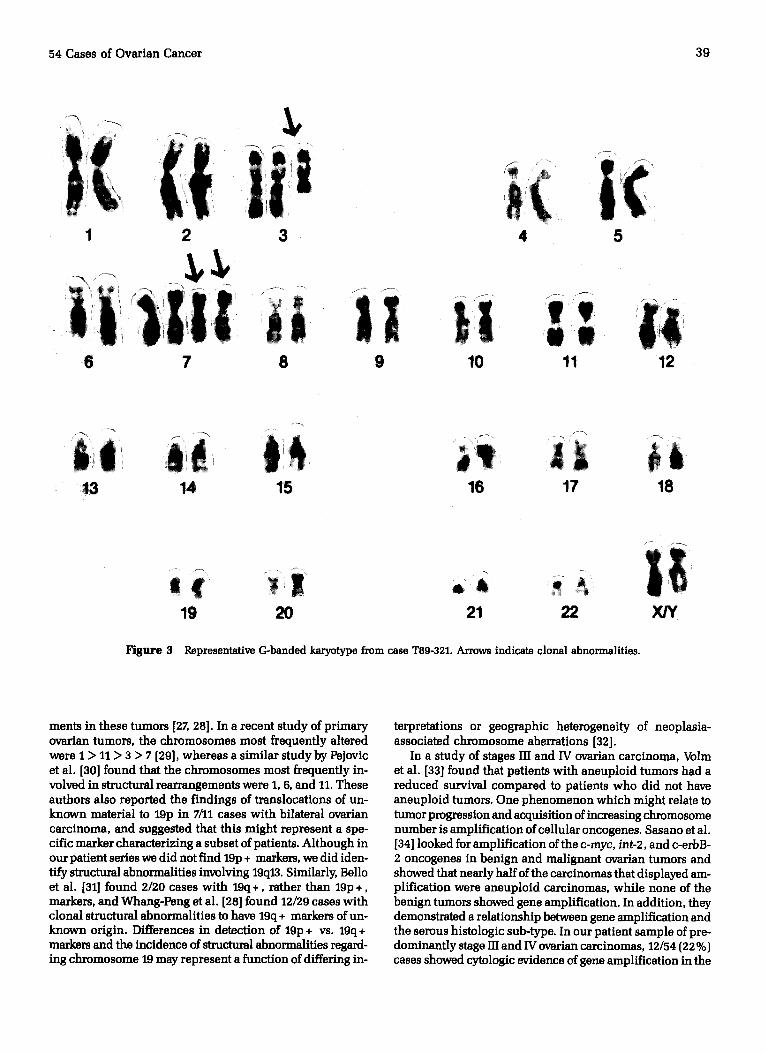
Figure 3 Representative G-banded karyotype from case T89-321. Arrows indicate clonal abnormalities.
ments in these tumors [27, 28]. In a recent study of primary ovarian tumors, the chromosomes most frequently altered were 1 > 11 > 3 > 7 [29], whereas a similar study by Pejovic et al. [30] found that the chromosomes most frequently in- volved in structural rearrangements were 1, 6, and 11. These authors also reported the findings of translocations of un- known material to 19p in 7/11 cases with bilateral ovarian carcinoma, and suggested that this might represent a spe- cific marker characterizing a subset of patients. Although in our patient series we did not find 19p + markers, we did iden- tify structural abnormalities involving 19q13. Similarly, Belle et al. [31] found 2/20 cases with 19q +, rather than 19p +, markers, and Whang-Peng et al. [28] found 12/29 cases with clonal structural abnormalities to have 19q + markers of un- known origin. Differences in detection of 19p + vs. 19q + markers and the incidence of structural abnormalities regard- ing chromosome 19 may represent a function of differing in-
terpretations or geographic heterogeneity of neoplasia- associated chromosome aberrations [32].
In a study of stages HI and IV ovarian carcinoma, Volta et al. [33] found that patients with aneuploid tumors had a reduced survival compared to patients who did not have aneuploid tumors. One phenomenon which might relate to tumor progression and acquisition of increasing chromosome number is amplification of cellular oncogenes. Sasano et al. [34] looked for amplification of the c-myc, int-2, and c-arbB- 2 oncogenes in benign and malignant ovarian tumors and showed that nearly half of the carcinomas that displayed am- plification were aneuploid carcinomas, while none of the benign tumors showed gene amplification. In addition, they demonstrated a relationship between gene amplification and the serous histologic sub-type. In our patient sample of pre- dominantly stage HI and IV ovarian carcinomas, 12/54 [22 %} cases showed cytologic evidence of gene amplification in the

40
Lo
c~
q b m
4mb4umBmllp ~
f4mD
C~l Tm
Q ,qrm
¢0 o ~
a b
~ vm
ONIe
tromp
dlplmmb
c~
s=
C~ c~
°~
¢0
41
1 1
- . )
,oi111
m
w n l l p
--)
¢0
T,m
-...) Tm
i t D ~ .,_ D m 8 ' , -
a m J ~
q U l m o
f I R
. . . ) j ao
' ¢ 0
k O
o o
T , -
,i,,-
i ~ 't m
'r.,.
T - -
i8
I D
i o
g~ I n ~a E
Q~
a~
0
0
u~
c~
0
0
a~
c~
I,,*
42 F .H. Thompson et al.
3 0
20
o
Z 10
20 I
Figure 6
B P [ ] q
2 3 4 5 6 7 8 9 10111213141516171819202122
Chromosome
Histogram summ~'izin R the distribution of clone] structural abnormalities in 52 cases of ovarian carcinoma.
X
form of homogeneously staining regions {I-ISRs}. "lbn of these {83%] were serous tumors, one was endometrioid, and one was an undifferentiated stage I tumor.
While most tumors in this series displayed numerous and complex karyotypic alterations, some cases showed few ab- normalities, and two of these had only numeric changes. Clonal gain of chromosomes 7 and 8 were the only abnor- malities in "1"89-232, and loss of X was found in the tumor from patient T90-064, who was shown to be a previously un- diagnosed Turner's syndrome mosaic. Although the - X was a constitutional abnormality in this patient, it is interesting that CSRP analysis showed that loss of the X chromosome was one of the most consistent findings in our series. Other cases were near-diploid and showed simple karyotypes with both numeric and structural abnormalities. In case T89-321, a well-differentiated stage I endometr/oid carcinoma, the mainline karyotype was 49,XX, +del{3)[q21), +7, +7. The stemline karyotype of T89-154, a wall-differentiated stage HI serous carcinoma, was 45,X, - X, i(1}{ql0}, + 7, - 22, while the mainline was 46,idem with random numeric gains. In a case of a poorly differentiated stage HI carcinoma with an un- differentiated histologic sub-type, T88-286, the modal karyo- type was hypodiploid [45] and displayed balanced, recipro- cal translocations, unbalanced translocations, i{lq], and clonal loss of 15, 16, 19, and both X chromosomes. A repre- sentative karyotype from this case is given in Figure 4.
These cases are of interest because they contained a sin- gle or few chromosome abnormalities, which may represent ~ r imar~ ' changes important in those tumors' in i t i~on, pro-
motion, and/or early development. Nine of 13 cases in which clonal numeric abnormalities were identified {six near- diploid and three near-triploid} showed loss of at least one X chromosome relative to ploidy level. Loss of X chromo- somes has also been previously reported by others. For in- stance, Pejovic et al. [30] found loss of the X in 3/4 abnormal near-diploid cases of bilateral ovarian carcinoma and, rela- tive to ploidy level, in 7/7 near-triploid and near-tetmploid tumors. Tanaka et al. [35] found loss of X in 8/9 ovarian can- cer cases, and Pejovic et al. [36] found loss of X in 8/9 karyo- typically abnormal carcinomas, relative to ploidy level. Fur- thermore, Pejovic et el. [37] found that loss ofX wes the only anomaly in the karyotype of an ovarian adenofibroma (al- though they also found + 12 alone or in combination with additional numeric changes in six other benign ovarian tumors). It is tempting to speculate that loss of X may be a primary and/or predisposing event in ovarian cancer initi- ation.
While clonal losses were more common than gains (rela- tive to ploidy levels in both diploid and triptoid tumors), the most common gain of at least one normal homolog in- volved chromosome 7 {3/13 cases, all diploid}. In the study by Pejovic et el. [38], cytnganetic studies of four well- differentiated malignant epithelial tumors {including a seropapfllary ovarian adenocarcinonm and a squamous cell carcinoma which developed on an ovarian dermoid cyst) showed only simple numeric karyotype aberrations. Gain of chromosome 7 was common to all four cases. Gain of chro- mosome 7 followed by structural abnormalities of that chro-
54 Cases of Ovarian Cancer 43
i ..... li:!!!i.illlll I, IIIi 2 3 4
....IIiI ]i!'[rllJI 5
ii, iii:lllJ 6
......... IIII'IlllIIIII i" :..... IIIIIl 8 9
IiIII ii!':':I lllIIlll l:
. I :'.. " : " ]Ill . . . . . . . . .
10 11 12
Ilii:llllllJ IIIi lll IIIi' II 13 14 15
I ..lll I IIII :llnlIl 19 20 21
I ::H [ iIIII 16 17
lllllllill 22 ×
Figure 7 Summary of all identified breakpoints involved in clonal structural abnormalities {dots) from 52 cases of ovarian carcinoma. Chromosome segment representation profiles {CSRPs) from 13 cases that had complete and well-defined modal karyutypes are also shown. Lines on the right side of each chromosome,s ideogram represent net gain of chromosomes or segments, and lines on the left represent net loss.
18
mosome may be an early, if not primary, change in at least a subset of patients. Trisomy 7 as a sole karyotype abnormal- ity has been reported in a number of epithelial, mesen- chymal, and neurogenic neoplasms, as well as in nonneoplas- tic tissues [39]. Although + 7 alone may not strictly be a marker of malignant cells, it is not known whether + 7 is a neutral karyotype imbalance or if it confers growth advan- tages on affected cells. Nonetheless, the significant number of chromosome 7 break points identified in our patient se- ries and over-representation rivaled only by chromosome 1 suggests that chromosome 7 imbalances are associated with, if not a contributing or predisposing factor to, early tumor progression.
The only other normal homologs gained in near-diploid tumors in this series were chromosomes 8 and 12 {one case each). We have previously reported on an ascites fluid from ovarian cancer in which the only karyotypic abnormalities were inv(3), + 8, + 12, + 20 [40]. Additionally, we have observed other cases of ovarian carcinoma with very simple karyotypes
which have numeric abnormalities, including + 8 and + 12, as sole abnormalities {unpublished data}. It is possible that gain of chromosome 12 followed by structural rearrangement accompanying tumor progression is an important character- istic of a subset of patients with ovarian carcinoma. Evidence for this includes the finding by Pejovic et al. [3Z 41] of + 12 in some benign ovarian tumors and ovarian carcinomas, and the finding of frequent structural abnormalities of chromo- some 12 in the present series.
Structural alterations involving chromosome I were found in 62 % of the samples in this case series, an observation reported frequently in all human solid tumors. However, two of our cases with very simple karyotypes, T89-154 and I"88- 286, displayed alterations of chromosome I [i{lq}], suggest- ing that rearrangements involving chromosome I may be an early event in tumor progression. In our patient series, we also noted a subset of patients with translocations of unknown material to chromosome band Ip36. These markers are similar in appearance to alp + marker in a published karyotype of
44 F.H. Thompson et al.
alterations of chromosome 6 in our patient series correlated with an undifferentiated histologic sub-type, rather than the much more common serous sub-type, and were quite fre- quently found in cases with the most complex ~ y p e s .
In summary, our findings are consistent with the idea that nonrandom chromosome abnormalities can be identified in ovarian carcinoma and frequently involve chromosomes 1, 3, 6, 7, 11, and 12. Some of these keryotypic abnormalities (involving chromosomes 1, 3, and 7, or loss of the X} may be important to tumor initiation, whereas others {possibly involving 6, 11, and 12) may be related to tumor progression, histology, or metastasis. While further cytogenetic studies are warranted, examination of possible clinicopathologic correlations are underway and may shed light on the bio- logic significance of these chromosome rearrangements.
We would like to acknowledge members of the Gynecologic Cancer Committee of the Southwest Oncology Group for forwarding fresh humAr~ ovarian cancers to the Arizona Cancer Center for cytogenctic analysis. The major tumor contributors include Drs. Earl Surwit, A1 Bonebrake, Darryl Wallace, Ken Hatch, and Francisco Ampuero. We would also like to thank Lee Wisner for technical assistance in the preparation of the figures. This work is supported by NCI grant CA-41183.
Figure 8 Examples of lp + markers with similar morphology and banding patterns from six different cases of ovarian carcinoma. The marker from T89-148 was identified as a der{1}t{1;ll){p36;q13), whereas the others have smaller pieces of chromosome] material from unknown origins.
Pejovic et al. [42], and another by Roberts and Tatersall [43]. Additionally, lp + markers were reported in 2/9 cases by Tanaka et al. [35] and 10/29 cases by Whang-Peng et al. [28]. In most cases, translocated chromosomal segments are too small to identify their origins by standard banding methods. Future studies are planned combining chromosome microdissection [44] and fluorescent in situ hybridization methods in the hopes of revealing whether these represent consistent, specific translocations or only a common break- point involving translocations with varying donor chromo- somes.
Karyotype abnormalities of chromosome 3 may also be important to early tumor progression in some patients be- cause + del{3) {resulting in gain of the 3p and praximal 3o.) was the only structural abnormality in our case "I"89-321 (a patient with stage I disease). Bel]o et al. [31] reported a case of a pleura] fluid from a patient with an ovarian primary whose karyotype only displayed + 3, and we have previously reported on the study of serial ascites fluid specimen sam- ples where the stemll-e karyotype was 50,XX,inv{3}, + inv(3), + 8, +12, + 20 [40]. A number of our cases with more com- plex ~ s also had rem'rangements of chromosome 3, a finding that is in agreement with other studies.
Our findings suggest that certain karyotypic alterations in ovarian cancer m a y be related to histology. Specifically,
REFERENCES
1. Boring CC, Squires TS, Tong T (1991): Cancer Statistics, 1991, American Cancer Society.
2. Parkin DM (1989): Cancers of the breast, endometrium ovary: geographic correlations. Eur J C]in Oncol 25:1917-1925.
3. Bertelsan K (1990): Tumor reduction surgery and long-term sur- vival in advanced ovarian cancer: a DACOVA study. Gynecol Oncol 38:203-209.
4. Omura GA, Brady MF, Homesley HD, Edgardo Y, Major FJ, Buchsbaum HJ, Parb RC (1991): Long-tarm follow-up and prog- nostic factor analysis in advanced ovarian carcinoma: the Gy- necologic Oncology Group experience. J C]in Oncol 9:1138-1150.
5. Piver MS, Lele SB, Marchetti DL, Baker TR, Yoshiaki T, Emrich LJ (1988): The impact of aggressive debulkin8 surgery and cisplatin-based chemotherapy on progression-free survival in stage HI and IV ovarian carcinoma. J C]in Oncol 6:983-989.
6. Alberts DS, Dahlber8 S, Green SJ, Garcia D, I-Iannigan EV, C)qbole R, Stock-Noveck D, Surwit EA, Malviya VK, Jones CJ (1993}: Analysis of patient age as an independent pmgnoatic factor for survival in a phase II study of cisplatin-c3r, lophosphemide versus carboplatin-c~lophosphamide in stages Ill {suboptimal} and IV ovarian cancer: a Southwest Oncology Group study. Cancer Suppl 71{2}'.618-827.
Z Friedlander ML, Dembo AJ {1991): Prognostic factors in ovar- ian cancer. Semln Oncol 18:205-212.
8. Alberts DS, Green S, Hannigan EV, o'roole R, Stock-Novack D, Anderson P, Surwit EA, Malviya VK, Nahhas WA, Jolles CJ {1992}: Improved therapeutic inde~ of car~platin plus cy- clophmphamide: final report by the Southtmst Oncolngy Group of a phase HI mudomi~,-sd trial in stages HI and IV ovarian can- cer. J Clin Oncol 10:706-717.
9. Alberts DS, Lin PY, HAnniglm E, Surwit EA, O'Sullivan J, Gar- cia D, Richardson M, Willl,m~ S, Young J, O~Toole R (1990): Rela- tionship between serum CA-125 levels at chemotherapy courses 3 and 6 and the outcome of second-look surgery status in ad- vanced overian cancer. Pmc Amer Assoc Cancer Res 31:189 {ab- stract #1121}.
10. Mogeusen O, Mogenson B, Jakobsen A (1990): Predictive value
54 Cases of Ovarian Cancer 45
of CA-125 during early chemotherapy of advanced ovarian can- cer. Gynecol Oncol 37:44-46.
11. Hunter VJ, Lee D, Helms M, Soper JT, Berchuck A, Clark-Rmmon DL, Bast Jr. RC (1990): The prognostic significance of CA-125 half-life in patients with ovarian cancer who have received pri- mary chemotherapy after surgical cytoreduction. Am J Obstet Gynecol 163:1164-1167.
12. Hawkins RE, Roberts K, Wiltshaw E, Mundy J, Fryatt IJ, McReady VR {1989}: The prognostic significance of the half-life of serum CA-125 in patients responding to chemotherapy for epithelial ovarian carcinoma. Br J Obstet Gyneacol 96:1395-1399.
13. Sevelda P, Schemper M, Spona J {1989}: CA-125 as an indepen- dent prognostic factor for survival in patients with epithelial ovarian cancer. Am J Obstet Gynecol 161:1213-1216.
14. Heim S; and Mitelman F {1967): Cancer Cytogenetics, Alan Liss, New York.
15. Mitelman F, Kaneko Y, Trent J {1991): Report of the committee on chromosome changes in neoplasia. Human Gene Mapping 11 {1991}. Cytogenet Cell Genet 58:1-2200.
16. Lee JH, Kavanagh JJ, Wildrick DM, Wharton JT, BlickM {1990}: Frequent loss of hetemzygosity on chromosomes 6q, 11, and 12 in human ovarian carcinomas. Cancer Ros 50:2724-2728.
17. Serov SF, Scully RE, Sobin LH (1973}: Histological typing of ovar- ian tumours. In: International; Histological Classification of Tumours. Geneva, World Health Organization.
18. Russell P, Bannatyne P {1989}: Surgical Pathology of the Ova- ries, Churchill Livingston, London.
19. FIGO: International Federation of Gynecology and Obstetrics {1971): Classification and staging of malignant tumors in the fe- male pelvis. Acta Obstet Gynaecol Scand 50:1-7.
20. Trent JM, Thompson FH {1967}: In: Molecular genetics of mam- malian cells. Vol. 151 of Methods in Enzymology, Gottesman M, ed. Academic Press, San Diego, pp. 267-279.
21. Leibovitz A {1985): In: Mammomsh K, ed. Advances in cell cul- ture. Vol. 4, Academic Press, New York, pp. 249-260.
22. ISCN {1985}: An International System for Human Cytogenetic Nomenclature, Harnden IX;, Klinger HP, eds, published in col- laborations with Cytogenet Cell Genet {Karger, Basel 1985); also in Birth Defects: Original Article Series, Vol. 121, No. 1 {March of Dimes Birth Defects Foundation, New York, 1985}.
23. ISCN (1991}: Guidelines for Cancer Cytogenetics, Supplement to An International System for Human Cytogenetic Nomencla- ture, F. Mitelman, ed. S. Karger, Basel {1991}.
24. Bmdeur GM, Tsiatis AA, Williams DL, Luthardt FW. Green AA {1982}: Statistical analysis of cytogenetic abnormalities in hu- man cancer cells. Cancer Genet Cytogenet 7:137-152.
25. Thompson F, Emerson J, Dalton WS, Yang J-M, McGee D, Villar H, Knox S, Massey K, Weinstein R, Bhattacharya A, Trent J {1994}: Clonal chromosomal abnormalities in bl]mAn breast car- cinomas: I: 28 cases with primary disease. Genes Chrom Can- cer {in press}.
26. Trent JM {1985}: Prevalence and clinical significance of cyto- genetic abnormalities in bllmAn ovarian cancer. In: Ovarian Can- cer, DS Albets, EA Surwit, eds. Martinus, Nijhof Publishers, New York, pp. 1-21.
2Z Bello MJ, Moreno S, Rey JA {1990): Involvement of 9p in me- tastatic ovarian adenocarcinomas. Cancer Genet Cytogenet 45: 223-229.
28. Whang-Peng J, Knutsan T, Douglass EC, Chu E, Ozols RF, Ho- gan W'M, Young RC {1984): Cytogenetic studies in ovarian can- cer. Cancer Genet Cytogenet 11:91-106.
29. Gallion HI-I, Powell DE, Smith LW, Morrow JK, Martin AW, van Nagell JR, Donaldson ES {1990}: Chromosome abnormalities in human epithelial ovarian malignancies. Gynecol Oncol 38: 473-47Z
30. Pejovic T, Helm S, Mandahl N, Elmfors B, Furgyik S, Floderus U-M, Helm G, Willan H, Mitelman F {1991}: Bilateral ovarian carcinoma: cytogenetic evidence of unicentric origin. Int J Can- cer 47:358-361.
31. Bello MJ, ReyJA {1990): Chromosome aberrations in metastatic ovarian cancer: relationship with abnormalities in primary tumors. Int J Cancer 45:50-54.
32. Johansson B, Martens F, Mitelman F (1991): Geographic hetero- geneRy of neoplasia-associeted aberrations. Genes Chrom Cancer 3:1-7.
33. Volta M, Bruggemann A, Marianne Gunther, Kleine W, Pflei- deter A, Vogt-Schaden M {1985}: Prognostic relevance of ploidy, pmifferation, and resistance-predictive tests in ovarian carci- noma. Cancer Res 45:5180-5185.
34. Sasano H, Garrett CT, Wilkinson DS, Silverberg S, Comerford J, Hyde J {199o}: Pmtooncogane amplification in human ovar- ian neoplasms. Hum Pathol 21:382-391.
35. Tanaka K, Boice CR, Tasta JR {1989}: Chromosome aberrations in nine patients with ovarian cancer. Cancer Genet Cytogenet 43:1-14.
36. Pejovic T, Helm S, Mandahl N, Elmfors B, Floddarus U-M, Fur- gyik S, Helm G, Willeu H, Mitelman F (1989): Consistent oc- currence of a 19p + marker chromosome and loss of 11p mate- rial in overian sempapfllary cystadenocarcinomas. Genes Chrom Cancer 1:167-171.
37. Pejovic T, Heim S, Mandahl N, Ehnfors B, Floderus U-M, Fur- gyik S, Helm G, Willen H, Mitelman F (1990}: Trisomy 12 is a consistent chromosomal aberration in benign ovarian tumors. Genes Chrom Cancer 2:48-52.
38. Pejovic T, Heim S, Orndahl C, Jin Y, Mandahl N, Willen H, Mitel- man F {1990}: Simple numerical chromosome aberrations in well-differentiated malignant epithelial tumors. Cancer Genet Cytogenet 49:95-101.
39. Johansson B, Heim S, Mandahl N, Mertens P, Mitelman F {1993): Trisomy 7 in nonneoplastic cells. Genes Chrom Cancer 6: 199-205.
40. Trent JM, Thompson FFI, Buick RN (1985}: Generation of clonal variants in a human ovarian carcinoma studied by chromosome banding analysis. Cancer Genet Cytogenet 14:153-161.
41. Pejovic T, Helm S, Mandahl N, Baldetorp B, Elmfors B, Floderus U-M, Furgyik S, Helm G, Himmelmaun A, Willen H, Mitelman F {1992): Chromosome aberrations in 35 primary ovarian carci- nomas. Genes Chrom Cancer 4:58-68.
42. Pejovic T, Helm S, Mandahl N, Floderus U-M, Willan H, Mitel- man F {1990}: Complex karyotypic anomalies, including an i{5p} marker chromosome, in malignant mixed mesodermal tumor of the ovary. Cancer Genet Cytogenet 46:65-69.
43. Roberts CG, Tatersall MHN {1990): Cytogenetic study of solid ovarian tumors. Cancer Genet Cytogenet 48:243-253.
44. Meltzer P, Guan X-Y, Trent JM (1994): Telomere capture stabi- lizes chromosome breakage. Nature Genet (in press).