clostridial toxins - potent poisons, potent … · clostridial toxins - potent poisons, potent...
TRANSCRIPT
Received: January 3, 2005 Accepted: January 3, 2005 Published online: October 30, 2005
J. Venom. Anim. Toxins incl. Trop. Dis. V.11, n.4, p.391 – 411, 2005.
Review article - ISSN 1678-9199.
CLOSTRIDIAL TOXINS - POTENT POISONS, POTENT MEDICINES
BALDASSI L. (1)
(1) Instituto Biológico, Centro de Sanidade Animal, APTA, Secretaria de Agricultura
e Abastecimento, São Paulo, Brazil.
ABSTRACT: Clostridium is an anaerobic bacterial genus. The clostridia produce
more protein toxins than any other bacterial genus and are a rich reservoir of toxins
for research and medicinal uses. Clostridia are widely spread in the environment:
soil, dust and water, presenting more than 120 described species, although few can
cause diseases. Diseases can grossly be divided into neurotropic disorders (nervous
system is primarily affected), enterotoxemias (affecting intestinal tract and
parenchymatous organs), and gas gangrene (myonecrosis with toxemia).
Undoubtedly the most widely recognized infection due to anaerobes was clostridial
myonecrosis, but recently interest has arisen for the role of clostridia in intestinal
diseases. This report describes the most important species, the diseases caused by
them, and their occurrence in Brazil, focusing on cattle raising.
KEY WORDS: Clostridiosis, Clostridium spp, toxins.
CORRESPONDENCE TO: LÚCIA BALDASSI, Instituto Biológico, Laboratório de Bacteriologia Geral, CSA, Av.
Conselheiro Rodrigues Alves, 1252, Vila Mariana, 04014-002 São Paulo, Brasil.
Email: [email protected]
L. Baldassi CLOSTRIDIAL TOXINS – POTENT POISONS, POTENT MEDICINES. J. Venom. Anim. Toxins incl. Trop. Dis., 2005, 11, 4, p. 392 INTRODUCTION History
Lavoisier (1743-1794) discovered the respiration process and at that time just the
oxygen-dependent creatures were known. Anaerobiosis was described in 1861,
when Louis Pasteur proved that butyric fermentation occurred in the absence of
oxygen, introducing the terms anaerobic and aerobic. Although Pasteur’s idea of life
without oxygen has faced much opposition, it was based on those studies that
Anaerobic Microbiology was born (39).
The history of Clostridia begins in 1871 with Bottini’s demonstration of the bacterial
nature of Gas Gangrene (GG), although the microorganism was not isolated. Almost
at the same time a similar type of infection, Black Leg (BL), was also recognized in
cattle, and in 1879 it was shown to be due to an anaerobic bacillus named Bacterium
chauvoei (today, C. chauvoei), constituting thus the first naturally occurring anaerobic
infection identified. After that, in 1891, Koch described an experimentally induced
infection, the Malignant Edema (ME), very similar to BL, identifying the agent as
Oedembazillus, the Vibrion septique described by Pasteur. Chauveau and Arloing
(1884) proved that GG and ME were the same disease and Oedembazillus and
Vibrion septique a unique bacterium. But it was only by 1919 that it became clear that
C. chauvoei infections were exclusive of animals, and ME and GG were slightly
different clinical manifestations that could be caused by many other Clostridium
species (39).
Clostridium spp is one of the genera that comprise the group of anaerobic bacteria of
considerable medical and economic importance. Clostridiosis is the general name
given to a variety of diseases caused by the bacteria of this genus, which can occur
in any part of the body that offers conditions for their development with consequent
toxin production during multiplication.
Clostridia are widely spread in the environment: soil, dust and water, and are part of
the flora. MacLennan (39) says that large number and wide distribution make
clostridia one of the commonest types of bacteria found in wounds”. More than 120
described species make up this genus, but few can produce diseases. Much of what
is known today about clostridial toxins is due to GG cases during wartime,
undoubtedly the most recognized infection due to anaerobes. More recently, interest
has arisen for its role in intestinal diseases. The genus Clostridium consists of a
diverse group of gram-positive bacteria which do not grow in the presence of oxygen,
L. Baldassi CLOSTRIDIAL TOXINS – POTENT POISONS, POTENT MEDICINES. J. Venom. Anim. Toxins incl. Trop. Dis., 2005, 11, 4, p. 393 being strict anaerobes, and having the ability to form heat-resistant endospores, an
important factor in the epidemiology of the wide range of diseases they can cause
(63).
Clostridia produce more protein toxins than any other bacterial genus, what makes
them a source of knowledge for researchers in many areas, from pathology to
molecular biology, allowing for the codification of gene proteins, and for medicinal
uses (33). In the past, those toxins were designated by the first letters of the Greek
alphabet in the order of their recognition; so for different species, despite the same
letter, the toxins do not have the same biological property.
Virulence - the capacity to damage or kill the host - of clostridia is attributed to the
properties of invasiveness and toxigenicity, expressed separately or in combination.
Although not directly linked with virulence, spores play an important role in its
expression, as they allow the bacteria to survive through adverse circumstances.
Other pathogen structures are also of great value: adhesins (fimbriae or pili),
permitting microorganisms adherence; and capsule, which confers resistance to
phagocytosis by neutrophils. Invasiveness is another characteristic, although in
general anaerobes are not invasive; in myonecrosis, when it is established, they can
be fiercely aggressive. Exotoxins, though, are a relevant factor, responsible for the
wide-ranging features of the diseases (16).
Based upon clinical aspects, this paper will consider clostridial species that can
cause neurotropic disorders (C. botulinum and C. tetani), those that affect the
intestinal tract and parenchymatous organs (C. perfringens and C. difficile), and
finally those that determine myonecrosis (C. chauvoei, C. septicum, C.
haemolyticum, C. novyi, C. hystolyticum, and C. sordellii). It also presents a review of
the literature on the genus Clostridium, the pathogenic species, the toxins produced,
their action, clinical aspects, and occurrence in Brazil livestock.
Clostridium botulinum
Clostridium botulinum spores commonly occur in soil, water, and marine sediments
throughout the world and are normal inhabitants of the intestinal tract of animals (66).
They produce one of the most highly toxic substances known, a neurotoxin with a
high fatality rate: botulinum toxin type A is about 1,000 times more toxic than tetanus
toxin at the myoneural junction. Seven toxinotypes (A, B, C, D, E, F, and G) are
responsible for botulism symptoms. Herbivores are more susceptible to the toxin than
L. Baldassi CLOSTRIDIAL TOXINS – POTENT POISONS, POTENT MEDICINES. J. Venom. Anim. Toxins incl. Trop. Dis., 2005, 11, 4, p. 394 carnivores. The turkey vulture is known to be the only resistant species; it is able to
resist 100,000 times as much type C toxin as it is required to kill a pigeon (35). Types
A, B, E, and F are mainly involved in botulism in humans, while types C and D are
mainly involved in animals (74). There has never been a confirmed outbreak of type
G botulism. For Brazil, the Central Laboratory of Food Microbiology of Adolfo Lutz
Institute has registered, from 1982 to 2001, 40 suspected cases/outbreaks. From
those, 8 were confirmed to be botulism, and in 7 of them the toxin was identified as
type A (21). Three categories of the disease are considered for human: foodborne,
infant, and wound botulism. Human and animal botulism most often are foodborne,
occurring after ingestion of toxin preformed in food (79). Human cases are associated
with the ingestion of improperly preserved food, meat, fish, vegetables such as
beans, peas or beets, and, more recently, of unusual food such as sautéed onions
stored unrefrigerated (75), and bottled chopped garlic (76, 80). When botulinum toxin
is ingested, part is destroyed by digestive processes and the part absorbed into the
blood stream disseminates throughout the body and reaches peripheral nerve
endings that have acetylcholine as a neurotransmitter. The central nervous system is
protected by the blood-brain barrier. The flaccid paralysis results from the inhibition of
the acetylcholine release at the neuromuscular junctions, and because of this no
stimulus reaches the motor endplates, resulting in double vision and salivation as
there is a difficulty in swallowing, leading to death by asphyxiation due to diaphragm
paralysis. Although the toxin binds rapidly and irreversibly to tissue receptors, it does
not immediately cause paralysis, a very important condition for the collection of
samples for laboratory diagnosis (65).
The quantity of toxin ingested in foodborne botulism is not sufficient to trigger
antibody formation, since recurrent cases in the same patient, with types B and C,
have been reported in the literature (65).
Serological detection of the specific toxin remains the essential procedure for
diagnosis in man and animals (73), and currently the most sensitive standard method
of botulism toxin detection is the mouse bioassay (18). Botulinum toxin can also be
produced by C. butyricum, C. barati, and C. argentiniense, causing infant botulism
(27).
C. botulinum type C is responsible for botulism in waterfowl, poultry, mink, cattle and
other animal species, although few human cases have been recorded (71).
L. Baldassi CLOSTRIDIAL TOXINS – POTENT POISONS, POTENT MEDICINES. J. Venom. Anim. Toxins incl. Trop. Dis., 2005, 11, 4, p. 395 Kalmbach (34) demonstrated experimentally that a variety of dead invertebrates and
their debris can support the growth of C. botulinum type C and toxin production.
Bovine botulism, usually involving C. botulinum type D, is an intoxication which
results from the eating (due to phosphorus deficiency) of carcasses left in the pasture
or other decomposed animal matter, hay, or silage contaminated by decomposing
carcasses of small animals or birds. It also results from the drinking of stagnant water
containing decaying vegetation or carcasses in which a toxin has been elaborated
(70). Less common sources are silage made of grains (brewer and corn) and grass,
and also fowl litter (type C), used as a dietary supplement (37, 51).
Avian botulism causes huge losses in waterfowl throughout the world (37) and in fowl
raising plants: layer (15, 20) and ornamental (58). The agent can colonize in the crop
or fowl cecum, but fly larvae is the most important source of toxin (26, 32, 74).
All the serotypes of toxin can poison, but type A has caused the severest illness and
many deaths. On the other hand, since the approval by the US Food and Drug
Administration in 1989, the use of type A toxin for some disorders (strabismus,
blepharospasm and hemifacial spasm) and the number of indications for treatment
have increased (33), including cosmetic applications of which Brazil is considered to
be the top user in volume injected according to Dr. Valcinir Bedin, President of the
Brazilian Society of Medicine in 2005.
Clostridium tetani
C. tetani is a strict anaerobe found in soil samples of all parts of the world and in
human and animal feces. It has terminal endospores with a characteristic drumstick
form. C. tetani is not very invasive, but can establish in wounds and produce enough
toxin to kill. Tetanus is an acute, often fatal, toxigenic disease that results from the
production and absorption of tetanospasmin in the contaminated wound, as the
participation of tetanolysin is questionable in the infection (45).
The tetanospasmin acts primarily in the central nervous system, blocks inhibitory
synapses of the spinal cord, and causes hyperactivity of the motor system and
spastic paralysis. The toxin spreads from the infected site by diffusing into the
adjacent muscle tissue by transport via lymphatic system, or by passage through the
nerves. The toxin enters the blood from the lymphatic system, attaches to a receptor
on the nerve ending, and a fragment of the bound toxin is taken into the nerve cell
and passes on to the central nervous system by retrograde movement through the
L. Baldassi CLOSTRIDIAL TOXINS – POTENT POISONS, POTENT MEDICINES. J. Venom. Anim. Toxins incl. Trop. Dis., 2005, 11, 4, p. 396 nerve axons (85). It is 2,000 times more toxic at central inhibitory nerves than at
peripheral synapses (46).
In humans, symptoms begin with facial stiffness in the jaw and abdomen, progressing
to the neck, trunk and extremities. The incidence of tetanus neonatorum is high in
developing countries because mothers are not immunized and deliveries are at
home. Recurrent sensitivity to tetanus toxin was observed in humans (65). Damage
to the muscular junction appears to be permanent and recovery requires the
formation of new synapses (46). In cattle, affected animals present muscular tremors,
stiff gait and a slightly raised tail. The third eyelid becomes prominent; death comes
in about a week with respiratory difficulty. Loud noises or touching elicit tetanic
contractions (70,79).
Fowl are considered resistant, and among domestic mammals, equine are
considered the most sensitive to tetanus (14), making their vaccination against
tetanus a common practice. In dogs and cats, it is rare. Ruminants have a naturally
acquired resistance but the disease can occur after delivery, castration or wounds by
contamination with C. tetani spores (43, 61).
Clostridium perfringens
Among the species in this genus, C. perfringens (Cp) is the most widely distributed
pathogen (62, 67), being recognized as the cause of many human and animal
diseases (22, 55, 63). In humans, it causes GG, food poisoning, and several other
diseases (5). In animals, various forms of acute enteritis, fatal enterotoxemias,
hemorrhagic enteritis, and sudden death have been reported (7, 49, 57, 63, 68, 69).
This species can produce up to 17 different antigens also named toxins, although
only a selection of these enzymes is produced by a given strain of this agent (63).
Four of these, alpha, beta, epsilon, and iota, are responsible for the tissue lesions
and the hosts death (17, 63, 77) and are considered to be major toxins, being used to
group the bacteria into their five toxigenic types, A, B, C, D, and E (Table 1) (77). All
five types of Cp produce alpha toxin, but type A strains usually produce it in great
quantity (55).
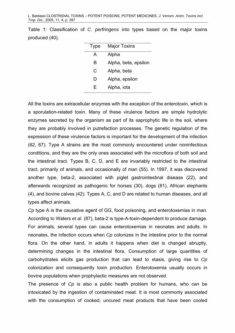
L. Baldassi CLOSTRIDIAL TOXINS – POTENT POISONS, POTENT MEDICINES. J. Venom. Anim. Toxins incl. Trop. Dis., 2005, 11, 4, p. 397 Table 1: Classification of C. perfringens into types based on the major toxins
produced (40).
Type Major Toxins
A Alpha
B Alpha, beta, epsilon
C Alpha, beta
D Alpha, epsilon
E Alpha, iota
All the toxins are extracellular enzymes with the exception of the enterotoxin, which is
a sporulation-related toxin. Many of these virulence factors are simple hydrolytic
enzymes secreted by the organism as part of its saprophytic life in the soil, where
they are probably involved in putrefaction processes. The genetic regulation of the
expression of these virulence factors is important for the development of the infection
(62, 67). Type A strains are the most commonly encountered under noninfectious
conditions, and they are the only ones associated with the microflora of both soil and
the intestinal tract. Types B, C, D, and E are invariably restricted to the intestinal
tract, primarily of animals, and occasionally of man (55). In 1997, it was discovered
another type, beta-2, associated with piglet gastrointestinal disease (22), and
afterwards recognized as pathogenic for horses (30), dogs (81), African elephants
(4), and bovine calves (42). Types A, C, and D are related to human diseases, and all
types affect animals.
Cp type A is the causative agent of GG, food poisoning, and enterotoxemias in man.
According to Waters et al. (87), beta-2 is type-A-toxin-dependent to produce damage.
For animals, several types can cause enterotoxemias in neonates and adults. In
neonates, the infection occurs when Cp colonizes in the intestine prior to the normal
flora. On the other hand, in adults it happens when diet is changed abruptly,
determining changes in the intestinal flora. Consumption of large quantities of
carbohydrates elicits gas production that can lead to stasis, giving rise to Cp
colonization and consequently toxin production. Enterotoxemia usually occurs in
bovine populations when prophylactic measures are not observed.
The presence of Cp is also a public health problem for humans, who can be
intoxicated by the ingestion of contaminated meat. It is most commonly associated
with the consumption of cooked, uncured meat products that have been cooled

L. Baldassi CLOSTRIDIAL TOXINS – POTENT POISONS, POTENT MEDICINES. J. Venom. Anim. Toxins incl. Trop. Dis., 2005, 11, 4, p. 398 slowly or stored under inadequate refrigeration and then consumed without
reheating. The prerequisite for diarrhea can be the consumption of large numbers of
vegetative cells that survive the acidity in the stomach and reach the intestine, where
they sporulate and thereby liberate the enterotoxin, which causes diarrhea.
Necrotizing enteritis has also been observed with sudden dietary changes in
Germany and Papua New Guinea due to type C Cp organisms consumed with the
meat, or with the multiplication of part of the intestinal flora after the ingestion of meat
(protein), producing beta-toxin. Because of dietary problems, low levels of proteases
are not sufficient to destroy the toxin produced (86).
Clostridium difficile
C. difficile is considered commensal of 40-50% of neonate intestines and rarely of
those of adults, where it is associated with antibiotic-induced enterocolitis,
pseudomembranous colitis, and antibiotic-associated colitis (86). Two toxins are
related to this organism, A and B, one is not produced without the other, and there is
a correlation between the amounts of toxin produced (38). Both are lethal for mice,
although type A potency is 50 to 400 times that of type B (36, 59, 64).
Toxin A has enterotoxic activity and is as cytotoxic as type B when tested in an
appropriate cell line. Toxin B is potently cytotoxic, but active just in damaged
intestine, and therefore not considered, by some, to be important in the disease.
When the gut mucosa is compromised, the toxins can disseminate from the intestine
to other target organs (38). The enterotoxic effect is seen when toxin A binds to its
receptor. The toxin is internalized, causing tissue damage and influx of inflammatory
cells. The inflammatory process leads to tissue damage and alteration of membrane
permeability, resulting in the extravasation of serous fluid and hemorrhagic necrosis
(38, 60, 82).
Other virulence factors, such as the motility-altering factor and other enterotoxins,
besides type A toxin, are being studied to confirm their participation in the disease
(38).
Clostridium chauvoei and Clostridium septicum
C. chauvoei and C. septicum are similar and considered, by some, to be members of
the same species (39, 89).
L. Baldassi CLOSTRIDIAL TOXINS – POTENT POISONS, POTENT MEDICINES. J. Venom. Anim. Toxins incl. Trop. Dis., 2005, 11, 4, p. 399 C. chauvoei and C. septicum produce many toxic substances. The number and type
of these vary with the strain, the culture medium, and the length of the incubation
period (47).
C. chauvoei affects only animals, is presumably ingested, and seems to occur
preferentially among the best calves in the herd. It produces four toxins: α-hemolisin,
necrotoxin; β-deoxyribonuclease; γ-hyaluronidase; and δ-hemolysin.
C. septicum produces five toxins: α-lethal, necrotic, hemolytic; β-deoxyribonuclease;
γ-hyaluronidase; δ-O2-labile hemolysin (serologically related to that of C. chauvoei but
not identical); and collagenase (86).
C. septicum was the first reported cause of GG due to war wounds (39). For a long
time it was thought to be the most important agent in human pathology. Nowadays, it
is associated with malignant disease, patient’s intestinal tract probably being the
source of infection (1, 53). It is also considered the agent of neutropenic enterocolitis
(12).
Both bacteria are associated with cattle and sheep GG (29), the former being
responsible for black leg, and the other causing ME (39). Black leg in cattle is a
nontraumatic endogenous infection, and a considerable proportion of bovines may
harbor C. chauvoei in their liver. Affected animals are anorectic, depressed, febrile,
and lame (one side limb), presenting a hot, painful swelling that becomes cold,
edematous with crepitation. Death is seen within 12 to 48 hours. This agent seems to
have a preference for big muscles (thigh, diaphragm, and heart), which at necropsy
are dark red, dry, and spongy (70). In sheep, it is associated with shearing,
castration, and dehorning; the muscles affected are dark, the edema restricted, and
death comes after 12 to 36 hours (29).
ME is characterized by fever and subcutaneous swelling; the infected muscles
become deep red (dark) with bloody gelatinous fluid accumulation in the body
cavities, not noted with C. chauvoei infection. In sheep, in which the disease is
named Braxy, C. septicum invades the mucosa and submucosa of the abomasum,
where it multiplies, producing toxin that leads to death (70, 79).
During the year of 1983, in the southern states of Brazil, there were many bovine
deaths caused by C. septicum. This happened at the same moment as the Ministry of
Agriculture has issued a law obliging the refrigeration of all products of animal origin.
As the commercial establishments were committed to selling Foot and Mouth
L. Baldassi CLOSTRIDIAL TOXINS – POTENT POISONS, POTENT MEDICINES. J. Venom. Anim. Toxins incl. Trop. Dis., 2005, 11, 4, p. 400 Disease vaccines (economically more convenient), the clostridial vaccines were not
available for use, thus the herds, not being protected, manifested the disease.
Clostridium novyi and Clostridium haemolyticum C. novyi, formerly named C. oedematiens, is (together with C. haemolyticum) among
the most O2-sensitive bacteria and fastidious microorganisms for cultivation and
isolation. This may be the reason why it is not so frequently recognized as an agent
of histotoxic human cases (39). It is usually associated with C. sporogenes and
considered by some to be the agent in one-third of the GG cases. It is responsible for
infections in domestic animals, Bacillary Hemoglobinuria (BH) of cattle, and sheep
necrotic hepatitis, liver parasitism being the infection origin (72).
C. haemolyticum is, by DNA studies, closely related to C. novyi type B (48). The
diseases caused by both primarily involve β-toxin of C. haemolyticum and α-toxin of
C. novyi. In human GG, C. novyi α-toxin produces capillary permeability that leads to
massive edema, while β-toxin of C. haemolyticum in BH destroys circulating
erythrocytes, leading to hemoglobin excretion in the urine and blood appearing in the
intestinal lumen due to endothelium capillary destruction, being a highly fatal disease
of cattle (72). These great differences in pathological aspects provide for the
maintenance of the two species.
Nakamura et al. (48) showed a genetic relatedness among C. novyi, C.
haemolyticum and C. botulinum.
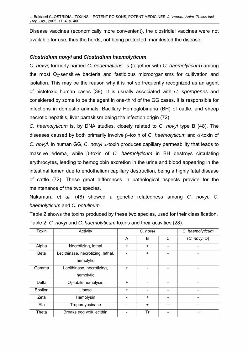
Table 2 shows the toxins produced by these two species, used for their classification.
Table 2: C. novyi and C. haemolyticum toxins and their activities (28). Toxin Activity C. novyi C. haemolyticum
A B C (C. novyi D)
Alpha Necrotizing, lethal + + - -
Beta Lecithinase, necrotizing, lethal,
hemolytic
- + - +
Gamma Lecithinase, necrotizing,
hemolytic
+ - - -
Delta O2-labile hemolysin + - - -
Epsilon Lipase + - - -
Zeta Hemolysin - + - -
Eta Tropomyosinase - + - -
Theta Breaks egg yolk lecithin - Tr - +
L. Baldassi CLOSTRIDIAL TOXINS – POTENT POISONS, POTENT MEDICINES. J. Venom. Anim. Toxins incl. Trop. Dis., 2005, 11, 4, p. 401 Clostridium sordellii and Clostridium bifermentans
C. sordellii was first isolated from a human edematous wound, and from then on it
was implicated in histotoxic diseases in cattle (25, 39). It can be a normal inhabitant,
with other clostridia, in the liver and elsewhere without being the primary causative
agent of the disease (70).
Recently, fatal human cases have been reported due to episiotomy, spontaneous
endometritis with maternal death, and deep laceration of a thigh infection (13, 31, 44,
78).
C. bifermentans is related to C. sordellii but does not have the lethal toxin. C. sordellii
produces four toxins, three in common with C. bifermentans: fibrinolysin, lecithinase,
and O2-labile hemolysin. Lethal toxin, responsible for the severe generalized
gelatinous edema, shock, and sudden death, is also produced. C. bifermentans
lecithinase has approximately 1/50 of the enzymatic and hemolytic activities and
lethality of C. perfringens alpha-toxin (83).
Beta-toxin (C. sordellii) has two activities: dermonecrotic and hemorrhagic, when
tested in the skin of laboratory animals. Dermonecrotic toxin causes massive edema,
small areas of bright-red hemorrhage (subcutaneously or intramuscularly), and death
in 24-36 hours, while the hemorrhagic toxin (intraperitoneally) produces little edema,
but confluent areas of brownish hemorrhage in the skin that can propagate to other
tissues. Lethality is not so common (3, 89).
Clostridium hystolyticum
C. hystolyticum was first described in 1916 in gangrenous and nongangrenous war
wounds but rarely reported, maybe due to its low heat resistance and growth
inhibition by the presence of sugar (50). In GG, its presence is easily identified
clinically because it has seven collagenases, and as collagen is the most abundant
protein in the animal body (constituting connective tissues) (88), there is a tissue
digestion including the soft parts of bones (39, 72).
C. hystolyticum produces five toxins: alpha - (unstable), lethal and necrotizing; beta -
a group of seven collagenases; epsilon - O2-labile hemolysin; gamma - proteinase;
delta - elastase (84).
The histopathologic aspect of lesions can suggest Clostridia species involvement in
the infection (Table 3).
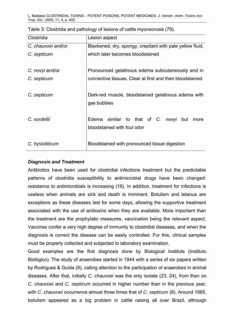
L. Baldassi CLOSTRIDIAL TOXINS – POTENT POISONS, POTENT MEDICINES. J. Venom. Anim. Toxins incl. Trop. Dis., 2005, 11, 4, p. 402 Table 3: Clostridia and pathology of lesions of cattle myonecrosis (79).
Clostridia Lesion aspect
C. chauvoei and/or
C. septicum
Blackened, dry, spongy, crepitant with pale yellow fluid,
which later becomes bloodstained
C. novyi and/or
C. septicum
Pronounced gelatinous edema subcutaneously and in
connective tissues. Clear at first and then bloodstained
C. septicum Dark-red muscle, bloodstained gelatinous edema with
gas bubbles
C. sordellii Edema similar to that of C. novyi but more
bloodstained with foul odor
C. hystoliticum Bloodstained with pronounced tissue digestion
Diagnosis and Treatment
Antibiotics have been used for clostridial infections treatment but the predictable
patterns of clostridia susceptibility to antimicrobial drugs have been changed:
resistance to antimicrobials is increasing (19). In addition, treatment for infections is
useless when animals are sick and death is imminent. Botulism and tetanus are
exceptions as these diseases last for some days, allowing the supportive treatment
associated with the use of antitoxins when they are available. More important than
the treatment are the prophylatic measures, vaccination being the relevant aspect.
Vaccines confer a very high degree of immunity to clostridial diseases, and when the
diagnosis is correct the disease can be easily controlled. For this, clinical samples
must be properly collected and subjected to laboratory examination.
Good examples are the first diagnosis done by Biological Institute (Instituto
Biológico). The study of anaerobes started in 1944 with a series of six papers written
by Rodrigues & Guida (8), calling attention to the participation of anaerobes in animal
diseases. After that, initially C. chauvoei was the only isolate (23, 24), from then on
C. chauvoei and C. septicum occurred in higher number than in the previous year,
with C. chauvoei occurrence almost three times that of C. septicum (8). Around 1985,
botulism appeared as a big problem in cattle raising all over Brazil, although
L. Baldassi CLOSTRIDIAL TOXINS – POTENT POISONS, POTENT MEDICINES. J. Venom. Anim. Toxins incl. Trop. Dis., 2005, 11, 4, p. 403 laboratorial diagnosis showed that other clostridia could be involved with the high
mortality, mainly C. perfringens, C. novyi, and C. haemolyticum (5, 9). From 1986 to
1989, in 517 suspected cases of botulism, toxin was detected in 37, leading to the
supposition of participation of other diseases. During the period from 1986 to 1995
and thereafter, the isolation of C. perfringens surpassed the other two (10, 11, 52).
Laboratory diagnosis allow the isolation of new species or strains that can be studied
and even added to the vaccines, which was achieved in Brazil in 1983, during the
outbreak of GG due to C. septicum.
What must be considered is that clostridia will be present in almost every sample
collected, mainly if it has suffered putrefaction. So the significance of the presence of
any clostridia will depend on the time elapsed between the death and the sample
collection. Once all diseases due to clostridia can be prevented by vaccination, if the
problem continues despite vaccination, the diagnosis must be wrong. So samples
must be collected as soon as possible after death or by sacrifice, selecting
compromised tissues, which must be well-packed, in order to limit deterioration, and
sent quickly to the laboratory.
Moreover, a case description is of fundamental importance to conduct laboratorial
analysis. Diagnosis must be based on the history and clinical data, number of heads
affected, type of feed, range of species involved, and age of diseased animals.
Diagnosis is commonly made by serologic testing, the most commonly used
screening. However, in Brazil, it is almost impossible to get antiserum for diagnosis or
treatment due to importation problems, no availability, and price. Therefore, other
forms of diagnosis, avoiding the use of both antitoxin and especially laboratory
animals, have been tried with success, including esterase electrophoretic
polymorphism for C. perfringens typing (6) and the widely used Polimerase Chain
Reaction (PCR) test (54, 56)
CONCLUSION The participation of clostridia in livestock diseases is well known, and vaccination
against clostridial diseases must be a practice in animal husbandry (86).
Vaccination effectively prevents all clostridial diseases, so any problem that exists
despite vaccination cannot be attributed to clostridia. Nevertheless, diseases caused
by clostridia currently remain a great economical problem in Brazil, being responsible
L. Baldassi CLOSTRIDIAL TOXINS – POTENT POISONS, POTENT MEDICINES. J. Venom. Anim. Toxins incl. Trop. Dis., 2005, 11, 4, p. 404 for great losses for cattle producers, and decreases in meat exports and in protein
availability to the population.
In veterinary practice, diagnosis of clostridiosis is based on history, clinical signs, and
findings in the post-mortem examination. However, laboratory analyses are essential
for confirmation of the presence of toxins, in some cases, toxigenic strains found in
bacterial cultures of recently collected clinical samples are confirmatory proofs, since
antitoxins are not available. In addition, PCR assay is being used to detect if isolated
strains possess genes that codify toxin production.
Economic impact is not usually evaluated in medical studies, but Arnon (2),
describing infant botulism, calls attention to the fact that the average hospital stay is
usually one month, and as there is a need for intensive care, the costs exceed US$
50,000 per case. He also mentions a complicated case of 10 months hospitalization,
which costs US$ 635,000. Beyond medical assistance, legal expenses must be
considered as these can reach 84.4% of the total expenses, what was well
documented by Man et al. (41) on a foodborne outbreak of botulism.
Molecular biology studies are so progressive that, certainly, more factors of
pathogenicity will be discovered as well as other uses for them. One example is
botulinal toxin type A, which, despite being the cause of a wide range of neurologic
disorders, is being used for treatment and relief of several human dystonias; and as
Schantz & Johnson (65) pointed out, in the future, a combination of botulinal toxin
and tetanospasmin will also be used to control neurologic disorders.
Researchers should take in mind Claude Bernard phrase written in 1875 (65):
“Poisons can be employed as a means for the destruction of life or as agents for the
treatment of the sick”, let’s not follow those sick minds that have used them as
biological weapons!
REFERENCES 1 ALPERN RH., DOWELL VR. C. septicum infection and malignancy. J. Am. Med.
Assoc., 1969, 209, 385-8.
2 ARNON SS. Infant botulism. In: ANAEROBE DISCUSSION GROUP
INTERNATIONAL SYMPOSIUM, 6, London, 1989. Proceedings... London:
Churchill College, 1989. p. 41-8.
L. Baldassi CLOSTRIDIAL TOXINS – POTENT POISONS, POTENT MEDICINES. J. Venom. Anim. Toxins incl. Trop. Dis., 2005, 11, 4, p. 405 3 ARSECULERATNE SN., PANABOKKE RG., WIJESUNDRA S. The toxins
responsible for the lesions of C. sordellii gas gangrene. J. Med. Microbiol.,
1969, 2, 237-53.
4 BACCIARNI LN., PAGAN O., FREY J., GRONE A. C. perfringens beta2 toxin in an
African elephant with ulcerative enteritis. Vet. Rec., 2001, 149, 618-20.
5 BALDASSI L. Isolamento de bactérias do gênero Clostridium e detecção de toxina
botulínica a partir de materiais obtidos de bovinos com suspeita clínica de
botulismo. São Paulo: Universidade de São Paulo, Faculdade de Saúde
Pública, 1986. 59p. [Dissertação – Mestrado]
6 BALDASSI L., BARBOSA ML., BACH EE., IARIA ST. Esterase electrophoresis of
Clostridium perfringens bovine strains. J. Venom. Anim. Toxins incl. Trop. Dis.,
2003, 9, 277-90.
7 BALDASSI L., CALIL EMB., PORTUGAL MASC., MOULIN AAP., MOURÃO MAF.
Morte súbita de caprinos por enterotoxemia. Braz. J. Vet. Res. Anim. Sci.,
1995, 32, 109-13.
8 BALDASSI L., HIPOLITO M., CALIL EMB., CHIBA S., MOULIN AAP. Observações
sobre a incidência da Gangrena Gasosa e do Carbúnculo Sintomático durante
10 anos, 1970-79, no estado de São Paulo. O Biológico, 1985, 51, 161-5.
9 BALDASSI L., HIPOLITO M., PORTUGAL MASC., MOULIN AAP., CALIL EMB.
Botulismo bovino: comprovação laboratorial do diagnóstico clínico, período de
1986-1989. Rev. Saúde Públ., 1991, 25, 371-4.
10 BALDASSI L., HIPOLITO M., PORTUGAL MASC., MOULIN AAP., CALIL EMB.,
PIRES DC. A botulism-like disease of catlle in Brazil. In: ANAEROBE
DISCUSSION GROUP INTERNATIONAL SYMPOSIUM, 6, London, 1989.
Proceedings… London: Churchill College, 1990. p.102-3.
11 BALDASSI L., ROJAS MVR., MARICATO JT., ANDRADE KA., RIBEIRO VB.,
CRUZ PR. Botulismo bovino: comprovação laboratorial da suspeita clínica no
período de 1992 a 2001. In: CONGRESSO BRASILEIRO DE MEDICINA
VETERINÁRIA, 29, Gramado, 2002. Anais... Gramado, 2002. In Press.
12 BORRIELLO SP. Newly described clostridial diseases of the gastrointestinal tract:
C. perfringens enterotoxin-associated diarrhea and neutropenic enterocolitis
due to C. septicum. In: BORRIELO SP. Ed. Clostridia in gastrointestinal
disease. Boca Raton: CRC Press, 1985: 223-9.
L. Baldassi CLOSTRIDIAL TOXINS – POTENT POISONS, POTENT MEDICINES. J. Venom. Anim. Toxins incl. Trop. Dis., 2005, 11, 4, p. 406 13 BROWDIE DA., DAVIS JH., KOPLEWITZ MJ., CORDAY L., LEADBETTER AW.
C. sordellii infection. J. Trauma, 1975, 15, 515-8.
14 CALIL EMB., BALDASSI L., PORTUGAL MASC., MACRUZ R., MOULIN AAP.
Tétano em eqüino puro sangue inglês – relato de uma ocorrência. O
Biológico, 1995, 57, 1-4.
15 CASTRO AGM., CARVALHO AM., BALDASSI L., PORTUGAL MASC.,
NARIMATSU MN. Botulismo em aves de postura no estado de São Paulo.
Arq. Inst. Biol., 1988, 55, 1-4.
16 COLLEE JG. Virulence factors of anaerobes: an overview. In: ANAEROBE
DISCUSSION GROUP INTERNATIONAL SYMPOSIUM, 6, London, 1989.
Proceedings… London: Churchill College, 1990. p.127-45.
17 DAUBE G. Clostridium perfringens et pathologies digestives. Ann. Méd. Vét.,
1992, 136, 5-30.
18 FACH P., GIBERT M., GRIFFAIS R., GUILLOU JP., POPOFF MR. PCR and gene
probe identification of botulinum neurotoxin A-, B-, E-, F- and G-producing
Clostridium spp and evaluation in food samples. Appl. Environ. Microbiol.,
1995, 61, 389-92.
19 FINEGOLD SM. Anaerobic infections in human: an overview. Anaerobes, 1995, 1,
3-9.
20 GAMA NMSQ., NAGAMATSU M., BALDASSI L., GAMA JRQ. Botulismo em aves
de postura na região noroeste do estado de São Paulo. Ars. Veterinária, 1992,
8, 58-63.
21 GELLI DS., JAKABI M., SOUZA A. Botulism: a laboratory investigation on
biological and food samples from cases and outbreaks in Brazil (1982-2001).
Rev. Inst. Med. Trop. S. Paulo, 2002, 44, 321-4.
22 GIBERT M., RENAUD CJ., POPOFF MR. Beta-2 toxin, a novel toxin produced by
C. perfringens. Gene, 1997, 203, 65-73.
23 GIORGI W., TROISE C. Prevalência de Salmonelose e Carbúnculo Sintomático
em materiais de bovinos remetidos para exame bacteriológico durante o
qüinqüênio 1963/67 no estado de São Paulo. O Biológico, 1968, 34, 229-31.
24 GIORGI W., TROISE C. Prevalência de Salmonelose e Carbúnculo Sintomático
em materiais de bovinos remetidos para exame bacteriológico durante o ano
de 1968 no estado de São Paulo. O Biológico, 1969, 35, 49-50.
L. Baldassi CLOSTRIDIAL TOXINS – POTENT POISONS, POTENT MEDICINES. J. Venom. Anim. Toxins incl. Trop. Dis., 2005, 11, 4, p. 407 25 HALL IC. The occurrence of Bacillus sordellii in icterohemoglobinuria of cattle in
Nevada. J. Infect Dis., 1929, 45, 156-62.
26 HARIHARAN H., MITCHELL WR. Type C botulism: the agent, host spectrum and
environment. Vet. Bull., 1977, 47, 95-102.
27 HATHEWAY CL. Bacterial sources of clostridial neurotoxins. In: SIMPSON LL.
Ed. Botulinum neurotoxin and tetanus toxin. San Diego: Academic Press,
1989: 3-24.
28 HATHEWAY CL. Toxigenic clostridia. Clin Microbiol. Rev., 1990, 3, 66-98.
29 HELLER HH. A etiology of acute gangrenous infections of animals: a discussion
of black leg, braxy, malignant edema and whale septicemia. J. Infect. Dis.,
1920, 27, 385-451.
30 HERHOLZ C., MISEREZ R., NICOLET J., FREY J., POPOFF MR., GIBERT M.,
GERBER H., STRAUB R. Prevalence of β2-toxigenic Clostridium perfringens
in horses with intestinal disorders. J. Clin. Microbiol., 1999, 37, 358-61.
31 HOGAN SF., IRELAND F. Fatal acute spontaneous endometritis resulting from C.
sordellii. Am. J. Clin. Pathol., 1989, 91, 104-6.
32 HUNTER BF. Ecology of waterfowl botulism toxin production. Trans. N. Am. Wildl.
Conf., 1970, 35, 64-72.
33 JOHNSON EA. Clostridial toxins as therapeutic agents: benefits of nature’s most
toxic proteins. Annu. Rev. Microbiol., 1999, 53, 551-75.
34 KALMBACH ER. Progress in western duck sickness studies. Science, 1932, 75,
57-8.
35 KALMBACH ER. American vultures and the toxin of C. botulinum. J. Am. Vet.
Med. Ass., 1939, 94, 187-91.
36 LIMA AA., LYERLY DM., WILKINS TD., INNES DJ., GUERRANT RL. Effects of C.
difficile toxins A and B in rabbit small and large intestine in vivo and ion
cultured cells in vitro. Infect. Immun., 1988, 56, 582-8.
37 LOBATO FCF., ALMEIDA AC., ABREU VLV., NASCIMENTO RA. Surto de
botulismo em bovinos alimentados com cama de frango no Brasil. Arq. Bras.
Med. Vet. Zootec., 1995, 47, 849-50.
38 LYERLY DM., KRIVAN HC., WILKINS TD. C. difficile: its disease and toxins. Clin.
Microbiol. Rev., 1988, 1, 1-18.
39 MACLENNAN JD. The histotoxic clostridial infections of man. Bacteriol. Rev.,
1962, 26, 177-276.
L. Baldassi CLOSTRIDIAL TOXINS – POTENT POISONS, POTENT MEDICINES. J. Venom. Anim. Toxins incl. Trop. Dis., 2005, 11, 4, p. 408 40 McDONEL JL. Clostridium perfringens toxins (type A, B, C, D, E). Pharmac. Ther.,
1980, 10, 617-55.
41 MANN JM., LATHROP GD., BANNERMAN JA. Economic impact of a botulism
outbreak. JAMA, 1983, 249, 1299-301.
42 MANTECA C., DAUBE G., JAUNIAUX T., LINDEN A., PIRSON V., DETILLEUX
J., GINTER A., COPPE P., KAECKENBEECK A., MAINIL JG. The role of C.
perfringens beta2-toxin in bovine enterotoxemia. Vet. Microbiol., 2002, 86,
191-202.
43 MARGATHO LFF., CARVALHO PR., PARENTE VLC., BALDASSI L. Ocorrência
de tétano em bovinos após castração. O Biológico, 1988, 54, 59-61.
44 MC GREGOR JA., SOPER DE., LOVELL G., TODD JK. Maternal deaths
associated with C. sordellii infection. Obstet. Gynecol., 1989, 161, 987-95.
45 MELLANBY J., GREEN J. How does tetanus toxin acts? Neuroscience, 1981, 6,
281-300.
46 MORTON VL., MEUNIER-POWELL JL. Coinfection with histotoxic and neurotoxic
Clostridia. Clin. Microbiol. Newsletter, 1997, 19, 93-4.
47 MOUSSA RS. Complexity of toxins from C. septicum and C. chauvoei. J.
Bacteriol., 1958, 76, 538-45.
48 NAKAMURA S., KIMURA I., YAMAKAWA K., NISHIDA S. Taxonomic
relationships among C. novyi types A and B, C. haemolyticum and C.
botulinum type C. J. Gen. Microbiol., 1983, 129, 1473-9.
49 NIILO L. C. perfringens in animal disease: a review of current knowledge. Can.
Vet. J., 1980, 21, 141-8.
50 NISHIDA S., IMAIZUMI M. Toxigenicity of C. histolyticum. J. Bacteriol., 1966, 91,
477-83.
51 ORTOLANI EL., BRITO LAB., MORI CS., SCHALCH U., PACHECO J.,
BALDASSI L. Botulism outbreak associated with poultry litter consumption in
three Brazilian cattle herds. Vet. Human Toxicol., 1997, 39, 89-93.
52 OTUKI AK., MEGGIOLARO MN., ROJAS MVR., GONTIJO FA., BALDASSI L.
Diagnóstico laboratorial de clostridoses em bovinos – período de 2002 a 2003.
In: REUNIÃO ANUAL DO INSTITUTO BIOLÓGICO, 17, São Paulo, 2004.
Anais... São Paulo, 2004. In Press.
53 PELFREY TM., TURK RP., PEOPLES JB., ELLIOT HDW. Surgical aspects of C.
septicum septicemia. Arch. Surg., 1984, 119, 546-50.
L. Baldassi CLOSTRIDIAL TOXINS – POTENT POISONS, POTENT MEDICINES. J. Venom. Anim. Toxins incl. Trop. Dis., 2005, 11, 4, p. 409 54 PENHA ML. Detecção dos genes das toxinas alfa, beta e epsilon de Clostridium
perfringens isolados a partir de amostras clínicas de bovinos pela reação em
cadeia da polimerase. São Paulo: Universidade de São Paulo, Faculdade de
Medicina Veterinária e Zootecnia, 2004. 59f. [Dissertação – Mestrado]
55 PETIT L., GILBERT M., POPOFF MR. Clostridium perfringens: toxinotype and
genotype. Trends Microbiol., 1999, 7, 104-10.
56 PIATTI RM., IKUNO AA., BALDASSI L. Detection of bovine Clostridium
perfringens by polymerase chain reaction. J. Venom. Anim. Toxins incl. Trop.
Dis., 2004, 10, 154-60.
57 PINEGAR JA., STRINGER MF. Outbreaks of food poisoning attributed to
lecithinase-negative Clostridium welchii. J. Clin. Pathol., 1977, 30, 491-2.
58 PORTUGAL MASC., BALDASSI L., CALIL EMB. Surto de botulismo em
anatídeos no município de Valinhos, São Paulo. Arq. Inst. Biol., 1995, 62, 45-
52.
59 POTHOULAKIS C., BARONE LM., ELY R., FARIS B., CLARK ME., FRANZBLAU
C., LAMONT JT. Purification and properties of C. difficile cytotoxin B. J. Biol.
Chem., 1986, 261, 1316-21.
60 POTHOULAKIS C., SULLIVAN DA., MELNICK AJ., GADENNE T., MESHULAM
T., LAMONT JT. C. difficile toxins A and B stimulates intracellular calcium
release in human neutrophils. Clin. Res., 1986, 134, 530.
61 ROGICK FA. Porque são os bovinos imunes à toxi-infecção tetânica? Ver. Soc.
Paulista Med. Vet., 1937, 4, 23-5.
62 ROOD JL. Virulence genes of Clostridium perfringens. Annu. Rev. Microbiol.,
1998, 52, 333-60.
63 ROOD JL., COLE ST. Molecular genetics and pathogenesis of Clostridium
perfringens. Microbiol. Rev., 1991, 55, 621-48.
64 ROTHMAN SW., BROWN JE., DIECIDUE A., FORET DA. Differential cytotoxic
effects of toxins A and B isolated from C. difficile. Infect. Immun., 1984, 46,
324-31.
65 SCHANTZ EJ., JOHNSON EA. Properties and use of botulinum toxin and other
microbial neurotoxins in medicine. Microbiol. Rev., 1992, 56, 80-99.
66 SEIFERT HS., BÖHNEL H. Clostridiosen. In: BLOBEL H., SCHLIEBER T.
Handbuch der bakteriellen infektionen bei Tieren. Stuttgart: Gustav Fischer,
1994: 89-153.
L. Baldassi CLOSTRIDIAL TOXINS – POTENT POISONS, POTENT MEDICINES. J. Venom. Anim. Toxins incl. Trop. Dis., 2005, 11, 4, p. 410 67 SHIMIZU T., OKABE A., ROOD JL. Regulation in toxin production in Clostridium
perfringens. In: ROOD JL., MAcCLANE BA., SONGER JE., TITBALL RW.
Eds. The clostridia: molecular biology and pathogenesis. London: Academic
Press, 1997: 451-70.
68 SIGURDARSON S., THORSTEINSSON T. Sudden death of Icelandic dairy cattle.
Vet. Rec., 1990, 127, 410.
69 SILVEIRA D., SOUZA AM., MESQUISTA AJ. Enterotoxemia em bovinos: uma
enfermidade de importância emergente. Bol. Tec. Inf. Rhodia-Mérieux, 1995,
2, 1-4.
70 SIPPEL WL. Diagnosis of Clostridial Diseases. JAVMA, 1982, 161, 1299-1305.
71 SMITH LDS. Botulism: the organism, its toxins, the disease. Springfield: Charles
C. Thomas, 1977. 236p.
72 SMITH LDS., WILLIAMS BL. The pathogenic anaerobic bacteria. 3.ed.
Springfield: Charles C. Thomas, 1984. 550p.
73 SMITH GR. Individual variation in botulism. Br. J. Exp. Path., 1986, 67, 617-21.
74 SMITH GR., TURNER A., TILL D. Factors affecting the toxicity of rotting
carcasses containing C. botulinum type E. Epidem. Inf., 1988, 100, 399-405.
75 SOLOMON HM, KAUTTER DA. Growth and toxin production by C. botulinum in
sauteed onions. J. Food Prot., 1986, 49, 618-20.
76 SOLOMON HM., KAUTTER DA. Outgrowth and toxin production by C. botulinum
in bottled chopped garlic. J. Food Prot., 1988, 51, 862-5.
77 SONGER JG. Clostridial diseases of domestic animals. Clin. Microbiol. Rev.,
1996, 9, 216-34.
78 SOPER DD. Clostridial myonecrosis arising from an episiotomy. Obstet. Gynecol.,
1986, 68, 265-85.
79 STERNE M. Clostridial infections. Brit. Vet. J., 1981, 137, 443-54.
80 ST. LOUIS ME., PECK SHS., BOWERING D., MORGAN GB., BLATHERWICK J.,
BANERJEE S., KETTYLS GDM., BLACK WA., MILLING ME., HAUSCHILD
AHW., TAUXE RV., BLAKE PA. Botulism from chopped garlic: delayed
recognition of a major outbreak. Annals of Internal Medicine, 1988, 108, 363-8.
81 THIEDE S., GOETHE R., AMTSBERG G. Prevalence of beta2-toxin gene in C.
perfringens type A from diarrheic dogs. Vet. Rec., 2001, 149, 273-4.
L. Baldassi CLOSTRIDIAL TOXINS – POTENT POISONS, POTENT MEDICINES. J. Venom. Anim. Toxins incl. Trop. Dis., 2005, 11, 4, p. 411 82 TRIADAFILOPOULOS G., POTHOULAKIS C., O’BRIEN MJ., LAMONT JT.
Differential effects of C. difficile toxins A and B on rabbit ileum.
Gastroenterology, 1987, 93, 273-9.
83 TSO JY., SEIBEL D. Cloning and expression of the phospholipase C gene from C.
perfringens and C. bifermentans. Infect. Immun., 1989, 57, 468-76.
84 VAN MART HE., STEINBRINK DR. Complementary substrate specificities of
class I and class II collagenases from C. hystoliticum. Biochemistry, 1985, 24,
2520-6.
85 VERONESI S., FOCACCIA R. The clinical picture. In: VERONESI R. Ed. Tetanus:
important new concepts. Amsterdam: Excerpta Medica, 1981: 183-206.
86 WALKER PD., BATTY I., FERNIE DS. Clostridial toxins and enteric diseases. Eur.
J. Chemo. Antib., 1982, 2, 107-14.
87 WATERS M., SAVOIE A., GARMORY HS., BUESCHEL D., POPOFF MR.,
SONGER JG., TITBALL RW., MCCLANE BA., SARKER MR. Genotyping and
phenotyping of beta-2 toxigenic C. perfringens fecal isolates associated with
gastrointestinal diseases in piglets. J. Clin. Microbiol., 2003, 41, 3584-91.
88 WHITE A., HANDLER P., SMITH EL., HILL RL., LEHMAN IR. Principles of
biochemistry. 6.ed. New York: McGraw Hill, 1978. 1492p.
89 WILLIS AT. Clostridia of wound infection. London: Butterworths, 1969. 156p.