clusters of circulating tumor cells traverse capillary ... · clusters of circulating tumor cells...
TRANSCRIPT
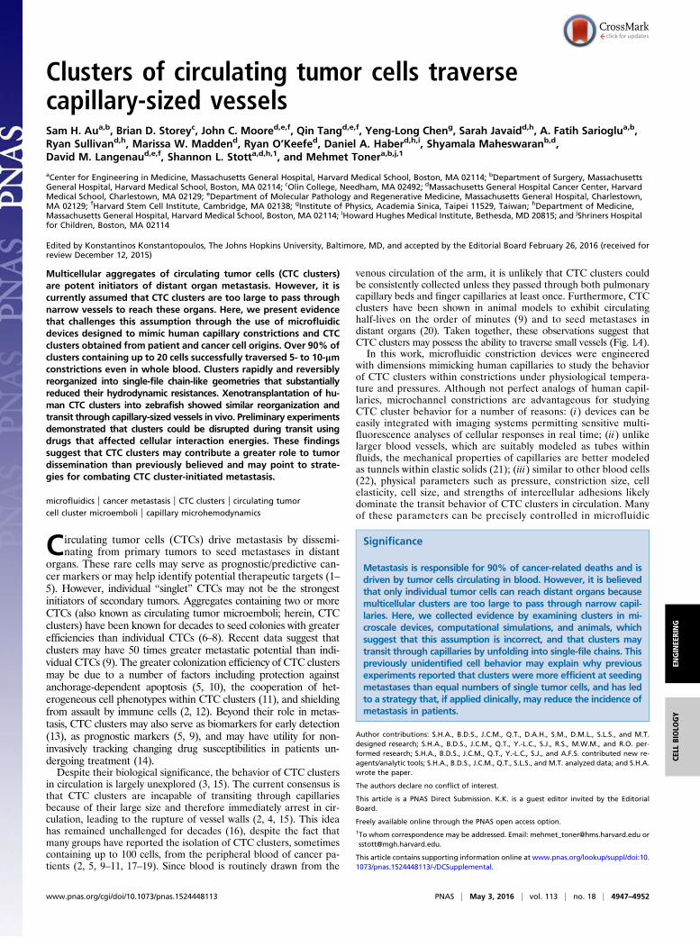
Clusters of circulating tumor cells traversecapillary-sized vesselsSam H. Aua,b, Brian D. Storeyc, John C. Moored,e,f, Qin Tangd,e,f, Yeng-Long Cheng, Sarah Javaidd,h, A. Fatih Sarioglua,b,Ryan Sullivand,h, Marissa W. Maddend, Ryan O’Keefed, Daniel A. Haberd,h,i, Shyamala Maheswaranb,d,David M. Langenaud,e,f, Shannon L. Stotta,d,h,1, and Mehmet Tonera,b,j,1
aCenter for Engineering in Medicine, Massachusetts General Hospital, Harvard Medical School, Boston, MA 02114; bDepartment of Surgery, MassachusettsGeneral Hospital, Harvard Medical School, Boston, MA 02114; cOlin College, Needham, MA 02492; dMassachusetts General Hospital Cancer Center, HarvardMedical School, Charlestown, MA 02129; eDepartment of Molecular Pathology and Regenerative Medicine, Massachusetts General Hospital, Charlestown,MA 02129; fHarvard Stem Cell Institute, Cambridge, MA 02138; gInstitute of Physics, Academia Sinica, Taipei 11529, Taiwan; hDepartment of Medicine,Massachusetts General Hospital, Harvard Medical School, Boston, MA 02114; iHoward Hughes Medical Institute, Bethesda, MD 20815; and jShriners Hospitalfor Children, Boston, MA 02114
Edited by Konstantinos Konstantopoulos, The Johns Hopkins University, Baltimore, MD, and accepted by the Editorial Board February 26, 2016 (received forreview December 12, 2015)
Multicellular aggregates of circulating tumor cells (CTC clusters)are potent initiators of distant organ metastasis. However, it iscurrently assumed that CTC clusters are too large to pass throughnarrow vessels to reach these organs. Here, we present evidencethat challenges this assumption through the use of microfluidicdevices designed to mimic human capillary constrictions and CTCclusters obtained from patient and cancer cell origins. Over 90% ofclusters containing up to 20 cells successfully traversed 5- to 10-μmconstrictions even in whole blood. Clusters rapidly and reversiblyreorganized into single-file chain-like geometries that substantiallyreduced their hydrodynamic resistances. Xenotransplantation of hu-man CTC clusters into zebrafish showed similar reorganization andtransit through capillary-sized vessels in vivo. Preliminary experimentsdemonstrated that clusters could be disrupted during transit usingdrugs that affected cellular interaction energies. These findingssuggest that CTC clusters may contribute a greater role to tumordissemination than previously believed and may point to strate-gies for combating CTC cluster-initiated metastasis.
microfluidics | cancer metastasis | CTC clusters | circulating tumorcell cluster microemboli | capillary microhemodynamics
Circulating tumor cells (CTCs) drive metastasis by dissemi-nating from primary tumors to seed metastases in distant
organs. These rare cells may serve as prognostic/predictive can-cer markers or may help identify potential therapeutic targets (1–5). However, individual “singlet” CTCs may not be the strongestinitiators of secondary tumors. Aggregates containing two or moreCTCs (also known as circulating tumor microemboli; herein, CTCclusters) have been known for decades to seed colonies with greaterefficiencies than individual CTCs (6–8). Recent data suggest thatclusters may have 50 times greater metastatic potential than indi-vidual CTCs (9). The greater colonization efficiency of CTC clustersmay be due to a number of factors including protection againstanchorage-dependent apoptosis (5, 10), the cooperation of het-erogeneous cell phenotypes within CTC clusters (11), and shieldingfrom assault by immune cells (2, 12). Beyond their role in metas-tasis, CTC clusters may also serve as biomarkers for early detection(13), as prognostic markers (5, 9), and may have utility for non-invasively tracking changing drug susceptibilities in patients un-dergoing treatment (14).
Despite their biological significance, the behavior of CTC clustersin circulation is largely unexplored (3, 15). The current consensus isthat CTC clusters are incapable of transiting through capillariesbecause of their large size and therefore immediately arrest in cir-culation, leading to the rupture of vessel walls (2, 4, 15). This ideahas remained unchallenged for decades (16), despite the fact thatmany groups have reported the isolation of CTC clusters, sometimescontaining up to 100 cells, from the peripheral blood of cancer pa-tients (2, 5, 9–11, 17–19). Since blood is routinely drawn from the
venous circulation of the arm, it is unlikely that CTC clusters couldbe consistently collected unless they passed through both pulmonarycapillary beds and finger capillaries at least once. Furthermore, CTCclusters have been shown in animal models to exhibit circulatinghalf-lives on the order of minutes (9) and to seed metastases indistant organs (20). Taken together, these observations suggest thatCTC clusters may possess the ability to traverse small vessels (Fig. 1A).
In this work, microfluidic constriction devices were engineeredwith dimensions mimicking human capillaries to study the behaviorof CTC clusters within constrictions under physiological tempera-ture and pressures. Although not perfect analogs of human capil-laries, microchannel constrictions are advantageous for studyingCTC cluster behavior for a number of reasons: (i) devices can beeasily integrated with imaging systems permitting sensitive multi-fluorescence analyses of cellular responses in real time; (ii) unlikelarger blood vessels, which are suitably modeled as tubes withinfluids, the mechanical properties of capillaries are better modeledas tunnels within elastic solids (21); (iii) similar to other blood cells(22), physical parameters such as pressure, constriction size, cellelasticity, cell size, and strengths of intercellular adhesions likelydominate the transit behavior of CTC clusters in circulation. Manyof these parameters can be precisely controlled in microfluidic
Significance
Metastasis is responsible for 90% of cancer-related deaths and isdriven by tumor cells circulating in blood. However, it is believedthat only individual tumor cells can reach distant organs becausemulticellular clusters are too large to pass through narrow capil-laries. Here, we collected evidence by examining clusters in mi-croscale devices, computational simulations, and animals, whichsuggest that this assumption is incorrect, and that clusters maytransit through capillaries by unfolding into single-file chains. Thispreviously unidentified cell behavior may explain why previousexperiments reported that clusters were more efficient at seedingmetastases than equal numbers of single tumor cells, and has ledto a strategy that, if applied clinically, may reduce the incidence ofmetastasis in patients.
Author contributions: S.H.A., B.D.S., J.C.M., Q.T., D.A.H., S.M., D.M.L., S.L.S., and M.T.designed research; S.H.A., B.D.S., J.C.M., Q.T., Y.-L.C., S.J., R.S., M.W.M., and R.O. per-formed research; S.H.A., B.D.S., J.C.M., Q.T., Y.-L.C., S.J., and A.F.S. contributed new re-agents/analytic tools; S.H.A., B.D.S., J.C.M., Q.T., S.L.S., and M.T. analyzed data; and S.H.A.wrote the paper.
The authors declare no conflict of interest.
This article is a PNAS Direct Submission. K.K. is a guest editor invited by the EditorialBoard.
Freely available online through the PNAS open access option.1To whom correspondence may be addressed. Email: [email protected] [email protected].
This article contains supporting information online at www.pnas.org/lookup/suppl/doi:10.1073/pnas.1524448113/-/DCSupplemental.
www.pnas.org/cgi/doi/10.1073/pnas.1524448113 PNAS | May 3, 2016 | vol. 113 | no. 18 | 4947–4952
ENGINEE
RING
CELL
BIOLO
GY
systems, and (iv) microfluidic constrictions of this sort have pre-viously demonstrated utility in exploring various biophysical cellularproperties such as the viscoelastic properties of cancer cells (23) andneutrophils (24), examining nuclear deformability during migration(25, 26) and for phenotypic discrimination (27). Although clustershave been demonstrated to transit through 50- to 300-μm micro-channels (28), capillary-sized microfluidic constrictions have yetto be studied. Observations of CTC clusters in microchannel con-strictions in conjunction and in vivo models organisms reveal howCTC clusters dynamically reorganize to pass through narrow bloodvessels.
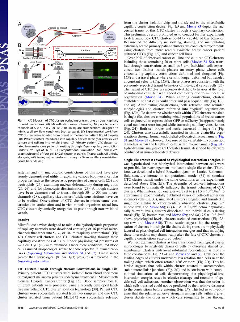
ResultsMicrofluidic devices designed to mimic the hydrodynamic propertiesof capillary networks were developed consisting of 16 parallel micro-channels that taper into 5-, 7-, or 10-μm “capillary constrictions” (Fig.1B). Cancer cell clusters and CTC clusters traveling through thesecapillary constrictions at 37 °C under physiological pressures of7–33 cm H2O (29) were examined. Under these conditions, red bloodcells assumed morphologies similar to those reported in human capil-laries (Supporting Information and Movies S1 and S2). Transit undergreater than physiological (83 cm H2O) pressures is presented in theSupporting Information.
CTC Clusters Transit Through Narrow Constrictions in Single File.Primary patient CTC clusters were isolated from blood specimensof malignant melanoma patients undergoing treatment at MassachusettsGeneral Hospital Cancer Center (Fig. 1C). Blood samples from 10different patients were processed using a recently developed label-free microfluidic CTC cluster isolation technology (30). Patient CTCclusters were successfully isolated from two samples, and one CTCcluster isolated from patient MEL-142 was successfully released
from the cluster isolation chip and transferred to the microfluidiccapillary constriction device. Fig. 1D and Movie S3 depict the suc-cessful transit of this CTC cluster through a capillary constriction.This preliminary result prompted us to conduct further experimentsto determine how CTC clusters could be capable of this behavior.Because of the difficulty in isolating, staining, and manipulatingextremely scarce primary patient clusters, we conducted experimentsusing clusters from more readily available breast cancer patientcultured CTCs (Fig. 1C) and cancer cell lines.
Over 90% of observed cancer cell line and cultured CTC clusters,including those containing 20 or more cells (Movies S4–S6), trans-ited through constrictions as small as 5 μm. Individual cells experi-enced two distinct transit phases: an entry phase where cellsencountering capillary constrictions deformed and elongated (Fig.1Eii) and a travel phase where cells no longer deformed but traveledat constant velocity (Fig. 1Eiii). These phases are consistent with thepreviously reported transit behaviors of individual cancer cells (23).The transit of CTC clusters incorporated these behaviors at the levelof individual cells, but with added complexity due to multicellularorganization (Movie S4). When entering constrictions, clusters“unfolded” so that cells could enter and pass sequentially (Fig. 1E iiand iii). After exiting constrictions, cells retracted into roundedmorphologies and clusters reformed into “typical” organizations(Fig. 1Eiv). To determine whether cells within CTC clusters transitedin single file, clusters containing mixed populations of breast cancercells engineered to express either GFP or mCherry (in approximatelyequal numbers) were imaged while traversing capillary constrictions(Fig. 2A). Both cell bodies and nuclei traversed in single file (Fig.2A). Clusters also successfully transited in similar chain-like orga-nizations through human endothelial cell-coated microchannels (Fig.S1 and Movie S7). However, because of the variability in constrictiondiameters across the lengths of cellularized microchannels (Fig. S1),hydrodynamic analyses of CTC cluster transit, described below, wereconducted in non–cell-coated devices.
Single-File Transit Is Favored at Physiological Interaction Energies. Itwas hypothesized that biophysical interactions between cells wereresponsible for rearrangement into stable single-file chains. There-fore, we developed a hybrid Brownian dynamics–Lattice Boltzmannfluid–structure interaction computational model (31) to simulateCTC cluster transit under the same conditions as the experimentsdescribed above (Fig. 2B). The strengths of cell–cell interactionswere found to dramatically influence the transit behaviors of CTCclusters. When interaction energies were set to (i) 1.5 × 10⁻³ J/m2
, toapproximate experimentally published values of cell–cell adhesionsin cancer cells (32, 33), simulated clusters elongated and transited insingle file similar to experimentally observed clusters (Fig. 2B,middle row, and Movie S8); (ii) 6.0 × 10⁻⁴ J/m2, below physiologi-cally relevant levels, clusters dissociated into individual cells duringtransit (Fig. 2B, bottom row, and Movie S9); and (iii) 7.5 × 10⁻³ J/m2,above physiological levels, clusters occluded constrictions (Fig. 2B,top row, and Movie S10). These results suggest that the reorgani-zation of clusters into single-file chains during transit is biophysicallyfavored at physiological cell interaction energies and that modifyingthese interactions may dramatically alter the fate of CTC clusters atcapillary constrictions (explored below).
We next examined clusters as they transitioned from typical clustermorphologies to single-file chains of cells by observing stained cellmembranes. Clusters underwent substantial reorientation as they en-tered constrictions (Fig. 2 C–F and Movies S5 and S6). Cells near theleading edges of clusters underwent less rotation than cells near thetrailing edges, which often rotated 180° or more (Fig. 2D). This be-havior suggests that cells within clusters rotated to accommodatestable intercellular junctions (Fig. 2C) and is consistent with compu-tational simulations of cells demonstrating that physiological-levelinteraction energies result in selective cleavage and retention of spe-cific cell–cell adhesions. Another observation was that the order inwhich cells transited could not be predicted by their relative distancesto the constrictions before entering (Fig. 2F). This led us to hypoth-esize that the relative adhesive strengths among cells within a givencluster dictate the order in which cells reorganize to pass through
Flow
Capillary Constrictions
5x5, 7x7, 10x10 μm 50 μm
Inlet OutletCapillary
ΔP = 5-58 mm Hg
oC
i ii iii iv
A
B
C
InvestigateTrafficking
Liquid Biopsy
Spike Into Whole Blood
PrimaryPatient
CulturedClusters
Culture
Isolate CTC-clusters
Capillary
Occlude or Transit
DistantOrgan
Metastasis
PrimaryTumor
CTC-Cluster
NG2/CD146
CD45DNA
D
E
Fig. 1. (A) Diagram of CTC clusters occluding or transiting through capillaryto seed metastases. (B) Microfluidic device schematic, 16 parallel micro-channels of 5 × 5, 7 × 7, or 10 × 10-μm square cross-sections, designed tomimic capillary flow conditions (not to scale). (C) Experimental workflow:CTC clusters were isolated from breast or melanoma patient liquid biopsies(30). Patient clusters introduced into capillary devices directly or after ex vivoculture and spiking into whole blood. (D) Primary patient CTC cluster iso-lated from melanoma patient transiting through 10-μm capillary constrictionunder 7 cm H2O at 37 °C. (E) Computational simulation (Top) and micro-graphs (Bottom) of four-cell LNCaP cluster in transit: (i) approach, (ii) unfold/elongate, (iii) travel, (iv) exit/reform through a 5-μm capillary constriction.(Scale bars: 50 μm.)
4948 | www.pnas.org/cgi/doi/10.1073/pnas.1524448113 Au et al.

(Fig. 2E). If cells must break intercellular adhesions with all cells ex-cept two adjacent neighbors, it is likely only the strongest cell–celladhesions are retained.
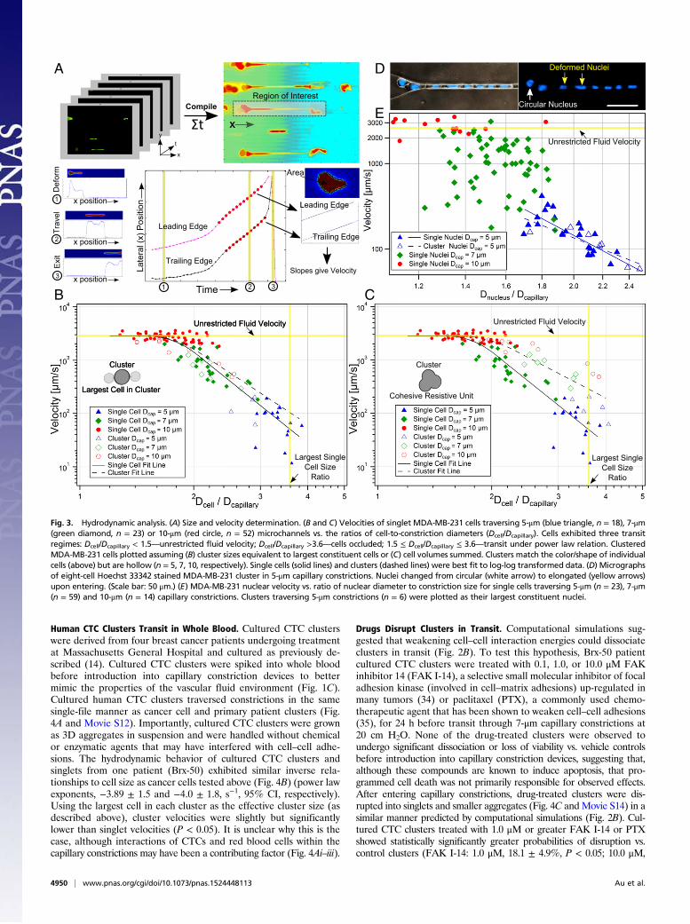
Cluster Reorganization Permits Transit by Reducing Resistance. In-spection of CTC clusters revealed qualitative characteristics ofcell reorganization during transit (above). However, the key tounderstanding the mechanisms that dominate cluster transit is toevaluate transit hydrodynamics. We therefore compared the transitbehaviors of singlet CTCs and CTC clusters traversing 5-, 7-, and10-μm capillary constrictions at 37 °C under 7- to 80-cm H2O appliedpressures. Cell diameter(s) and transit velocities of single cell andcluster events were computed using custom Matlab scripts (Fig. 3Aand Movie S11).
First, the velocities of singlet MDA-MB-231-LM2 cells (solidsymbols) transiting under 33-cm H2O pressures through 5-μm (n =18), 7-μm (n = 23), and 10-μm (n = 52) constrictions were plotted vs.the ratio of cells to constriction sizes (Fig. 3 B and C). Cell velocitiesranged from 5 μm/s to unrestricted fluid velocities of ∼2,850 μm/s,
which encompassed the physiological capillary velocity of ∼200 μm/s.Singlets exhibited three distinct transit regimes dictated by relativesize of the cell to the constriction (Fig. 3 B and C, solid symbols).(i) Small cells (Dcell/Dcapillary < 1.5) transited at unrestricted fluidvelocities. (ii) Large cells (Dcell/Dcapillary > 3.6) occluded capillaryconstrictions (the mean ratio for occluding cells was 3.8 ± 0.5 [95%confidence interval (CI)]; occlusions occurred almost exclusivelyin 5-μm constrictions) and were not included in this analysis.(iii) Moderately sized cells (1.5 ≤Dcell/Dcapillary ≤ 3.6) transited accordingto a power law relation (23) with a power law exponent of −5.7 ± 0.6(95% CI). This is consistent with theoretical scaling analyses (Sup-porting Information) and previously reported values, which suggest apower law exponent of approximately −5 (23). The strong powerrelation between size and velocity means that small changes in in-dividual cell sizes dramatically affect hydrodynamic resistance (Fig. 2A).
The transit behavior of clusters was then investigated by over-laying cluster data points onto singlet data. The effective hydrody-namic diameters of clusters were calculated in two manners forcomparison. When effective cluster diameters were set equal tospheres of equivalent volumes as all constituent cells, cluster veloc-ities deviated significantly from singlet velocities (Fig. 3C) (powerlaw exponents of −3.3 ± 0.9 vs. −5.7 ± 0.6, 95% CI), indicating thatclusters did not act as cohesive resistive units. However, when clusterdiameters were assumed to equal the diameters of their largestconstituent cells, the transit of CTC clusters was statistically in-distinguishably from singlet counterparts (Fig. 3B) (power law ex-ponents, −4.7 ± 1.2 vs. −5.7 ± 0.6, 95% CI). This agreement suggeststhat the resistances of examined clusters were dominated by thelargest cell within each cluster; a result consistent with the singlet cellfindings that transit velocities depended strongly on size (above). Itshould be noted that this approximation was accurate for clusterscontaining relatively few cells (less than five in most cases). In thegeneral case, the behavior of clusters in constrictions was betterapproximated as the sum of the resistances of each individual cell inthe cluster (Supporting Information and Fig. S2). Detailed hydrody-namic analyses of CTC cluster transit using an expanded dataset withtransit scaling analyses are included in the Supporting Informationand Figs. S2–S4. Clusters that passed through constrictions wereobserved to return to spherical morphologies and reassemble intotypical “cluster” morphologies within seconds (Supporting Informa-tion, Fig. S5, and Movie S12). Transited clusters also remained viableafter exiting constrictions (Fig. S5) and proliferated at rates indis-tinguishable from controls (Fig. S6). Theoretical cell and clusterresistances derived from hydrodynamic analyses are presented forcomparison in Fig. S7.
Cell Nuclei Contribute Resistance in Very Narrow Vessels. To in-vestigate the role of nuclei in cluster transit, the sizes and velocitiesof nuclei within singlets and clusters were analyzed as describedabove. Nuclei deformed from rounded to elongated ellipsoidalmorphologies upon entering constrictions (Fig. 3D and Movie S13).The dependence of nuclear size on transit behavior is plotted in Fig.3E. In 10-μm channels (n = 14), cell nuclei traversed capillaries atunrestricted fluid velocities similar to whole-cell studies (Fig. 3 B andC). In 7-μm channels (n = 29), nuclear diameters were not correlatedwith cell velocities, suggesting that nuclei were not large enough tooffer significant resistance. However, in 5-μm channels, nuclei inboth singlets/clusters (n = 35/13, respectively) were inversely corre-lated with cell velocities and were statistically indistinguishable fromone another (power law exponents, −4.3 ± 1.6 vs. −3.2 ± 2.2, re-spectively, 95% CI).
Interestingly, the diameter-to-constriction ratios that dictatedtransit regimes for whole cells (Fig. 3 B and C) (above) alsoappeared to be valid for nuclei-to-constriction ratios. Nuclei tra-versing 5-μm constrictions had diameter ratios within the power lawregime calculated for whole cells (1.7 ≤ Dnucleus/Dcapillary ≤ 2.5) butbelow the whole-cell power law regime for most cells in 7-μm con-strictions (1.2 ≤ Dnucleus/Dcapillary ≤ 1.8) and all nuclei in 10-μmconstrictions (0.9 ≤ Dnucleus/Dcapillary ≤ 1.3). These findings suggestthat nuclei may play a role in the transit of CTCs and CTC clusters innarrow (∼5 μm) but not larger capillaries.
Strong Adhesions
Cohesive Resistive Unit
Single File Chain
126
5 34
1265
34
1265 3
4
12653 4
1265
34
i ii iii
iv v vi
2653 4
Cluster
Individual Cells
Strong Adhesions
Weak Adhesions
Moderate Adhesions
Midline
Stretch
Midline
Cohesive Resistive Unit
UnfoldModerateAdhesions
Midline
Distance to Constriction Dominated
Cell AdhesionStrength
Dominated
12
312
3123 Equal Adhesive
Strengths12
3
12
312 3
Stronger AdhesionWeaker Adhesion1
2
31
2
3
1.5 x 10 -4 J/m 2
15.0 x 10 -4 J/m 2
i ii iii
iv v vi
Interaction Strengths [J/m2]
75.0 x 10 -4 J/m 2
A
B
C
D
E
F
Fig. 2. Cluster organization. (A) Micrographs of three-cell (left) and eight-cell (right) MDA-MB-231 GFP or mCherry tagged cells in clusters stained withHoechst 33342 transiting through 5-μm capillary constriction. (B) Conceptualcluster behavior (left) and computational transit simulations (right). Strongintercellular adhesions (75.0 × 10⁻⁴ J/m2)—cluster occludes; moderate ad-hesions (15.0 × 10⁻⁴ J/m2)—single-file transit; weak adhesions (1.5 × 10⁻⁴ J/m2)—dissociation. (C) Conceptual responses: strong adhesions—minimal cell rotation;moderate adhesions—unfolding and cellular rotation. Greens arrows indicateleading edges of cells in the flow direction in frame i. (D) Time-lapse images ofsix-cell LNCaP cluster membrane stained with CellMask DeepRed. (E) Concep-tual transit depends on distance to constriction (equal strength adhesions) orsomewhat independent (heterogeneous adhesions). (F) Time-lapse images ofsix-cell LNCaP cluster demonstrating transit order. Cells numbered by distanceto constriction in frame i. Experiments were conducted under 33 cm H2O at37 °C. (Scale bars: 50 μm.)
Au et al. PNAS | May 3, 2016 | vol. 113 | no. 18 | 4949
ENGINEE
RING
CELL
BIOLO
GY
Human CTC Clusters Transit in Whole Blood. Cultured CTC clusterswere derived from four breast cancer patients undergoing treatmentat Massachusetts General Hospital and cultured as previously de-scribed (14). Cultured CTC clusters were spiked into whole bloodbefore introduction into capillary constriction devices to bettermimic the properties of the vascular fluid environment (Fig. 1C).Cultured human CTC clusters traversed constrictions in the samesingle-file manner as cancer cell and primary patient clusters (Fig.4A and Movie S12). Importantly, cultured CTC clusters were grownas 3D aggregates in suspension and were handled without chemicalor enzymatic agents that may have interfered with cell–cell adhe-sions. The hydrodynamic behavior of cultured CTC clusters andsinglets from one patient (Brx-50) exhibited similar inverse rela-tionships to cell size as cancer cells tested above (Fig. 4B) (power lawexponents, −3.89 ± 1.5 and −4.0 ± 1.8, s−1, 95% CI, respectively).Using the largest cell in each cluster as the effective cluster size (asdescribed above), cluster velocities were slightly but significantlylower than singlet velocities (P < 0.05). It is unclear why this is thecase, although interactions of CTCs and red blood cells within thecapillary constrictions may have been a contributing factor (Fig. 4Ai–iii).
Drugs Disrupt Clusters in Transit. Computational simulations sug-gested that weakening cell–cell interaction energies could dissociateclusters in transit (Fig. 2B). To test this hypothesis, Brx-50 patientcultured CTC clusters were treated with 0.1, 1.0, or 10.0 μM FAKinhibitor 14 (FAK I-14), a selective small molecular inhibitor of focaladhesion kinase (involved in cell–matrix adhesions) up-regulated inmany tumors (34) or paclitaxel (PTX), a commonly used chemo-therapeutic agent that has been shown to weaken cell–cell adhesions(35), for 24 h before transit through 7-μm capillary constrictions at20 cm H2O. None of the drug-treated clusters were observed toundergo significant dissociation or loss of viability vs. vehicle controlsbefore introduction into capillary constriction devices, suggesting that,although these compounds are known to induce apoptosis, that pro-grammed cell death was not primarily responsible for observed effects.After entering capillary constrictions, drug-treated clusters were dis-rupted into singlets and smaller aggregates (Fig. 4C and Movie S14) in asimilar manner predicted by computational simulations (Fig. 2B). Cul-tured CTC clusters treated with 1.0 μM or greater FAK I-14 or PTXshowed statistically significantly greater probabilities of disruption vs.control clusters (FAK I-14: 1.0 μM, 18.1 ± 4.9%, P < 0.05; 10.0 μM,
Circular Nucleus
Deformed Nuclei
Unrestricted Fluid Velocity
Largest Cell in Cluster
Cluster
Unrestricted Fluid Velocity
Largest Single Cell Size
Ratio
B
CompileRegion of Interest
Leading Edge
Trailing Edge
Area
Leading Edge
Trailing Edge
Slopes give VelocityLate
ral (
x) P
ositi
on
Time
Def
orm
Trav
elE
xit
x
x position
Σt
A
y
x
t
1
2
3321
D
E
Cohesive Resistive Unit
Cluster
Unrestricted Fluid Velocity
Largest Single Cell Size
Ratio
C
x position
x position
Fig. 3. Hydrodynamic analysis. (A) Size and velocity determination. (B and C) Velocities of singlet MDA-MB-231 cells traversing 5-μm (blue triangle, n = 18), 7-μm(green diamond, n = 23) or 10-μm (red circle, n = 52) microchannels vs. the ratios of cell-to-constriction diameters (Dcell/Dcapillary). Cells exhibited three transitregimes: Dcell/Dcapillary < 1.5—unrestricted fluid velocity; Dcell/Dcapillary >3.6—cells occluded; 1.5 ≤ Dcell/Dcapillary ≤ 3.6—transit under power law relation. ClusteredMDA-MB-231 cells plotted assuming (B) cluster sizes equivalent to largest constituent cells or (C) cell volumes summed. Clusters match the color/shape of individualcells (above) but are hollow (n = 5, 7, 10, respectively). Single cells (solid lines) and clusters (dashed lines) were best fit to log-log transformed data. (D) Micrographsof eight-cell Hoechst 33342 stained MDA-MB-231 cluster in 5-μm capillary constrictions. Nuclei changed from circular (white arrow) to elongated (yellow arrows)upon entering. (Scale bar: 50 μm.) (E) MDA-MB-231 nuclear velocity vs. ratio of nuclear diameter to constriction size for single cells traversing 5-μm (n = 23), 7-μm(n = 59) and 10-μm (n = 14) capillary constrictions. Clusters traversing 5-μm constrictions (n = 6) were plotted as their largest constituent nuclei.
4950 | www.pnas.org/cgi/doi/10.1073/pnas.1524448113 Au et al.
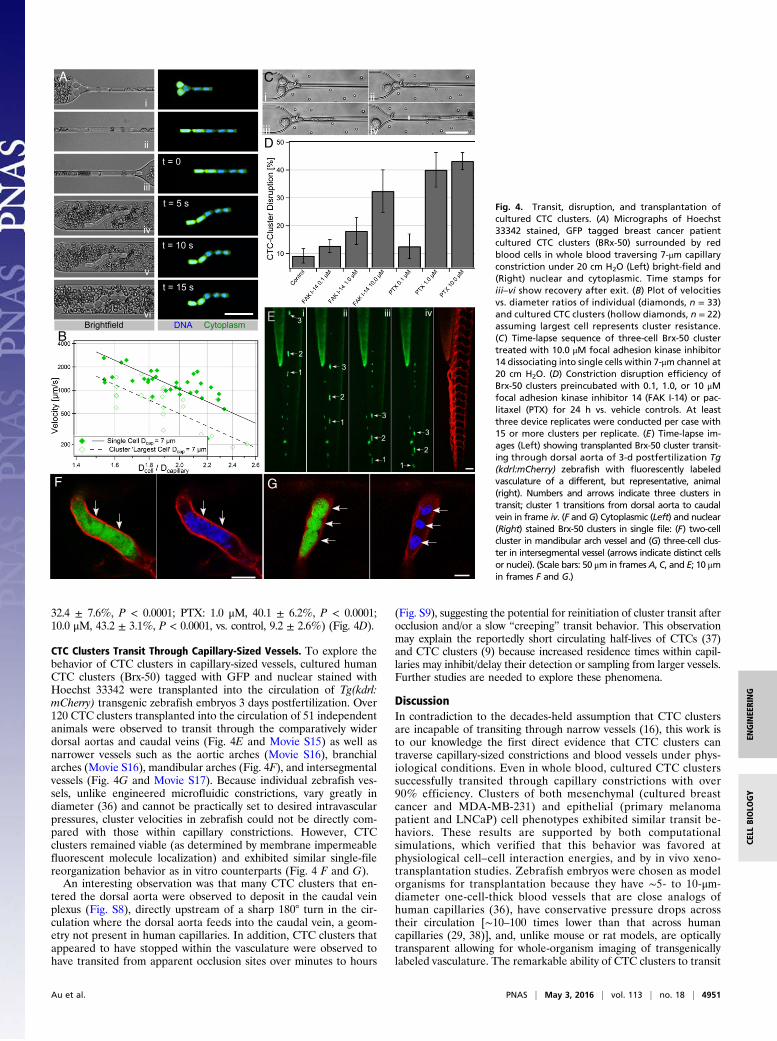
32.4 ± 7.6%, P < 0.0001; PTX: 1.0 μM, 40.1 ± 6.2%, P < 0.0001;10.0 μM, 43.2 ± 3.1%, P < 0.0001, vs. control, 9.2 ± 2.6%) (Fig. 4D).
CTC Clusters Transit Through Capillary-Sized Vessels. To explore thebehavior of CTC clusters in capillary-sized vessels, cultured humanCTC clusters (Brx-50) tagged with GFP and nuclear stained withHoechst 33342 were transplanted into the circulation of Tg(kdrl:mCherry) transgenic zebrafish embryos 3 days postfertilization. Over120 CTC clusters transplanted into the circulation of 51 independentanimals were observed to transit through the comparatively widerdorsal aortas and caudal veins (Fig. 4E and Movie S15) as well asnarrower vessels such as the aortic arches (Movie S16), branchialarches (Movie S16), mandibular arches (Fig. 4F), and intersegmentalvessels (Fig. 4G and Movie S17). Because individual zebrafish ves-sels, unlike engineered microfluidic constrictions, vary greatly indiameter (36) and cannot be practically set to desired intravascularpressures, cluster velocities in zebrafish could not be directly com-pared with those within capillary constrictions. However, CTCclusters remained viable (as determined by membrane impermeablefluorescent molecule localization) and exhibited similar single-filereorganization behavior as in vitro counterparts (Fig. 4 F and G).
An interesting observation was that many CTC clusters that en-tered the dorsal aorta were observed to deposit in the caudal veinplexus (Fig. S8), directly upstream of a sharp 180° turn in the cir-culation where the dorsal aorta feeds into the caudal vein, a geom-etry not present in human capillaries. In addition, CTC clusters thatappeared to have stopped within the vasculature were observed tohave transited from apparent occlusion sites over minutes to hours
(Fig. S9), suggesting the potential for reinitiation of cluster transit afterocclusion and/or a slow “creeping” transit behavior. This observationmay explain the reportedly short circulating half-lives of CTCs (37)and CTC clusters (9) because increased residence times within capil-laries may inhibit/delay their detection or sampling from larger vessels.Further studies are needed to explore these phenomena.
DiscussionIn contradiction to the decades-held assumption that CTC clustersare incapable of transiting through narrow vessels (16), this work isto our knowledge the first direct evidence that CTC clusters cantraverse capillary-sized constrictions and blood vessels under phys-iological conditions. Even in whole blood, cultured CTC clusterssuccessfully transited through capillary constrictions with over90% efficiency. Clusters of both mesenchymal (cultured breastcancer and MDA-MB-231) and epithelial (primary melanomapatient and LNCaP) cell phenotypes exhibited similar transit be-haviors. These results are supported by both computationalsimulations, which verified that this behavior was favored atphysiological cell–cell interaction energies, and by in vivo xeno-transplantation studies. Zebrafish embryos were chosen as modelorganisms for transplantation because they have ∼5- to 10-μm-diameter one-cell-thick blood vessels that are close analogs ofhuman capillaries (36), have conservative pressure drops acrosstheir circulation [∼10–100 times lower than that across humancapillaries (29, 38)], and, unlike mouse or rat models, are opticallytransparent allowing for whole-organism imaging of transgenicallylabeled vasculature. The remarkable ability of CTC clusters to transit
E 3
2
13
2
13
2
1
3
2
1
i ii iii iv
t = 0
t = 5 s
t = 10 s
t = 15 s
i
ii
iii
iv
v
vi
A
Brightfield DNA Cytoplasm
iii
i ii
iv
B
D
C
F G
Fig. 4. Transit, disruption, and transplantation ofcultured CTC clusters. (A) Micrographs of Hoechst33342 stained, GFP tagged breast cancer patientcultured CTC clusters (BRx-50) surrounded by redblood cells in whole blood traversing 7-μm capillaryconstriction under 20 cm H2O (Left) bright-field and(Right) nuclear and cytoplasmic. Time stamps foriii–vi show recovery after exit. (B) Plot of velocitiesvs. diameter ratios of individual (diamonds, n = 33)and cultured CTC clusters (hollow diamonds, n = 22)assuming largest cell represents cluster resistance.(C) Time-lapse sequence of three-cell Brx-50 clustertreated with 10.0 μM focal adhesion kinase inhibitor14 dissociating into single cells within 7-μm channel at20 cm H2O. (D) Constriction disruption efficiency ofBrx-50 clusters preincubated with 0.1, 1.0, or 10 μMfocal adhesion kinase inhibitor 14 (FAK I-14) or pac-litaxel (PTX) for 24 h vs. vehicle controls. At leastthree device replicates were conducted per case with15 or more clusters per replicate. (E) Time-lapse im-ages (Left) showing transplanted Brx-50 cluster transit-ing through dorsal aorta of 3-d postfertilization Tg(kdrl:mCherry) zebrafish with fluorescently labeledvasculature of a different, but representative, animal(right). Numbers and arrows indicate three clusters intransit; cluster 1 transitions from dorsal aorta to caudalvein in frame iv. (F and G) Cytoplasmic (Left) and nuclear(Right) stained Brx-50 clusters in single file: (F) two-cellcluster in mandibular arch vessel and (G) three-cell clus-ter in intersegmental vessel (arrows indicate distinct cellsor nuclei). (Scale bars: 50 μm in framesA, C, and E; 10 μmin frames F and G.)
Au et al. PNAS | May 3, 2016 | vol. 113 | no. 18 | 4951
ENGINEE
RING
CELL
BIOLO
GY
through capillary-sized vessels suggests that the term circulating tu-mor “microemboli” may be a misnomer and that the disseminationof CTC clusters to distant organs may contribute to their greatermetastatic potential.
The key to CTC cluster transit through narrow blood vessels ap-pears to be their ability to rapidly and reversibly unfold into single-file chains through selective cleavage of intercellular adhesions.This unfolding behavior is critically important for successful transitbecause, instead of acting as cohesive resistive units, CTC clustersact as individual cells in series, which significantly reduce theiroverall resistances to flow. Because of the timescales at which clus-ters unfold to enter constrictions and reorganize after exiting (ap-proximately milliseconds to seconds), the responses of CTC clustersare likely dominated by the state of existing intercellular adhesionsand cytoskeletal elements. An interesting area of investigation iswhether the physical forces exerted on clusters during transit maycontribute to the greater metastatic ability of CTC clusters vs. sin-glets (6–9). For example, mechanotransduction pathways involved inthe metastatic progression of cancer cells (39, 40) may provide CTCclusters with biophysical cues that promote the extravasation, mi-gration, and eventual colonization of new tumors.
Finally, strategies that interfere with cell–cell or cell–matrix ad-hesions may lead to the disruption of clusters in constrictions wherehigh intravascular shear forces are present. Because of the greatermetastatic potential of clustered circulating tumor cells than singlets(9) and of larger clusters vs. smaller clusters (7), this strategy may be
an effective method of reducing the probability of metastasis, theleading cause of cancer mortality worldwide.
Materials and MethodsSingle CTCs and CTC clusters containing ∼2–20 cells of patient (obtained withinformed consent according to Massachusetts General Hospital InstitutionalReview Board Protocol 05-300) and cancer cell line origins were introduced into5- to 10-μm microfluidic constrictions under pressure drops of 7–83 cm H2O at37 °C and analyzed using custom Matlab scripts to calculate cell/nuclear sizes(Fig. S10) and velocities. Human cultured CTC clusters were introduced into thebloodstream of 3 d postfertilization Tg(kdrl:mCherry) transgenic zebrafish(Massachusetts General Hospital Subcommittee on Research Animal Care Pro-tocol 2011N000127) for observation. Complete materials and methods areavailable in Supporting Information.
ACKNOWLEDGMENTS. We thank E. Reategui, X. Hong, S. Pan, D. Miyamoto,N. V. Jordan, M. Choz, M. Zeinali, R. Oklu, T. Todorova, and L. Sequist forhelping with patient sample acquisition, coordination, and processing. Weare grateful to A. Khankhel and A. Chandrasekaran for microfabrication ofmasters; C. Angpraseuth, R. Desai, and R. O’Keefe for device fabrication; andX. Jiang, A. Stoddard, J. Edd, F. Ellett, S. Angione, N. Aceto, L. Libby, andC. Mackenzie for helpful discussions. We are indebted to O. Hurtado formicrofabrication guidance. This work was financially supported by NIH GrantF33-GM109574 (to B.D.S.), Howard Hughes Medical Institute (D.A.H.), AlexLemonade Stand Foundation (D.M.L.), Live Like Bella Foundation (D.M.L.),NIH Grant R24OD016761 (to D.M.L.), NIH Grant P41 EB002503-11 (to M.T.),and NIH National Institute of Biomedical Imaging and Bioengineering Quan-tum Grant (to M.T. and D.A.H.).
1. Yu M, Stott S, Toner M, Maheswaran S, Haber DA (2011) Circulating tumor cells:Approaches to isolation and characterization. J Cell Biol 192(3):373–382.
2. Hong B, Zu Y (2013) Detecting circulating tumor cells: Current challenges and newtrends. Theranostics 3(6):377–394.
3. Krebs MG, et al. (2014) Molecular analysis of circulating tumour cells—biology andbiomarkers. Nat Rev Clin Oncol 11(3):129–144.
4. Paterlini-Brechot P, Benali NL (2007) Circulating tumor cells (CTC) detection: Clinicalimpact and future directions. Cancer Lett 253(2):180–204.
5. Hou JM, et al. (2012) Clinical significance and molecular characteristics of circulatingtumor cells and circulating tumor microemboli in patients with small-cell lung cancer.J Clin Oncol 30(5):525–532.
6. Fidler IJ (1973) The relationship of embolic homogeneity, number, size and viability tothe incidence of experimental metastasis. Eur J Cancer 9(3):223–227.
7. Liotta LA, Saidel MG, Kleinerman J (1976) The significance of hematogenous tumorcell clumps in the metastatic process. Cancer Res 36(3):889–894.
8. Fidler IJ (1978) Tumor heterogeneity and the biology of cancer invasion and metas-tasis. Cancer Res 38(9):2651–2660.
9. Aceto N, et al. (2014) Circulating tumor cell clusters are oligoclonal precursors ofbreast cancer metastasis. Cell 158(5):1110–1122.
10. Hou JM, et al. (2011) Circulating tumor cells as a window on metastasis biology inlung cancer. Am J Pathol 178(3):989–996.
11. Yu M, et al. (2013) Circulating breast tumor cells exhibit dynamic changes in epithelialand mesenchymal composition. Science 339(6119):580–584.
12. Balzer EM, Konstantopoulos K (2012) Intercellular adhesion: Mechanisms for growthand metastasis of epithelial cancers. Wiley Interdiscip Rev Syst Biol Med 4(2):171–181.
13. Carlsson A, et al. (2014) Circulating tumor microemboli diagnostics for patients withnon-small-cell lung cancer. J Thorac Oncol 9(8):1111–1119.
14. Yu M, et al. (2014) Cancer therapy. Ex vivo culture of circulating breast tumor cells forindividualized testing of drug susceptibility. Science 345(6193):216–220.
15. Chaffer CL, Weinberg RA (2011) A perspective on cancer cell metastasis. Science331(6024):1559–1564.
16. Weiss L (1987) The hemodynamic destruction of circulating cancer cells. Biorheology24(2):105–115.
17. Molnar B, Ladanyi A, Tanko L, Sréter L, Tulassay Z (2001) Circulating tumor cell clustersin the peripheral blood of colorectal cancer patients. Clin Cancer Res 7(12):4080–4085.
18. Cho EH, et al. (2012) Characterization of circulating tumor cell aggregates identifiedin patients with epithelial tumors. Phys Biol 9(1):016001.
19. Hou JM, et al. (2011) Molecular features and clinical relevance of circulating tumorcells (CTC) and circulating tumor microemboli (CTM) in patients with small cell lungcancer (SCLC). Clin Exp Metastasis 28(2):221–222.
20. Zeidman I, Buss JM (1952) Transpulmonary passage of tumor cell emboli. Cancer Res12(10):731–733.
21. Fung YC, Zweifach BW, Intaglietta M (1966) Elastic environment of the capillary bed.Circ Res 19(2):441–461.
22. Baskurt OK, Meiselman HJ (2003) Blood rheology and hemodynamics. Semin ThrombHemost 29(5):435–450.
23. Byun S, et al. (2013) Characterizing deformability and surface friction of cancer cells.Proc Natl Acad Sci USA 110(19):7580–7585.
24. Bathe M, Shirai A, Doerschuk CM, Kamm RD (2002) Neutrophil transit times throughpulmonary capillaries: The effects of capillary geometry and fMLP-stimulation. Biophys J83(4):1917–1933.
25. Davidson PM, Denais C, BakshiMC, Lammerding J (2014) Nuclear deformability constitutes arate-limiting step during cell migration in 3-D environments. Cell Mol Bioeng 7(3):293–306.
26. Harada T, et al. (2014) Nuclear lamin stiffness is a barrier to 3D migration, but softnesscan limit survival. J Cell Biol 204(5):669–682.
27. Chen J, et al. (2011) Classification of cell types using a microfluidic device for me-chanical and electrical measurement on single cells. Lab Chip 11(18):3174–3181.
28. King MR, et al. (2015) A physical sciences network characterization of circulatingtumor cell aggregate transport. Am J Physiol Cell Physiol 308(10):C792–C802.
29. Williams SA, et al. (1988) Dynamic measurement of human capillary blood pressure.Clin Sci (Lond) 74(5):507–512.
30. Sarioglu AF, et al. (2015) A microfluidic device for label-free, physical capture of cir-culating tumor cell clusters. Nat Methods 12(7):685–691.
31. Chen Y-L (2014) Inertia- and deformation-driven migration of a soft particle in con-fined shear and Poiseuille flow. RSC Advances 4(34):17908–17916.
32. Maître JL, et al. (2012) Adhesion functions in cell sorting by mechanically coupling thecortices of adhering cells. Science 338(6104):253–256.
33. Duguay D, Foty RA, Steinberg MS (2003) Cadherin-mediated cell adhesion and tissuesegregation: Qualitative and quantitative determinants. Dev Biol 253(2):309–323.
34. Golubovskaya VM, et al. (2008) A small molecule inhibitor, 1,2,4,5-benzenetetra-amine tetrahydrochloride, targeting the Y397 site of focal adhesion kinase decreasestumor growth. J Med Chem 51(23):7405–7416.
35. Ling Y, Zhong Y, Perez-Soler R (2001) Disruption of cell adhesion and caspase-medi-ated proteolysis of beta- and gamma-catenins and APC protein in paclitaxel-inducedapoptosis. Mol Pharmacol 59(3):593–603.
36. Isogai S, Horiguchi M, Weinstein BM (2001) The vascular anatomy of the developingzebrafish: An atlas of embryonic and early larval development. Dev Biol 230(2):278–301.
37. Sasportas LS, Gambhir SS (2014) Imaging circulating tumor cells in freely movingawake small animals using a miniaturized intravital microscope. PLoS One 9(1):e86759.
38. Hu N, Sedmera D, Yost HJ, Clark EB (2000) Structure and function of the developingzebrafish heart. Anat Rec 260(2):148–157.
39. Stroka KM, Konstantopoulos K (2014) Physical biology in cancer. 4. Physical cues guidetumor cell adhesion and migration. Am J Physiol Cell Physiol 306(2):C98–C109.
40. Jaalouk DE, Lammerding J (2009) Mechanotransduction gone awry. Nat Rev Mol CellBiol 10(1):63–73.
41. Stott SL, et al. (2010) Isolation of circulating tumor cells using a microvortex-gener-ating herringbone-chip. Proc Natl Acad Sci USA 107(43):18392–18397.
42. Abdelgawad M, et al. (2011) A fast and simple method to fabricate circular micro-channels in polydimethylsiloxane (PDMS). Lab Chip 11(3):545–551.
43. Fiddes LK, et al. (2010) A circular cross-section PDMS microfluidics system for repli-cation of cardiovascular flow conditions. Biomaterials 31(13):3459–3464.
44. Wang Y, et al. (2010) Moesin1 and Ve-cadherin are required in endothelial cellsduring in vivo tubulogenesis. Development 137(18):3119–3128.
45. Hsu CW, Chen YL (2010) Migration and fractionation of deformable particles in mi-crochannel. J Chem Phys 133(3):034906.
46. Skalak R, Branemark PI (1969) Deformation of red blood cells in capillaries. Science164(3880):717–719.
47. Batchelor GK (1967) An Introduction to Fluid Dynamics (Cambridge Univ Press,Cambridge, UK).
48. Lighthill MJ (1968) Pressure-forcing of tightly fitting pellets along fluid-filled elastictubes. J Fluid Mech 34(1):113–143.
49. Zhang Z, Xu J, Hong B, Chen X (2014) The effects of 3D channel geometry on CTC passingpressure—towards deformability-based cancer cell separation. Lab Chip 14(14):2576–2584.
4952 | www.pnas.org/cgi/doi/10.1073/pnas.1524448113 Au et al.