cns infections j. ned pruitt ii associate professor of neurology medical college of georgia
TRANSCRIPT

CNS InfectionsCNS Infections
J. Ned Pruitt II
Associate Professor of Neurology
Medical College of Georgia

Case 1Case 1
A 35 yo man is brought to the ER after 5 days of fever and chills. His wife relates that he has been very confused today and she called 911 after a seizure.
PMHx is unremarkable except for a splenectomy at age 14 after a traumatic injury.
Meds – prn tylenol in the last week. NKDA Vaccinations are up to date.

Case 1Case 1
Exam – Ill appearing man. Temp 39 C. Lethargic and can answer simple questions but can give no meaningful history. Neck is stiff to flexion and extension. A fine petechial rash is on his chest and upper arms.

Case 1 – What next?Case 1 – What next?
More examination or history?Labs?Radiology?Medications?

CNS InfectionsCNS Infections
Meningitis– Bacterial, viral, fungal, chemical,
carcinomatousEncephalitis
– Bacterial, viralMeningoencephalitisAbscess
– Parenchymal, subdural, epidural

CNS InfectionsCNS Infections
Signs and symptoms– Fever– Headache– Altered mental status -lethargy to coma– Neck stiffness – meningismus – flex/ext– Increased intracranial pressure – papilledema,
nausea/vomiting, abducens palsies, bulging fontanelle in infants

Exam in suspected CNS Exam in suspected CNS InfectionInfection
Mental StatusCranial nerve and fundiscopic examMeningeal SignsGeneral exam – rashes, lymphadenpathyLabs – CBCD, BMP, PT/PTT, bHCG,
blood cultures, UA C&SRadiology – CT head - uncontrasted if no
focal signs, contrast if mass suspected

LPLP
Increased intracranial pressure is expected – but LP contraindicated if a mass is present or if epidural spinal abscess is suspected
Left lateral decubitus position
L3-L4 interspace or L4-L5 interspace
Think about your studies before the LP

LPLP
Tube #1 – glucose and proteinTube #2 – cell count and differentialTube #3 – gram stain and rountine culture,
cyrptococcal antigen, AFB stain and cultureTube #4 – VDRL, or viral studies (PCR)
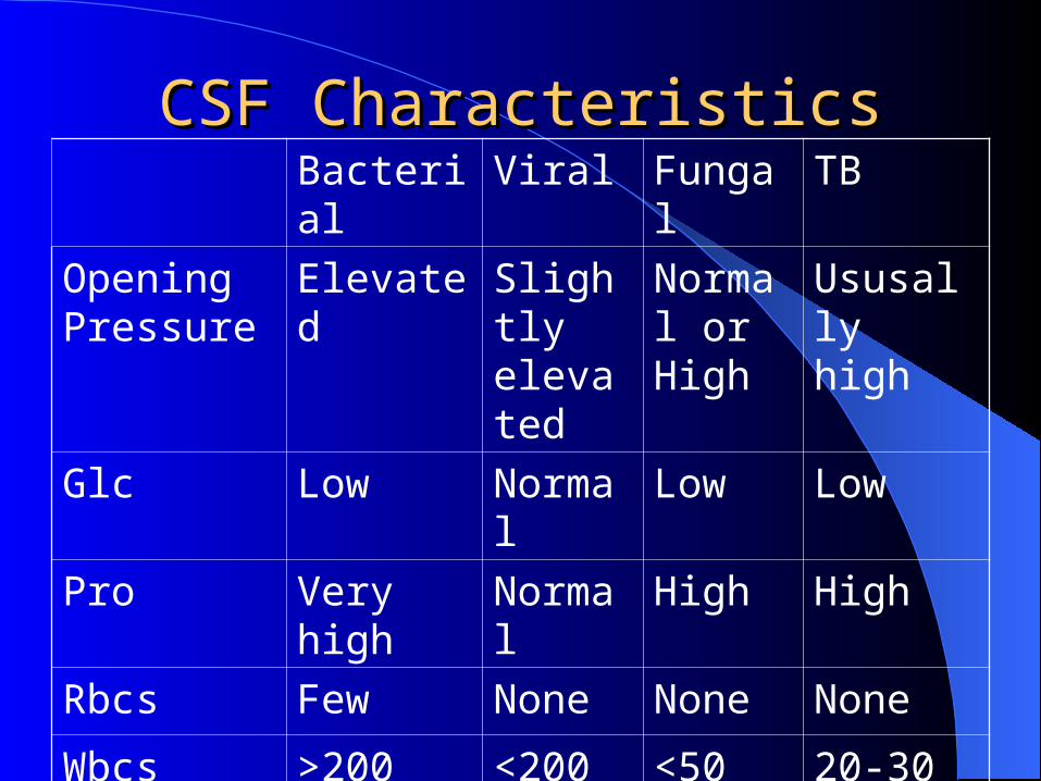
CSF CharacteristicsCSF CharacteristicsBacterial Viral Fungal TB
Opening Pressure
Elevated Slightly elevated
Normal or High
Ususally high
Glc Low Normal Low Low
Pro Very high Normal High High
Rbcs Few None None None
Wbcs (c/mm3)
>200 <200 <50 20-30
Diff PMNs Mono Mono Mono

Key CSF FeaturesKey CSF Features
CSF is not liquid gold – get enough to get your answer CSF Glucose is 2/3 of serum glucose
– Important in diabetic patients Traumatic LPs –
– CSF pro increases by 1 for every 1000 rbcs– Tube #1 and Tube#4 for rbcs when SAH is in the
differential not as a routine Very high CSF Protein levels will make CSF yellow Send a full tube of CSF for cytology not just a few cc’s

Case 1Case 1
CT of head negative.LP - OP (opening pressure) 250mm,
glucose 17, protein 92, Rbcs 3, Wbcs 280 with 89% pmns, 11% lymphocytes
Gram stain - + for Gram neg organisms

Bacterial MeningitisBacterial Meningitis
Streptococcus pneumoniaeHemophilus influenzaeListeria moncytogenesGroup B streptococcusNiesseria meningitidis

Bacterial MenigitisBacterial Menigitis
Age less than 3 months-– Group B strep– L. Monocytogenes– E. coli– Strep pneumoniae

Bacterial MeningitisBacterial Meningitis
3 Months to 18 years –– N. meningitidis– S. pneumoniae– H. influenzae

Bacterial MeningitisBacterial Meningitis
Age 18 to 50 years– S. pneumoniae– N. meningitidis– H. influenzae

Bacterial MeningitisBacterial Meningitis
Over age 50 years– S. pnemoniae– L. monocytogenes– Gram (-) bacilli

Treatment of Bacterial Treatment of Bacterial MeningitisMeningitis
PCN G or 3rd generation cephalosporin and consult ID
Steroids – Dexamethasone IV q6 for 4 days

Viral MeningitisViral Meningitis
Very common
Often caused by enteroviruses
Treatment is supportive

Viral Encephalitis Viral Encephalitis
Encephalitis (Meningoencephalitis)– Altered mental status and seizures– Herpes Simplex virus – medial temporal lobe
Acyclovir Management of seizures Very high morbidity and mortality PCR diagnosis of CSF
– West Nile, St Lousi E, EEE, CMV

Chronic MeningitisChronic Meningitis
Immunocompromised patients– Cryptococcus neoformans– HIV– M. tuberculosis– M. avium
Carcinomatous meningitis– Lung, breast

Case 1Case 1
Meningitis caused by N. Meningitidis– Treatment with 3rd generation cephalosporin for
10 days– Dexamethasone– Prophlaxis with Rifampin for contacts