cognitive neuroscience - kaistraphe.kaist.ac.kr/lecture/2015fallbis451/ch2 structure... ·...
TRANSCRIPT

Cognitive Neuroscience
Chapter 2
Structure and Function of the Nervous System

Overview
• The Structure of Neurons
• Neuronal Signaling
• Synaptic Transmission
• The Role of Glial Cells
• The Bigger Picture
• Overview of the Nervous System
• Structure
• A Guided Tour of the Brain
• The Cerebral Cortex
• Development of the Nervous System

Neurons
• Neurons
• Soma
• Axons
• Dendrites
Axons can take different forms. A neuron and its axon collaterals are shown stained in yellow. The cell body (far right) gives rise to an axon, which branches, forming axon collaterals that can make contact with many different neurons.

Neurons as a unit element of the brain
Neurons: The function of a neuron is to receive, assimilate and analyze, and finally transmit information. Within the neuron action potentials transmit information.
There are sensory neurons (carry sensory information to the brain), motor neurons (transmit to muscles and glands), and interneurons (also called intrinsic or association neurons) found in the central nervous system (CNS), which transmit between sensory and motor neurons.
Neurons can have any number of dendrites but only one axon. There are anaxonic, unipolar, bipolar (one axon and one dendrite), and multipolar neurons.

Neuronal structure
Soma: The soma (cell body) contains the chromosomes.
Dendrites: Branching fibers that form the information-receiving pole of the nerve cell. The surface may be lined with synaptic receptors. The larger the surface area, the more information a dendrite can receive. Some dendrites contain additional short outgrowths called dendritic spines, which are believed to increase the surface area available for synapses.
Axons: Information flows from the dendrites through the cell body to the axon. At the axon hillock, information is summed, and if there is sufficient impetus, then the axons will carry information away to the sites of other neurons. Some axons (in vertebrates) are covered with myelin. The breaks or gaps in the myelin are the nodes of Ranvier.

Synaptic Terminals
Presynaptic terminal:The presynaptic terminal is the point from which an axon releases
chemicals into the synapse. (i.e., an end bulb.) Neurotransmitters are released by the presynaptic neuron when action
potentials depolarize its axon terminal. Calcium ions are known to mediate the release of neurotransmitter
molecules from the presynaptic neuron.
Postsynaptic terminal: Neurotransmitters may then bind to receptors on the membrane of
the postsynaptic neuron.Binding is dependent upon many factors, including the properties of
the postsynaptic neuron and the presence or absence of another neurotransmitter.
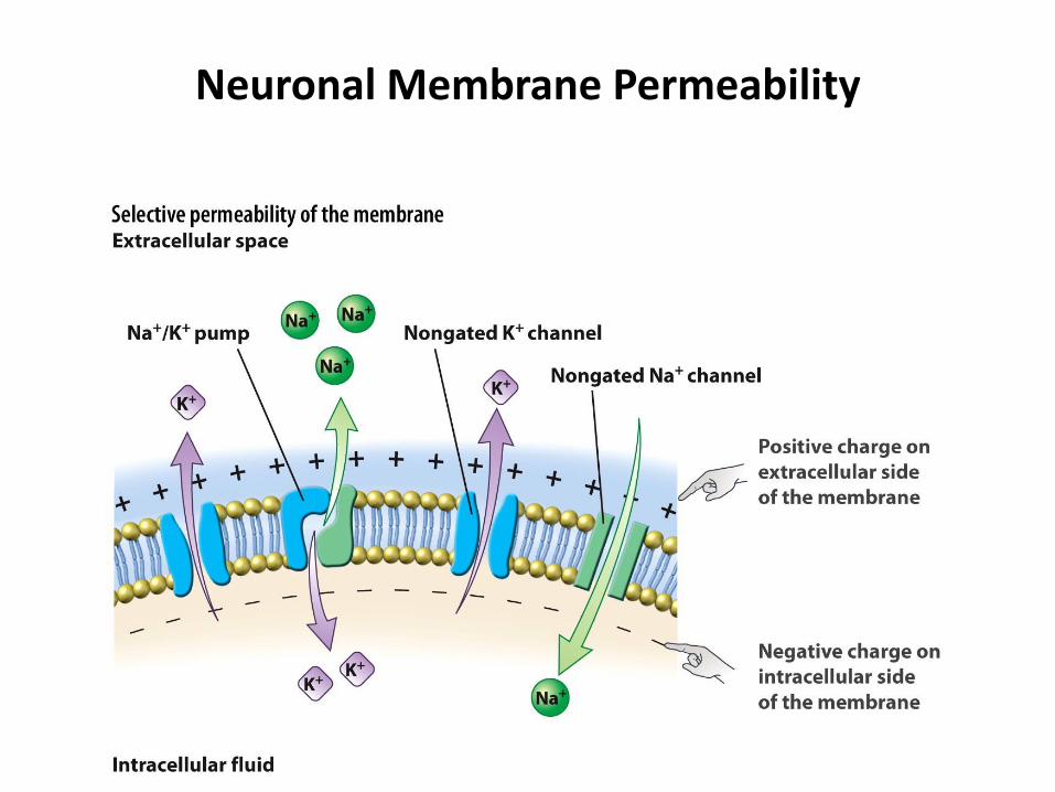
Neuronal Membrane Permeability

Selective permeability of the membrane.
• The membrane’s selective permeability to some ions, along with the concentration gradients formed by active pumping, leads to a difference in electrical potential across the membrane; this is the resting membrane potential.
• The membrane potential, represented here by the positive charges outside the neuron along the membrane and the negative charges inside along the membrane, is the basis for the transmembranevoltage difference.
• Because the concentration gradient for positively charged potassium (K+) forces it out of the cell, a net negative charge develops inside the neuron.
The neuronal membrane’s purpose is to control the exchange of chemicals between the inside and outside of the cell.

Selective permeability (SP)
Selective permeability (SP) refers to the fact that a cell membrane will allow some ions, including water, oxygen, and carbon dioxide, to pass through more readily than others. Small uncharged particles can usually pass via simple diffusion. Those ions that cannot flow freely may cross through specialized protein channels. Without SP, the sodium-potassium pump would be far less effective in creating a concentration gradient.
Note: A prolonged increase in the permeability of the membrane to sodium ions will interfere with a neuron's ability to have an action potential.

Voltage and the Membrane
• Membrane Potential
• Resting Potential
Membrane potential is the voltage across the neuronal membrane at any moment (Vm). When there is no net flow of ions in or out, then the membrane is said to have reached its equilibrium potential.
The difference in voltage that commonly exists between the inside and the outside of a neuron is the resting potential. At rest, the inside of a neuron's membrane is more negative than the outside. The approximate resting potential of the inside of a neuron's membrane is -70 mV.

Channels and Gradients
• Ion Channels
• Ion Pumps
• Electrical Gradient

Ion channels pump ions across the membrane.
• Ion channels are pore-forming proteins created from an amino acid chain. Channels differ in their selectivity. They need to be able to move ions with speed and selectivity.
The channels move across the lipid bilayer. As they are selective, they choose among the available ions. They open, close, or inactivate based on stimuli in their environment. Channels can be altered or modulated based on cellular needs. For example, when a neuron is at rest, the sodium channels are closed and potassium crosses very slowly. Two major ions that carry charge and change the membrane potential are voltage-gated calcium and voltage-gated sodium.

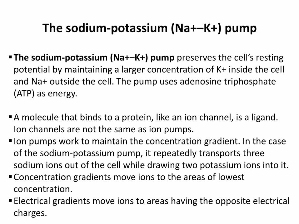
The sodium-potassium (Na+–K+) pump
The sodium-potassium (Na+–K+) pump preserves the cell’s resting potential by maintaining a larger concentration of K+ inside the cell and Na+ outside the cell. The pump uses adenosine triphosphate (ATP) as energy.
A molecule that binds to a protein, like an ion channel, is a ligand. Ion channels are not the same as ion pumps. Ion pumps work to maintain the concentration gradient. In the case
of the sodium-potassium pump, it repeatedly transports three sodium ions out of the cell while drawing two potassium ions into it.Concentration gradients move ions to the areas of lowest
concentration. Electrical gradients move ions to areas having the opposite electrical
charges.

Action Potential
• Why do they occur?
• What is the all-or-none law?
• Why is there an overshoot?
• Why don’t action potentials remain depolarized?

Relative time course of changes in membrane voltage during an action potential, and the underlying causative changes in membrane
conductance to Na+ (gNa) and K+ (gK).
•The initial depolarizing phase of the action potential is mediated by Na+ current, and the later repolarizing, descending phase of the action potential is mediated by an increase in K+ conductance that occurs when the K+ channels open. The Na+ channels close during the last part of the action potential, when repolarization by the K+ current is taking place. The action potential undershoots the resting membrane potential at the point where the membrane becomes more negative than the resting membrane potential.
Universal features: rising phase, overshoot, and falling phase (repolarization)
Signaling by another neuron or a sensory event may initiate an action potential. During an action potential there is a transitory change in the polarity of the electrical charge across the cell membrane. The membrane then alters its permeability to the charged ions, and the charge across the cell membrane becomes briefly less positive or negative. Action potentials result in the positively charged sodium ions flow rapidly into the neuron. Then the charge inside the neuron reverses polarity to become positive. When examined with an oscilloscope, action potentials are seen as brief spikes (+40 mV).
Depolarization: neurons will produce an action potential only if the depolarization exceeds threshold of excitation (about 15 mV from resting potential). Thus, depolarization is the electrical potential change that results in the propagation of an action (due to the depletion of positively charged potassium ions). After depolarization, voltage-gated sodium channels inactivate. After depolarization, both potassium-leak channels and voltage-gated potassium channels activate, allowing potassium to leave and to repolarize Vm and close.
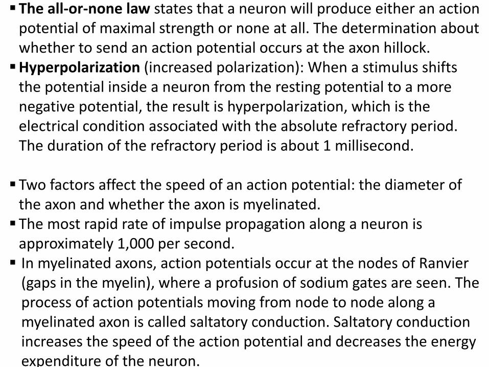
The all-or-none law states that a neuron will produce either an action potential of maximal strength or none at all. The determination about whether to send an action potential occurs at the axon hillock.Hyperpolarization (increased polarization): When a stimulus shifts
the potential inside a neuron from the resting potential to a more negative potential, the result is hyperpolarization, which is the electrical condition associated with the absolute refractory period. The duration of the refractory period is about 1 millisecond.
Two factors affect the speed of an action potential: the diameter of the axon and whether the axon is myelinated.The most rapid rate of impulse propagation along a neuron is
approximately 1,000 per second. In myelinated axons, action potentials occur at the nodes of Ranvier
(gaps in the myelin), where a profusion of sodium gates are seen. The process of action potentials moving from node to node along a myelinated axon is called saltatory conduction. Saltatory conduction increases the speed of the action potential and decreases the energy expenditure of the neuron.

Neurotransmitters

• The synapse consists of various specializations where the presynaptic and postsynaptic membranes are in close apposition. When the action potential invades the axon terminals, it (1) causes voltage-gated Ca2+ channels to open, which (2) triggers vesicles to bind to the presynaptic membrane. Then, (3) neurotransmitter is released into the synaptic cleft by exocytosis and diffuses across the cleft. Finally, (4) binding of the neurotransmitter to receptor molecules in the postsynaptic membrane completes the process of transmission.
• Neurotransmitter leading to postsynaptic potential.The binding of neurotransmitter to the postsynaptic membrane receptors changes the membrane potential (Vm). These postsynaptic potentials can be either excitatory (depolarizing the membrane), as shown here, or inhibitory (hyperpolarizing the membrane).

Neurotransmitters
Neurotransmitter: Any substance, such
as acetylcholine or dopamine,
responsible for sending nerve signals
across a synapse between two
neurons

Synapse and neurotransmitters
• The term synapse was coined by Sherrington. During synaptic transmission, neurotransmitters are released from the presynaptic cell.
• The neurotransmitter, if unimpeded, will diffuse across the synapse and bind to the postsynaptic membrane.
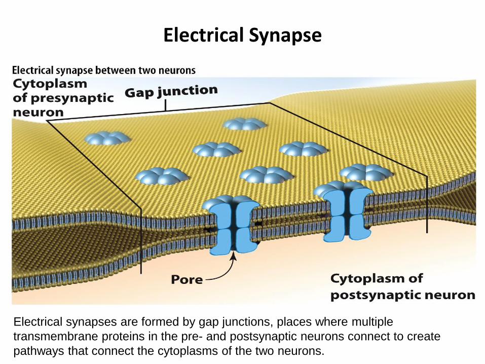
Electrical Synapse
Electrical synapses are formed by gap junctions, places where multiple
transmembrane proteins in the pre- and postsynaptic neurons connect to create
pathways that connect the cytoplasms of the two neurons.

Electrical synapses and Gap junctions
Within a neuron, the transmission of information is usually electrical. Between neurons, the transmission of information is usually chemical. When the signal is electrical, two neurons communicate physically through gap junctions. Synchronicity among the neurons is thereby maintained as the junctions permit alterations in the electrical properties of one neuron to affect another neuron.
Electricity: Ions are electrically charged particles.
Electrical potential (voltage): Force exerted on a charged particle
Electrical conductance (g): Ability of an electrical charge to move from one place to another
Electrical resistance: Relative inability of an electrical charge to move from one place to another

Brain is an information processor
iui
j
Spike reception: EPSP,
summation of EPSPs
Spike reception: EPSP
Threshold Spike emission
(Action potential)
threshold -> Spike

Glial Cells
• In the nervous system, glia provide structural support and insulation for neurons. Radial glia guide the migration of neurons during embryonic development.
• Oligodendrocytes myelinate axons in the brain and spinal cord.
• Schwann cells myelinate axons in the periphery of the body.

Neuronal Connectivity
Convergence: Single neurons connect to many other neurons.
Divergence: Neurons from divergent brain areas may synapse onto a single neuron.

Nervous System
• Peripheral Nervous System (PNS)
• Central Nervous System (CNS)
• Autonomic Nervous System (ANS)
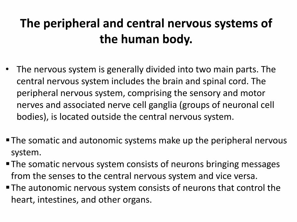
The peripheral and central nervous systems of the human body.
• The nervous system is generally divided into two main parts. The central nervous system includes the brain and spinal cord. The peripheral nervous system, comprising the sensory and motor nerves and associated nerve cell ganglia (groups of neuronal cell bodies), is located outside the central nervous system.
The somatic and autonomic systems make up the peripheral nervous system.The somatic nervous system consists of neurons bringing messages
from the senses to the central nervous system and vice versa.The autonomic nervous system consists of neurons that control the
heart, intestines, and other organs.
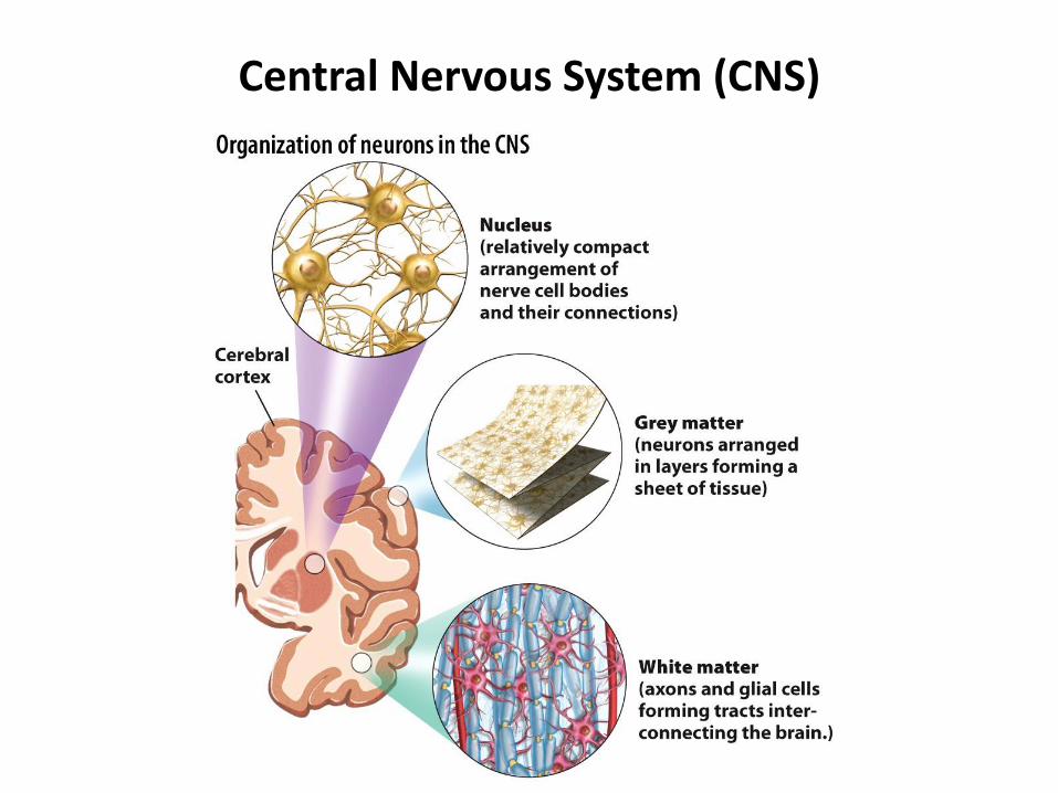
Central Nervous System (CNS)

Organization of neurons in the CNS.
• In the CNS, neurons can be organized in clumps called nuclei (top—not to be confused with the nucleus inside each neuron), which are most commonly found in subcortical and spinal structures, or sheets called layers (middle), which are most commonly found in the cortex.
• The cell bodies of glial cells are located in the white matter (e.g., oligodendrocytes), and in the cortex.
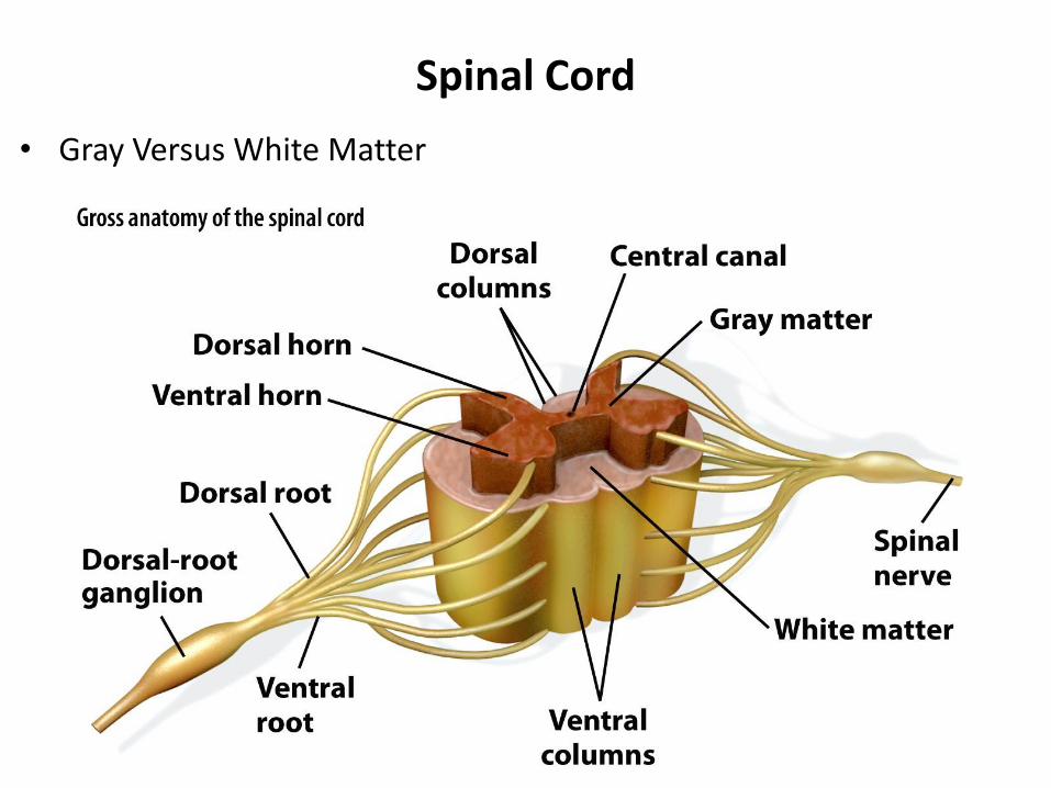
Spinal Cord
• Gray Versus White Matter

Spinal cord
• This cross-sectional and three-dimensional representation of the spinal cord shows the central butterfly-shaped gray matter, which contains neuronal cell bodies, and the surrounding white matter axon tracts, which convey information down the spinal cord from the brain to the peripheral neurons and up the spinal cord from peripheral receptors to the brain.
• The dorsal and ventral nerve roots are shown exiting and entering the cord; they fuse to form peripheral nerves. The cell bodies of peripheral sensory inputs reside in the dorsal-root ganglion and project their axons into the central nervous system via the dorsal root.
• The ventral horn of the spinal cord houses motor neurons that project their axons out the ventral roots to innervate peripheral muscles.

Navigating the human brain.
Anterior/rostral: front endSuperior: above Inferior: belowPosterior/caudal: behind (tail/rear)Dorsal: back or top sideVentral: belly
Lateral = away from the midline Medial = toward the midline Proximal = closer Distal = farther away

The coronal plane shows brain structures as they would be seen from the front.The horizontal plane shows brain
structures as they would be seen from above.The saggital plane shows brain
structures as they would be seen from the side.The central sulcus separates the
frontal and parietal lobes.The two hemispheres
communicate predominantly through the corpus callosum.
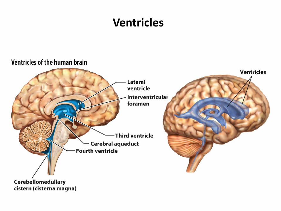
Ventricles
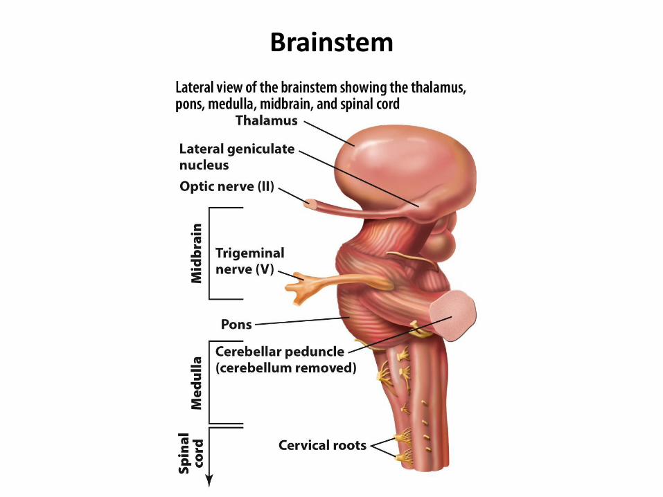
Brainstem

Cerebellum

Limbic System


Cerebral Cortex


White and Gray Matter

Brodmann’s Areas
Brodmann’s original cytoarchitectonic map (52 areas) from his
work around the start of the 20th century. Different regions of
cortex have been demarcated by histological examination of the
cellular microanatomy.

Brodmann’s Areas
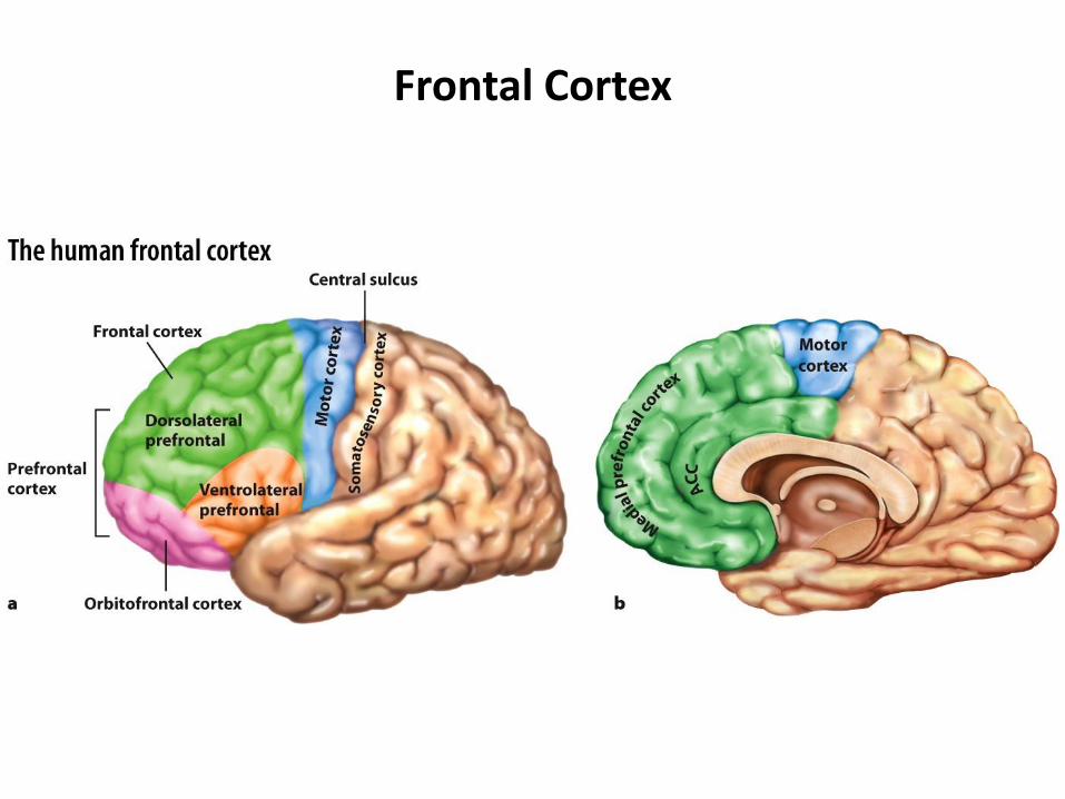
Frontal Cortex

Somatosensory Cortex

Topographical Memory

Visual Cortex

Auditory Cortex

Sensory and Association Cortices

Embryonic Development
• What changes occur in brain development?

Early stages of embryonic development in mammals.
(a) Developing embryo. The embryo goes through a series of folds, or flexures, during development. These alterations in the gross structure of the nervous system give rise to the compact organization of the adult brain and brainstem in which the cerebral cortex overlays the diencephalon and midbrain within the human skull.
(b) There is significant similarity between the gross features of the developing fetuses of mammals, as shown by this comparison of a human fetus (top) and pig (bottom) fetuses.

Neuronal Migration
Radial glia guide the migration of neurons during embryonic
development.

Neuronal Proliferation and Determination
• Neuronal proliferation is the process of rapid cell division that occurs early in development of the nervous system.


Radial Unit Hypothesis

Radial Unit Hypothesis
• Radial glial cells in the ventricular zone project their processes in an orderly map through the various cortical layers, thus maintaining the organizational structure specified in the ventricular layer. (E = Embryonic day.)
The radial unit hypothesis says that the columnar organization in the adult brain arises during development from the cells dividing in the ventricular (germinal) region.

Neurogenesis
Baby Brain
• Synaptogenesis
Synaptogenesis is the formation of new synapses in the central nervous system
• Synapse Elimination
Synapse Elimination – many neural circuits are redundant in a newborn. Unneeded circuits are eliminated. This elimination process includes strengthening single climbing fibres(CF) to purkinjecells(PC) and leaving weaker fibres to be eliminated.