coinfections acquired from ixodes ticks - clinical ...cmr.asm.org/content/19/4/708.full.pdflaris...
TRANSCRIPT

CLINICAL MICROBIOLOGY REVIEWS, Oct. 2006, p. 708–727 Vol. 19, No. 40893-8512/06/$08.00�0 doi:10.1128/CMR.00011-06Copyright © 2006, American Society for Microbiology. All Rights Reserved.
Coinfections Acquired from Ixodes TicksStephen J. Swanson,1,2 David Neitzel,2 Kurt D. Reed,3 and Edward A. Belongia3*
Epidemic Intelligence Service Program, Office of Workforce and Career Development, Centers for Disease Control and Prevention,Atlanta, Georgia1; Acute Disease Investigation and Control, Minnesota Department of Health, St. Paul, Minnesota2;
and Marshfield Clinic Research Foundation, Marshfield, Wisconsin3
INTRODUCTION .......................................................................................................................................................708BIOLOGY AND ECOLOGY OF IXODES TICKS .................................................................................................709COINFECTIONS AMONG IXODES TICKS AND MAMMALIAN HOSTS ......................................................710
Prevalence of Coinfecting Pathogens among Ixodes Ticks ................................................................................710North America.....................................................................................................................................................710Europe ..................................................................................................................................................................712Asia and the remainder of the world ...............................................................................................................712
Prevalence of Coinfecting Pathogens among Nonhuman Mammalian Hosts ................................................712Transmission Dynamics of Coinfections among Ticks and Reservoir Hosts .................................................713Effects of Strain Diversity......................................................................................................................................713
COINFECTIONS AMONG HUMANS ....................................................................................................................714Epidemiology of Coinfections among Humans ...................................................................................................714
Prospective studies .............................................................................................................................................714(i) Molecular evidence of coinfection...........................................................................................................714(ii) Serologic evidence of coinfection ...........................................................................................................715
Serologic studies .................................................................................................................................................715(i) Lyme disease-babesiosis coinfection .......................................................................................................715(ii) Lyme disease-HA coinfection..................................................................................................................715(iii) HA, babesiosis, and triple coinfection .................................................................................................717
Laboratory Diagnosis of Coinfections..................................................................................................................718Pathogenesis and Immunologic Effects................................................................................................................719Clinical Manifestations ..........................................................................................................................................719
Lyme disease and babesiosis.............................................................................................................................719Lyme disease and HA.........................................................................................................................................720
Transfusion-Related Tick-Borne Illness ..............................................................................................................720THERAPY ....................................................................................................................................................................720
Treatment of HA and LD ......................................................................................................................................720Treatment of Babesiosis.........................................................................................................................................721
STRATEGIES FOR PREVENTING COINFECTIONS FROM IXODES TICKS ..............................................721RESEARCH NEEDS ..................................................................................................................................................722ACKNOWLEDGMENTS ...........................................................................................................................................722REFERENCES ............................................................................................................................................................722
INTRODUCTION
Ticks have been implicated as a source of disease for �100years. In 1893 Smith and Kilbourne offered the first descriptionof a tick-borne disease, establishing that the cattle tick(Boophilus microplus) transmits the protozoan Babesia bigem-ina, the causative pathogen of Texas cattle fever (182). Thisdramatic report became the foundation for subsequent workon vertebrate hosts and arthropod vectors. Later work in 1909by Ricketts recognized the role of ticks as vectors of humandisease, with his description of the wood tick, Dermacentorandersoni, transmitting Rocky Mountain spotted fever (165).The first recognition of disease caused by Ixodes ticks occurredin the early 20th century when a Swedish dermatologist re-ported that the bite of an Ixodes ricinus tick was associated with
a characteristic skin lesion near tick bites, termed erythemachronicum migrans (2). In the 1940s, spirochetes were ob-served in skin lesions, but only isolated cases of erythemamigrans (EM) were reported until 1975, when Steere and col-leagues investigated a cluster of children with juvenile rheu-matoid arthritis living in Old Lyme, Connecticut (192, 193).They observed that the majority of children had illness onset inthe summer or fall, and many recalled an expanding rash be-fore the onset of arthritis. Further epidemiologic investigationsstrongly implicated Ixodes scapularis as the tick vector forLyme disease (LD) (191). Not until 7 years after the initialrecognition was a spirochete (Borrelia burgdorferi) finally iso-lated from Ixodes ticks by Burgdorfer and colleagues at theRocky Mountain Laboratories of the U.S. Public Health Ser-vice (31).
Since then, newly recognized pathogens and health hazardsassociated with Ixodes ticks have increased dramatically. Wenow realize that B. burgdorferi is a genogroup of multipleclosely related spirochetes, which have been described
* Corresponding author. Mailing address: Epidemiology ResearchCenter (ML2), Marshfield Clinic Research Foundation, 1000 NorthOak Ave., Marshfield, WI 54449. Phone: (715) 389-3783. Fax: (715)389-3880. E-mail: [email protected].
708
on July 9, 2018 by guesthttp://cm
r.asm.org/
Dow
nloaded from
throughout the world. The first documented human case ofbabesiosis occurred in 1957 (181), but only a few isolated caseswere reported before 1977, when five cases of Babesia microtiinfection were identified among residents of Nantucket Island(167). In 1979 the vector for B. microti was identified as anIxodes tick, and the white-footed mouse (Peromyscus leucopus)was thereafter identified as being a common reservoir for bothB. microti and B. burgdorferi (184, 186). Human infections withother Babesia species have since been reported, includingBabesia divergens and the unnamed species WA1, CA1, MO1,and TW1 (82, 150, 160, 177).
Human anaplasmosis (HA; previously known as humangranulocytic ehrlichiosis) was first reported among patientsfrom Minnesota and Wisconsin in 1994 (12, 39). The etiologicagent, Anaplasma phagocytophilum (previously known as Ehr-lichia equi and E. phagocytophila), was detected in blood sam-ples from 12 patients presenting with fever, headache, andmyalgias. Subsequent studies confirmed I. scapularis as thevector (147). HA is now known to occur in regions of NorthAmerica and Europe inhabited by vector-competent species ofIxodes (24, 25, 49, 170, 201, 213). Certain species of Ixodes ticksin Europe (I. ricinus and I. persulcatus) are also capable oftransmitting tick-borne encephalitis (TBE) virus, a flavivirusthat can cause fatal brain infection among humans (47, 142,226).
Not surprisingly, because all of these agents can coexist inIxodes ticks, coinfections have been reported. However, theepidemiology and natural history of coinfections are not fullyunderstood, and the majority of clinicians have limited expe-rience in recognizing or managing them. The purpose of this
review is to summarize relevant findings from the medicalliterature on the occurrence, natural history, and outcomes ofcoinfections acquired from Ixodes ticks.
BIOLOGY AND ECOLOGY OF IXODES TICKS
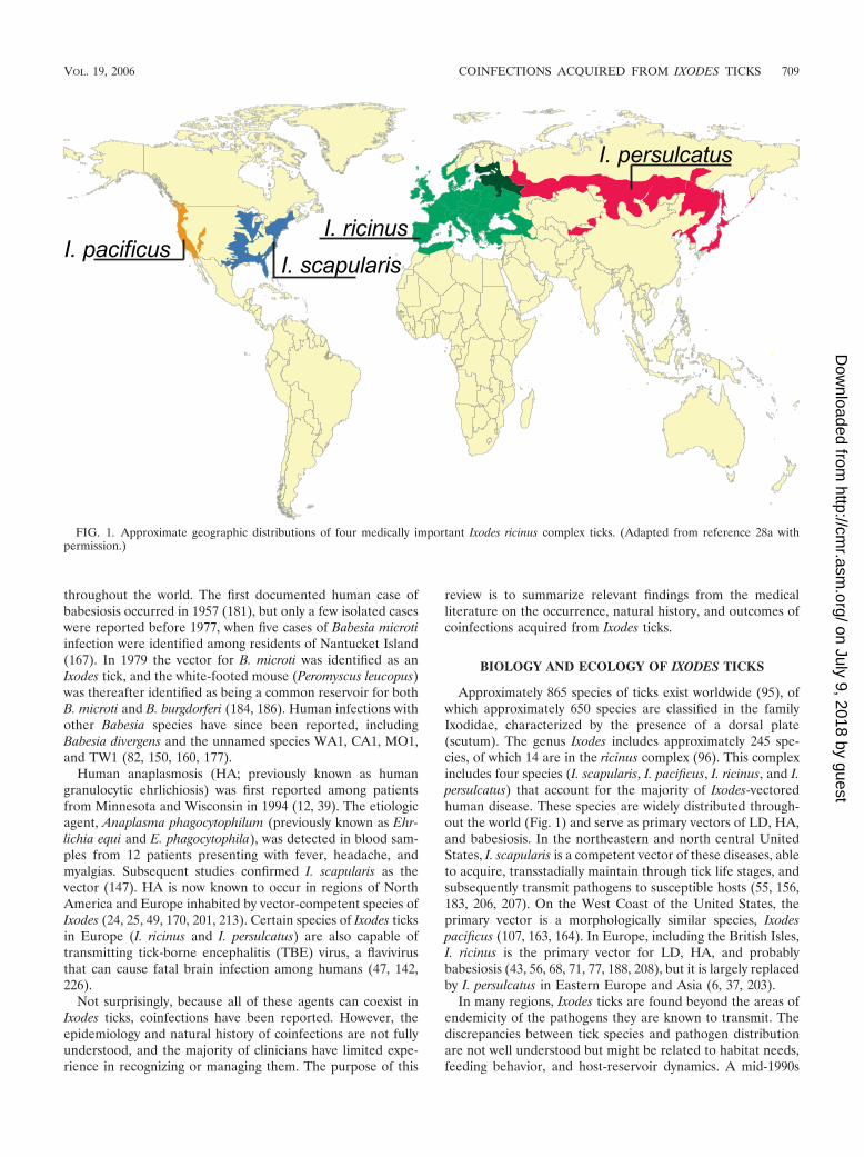
Approximately 865 species of ticks exist worldwide (95), ofwhich approximately 650 species are classified in the familyIxodidae, characterized by the presence of a dorsal plate(scutum). The genus Ixodes includes approximately 245 spe-cies, of which 14 are in the ricinus complex (96). This complexincludes four species (I. scapularis, I. pacificus, I. ricinus, and I.persulcatus) that account for the majority of Ixodes-vectoredhuman disease. These species are widely distributed through-out the world (Fig. 1) and serve as primary vectors of LD, HA,and babesiosis. In the northeastern and north central UnitedStates, I. scapularis is a competent vector of these diseases, ableto acquire, transstadially maintain through tick life stages, andsubsequently transmit pathogens to susceptible hosts (55, 156,183, 206, 207). On the West Coast of the United States, theprimary vector is a morphologically similar species, Ixodespacificus (107, 163, 164). In Europe, including the British Isles,I. ricinus is the primary vector for LD, HA, and probablybabesiosis (43, 56, 68, 71, 77, 188, 208), but it is largely replacedby I. persulcatus in Eastern Europe and Asia (6, 37, 203).
In many regions, Ixodes ticks are found beyond the areas ofendemicity of the pathogens they are known to transmit. Thediscrepancies between tick species and pathogen distributionare not well understood but might be related to habitat needs,feeding behavior, and host-reservoir dynamics. A mid-1990s
FIG. 1. Approximate geographic distributions of four medically important Ixodes ricinus complex ticks. (Adapted from reference 28a withpermission.)
VOL. 19, 2006 COINFECTIONS ACQUIRED FROM IXODES TICKS 709
on July 9, 2018 by guesthttp://cm
r.asm.org/
Dow
nloaded from

review of distribution records in the United States (51) dem-onstrated the establishment of I. scapularis or I. pacificus pop-ulations in 1,058 of 3,141 (34%) U.S. counties, an area includ-ing the West Coast and much of the United States east of theGreat Plains. However, only a limited proportion of counties(63, or 2%) accounted for the majority (78%) of nationallyreported LD cases in 1995 (38).
The distribution and abundance of Ixodes ticks are related tomultiple factors, including the presence of suitable wooded orbrushy habitat and the abundance of hosts for all life stages ofthe ticks. The resurgence in white-tailed deer populations dur-ing the past 30 years might have allowed I. scapularis to expandits range in much of the eastern United States (80, 186, 220).The distributions of tick-borne pathogens and resulting humaninfections often depend on local tick feeding habits and thedistribution and density of small-mammal species that act ascompetent pathogen reservoirs. For example, the lack of hu-man LD cases in the southern United States might be partiallythe result of immature I. scapularis ticks commonly feeding onlizards (144), which are incompetent reservoir species for B.burgdorferi (108, 186); in addition, for unknown reasons, I.scapularis ticks in that region do not commonly bite humans(66). Conversely, northern populations of immature I. scapu-laris feed on reservoir-competent small mammals (e.g., P. leu-copus and eastern chipmunks [Tamias striatus]) as well as hu-mans. Reservoir competence, local tick vector feeding habits,and pathogen strain variations each contribute to differences inthe geographic distribution of tick-borne diseases.
The risk for tick-borne disease is also closely linked with thelife cycle of the Ixodes tick and with vector competency at eachlife stage. This life cycle involves four life stages (egg, larva,nymph, and adult) and spans �2 years, with tick activity dif-fering dramatically by season and life stage. For example, lar-val I. scapularis ticks often have peaks in seasonal activityduring early and late summer, whereas the nymph stage is mostactive from late spring through midsummer (137, 221). Adult I.scapularis ticks are abundant during the early fall and areactive again during spring months if they did not feed in thefall. Transmission of LD, HA, and babesiosis usually occursduring the relatively short period of the nymph stage when thetick is active (145). The nymphs’ small size (approximately 1mm) allows them to often feed undetected on humans longenough to transmit these pathogens. Adult ticks are larger andmore likely to be detected and removed before disease trans-mission, whereas host-seeking larvae are uninfected and thusepidemiologically unimportant.
The feeding behavior of Ixodes ticks at each life stage has animpact on the risk for tick-borne infection and coinfectionamong humans. All Ixodes species of public health importanceare three-host ticks that must find a new host at each life stage.During each life stage after hatching (larva, nymph, and adult),an Ixodes tick takes one blood meal, which typically requires 3to 5 days to complete. Certain Ixodes ticks are host specific,whereas others feed on different host species. Those with non-specific feeding habits, (e.g., I. scapularis, I. pacificus, I. ricinus,and I. persulcatus) not only feed on species that are reservoirsfor multiple tick-borne pathogens (e.g., small mammals) butalso will readily bite humans. Therefore, nonspecific feedersmight be more important as vectors of human disease thanhost-specific ticks, which are less likely to bite humans.
When feeding on an infected small-mammal host, tick larvaeand nymphs can take up one or more pathogens, which mightbe transmissible during subsequent blood meals. Larvae aregenerally not infected with B. burgdorferi, A. phagocytophilum,or B. microti upon hatching; transovarial passage of thesepathogens from adult females to eggs has not been consistentlydemonstrated or is considered insignificant (91, 136, 148, 154,171, 228). However, transovarial transmission of B. divergensfrom adult I. ricinus ticks to larvae does occur (57, 207) and isalso believed to be important in maintaining the life cycle ofother tick-borne viral and rickettsial pathogens (e.g., TBE vi-rus, spotted fever group rickettsia) (32, 162). Following acqui-sition of either LD, HA, or Babesia, transstadial transmission(i.e., from larva to nymph or from nymph to adult tick) occurs.After molting, nymphs and adult ticks infected in a previouslife stage emerge infective and may transmit disease to suscep-tible hosts during subsequent feedings. Adult female ticks re-quire a blood meal to develop their egg mass and commonlyseek a large-mammal host for their third and final blood meal.
COINFECTIONS AMONG IXODES TICKSAND MAMMALIAN HOSTS
Prevalence of Coinfecting Pathogens among Ixodes Ticks
The risk for human coinfection with multiple pathogensafter an Ixodes tick bite differs by geographic location anddepends on the prevalence of pathogens within the reservoirhost and Ixodes ticks. The distribution of pathogens withinIxodes ticks has been derived largely from epidemiologic re-ports of human disease. Systematic or large-scale surveys oftick-borne pathogens are lacking. Numerous smaller studieshave attempted to identify the prevalence of pathogens amongticks through PCR analysis of DNA isolated from individualticks. These studies remain difficult to compare because ofconsiderable differences in the methods of tick collection, sam-ple size, specimen preparation, DNA extraction, and selectionof nucleic acid probes (primers). Less specific PCR primerspotentially yield higher reported prevalence rates amongIxodes ticks as a result of the detection of additional strainvariants not associated with human illness (120, 178). Thus, thetrue prevalence of coinfecting human pathogens among Ixodesticks remains largely unknown in the majority of geographiclocations. Nonetheless, infection of both ticks and humans withB. burgdorferi appears to be substantially more widespread inNorth America and Europe than infection with Babesia orAnaplasma, and the reasons for this difference are poorly un-derstood.
North America. Molecular evidence of coinfection with mul-tiple human pathogens has been demonstrated for Ixodes tickssampled from select geographic areas of California, Wisconsin,and the northeastern United States (Table 1). The prevalenceof dually infected ticks appears highest among I. scapularisticks from regions of LD endemicity in the northeasternUnited States, with reported prevalences of �28%. Studiesfrom other North American regions have generally reportedlower prevalences of dually infected Ixodes ticks. In Wisconsin,2% of I. scapularis adult ticks were coinfected with B. burgdor-feri and A. phagocytophilum (147). In northern California, ap-proximately 1% of both I. pacificus nymph ticks from decidu-
710 SWANSON ET AL. CLIN. MICROBIOL. REV.
on July 9, 2018 by guesthttp://cm
r.asm.org/
Dow
nloaded from
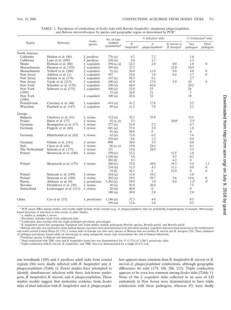
ous woodlands (109) and I. pacificus adult ticks from coastalregions (86) were dually infected with B. burgdorferi and A.phagocytophilum (Table 1). Fewer studies have attempted toidentify simultaneous infection with three tick-borne patho-gens, B. burgdorferi, B. microti, and A. phagocytophilum. Thesestudies weakly suggest that molecular evidence from Ixodesticks of dual infection with B. burgdorferi and A. phagocytophi-
lum appears more common than B. burgdorferi-B. microti or B.microti-A. phagocytophilum coinfections, although geographicdifferences do exist (179, 188, 206, 213). Triple coinfectionappears to be even less common among Ixodes ticks (Table 1).None of the I. scapularis ticks collected in an area of LDendemicity in New Jersey were demonstrated to have triplecoinfection with these pathogens, whereas 4% were dually
TABLE 1. Prevalences of coinfections of Ixodes ticks with Borrelia burgdorferi, Anaplasma phagocytophilum,and Babesia microti/divergens by species and geographic region as determined by PCRa
Region Reference Ixodesspecies
No. of tickssampled
(population)b
% Infectionc with: % Coinfectiond with:
B.burgdorferie
A.phagocytophiluma
B. microti orB. divergensf
Twopathogens
Threepathogens
North AmericaCalifornia Holden et al. (86) I. pacificus 776 (a) 6.7 7.2 1.0California Lane et al. (109) I. pacificus 158 (n) 3.8 3.2 1.3Maine Holman et al. (88) I. scapularis 394 (a, n) 22.3 2.8 0.8 1.0 0Massachusetts Piesman et al. (155) I. scapularis 395 (n) 27.3 23.0 10.9Massachusetts Telford et al. (206) I. scapularis 51 (a) 36.0 11.0 9.0 4.0 0New Jersey Adelson et al. (1) I. scapularis 107 33.6 1.9 8.4 3.7 0New Jersey Schulze et al. (174) I. scapularis 147 (a) 50.3 6.1 2.7New Jersey Varde et al. (213) I. scapularis 100 (a) 43.0 17.0 5.0 10 0New York Schauber et al. (170) I. scapularis 188 (a) 66.0 42.6 28.2New York(1995)
Schwartz et al. (175) I. scapularis 100 (a) 52.0 53 2673 (n) 26.0 21 5
New York(1984)
I. scapularis 100 (a) 45.0 32 19
Pennsylvania Courtney et al. (46) I. scapularis 454 (a) 41.2 17.8 3.5Wisconsin Pancholi et al. (147) I. scapularis 89 (a) 11.2 7.9 2.2
EuropeBulgaria Christova et al. (41) I. ricinus 112 (a) 32.1 33.9 13.4France Halos et al. (77) I. ricinus 92 (a, n) 3.3 20.6g 2.1g
Germany Baumgarten et al. (17) I. ricinus 275 (a) 21.8 2.2 0.7Germany Fingerle et al. (68) I. ricinus 401 (a) 37.4 2.0 1.0
91 (n) 30.8 0 0Germany Hildebrandt et al. (83) I. ricinus 62 (a) 21.0 6.5 1.6
243 (n) 8.6 1.2 0.4Germany Oehme et al. (141) I. ricinus 898 20.4 2.9 0.8Italy Cinco et al. (44) I. ricinus 86 (a, n) 19.8 24.4 8.1The Netherlands Schouls et al. (173) I. ricinus 121 13.0 28.9 3.3Poland Skotarczak et al. (180) I. ricinus 550 (a) 12.2 12.5 1.8
1,160 (n) 5.8 9.5 0.2385 (l) 5.5 4.2 0
Poland Skotarczak et al. (179) I. ricinus 280 (a) 25.0 10.0 19.3 5.4 1.1234 (n) 11.5 4.3 11.1 0.8 019 (l) 42.1 0 21.0 0 0
Poland Stanczak et al. (189) I. ricinus 424 (a) 11.6 19.2 5.0Poland Stanczak et al. (188) I. ricinus 303 (a) 19.5 29.7 3.6 10.6 0Russia Alekseev et al. (6) I. persulcatus 1,282 (a) 29.9 1.0 0.9 1.2h 0i
Slovakia Derdakova et al. (54) I. ricinus 40 (a) 45.0 20.0 7.5Switzerland Leutenegger et al. (111) I. ricinus 20 (n) 40.0 0 0
100 (a) 49.0 2.0 2.0
China Cao et al. (37) I. persulcatus 1,146 (a) 37.3 4.9 0.5199 (n) 13.6 3.0 0.5
a PCR assays differ among studies, and results might include strain variants (e.g., A. phagocytophilum) that are potentially nonpathogenic in humans. Microscopy-based detection of infection in ticks occurs in older studies.
b a, adults; n, nymphs; l, larvae.c Prevalence includes totals from coinfected ticks.d Coinfection data overlap with the single-pathogen prevalence percentages.e B. burgdorferi sensu lato genogroup; European and Asian studies include pathogenic Borrelia species, Borrelia garinii, and Borrelia afzelii.f Babesia odocoilei, not reported to cause human disease, has since been demonstrated to be prevalent among I. scapularis ticks in certain locations in the northeastern
and north central United States (9, 172). I. ricinus ticks in Europe can also carry species of Babesia that are neither B. microti nor B. divergens (59). Thus, estimatesof pathogen prevalence based solely on microscopy or using nonspecific assays may overestimate the risk of human babesiosis.
g Particular species of Babesia not determined.h Dual coinfection with TBE virus and B. burgdorferi sensu lato was demonstrated for 15 (1.2%) of 1,280 I. persulcatus ticks.i Triple coinfection with B. microti, B. burgdorferi, and TBE virus was demonstrated for a single (0.1%) tick.
VOL. 19, 2006 COINFECTIONS ACQUIRED FROM IXODES TICKS 711
on July 9, 2018 by guesthttp://cm
r.asm.org/
Dow
nloaded from

infected (1). Other researchers have not identified molecularevidence of triple coinfection among Ixodes ticks, despite re-porting a dual-pathogen prevalence of 1% to 10% (88, 206,213). Taken together, these few studies indicate that dual in-fection with any combination of B. burgdorferi, B. microti, andA. phagocytophilum occurs in 1% to 28% of Ixodes ticks fromregions of LD endemicity in the United States and in �1% to13% of sampled European Ixodes ticks (Table 1). Triple coin-fection is rarely detected in geographic regions where all threetick-borne diseases are endemic and likely represents an inci-dent occurrence of �1%.
Europe. European studies have predominantly examined I.ricinus for molecular evidence of coinfecting pathogens. Mul-tiple studies have involved ticks collected in Germany andPoland (Table 1); individual studies also exist from geographicareas within Bulgaria, France, Italy, The Netherlands, Slova-kia, and Switzerland. The prevalence of dual pathogens in I.ricinus ticks differs depending on the geographic site of ticksampling and the methodology, with the highest reportedprevalences occurring in Bulgaria (13%) (41) and Poland (2%to 11%) (188). European studies demonstrated DNA evidenceof B. burgdorferi sensu lato genogroup and A. phagocytophilumin 0.5% to 13% of I. ricinus adult ticks. The B. burgdorferi sensulato genogroup includes 11 Borrelia species worldwide, whichcan be identified and differentiated by using molecular ap-proaches described elsewhere (216). Of these 11 B. burgdorferisensu lato species, only 3 species (B. burgdorferi sensu stricto,Borrelia afzelii, and Borrelia garinii) are known to causedisease among humans (4, 190). All three pathogenic spe-cies inhabit Europe, whereas primarily B. afzelii and B. ga-rinii are thought to cause disease in Asia; B. burgdorferisensu stricto is the sole pathogenic species identified in theUnited States (15, 212, 216).
A limited number of studies have attempted to detect mo-lecular evidence of tick coinfection with Babesia species. Fromnorthwestern Poland, coexistent DNA of B. burgdorferi sensulato species and B. microti was identified for 3% of sampledI. ricinus female adult ticks but only 0.1% of nymphs (180).Among questing I. ricinus ticks collected in northern France,2% had evidence of coinfection with B. burgdorferi sensu latoand Babesia species (77). Of note, a limited number of Euro-pean studies have attempted PCR detection of all three coin-fecting pathogens in I. ricinus ticks; among these studies, onlyone report, from northwestern Poland, demonstrated a 1%prevalence of all three pathogens—B. burgdorferi sensu lato, B.microti, and A. phagocytophilum—among I. ricinus ticks (179).
Substantially less is known about the prevalence of dualpathogens among I. persulcatus ticks. Among I. persulcatusadult ticks collected from St. Petersburg, Russia (6), 1% hadmolecular evidence of coinfection with B. burgdorferi sensu latoand either B. microti or A. phagocytophilum. Triple infectionwas rarely demonstrated; 0.3% of sampled I. persulcatus tickshad evidence by PCR of TBE virus, B. burgdorferi sensu lato,and either B. microti or A. phagocytophilum. None of the I.persulcatus ticks had triple coinfection with B. burgdorferi sensulato, B. microti, and A. phagocytophilum.
Asia and the remainder of the world. Coinfection of I. per-sulcatus ticks has been reported from the forest areas of north-eastern China (37), where LD is highly endemic (5, 205). Of1,345 adult and nymph I. persulcatus ticks, 33.8% were infected
with B. burgdorferi, 4.6% with A. phagocytophilum, and 0.5%with both pathogens (37). Coexistence of both pathogens hadnot been previously reported for I. persulcatus ticks from Asia.Korenberg and colleagues reported a 6% prevalence of coin-fection with TBE virus and Borrelia species among I. persulca-tus Eurasian ticks (99). The prevalences of TBE virus andBorrelia in ticks appeared independent, with no apparent effecton each other (98).
Overall, information is limited or nonexistent on the preva-lence of pathogens among Ixodes ticks in Asia, Central andSouth America, Oceania, and Africa. Furthermore, despitereports of human babesiosis from countries such as China (71),Taiwan (177), Japan (8, 168), Colombia (166), Mexico (71),Egypt (127), and South Africa (35), coinfection of Ixodes tickswith Babesia species and B. burgdorferi or A. phagocytophilumhas not been reported outside sampled regions of LD ende-micity in Europe and the United States.
Prevalence of Coinfecting Pathogens amongNonhuman Mammalian Hosts
Ticks can become infected with multiple pathogens after asingle blood meal from a coinfected host or by feeding onsingle infected hosts during sequential life stages (113, 114,155, 209). Numerous wild rodent species have been demon-strated to be naturally infected with B. burgdorferi, B. microti,and A. phagocytophilum, serving as key reservoirs for Ixodestick species. In focused regions of the northeastern UnitedStates where LD is highly endemic, the proportion of rodentsinfected with either B. burgdorferi or B. microti differed signif-icantly by season, at times exceeding 75% (7, 185). Antibodiesto A. phagocytophilum have also been identified among dif-ferent rodent species in California, Colorado, Connecticut,Florida, New Jersey, New York, Maryland, Minnesota, andWisconsin (138, 215).
Studies have reported the prevalence of coexisting tick-borne pathogens among nonhuman mammalian hosts. Amongwhite-footed mice (P. leucopus) captured in Lyme, Connecti-cut, 50% had evidence of past or present infection with B.burgdorferi, B. microti, and A. phagocytophilum (187), confirm-ing earlier findings of antibodies to these pathogens amongmice from Connecticut (116). B. burgdorferi and A. phagocyto-philum DNAs were simultaneously detected among 7% of I.scapularis ticks allowed to feed as nymphs on wild-caught P.leucopus in Connecticut (112). Naturally occurring coinfectionwith B. burgdorferi and B. microti has also been documented forP. leucopus mice captured in the upper Midwest (85). Amongthese and perhaps other populations of P. leucopus mice, B.microti infection was strongly associated with concurrent B.burgdorferi infection (85). In areas of the western UnitedStates, coinfection with A. phagocytophilum and B. burgdorferihas been demonstrated among additional rodent species, in-cluding deer mice (Peromyscus maniculatus), Mexican woodrats (Neotoma mexicana), and prairie voles (Microtus ochro-gaster) (224). In Colorado, B. microti DNA has been commonlydetected among prairie voles as well (33). Both B. burgdorferiand B. microti are considered to cause long-lived infectionsamong rodent reservoir hosts (153, 185), but less is knownabout the duration of A. phagocytophilum infections amongreservoir hosts.
712 SWANSON ET AL. CLIN. MICROBIOL. REV.
on July 9, 2018 by guesthttp://cm
r.asm.org/
Dow
nloaded from

In Europe, additional studies have demonstrated the pres-ence of Francisella tularensis as a coinfecting pathogen amongreservoir animals. Christova and Gladnishka evaluated cap-tured urban rodents (e.g., Rattus rattus, Mus musculus, andApodemus agrarius) for infection with F. tularensis, B. burgdor-feri sensu lato, and A. phagocytophilum (40). PCR assaysyielded evidence of F. tularensis in 22% of captured rodents,whereas B. burgdorferi and A. phagocytophilum DNAs weredetected in specimens from 26% and 8% of rodents, respec-tively. Overall, the prevalence of coinfection with F. tularensisand either B. burgdorferi or A. phagocytophilum was 7%. Asimilar study of small terrestrial mammals captured from aregion of the Austrian and Slovakian borderland where LDand TBE are endemic revealed a coinfection prevalence of0.5% with B. burgdorferi sensu lato and F. tularensis (214).Taken together, evidence of coinfection among rodent hostshas increased, yet information on the prevalence, intensity, orduration of dual and triple infections among these and otherreservoir hosts remains limited.
Transmission Dynamics of Coinfections among Ticksand Reservoir Hosts
All Ixodes-vectored diseases of humans require a vertebratehost reservoir other than humans for maintenance of thepathogen in nature (52). The transmission dynamics are com-plex, in part because at least three conditions must be metbefore transmission cycles can be sustained. First, a vertebratehost that is susceptible to infection with the pathogen must bepresent, and that host must experience a sufficient level ofinfection in the blood so that the pathogen can be passed on toa tick during bloodfeeding. Second, Ixodes ticks that acquirethe pathogen must be able to maintain infection for extendedperiods of nonfeeding, including molting into subsequent lifestages, and then pass the infection on to other vertebratereservoir hosts or humans. Last, sufficient numbers of suscep-tible vertebrate hosts must be present to maintain enzootictransmission cycles.
Transmission cycles among ticks and vertebrate hosts areperpetuated when ticks transfer pathogens between suscepti-ble hosts (horizontal transmission) but cannot be sustainedwhen transmission is directed toward dead-end hosts incapableof experiencing high levels of the organism in blood (tangentialtransmission). Reservoir host responses to infection with atick-borne pathogen differ, depending on the specific agent andhost, and this interaction has a direct impact on transmissiondynamics. For example, parasites of red blood cells (e.g., Babe-sia spp.) are often associated with long-term, relatively asymp-tomatic infection of the reservoir host. These chronically in-fected animals can provide numerous opportunities for feedingticks to acquire infection. In contrast, viral and bacterial infec-tions often either are fatal or induce an immune response inthe reservoir host that limits the time during which the patho-gen is circulating in high numbers in the peripheral blood. Inthose situations where fewer opportunities exist for feedingticks to acquire infection, the tick becomes the crucial link inmaintaining the enzootic cycle in nature, by passing organismseither between different stages of tick development (transsta-dial maintenance from larva to nymph or from nymph toadult), between generations (transovarial transmission from an
adult female to her eggs), or from one tick to another duringcofeeding in close proximity on the same host (149).
Theoretically, coinfection with Ixodes-associated pathogenshas the potential to modulate transmission dynamics at multi-ple points in the transmission chain. These include alterationsin the efficiency of transmission from rodent to tick or fromtick to vertebrate, cooperative or competitive pathogen inter-actions, and increasing or decreasing disease severity amonghosts (210). Several laboratory studies have been used to quan-tify these potential interactions, and the results have beenconflicting.
For example, Levin and Fish investigated whether previousinfection of ticks with either Borrelia or Anaplasma affects theacquisition and transmission of a second pathogen. They fedAnaplasma-infected I. scapularis nymphs on Borrelia-infectedmice (and vice versa) and measured the efficiency of previouslyinfected nymphal ticks at acquiring a second pathogen andtransmitting one or both agents to susceptible hosts. No evi-dence of interaction between the agents of LD and humananaplasmosis among I. scapularis ticks was found with regardto acquiring or transmitting these infections (113). A murinemodel of coinfection, however, reveals that dual infection withB. burgdorferi and A. phagocytophilum alters immune responsesand increases the pathogen burden, such that an increasedbacterial burden resulted in increased pathogen transmissionto the vector (87, 209).
Effects of Strain Diversity
Tick-borne pathogens undergo substantial selection pres-sures to survive in the different environments of a mammalianhost and a tick vector. In the host, pathogens must overcomethe inflammatory and immunologic defenses of the mammal(140), and in the tick, pathogens must survive extreme fluctu-ations in temperature, pH, hemolymph osmotic pressure, andother factors related to the physiological status of the tick(130). Strain diversity has been demonstrated to be a criticaloutcome of this selective pressure, allowing a pathogen toevade host immune responses and to increase the number ofdifferent mammalian host species that can be infected. In thelaboratory, different strains of microorganisms are distin-guished by identifying differences in immunodominant anti-gens or by detecting changes in nucleic acid sequences at dif-ferent gene loci. During the past 2 decades, considerableprogress has been made in documenting the diversity of strainsamong pathogens associated with Ixodes ticks, as well as inunderstanding the genetic mechanisms behind these varia-tions.
Antigenic variation in major surface proteins of tick-bornebacterial pathogens is one of the most important mechanismsfor evasion of the host immune response and can result inpersistent infection. This can be accomplished by differentmechanisms. For example, borreliae generate antigenic diver-sity of specific coat proteins (vmp/vls) through a process ofrecombination termed gene conversion (16, 227). Gene con-version is usually widespread among tick-borne bacterialpathogens and allows organisms to retain a complete set ofvariable antigen genes. In selected instances, gene conversionis complete, and all epitopes of an antigen are replaced. On
VOL. 19, 2006 COINFECTIONS ACQUIRED FROM IXODES TICKS 713
on July 9, 2018 by guesthttp://cm
r.asm.org/
Dow
nloaded from

other occasions, partial replacement occurs at hypervariableregions of proteins.
Antigenic variation can also occur at the level of gene tran-scripts. Gene expression of a variable antigen can be activatedat one locus and inactivated at another. This is a reversibleprocess that does not involve changes in DNA at the locithemselves. Conversely, certain DNA rearrangements involverecombination between short direct repeats common to two ormore alleles and result in the loss of an allele in the process.Finally, antigenic variation can be generated by accumulationof point mutations among multiple genes. These mutations,along with recombination or reassortment between two differ-ent strains infecting the same host, are essential for generatinggenetic variation among select tick-borne pathogens.
In animal models, B. burgdorferi strain variation has beendemonstrated to alter the risk of disease transmission. Derda-kova and colleagues investigated the interaction between twostrains of B. burgdorferi in a laboratory system of P. leucopusmice and I. scapularis ticks. Two groups of mice were infectedwith either strain BL206 or strain B348 of B. burgdorferi. Twoweeks later, experimental mice were challenged with the op-posite strain. Transmission of both strains was assessed byxenodiagnosis with uninfected larval ticks at weekly intervals.Fewer dual infections were observed among xenodiagnosticticks, and BL206 was transmitted more efficiently than B348.These findings suggest that certain B. burgdorferi strains (e.g.,BL206) might be preferentially maintained in transmission cy-cles between Peromyscus mice and ticks, whereas other strainsare maintained in alternate tick-vertebrate host transmissioncycles (53). However, whether strain variation in B. burgdorferiaffects the transmission dynamics of other tick-borne patho-gens is unclear.
Strain variation has critical implications for preventing tick-borne infections, including vaccine development and serologictests. If variable antigens are the intended targets for immuneprophylaxis, then certain vaccines for pathogens transmitted byIxodes ticks will need to be multivalent. B. burgdorferi strainand genospecies diversity is a more acute issue in Europe thanin North America and therefore presents greater challengesfor vaccine development. Which epitopes to include or excludein vaccines might not be obvious; too few antigens might pro-vide insufficient protection, while too many epitopes mightrender development of an effective vaccine impractical. Fur-thermore, when different geographic areas require differentvaccine formulations, the market might not be sufficiently largeto support product development. Similar concerns surroundthe laboratory diagnosis of tick-borne infections, especiallywith regard to immunoserologic testing; determining the bestcombinations of epitopes to include in an enzyme-linked im-munosorbent assay (ELISA) or similar assay for optimal sen-sitivity and specificity is difficult (161).
COINFECTIONS AMONG HUMANS
Human coinfection with tick-borne pathogens can occur af-ter attachment of a single tick infected with multiple pathogensor from concurrent single-pathogen tick attachments. Both ofthese scenarios potentially can result in human coinfection andmight not be easily differentiated from sequential infection bypathogens occurring at different points in time. Individual dif-
ferences in innate and acquired immunity, as well as differ-ences in personal behaviors, occupation, activities, and place ofresidence, contribute to one’s risk for acquiring tick-borneinfections. Studies have reported that age-related differencesexist among patients with diagnosed babesiosis alone (104),those with HA alone (18), and those at risk for coinfection withLD and HA (3). However, at least one prospective study oftick-borne coinfections demonstrated no substantial differ-ences by age or sex (104).
Epidemiology of Coinfections among Humans
The epidemiology of tick-borne coinfections is ascertainedlargely from serologic studies of patients with suspected orconfirmed LD from limited regions of LD endemicity withinthe United States and Europe. In many geographic regions(e.g., Africa, Oceania, Central and South America, and largeregions of Asia), it is doubtful whether human babesiosis, LD,or HA occurs. In tropical regions, cross-reactivity to B. burg-dorferi proteins has been observed (34). Antigenic cross-reac-tivity, combined with the diverse clinical manifestations of LD,likely contributes to an overdiagnosis of LD; this problem isparticularly evident in geographic regions where neither com-petent vectors nor known LD spirochetes have been isolated(197).
Epidemiologic knowledge is further limited in Europeand North America by the common use of seroprevalencedata, with little ability to differentiate sequential or pastinfections from simultaneous infections. Additional limita-tions of seroprevalence studies exist (e.g., inappropriate cut-off values, false-positive and false-negative reactions, andpossible cross-reactivity between tick-borne pathogens such asA. phagocytophilum and B. burgdorferi) which should be con-sidered in interpreting the epidemiologic conclusions of thesestudies. In contrast, epidemiologic studies that use prospectiveseroincidence data or molecular methods of DNA detectionprovide a more accurate picture of the incidence of coinfec-tions; these studies, however, are less common. Taken to-gether, epidemiologic studies demonstrate that the majority ofcoinfections acquired from Ixodes ticks in North America andEurope include infection with B. burgdorferi, for reasons thatneed further investigation.
Prospective studies. (i) Molecular evidence of coinfection. Inprospective studies, the incidence of coinfection appears high-est among persons with LD; 4% to 45% of LD patients fromregions where LD is endemic are coinfected with either HA orbabesiosis. In a 1997-to-2000 New England study, patients whopresented during the summer months with an EM rash orinfluenza-like illness were prospectively enrolled; they submit-ted blood samples for tick-borne, pathogen-specific serologicand PCR assays (104). One hundred ninety-two (62%) of 310patients in this study had at least one tick-borne disease; 75(39%) of these 192 patients had coinfections. LD and babesio-sis accounted for the majority (81%) of tick-borne coinfectionscenarios, followed by LD-HA coinfection (9%), triple coin-fection (LD, HA, and babesiosis [5%]), and lastly babesio-sis-HA coinfection (4%). In this particular study, 161 patientshad diagnoses of acute LD; 45% of these LD patients demon-strated simultaneous evidence of coinfection with B. microti or
714 SWANSON ET AL. CLIN. MICROBIOL. REV.
on July 9, 2018 by guesthttp://cm
r.asm.org/
Dow
nloaded from

A. phagocytophilum.Other prospective studies have reported lower rates of acute
coinfection. Approximately 10% of 240 LD patients fromsouthern New England had either PCR, serologic, or directmicroscopic evidence of coinfection with B. microti (106). In a4-year prospective study in Rhode Island and Connecticut, 2(2%) of 93 patients with a culture-proven Borrelia burgdorferiEM skin lesion had PCR or immunoglobulin G (IgG) serocon-version evidence of coinfection with B. microti, and 2 (2%) hadevidence of coinfection with A. phagocytophilum (194). A pro-spective Wisconsin study of patients with EM indicated ahigher prevalence of coinfection with A. phagocytophilum, with11 (12%) of 94 patients with EM demonstrating laboratoryevidence (serologic or molecular) of dual infections (20). No-tably, approximately 20% of patients with LD do not developa rash (195, 200), and these persons were not included in eitherprospective study.
(ii) Serologic evidence of coinfection. In the only prospectiveseroincidence study performed to date, 671 persons with high-risk exposures in a region of New York where Lyme borreliosisis endemic participated in a 1-year study (84). Nineteen per-sons (2.8%) seroconverted to A. phagocytophilum, B. burgdor-feri, B. microti, or Rickettsia rickettsii. However, incident casesof coinfection were not observed, because no participants se-roconverted to dual pathogens during the 1-year follow-up.Five participants (0.7%) had evidence of prior exposure todual pathogens on their baseline sera. This study suggestedthat the absolute risk for dual infections is low, even amongpopulations at high risk. Although the absolute risk for coin-fection appears to be low, this risk differs by geographic regionand by level of human and tick activity. Not surprisingly, whencoinfection is reported, it is from regions of Lyme borreliosisendemicity, and coinfection occurs most commonly among pa-tients with LD. This indicates that patients with one docu-mented tick-transmitted infection might be at increased riskfor infection with another pathogen. At present, coinfectionwith A. phagocytophilum and B. microti and triple coinfectionsare rarely reported, even in prospective studies.
Serologic studies. (i) Lyme disease-babesiosis coinfection.Geographic areas where LD and babesiosis are endemic, par-ticularly regions of New England and the mid-Atlantic states,have long been associated with reported serologic evidence ofboth B. burgdorferi and B. microti among humans. Serologicconfirmation of concurrent babesiosis and LD was first re-ported in 1983 for an asplenic male aged 36 years, from ShelterIsland, N.Y., who experienced recurrent fevers, erythemachronicum migrans, and monoarticular arthritis (72). Within 2years, additional reports confirmed the simultaneous occur-rence of Lyme borreliosis and babesiosis (119, 198). In a ret-rospective study of persons residing in areas of LD endemicityin New York and Massachusetts during 1978 to 1984, approx-imately 50% of patients with confirmed babesiosis had anti-bodies to B. burgdorferi (22). In the same study, 66% of pa-tients who fulfilled clinical and serologic criteria for LD hadIgM and IgG antibodies to B. microti (22). Additional studieshave reached similar conclusions, namely, that the seropreva-lence of B. microti is highest among persons with prior or activeLD (105, 118, 217). For instance, on Nantucket Island, theestimated population seroprevalence of both B. burgdorferi and
B. microti is 3.5%; however, 26% of Nantucket Island residentswho were seropositive for LD also had serologic evidence ofprior B. microti infection (217).
Other studies from regions of Babesia and LD endemicity inthe northeast and mid-Atlantic United States have also dem-onstrated serologic evidence of B. microti infection amongpersons with LD, although generally in the 2%-to-12% range(Table 2). Febrile Connecticut residents with hematologic ab-normalities and exposure to tick-infested areas were evaluatedfor antibodies to tick-borne pathogens (118). Twenty-two of180 (12.2%) seropositive persons had dual antibodies to B.microti and B. burgdorferi, and 15 (8.3%) had antibodies to E.equi (A. phagocytophilum) and B. burgdorferi. In Wisconsin andMinnesota, 2 (2%) of 96 patients with laboratory-confirmedLD demonstrated immunoserologic evidence of B. microti in-fection (128). On the West Coast, the recently identified Babe-sia species WA-1 was determined in one study to be a coin-fecting pathogen; 60 (23.5%) of 255 LD patients tested positivefor antibodies to the WA-1 piroplasm (199).
In Europe, a limited number of English-language reportsexist on human coinfection, and epidemiologic studies of coin-fection with B. burgdorferi sensu lato and B. microti or B.divergens are limited. Most human babesiosis in North Americais due to infection with B. microti, whereas in Europe B. microtiinfections are rare and B. divergens appears to cause mosthuman babesiosis. A single case report of babesiosis (B. mi-croti) was described regarding a Swiss adult diagnosed withLD, though sequential infection could not be ruled out (125).Despite molecular evidence of Babesia species existing in Eu-ropean Ixodes ticks, two European studies involving humansfailed to demonstrate evidence of coinfection with Babesiaspecies; neither B. microti nor B. divergens was present amonghospitalized patients with LD in Poland (81) or febrile pedi-atric patients with tick-borne infections in Slovenia (10).
(ii) Lyme disease-HA coinfection. Serosurveys indicate thatsimultaneous occurrence of antibodies to B. burgdorferi and A.phagocytophilum is relatively common. In Wisconsin, Minne-sota (20, 128), and regions of the northeastern United States(3, 50, 84), seropositivity for both pathogens ranged from �3%to 26% (Table 2). In a serosurvey of residents of Connecticutand Rhode Island performed by an ELISA and Western blot-ting for B. burgdorferi and an ELISA (with a recombinantHGE-44 protein) for A. phagocytophilum, 2 (4%) of 52 patientshad a positive IgG response to each, and 7 (21%) of 34 patientswith a positive IgM response to B. burgdorferi also had a pos-itive IgM response to A. phagocytophilum (50). In a study fromWestchester County, New York, Aguero-Rosenfeld and col-leagues demonstrated that 45 (26%) of 175 B. burgdorferi-seropositive subjects had antibodies to A. phagocytophilum (3).The same study found that 9 (21%) of 42 patients with culture-confirmed Lyme borreliosis were seropositive for A. phagocy-tophilum. It should be noted, however, that this study alsodemonstrated a 5%-to-11% background rate of seropositivityfor A. phagocytophilum among healthy B. burgdorferi-negativechildren and adults, suggesting potential limitations of sero-logic testing (e.g., false-positive reactivity, low cutoff values)(3). False-positive IgM responses to B. burgdorferi are nowrecognized to occur also in response to A. phagocytophiluminfection (222), such that determining B. burgdorferi-A. phago-
VOL. 19, 2006 COINFECTIONS ACQUIRED FROM IXODES TICKS 715
on July 9, 2018 by guesthttp://cm
r.asm.org/
Dow
nloaded from

TA
BL
E2.
Prev
alen
ces
ofre
port
edco
infe
ctio
nsof
hum
ans
with
Bor
relia
burg
dorf
eri(
LD
),H
A,a
ndba
besi
osis
byge
ogra
phic
regi
on
Reg
ion
Stud
ypo
pula
tion
char
acte
rist
ics
Ref
eren
ceN
o.te
sted
Met
hod
ofde
term
inat
iona
No.
(%)
with
:
LD
Bab
esia
HA
LD
�ba
besi
osis
LD
�H
AH
A�
babe
sios
isT
ripl
eco
infe
ctio
n
Nor
thA
mer
ica
LD
patie
nts
Cal
iforn
iaL
Dpa
tient
ssc
reen
edfo
rB
abes
iast
rain
WA
-1St
rick
eret
al.(
199)
255
S60
(24)
Con
nect
icut
LD
-ser
opos
itive
pers
ons
Kra
use
etal
.(10
5)73
5S
S—
f(9
.5)
Con
nect
icut
LD
-ser
opos
itive
patie
nts
with
EM
rash
Mag
nare
lliet
al.(
117)
40S
SS
3(8
)3
(8)
Con
nect
icut
,Rho
deIs
land
Pers
ons
with
posi
tive
IgM
titer
sfo
rB
.bur
gdor
feri
De
Mar
tino
etal
.(50
)34
SS
7(2
1)
Con
nect
icut
,Rho
deIs
land
Patie
nts
diag
nose
dw
ithL
DK
raus
eet
al.(
106)
240
PCR
,SPC
R,S
26(1
1)C
onne
ctic
ut,R
hode
Isla
nd4-
yrpr
ospe
ctiv
est
udy
ofpa
tient
sw
ithcu
lture
-pro
ven
EM
rash
Stee
reet
al.(
194)
93C
PCR
,SPC
R,S
2(2
)2
(2)
00
Nan
tuck
etIs
land
LD
patie
nts
acco
rdin
gto
CD
Csu
rvei
llanc
eca
sede
finiti
onW
ang
etal
.(21
7)17
1D
,SM
,S37
(22)
New
Yor
kL
D-s
erop
ositi
vepe
rson
sA
guer
o-R
osen
feld
etal
.(3)
175
SS
45(2
6)L
Dpa
tient
sw
ithcu
lture
-co
nfirm
edE
M42
CS
9(2
1)
New
Yor
kL
Dpa
tient
sfr
omar
eas
ofB
abes
iaen
dem
icity
Ben
ach
etal
.(22
)30
SS
20(6
7)
Wis
cons
in,M
inne
sota
Patie
nts
with
EM
and
labo
rato
ry-c
onfir
mat
ion
ofL
DM
itche
llet
al.(
128)
96C
,SS
S2
(2)
5(5
)2
(2)
HA
patie
nts
Wis
cons
in,M
inne
sota
HA
patie
nts
posi
tive
byPC
RM
itche
llet
al.(
128)
19S
SPC
R1
(5)
1(5
)1
(5)
Wis
cons
inPa
tient
sw
ithH
Aor
LD
Bel
ongi
aet
al.(
20)
121
SM
,PC
R,S
11(9
)W
isco
nsin
HA
patie
nts
iden
tified
thro
ugh
activ
esu
rvei
llanc
eB
elon
gia
etal
.(19
)14
2S
SM
,PC
R,S
7(5
)7/
102
(7)
Oth
ertic
k-bo
rne
illne
ssC
onne
ctic
ut,N
antu
cket
Isla
nd,R
hode
Isla
ndSy
mpt
omat
icpa
tient
sw
ithla
bora
tory
evid
ence
of�
1tic
k-bo
rne
path
ogen
Kra
use
etal
.(10
4)19
2bPC
R,S
M,P
CR
,SM
,PC
R,S
61(3
2)7
(4)
3(2
)4
(2)
Mas
sach
uset
ts,N
ewY
ork
Bab
esio
sis
patie
nts
Ben
ach
etal
.(22
)41
SS
14(3
4)
Feb
rile
patie
nts
Con
nect
icut
Feb
rile
patie
nts
with
tick
expo
sure
and
leuc
open
iaor
thro
mbo
cyto
peni
a
Mag
nare
lliet
al.(
118)
375c
SS
S22
(6)
15(4
)2
(0.5
)2
(0.5
)
Wis
cons
inPa
tient
sw
ithun
expl
aine
dfe
brile
illne
ssdu
ring
tick
seas
onB
elon
gia
etal
.(21
)62
SM
,SM
,PC
R,S
02
(3)
00
New
Yor
kPr
ospe
ctiv
ese
roin
cide
nce:
1-yr
stud
yof
adul
tsw
ithhi
gh-r
isk
expo
sure
s
Hilt
onet
al.(
84)
671
SS
S0
00
0
Eur
ope
LD
patie
nts
Bul
gari
aPa
tient
sw
ithE
Mra
shC
hris
tova
and
Dum
ler
(42)
145
DS
14(1
0)N
orw
ayPa
tient
sw
ithac
ute
LD
Bak
ken
etal
.(13
)58
D,S
S6
(10)
Pola
ndH
ospi
taliz
edL
Dpa
tient
sH
erm
anow
ska-
Szpa
kow
icz
etal
.(81
)74
D,P
CR
,SPC
RPC
R0
8(1
1)0
0
Switz
erla
ndL
D-s
erop
ositi
vepe
rson
sB
rouq
uiet
al.(
28)
70S
S12
(17)
Switz
erla
ndPa
tient
spr
evio
usly
diag
nose
dw
ithL
DPu
ster
laet
al.(
159)
d14
9S
S19
(13)
716 SWANSON ET AL. CLIN. MICROBIOL. REV.
on July 9, 2018 by guesthttp://cm
r.asm.org/
Dow
nloaded from
cytophilum coinfection from serology alone is problematic.In Europe, human HA infection was first reported for a
Slovenian woman, aged 70 years, with evidence of potentialcoinfection with B. burgdorferi sensu lato determined through arise in the IgG antibody titer (151). Serologic evidence of HAinfection has since been reported widely across Europe, inmore than 17 countries. Seroprevalence rates among examinedpopulations range from zero or low to 28% (201); however,nonstandardized serologic tests for A. phagocytophilum anddifferent diagnostic approaches make it difficult to fully inter-pret and compare these different European studies. The high-est number of incident cases of HA has been reported inCentral Europe (Slovenia) and Sweden, and seroepidemio-logic evidence of HA infection has been reported to be higheramong persons frequently exposed to ticks (e.g., forestry work-ers) and among patients with Lyme borreliosis or TBE. De-spite this, well-documented, clinically compatible cases of HAhave rarely been reported from Europe, and A. phagocytophi-lum has yet to be isolated from European patients. Potentiallyinfected persons identified by serologic testing often appear tohave had asymptomatic infections (48, 67, 146, 158). Thesefactors have led to speculation that European HA might rep-resent a milder illness, possibly related to strain variants, orthat serologic testing might be detecting cross-reacting patho-gens rather than A. phagocytophilum (25, 70).
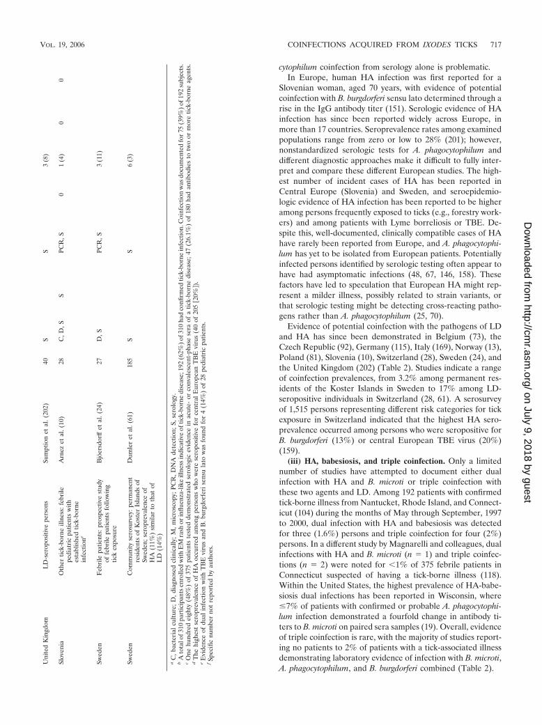
Evidence of potential coinfection with the pathogens of LDand HA has since been demonstrated in Belgium (73), theCzech Republic (92), Germany (115), Italy (169), Norway (13),Poland (81), Slovenia (10), Switzerland (28), Sweden (24), andthe United Kingdom (202) (Table 2). Studies indicate a rangeof coinfection prevalences, from 3.2% among permanent res-idents of the Koster Islands in Sweden to 17% among LD-seropositive individuals in Switzerland (28, 61). A serosurveyof 1,515 persons representing different risk categories for tickexposure in Switzerland indicated that the highest HA sero-prevalence occurred among persons who were seropositive forB. burgdorferi (13%) or central European TBE virus (20%)(159).
(iii) HA, babesiosis, and triple coinfection. Only a limitednumber of studies have attempted to document either dualinfection with HA and B. microti or triple coinfection withthese two agents and LD. Among 192 patients with confirmedtick-borne illness from Nantucket, Rhode Island, and Connect-icut (104) during the months of May through September, 1997to 2000, dual infection with HA and babesiosis was detectedfor three (1.6%) persons and triple coinfection for four (2%)persons. In a different study by Magnarelli and colleagues, dualinfections with HA and B. microti (n � 1) and triple coinfec-tions (n � 2) were noted for �1% of 375 febrile patients inConnecticut suspected of having a tick-borne illness (118).Within the United States, the highest prevalence of HA-babe-siosis dual infections has been reported in Wisconsin, where�7% of patients with confirmed or probable A. phagocytophi-lum infection demonstrated a fourfold change in antibody ti-ters to B. microti on paired sera samples (19). Overall, evidenceof triple coinfection is rare, with the majority of studies report-ing no patients to 2% of patients with a tick-associated illnessdemonstrating laboratory evidence of infection with B. microti,A. phagocytophilum, and B. burgdorferi combined (Table 2).
Uni
ted
Kin
gdom
LD
-ser
opos
itive
pers
ons
Sum
ptio
net
al.(
202)
40S
S3
(8)
Slov
enia
Oth
ertic
k-bo
rne
illne
ss:f
ebri
lepe
diat
ric
patie
nts
with
esta
blis
hed
tick-
born
ein
fect
ione
Arn
ezet
al.(
10)
28C
,D,S
SPC
R,S
01
(4)
00
Swed
enF
ebri
lepa
tient
s:pr
ospe
ctiv
est
udy
offe
brile
patie
nts
follo
win
gtic
kex
posu
re
Bjo
ersd
orff
etal
.(24
)27
D,S
PCR
,S3
(11)
Swed
enC
omm
unity
sero
surv
ey:p
erm
anen
tre
side
nts
ofK
oste
rIs
land
sof
Swed
en;s
erop
reva
lenc
eof
HA
(11%
)si
mila
rto
that
ofL
D(1
4%)
Dum
ler
etal
.(61
)18
5S
S6
(3)
aC
,bac
teri
alcu
lture
;D,d
iagn
osed
clin
ical
ly;M
,mic
rosc
opy;
PCR
,DN
Ade
tect
ion;
S,se
rolo
gy.
bA
tota
lof3
10pa
rtic
ipan
tsen
rolle
dw
ithE
Mra
shor
influ
enza
-like
illne
ssin
dica
tive
oftic
k-bo
rne
dise
ase;
192
(62%
)of3
10ha
dco
nfirm
edtic
k-bo
rne
infe
ctio
n.C
oinf
ectio
nw
asdo
cum
ente
dfo
r75
(39%
)of1
92su
bjec
ts.
cO
nehu
ndre
dei
ghty
(48%
)of
375
patie
nts
test
edde
mon
stra
ted
sero
logi
cev
iden
cein
acut
e-or
conv
ales
cent
-pha
sese
raof
atic
k-bo
rne
dise
ase;
47(2
6.1%
)of
180
had
antib
odie
sto
two
orm
ore
tick-
born
eag
ents
.d
The
high
est
sero
prev
alen
ceof
HA
occu
rred
amon
gpe
rson
sw
how
ere
sero
posi
tive
for
cent
ralE
urop
ean
TB
Evi
rus
(40
of20
5�2
0%�)
.e
Evi
denc
eof
dual
infe
ctio
nw
ithT
BE
viru
san
dB
.bur
gdor
feri
sens
ula
tow
asfo
und
for
4(1
4%)
of28
pedi
atri
cpa
tient
s.fSp
ecifi
cnu
mbe
rno
tre
port
edby
auth
ors.
VOL. 19, 2006 COINFECTIONS ACQUIRED FROM IXODES TICKS 717
on July 9, 2018 by guesthttp://cm
r.asm.org/
Dow
nloaded from
Laboratory Diagnosis of Coinfections
A number of testing methodologies are available in theclinical laboratory to aid in diagnosis and patient managementof tick-borne diseases. These methods include light microscopyfor detection of organisms in tissues or peripheral blood, mea-surement of specific antibody responses, culture isolation, andmolecularly based assays. Given the differences in geographicdistribution and prevalence of tick-borne pathogens, advocat-ing for a single testing algorithm that can be applied universallyfor the laboratory diagnosis of infections transmitted by Ixodesticks is difficult. However, consideration of disease features ina sequential manner can help narrow the differential diagnosisand effectively guide laboratory diagnosis and treatment.
A common theme is that patients with LD, HA, babesiosis,TBE virus, and other tick-transmitted diseases can all presentwith relatively nonspecific influenza-like illnesses. In these in-stances, the choice of laboratory tests should be guided by athorough patient history and physical examination that docu-ments evidence of tick exposure, place of residence, recenttravel, and objective signs and symptoms of tick-borne infec-tion. The decision whether or not to pursue specific laboratorytesting can then be based on knowledge of which tick-bornediseases are endemic in a particular area and, more impor-tantly, can be made after thoughtful assessment of the proba-bility that the patient actually has one or more tick-transmittedinfections. For example, according to the guidelines of theAmerican College of Physicians, patients with vague subjectivecomplaints (e.g., headache, fatigue, and myalgia) are consid-ered to have a low pretest probability for LD and should notundergo antibody screening by ELISA or immunofluorescenceassay (IFA), because the majority of positive results will befalse positives as a result of cross-reactivity with other micro-organisms or disease conditions (211). This is a critical con-cept, because an exaggerated perception of risk by patients andhealth care providers can result in substantial amounts of un-necessary testing and associated expense.
Despite those limitations, after it is established that a patientis at moderate to high risk for having one or more tick-trans-mitted infections, laboratory testing is indicated with only lim-ited exceptions. Patients presenting with typical primary orsecondary EM in areas where LD is endemic can often betreated empirically, and the diagnosis does not require labo-ratory confirmation. Additionally, when these patients aretreated with an antimicrobial agent (e.g., doxycycline), per-forming laboratory testing to document coinfection with HA isneither cost-effective nor necessary, because therapy is highlyeffective for both agents. In the majority of other clinical sit-uations, laboratory testing is required for definitive diagnosisand to guide therapy.
Among the most useful laboratory tests for tick-borne infec-tions are complete blood counts and peripheral blood smearsalong with tests of liver function. For patients with HA, leu-kopenia, lymphopenia, granulocytopenia, and elevated liverenzymes are commonly observed. Anemia is common in babe-siosis, and thrombocytopenia is frequently evident in babesio-sis and HA infections. Babesiosis and HA can often be diag-nosed directly by observing organisms on Giemsa-stainedsmears of peripheral blood. For patients with intact spleens,1% to 10% of erythrocytes might show B. microti ring forms on
thin blood smears; this proportion may be as high as 80% forasplenic patients (89, 126). The laboratory should screen mul-tiple slides before considering a smear to be negative. Manualmicroscopy is a subjective process, and the accuracy of theexamination depends on the vigilance and experience of theobserver, the intensity of parasitemia, and the timing of eval-uation relative to illness onset. Intracellular babesia might beconfused with Howell-Jolly bodies (121); conversely, false-pos-itive results can occur when inexperienced observers mistakeplatelets or staining artifacts for piroplasms within erythrocytesor anaplasmal morulae within granulocytes. A minority of HApatients with early, mild infection had intragranulocytic moru-lae detected on smears (21), in contrast with symptomatic,untreated patients whose smear results were evaluated afterseveral days of fever (12). With development of more sensitivemolecularly based techniques for diagnosing B. microti and A.phagocytophilum, PCR is increasingly relied on to detect infec-tion among patients with low pathogen loads and negativeblood smears.
For the majority of tick-borne infections, laboratory diagno-sis is often made by detection of IgM antibodies in specimensobtained during acute illness or by observing an increase inIgG antibody titers between acute- and convalescent-phasesamples taken 10 to 14 days apart. A significant advantage ofthis approach is that immunoserologic testing is widely avail-able and is usually cost-effective. When seeking laboratoryconfirmation of a tick-borne disease, health care professionalsshould utilize a licensed laboratory that employs strict qualitycontrol and is experienced in antibody testing. Multiple testingformats are available, including ELISA, IFA, and immunoblot-ting. For such diseases as LD, the interpretation of immuno-serologic testing has become standardized (29).
In the United States, suspected cases of extracutaneous LDare often evaluated by the CDC’s two-step protocol, wherepositive or equivocal screening results by ELISA or IFA areconfirmed by a standardized immunoblot. This approach im-proves specificity and provides sufficient information to allowrational patient management decisions in the majority of cases.In contrast, for Europe and Asia, development of a uniformapproach for the immunoserologic evaluation of LD is com-plicated, because organisms from three species of the B. burg-dorferi sensu lato genogroup can cause infections. Within thesespecies, substantial antigenic variation exists (75). For the bestperformance, immunoserologic assays need to be developedfor defined geographic areas on the basis of specific speciesand strains of B. burgdorferi sensu lato genogroup organismsthat are endemic to each area.
This review does not discuss the performance characteristicsof each of the immunoserologic assays available for Ixodes-associated infections, but a number of concerns related to thistype of testing warrant emphasis. Clinicians should recognizethat performance characteristics for these tests differ, depend-ing on the type and quality of the antigens incorporated intoeach test. Cutoff values for positive results differ among labo-ratories; furthermore, the patient population that is beingtested and the prevalence of specific infections in a particulargeographic area will affect the sensitivity and specificity of thetest. Laboratories should provide physicians with detailed in-formation on the performance characteristics for each of theassays they provide.
718 SWANSON ET AL. CLIN. MICROBIOL. REV.
on July 9, 2018 by guesthttp://cm
r.asm.org/
Dow
nloaded from
Culture isolation of Ixodes-associated pathogens from clini-cal specimens provides direct evidence of infection but can betime-consuming and expensive and is usually limited to specialcircumstances. Examples of this include confirmation of infec-tion caused by pathogens in areas where they have not previ-ously been endemic and diagnosis of reinfection for patientsfor whom the results of immunoserologic testing might not beinterpretable. The techniques involved often require specialculture media, cell lines, animal inoculation, and a high level ofbiocontainment that is not practical for the majority of clinicallaboratories. Sample requirements can also be stringent. Cli-nicians should contact their reference laboratory for guidanceif isolation of these pathogens is being considered.
Laboratories have often turned to molecular assays in anattempt to increase sensitivity and specificity and to decreasethe turnaround time for laboratory results. PCR assays areavailable for detecting nucleic acids of the agents for LD, HA,babesiosis, and TBE. An advantage of molecular detection isthe ability to diagnose early infections before the appearanceof serum antibodies, without the delay associated with cultureisolation. However, these assays have limitations as well, andassessing the probability of a tick-borne infection for PCR-based tests is as important as assessment for immunoserologicassays. False-positive and false-negative results can occur fordifferent reasons, and results need to be interpreted in thecontext of the clinical situation. Transient or limited numbersof tick-borne organisms (e.g., HA or babesiosis) within thesampled material might yield a false-negative test result. Pro-viders should be aware that there has been little standardiza-tion of molecular assays for tick-borne infections across labo-ratories, and performance characteristics are highly variable.Therefore, in the majority of cases, a negative PCR or othermolecular test result does not exclude the possibility that aninfection is present. One circumstance where PCR has beenevaluated extensively and is especially useful is the case ofLD-associated arthritis. Determining whether chronic arthritisis caused by persistent infection or a prolonged immunologicresponse is difficult. A negative PCR result from joint fluid inthis instance supports an immune-mediated etiology for per-sistent arthritis rather than an active infection requiring addi-tional antimicrobial therapy.
Pathogenesis and Immunologic Effects
Uncertainties remain regarding the pathogenesis and immu-nologic effects of coinfections among humans. No prospectivestudies have been conducted to assess the immunologic effectson humans, but experimental studies on animals reveal thatsimultaneous infection with B. burgdorferi and A. phagocytophi-lum modulates the immune response and affects the develop-ment of arthritis. In a mouse model, coinfection increases thenumber of CD4� cells and drives cytokine release toward a Thelper 1 (Th1) lymphocyte response (elevated interleukin-4[IL-4] and decreased IL-2 levels) (225). There is also evidencefrom animal models that coinfection with B. burgdorferi and A.phagocytophilum leads to increased pathogen loads in bloodand tissue, and to more-severe LD-associated arthritis, thansingle infection with B. burgdorferi alone (209). During coin-fection, murine levels of IL-12, gamma interferon, and tumornecrosis factor in serum were paradoxically decreased and
levels of IL-6 were elevated. These findings suggest that dualinfection may modulate host immune responses, such that anincreased spirochete burden results in more-severe Lyme ar-thritis. A similar study of coinfection among C3H/HeN miceevaluated the population distribution of tick-transmitted B.burgdorferi and A. phagocytophilum infection by quantitativePCR of multiple organ tissues as well as by serologic responsesto both agents (87). Among coinfected animals, spirochetenumbers increased in multiple tissues but Anaplasma numbersremained constant. Antibody responses decreased for A.phagocytophilum but not for B. burgdorferi. The researcherspostulated that coinfection modulated pathogen burdens andhost antibody responses, possibly by the ability of A. phagocy-tophilum to functionally impair neutrophils in the early defenseagainst B. burgdorferi infection.
Coinfection with B. burgdorferi and B. microti has also beendemonstrated to have immunologic effects in animal models,including alteration of the Th1 cell response and increasedseverity of arthritis (129). In another mouse model, dual infec-tion with B. burgdorferi and B. microti appeared to follow in-dependent courses (45). When young immunocompetent,young asplenic, young BALB/c, and aged C3H/HeN mice werecoinfected with B. burgdorferi and B. microti, babesiosis fol-lowed its normal course of infection without evidence of in-creased severity, as determined by the percentage of para-sitemia and other clinical and laboratory parameters. LD alsofollowed its usual course and severity among coinfected micecompared with singly infected control subjects, with no in-crease in spirochete dissemination or arthritis. In summary, theimmunologic and pathological effects in animal models areoften not generalizable to humans, and further investigation isneeded to determine the clinical implications of these findings.
Clinical Manifestations
Lyme disease and babesiosis. Early reports of babesiosisdescribed the occurrence of initial and secondary EM-like skinlesions and recurring monoarticular arthritis for some patients(23, 72), now widely recognized as clinical manifestations ofLD and indicative of undiagnosed coinfection. With the excep-tion of EM, the initial symptoms of LD can overlap withsymptoms of babesiosis. However, patients simultaneously in-fected with B. burgdorferi and B. microti appear to have morediverse, intense, and persistent symptoms. In a prospectivestudy, coinfected patients were twice as likely to report influ-enza-like symptoms (e.g., fever, chills, sweats, headaches, fa-tigue, and nausea) than patients with LD alone; they also hada higher incidence of splenomegaly (104, 106). EM was morefrequent among patients with LD alone (88%; n � 214) thanamong coinfected patients (62%; n � 26) (106). Not surpris-ingly, concurrent infection often resulted in a longer durationof illness, which is attributable, at least in part, to a delay indiagnosis of, and lack of treatment for, babesiosis (104, 106).Persistent symptoms appear to be more associated with coin-fection than with either babesiosis or LD alone, and 50% ofcoinfected patients report at least one symptom (most com-monly, extreme fatigue) for �3 months (106).
Persons coinfected with B. burgdorferi and B. microti do notappear to be at greater risk for spirochete dissemination or LDcomplications, despite the fact that B. burgdorferi DNA persists
VOL. 19, 2006 COINFECTIONS ACQUIRED FROM IXODES TICKS 719
on July 9, 2018 by guesthttp://cm
r.asm.org/
Dow
nloaded from

longer in the blood of coinfected patients after treatment(106). At least one prospective study demonstrated no evi-dence of increased joint, cardiac, or neurologic disease orhospitalization among coinfected patients compared with pa-tients with LD alone (104). This study’s findings are consistentwith a similar lack of increase in musculoskeletal and neuro-logic effects noted in other published reports of coinfectedpatients (106, 217). Case reports have discussed a more dra-matic, severe clinical course among coinfected patients, butthese likely reflect a bias toward reporting cases with moresevere or unusual manifestations (119, 131, 143, 204). All stud-ies to date have been limited in their power to detect poten-tially subtle differences in outcomes because of their relativelysmall numbers of patients. Whether clinical outcomes differamong partially treated or untreated patients with coinfectioncompared with those with LD alone remains unanswered.
In summary, possible dual infections with B. burgdorferi andB. microti should be considered for patients with a diagnosedtick-borne illness whose exposure occurred in areas where bothdiseases are endemic. Hematologic findings, such as anemiaand thrombocytopenia, are uncommon among patients withLD alone and can be indicators of Babesia coinfection (20, 76,104). Tick-borne diseases rarely occur during influenza season.Thus, the clinician should consider coinfection for any LDpatient complaining of marked influenza-like symptoms, or ifthe patient demonstrates unexplained splenomegaly, anemia,thrombocytopenia, or a failure to respond to antimicrobialtherapy directed against B. burgdorferi.
Lyme disease and HA. A limited number of case reportsdescribe B. burgdorferi-A. phagocytophilum coinfections thathave been confirmed by molecular methods or visualization ofintragranulocytic morulae. Serologic evidence alone is ofteninsufficient for confirming dual infection, because patients withacute HA infection might have a false-positive LD serology(222). The presence of IgM or IgG antibodies to B. burgdorferimight also represent prior asymptomatic infection. In an LDvaccine trial in the United States, 30 (11%) of the 269 studyparticipants who acquired LD had asymptomatic IgG serocon-version to B. burgdorferi (196). Several seroprevalence studiesin Europe have demonstrated that �50% of B. burgdorferi-seropositive persons do not recall having any symptoms of LD(64, 74, 139). Furthermore, levels of IgM and IgG antibodies toB. burgdorferi decline slowly after treatment, and these anti-bodies have been demonstrated to persist for months to yearsafter clinical cure (65, 94). Thus, LD infection might be incor-rectly diagnosed on the basis of a positive serologic test andshould be considered significant only if accompanied by a char-acteristic clinical picture.
A small cohort of 7 Rhode Island patients coinfected with B.burgdorferi and A. phagocytophilum reported significantly morechills, sweats, headaches, arthralgias, and sore throats than 89patients with LD alone (104). Coinfected patients reported asignificantly higher mean number of symptoms and a longerduration of symptoms. Although coinfection with B. burgdorferiand A. phagocytophilum might result in more severe or persis-tent symptoms (131, 147), no evidence exists of increased dis-semination of the LD spirochete among these coinfected pa-tients (104). One-third of patients with HA might have a cough(12), which is rarely reported in cases of LD (133). Leukopeniaand thrombocytopenia are commonly reported in HA (12, 104)
but would be unusual for LD patients (133, 135, 218). Elevatedliver transaminase levels can occur in both Lyme borreliosisand HA (12, 133).
As with babesiosis, clinicians should consider additional lab-oratory tests for HA when LD patients demonstrate a moreintense and persistent array of nonspecific, influenza-likesymptoms, especially fever, chills, and headache. Coinfectionwith HA is also suggested when LD patients fail to respond toappropriate �-lactam antimicrobial therapy (amoxicillin, am-picillin, or ceftriaxone) (104, 131) or demonstrate laboratoryevidence of neutropenia and thrombocytopenia (14, 104). Forsuspected coinfection with HA or babesiosis, clinicians mightconsider a complete blood count with a Giemsa-stained bloodsmear a reasonable initial evaluation.
Transfusion-Related Tick-Borne Illness
Tick-borne pathogens have been demonstrated to survivefor prolonged periods in refrigerated blood components, lead-ing to concern about the possibility of transmission of tick-borne diseases by transfusion (36, 110). More than 50 transfu-sion-related cases of infection with Babesia species (B. microti,WA1, MO1) (110, 123) and at least 1 potential case of A.phagocytophilum infection have been reported in the UnitedStates (62). Somewhat surprisingly, there are no confirmedreports of B. burgdorferi transmission by blood transfusions,despite the fact that tens of thousands of cases of LD arereported annually in North America and Europe (110, 123).
Nonetheless, viable B. burgdorferi has been identified in re-frigerated packed red blood cells, whole blood, and frozenplasma stored for �1 month (11, 63, 134), and public healthconcern exists regarding the possible risk for transfusion-re-lated transmission of LD. Less documentation exists on theviability of A. phagocytophilum in stored blood products; atleast one study documented that presumptive A. phagocytophi-lum remained viable in refrigerated blood for �18 days (93).At present, there are no known case reports on dual tick-borneinfections transmitted through transfusion of blood products.
THERAPY
Treatment of HA and LD
Doxycycline, 100 mg twice a day, has been used successfullyto treat HA and remains the treatment of choice for the ma-jority of HA infections among both children and adults. Doxy-cycline is highly active against A. phagocytophilum in vitro, andneither acquired nor inherent resistance has been described(90, 97, 122). Doxycycline is typically administered orally for 7days but can be intravenously administered for more severecases. Despite concerns about dental staining in children, doxy-cycline remains the drug of choice for treating HA infections,providing superior therapy for a potentially life-threateningdisease; further, available data suggest that short courses ofdoxycycline (�14 days) do not cause significant discolorationof permanent teeth (152). However, caution should be exer-cised during pregnancy, because of concerns of possible ad-verse effects on fetal skeletal development, bone growth, andenamel hypoplasia.
Under special non-life-threatening circumstances, rifampin
720 SWANSON ET AL. CLIN. MICROBIOL. REV.
on July 9, 2018 by guesthttp://cm
r.asm.org/
Dow
nloaded from

can be considered an alternative to doxycycline for treatmentof HA (70). Both rifampin and levofloxacin demonstrate ex-cellent in vitro activity against A. phagocytophilum (90); how-ever, clinical experience and documentation of effectivenessare limited for rifampin and nonexistent for levofloxacin. Theuse of rifampin in treating HA during pregnancy (30) and fortwo pediatric patients (102) has been described; no data existregarding the effectiveness of levofloxacin in treating HA.Chloramphenicol demonstrates suboptimal in vitro activityagainst A. phagocytophilum (122), and treatment failures withchloramphenicol have been reported (60). A. phagocytophilumis resistant to numerous antimicrobials commonly used in clin-ical settings, including aminoglycosides (gentamicin, amika-cin), �-lactam antibiotics (amoxicillin, ampicillin, cefuroximeaxetil, and ceftriaxone), macrolides (erythromycin, azithromy-cin, clarithromycin), clindamycin, and trimethoprim-sulfa-methoxazole (90, 122).
Doxycycline has the additional benefit of being highly activeagainst B. burgdorferi. Longer courses of doxycycline therapyare required for treatment of LD coinfection, with a 14-daycourse for early localized infection with B. burgdorferi (EM)and longer regimens (�28 days) for disseminated or late com-plications. Other antibiotics commonly used in treating earlyand late LD among both pediatric and adult populations (e.g.,amoxicillin and ceftriaxone) are ineffective in treating HA. Incontrast, doxycycline not only is highly active against both A.phagocytophilum and B. burgdorferi but also has the advantageof being active against other tick-borne infections, such asehrlichiosis, Rocky Mountain spotted fever, other spotted fe-ver group rickettsioses, and tick-borne relapsing fever (causedby Borrelia species). For patients with HA who have clinical orlaboratory features indicative of coinfection with B. burgdorferi,a longer course of doxycycline (14 to 21 days) is recommended,according to evidence-based treatment recommendations forLD presented by the Infectious Diseases Society of America(223). The ultimate choice and duration of antimicrobial ther-apy should be guided by the stage of LD. Clinicians might wantto empirically treat HA patients from regions of LD endemic-ity with 14 days of doxycycline, because B. burgdorferi coinfec-tion is difficult to rule out during the acute illness phase, and ithas been reported for as many as 10% of HA patients (128).
Treatment of Babesiosis
Patients infected by B. microti often remain asymptomatic orexperience only mild illness and recover without specific che-motherapy. The combination of oral clindamycin (for adults,600 mg every 8 h; for children, 20 to 40 mg/kg of body weight/day, divided into three daily doses) with oral quinine (foradults, 650 mg every 8 h; for children, 25 mg/kg/day, dividedinto three daily doses), taken for 7 to 10 days, appears to be aneffective chemotherapeutic regimen for the less severely illpatient, although potential toxicities with this regimen are clin-ically significant (103, 124). Use of intravenous clindamycin(for adults, 300 to 600 mg every 6 h; for children, 20 to 40mg/kg/day in three doses) with oral quinine remains the rec-ommended and preferred regimen for the more severely illpatient (101). In treating children, careful attention must begiven to the calculation of dosing. The pediatric dose of med-
ication, as a general principle, must not exceed the recom-mended adult dose.
More recently, combination therapy with atovaquone (foradults, 750 mg every 12 h with meals; for children, 20 mg/kgevery 12 h with meals) and azithromycin (for adults, 500 mg onday 1 and 250 mg/day thereafter; for children, 12 mg/kg/day)for 7 days has been demonstrated to be as effective as 7 daysof combination therapy with clindamycin-quinine for patientswith non-life-threatening B. microti infections (103, 124).Higher doses of azithromycin (500 to 1,000 mg daily) and 10days of total combination therapy with atovaquone have beenemployed also (124, 219). Fifteen percent of patients on atova-quone-azithromycin reported adverse reactions, most com-monly diarrhea and rash; clindamycin-quinine was associatedwith adverse reactions (tinnitus, diarrhea, and decreased hear-ing) for 72% of patients (103).
Partial or complete exchange transfusions have been usedfor severely ill babesiosis patients with substantial hemolysis,renal or pulmonary compromise, or high levels of parasitemia(�10%) (26, 78, 101). When given concurrently with appro-priate chemotherapy, whole-blood exchange transfusion mightbe life-saving (58). Human infections with the bovine pathogenB. divergens should also be regarded as a medical emergency,and prompt therapy is indicated. Exchange transfusions incombination with intravenous clindamycin and intravenous ororal quinine have been successful in treating B. divergens inEurope (26, 27).
Certain antiprotozoal and antimalarial therapeutic regimenshave been largely unsuccessful, including chloroquine, prima-quine, pyrimethamine, pyrimethamine-sulfadoxine, tetracycline,minocycline, pentamidine isethionate, and trimethoprim-sulfa-methoxazole (100, 101). Clinicians should be aware that treat-ment for babesiosis will not provide effective treatment for LD orHA. Neither clindamycin, quinine, atovaquone, nor azithromycinprovides any activity against B. burgdorferi or A. phagocytophilum.Under these circumstances, patients with B. microti/divergens in-fections who are suspected of also having LD or HA require anadditional antimicrobial (e.g., doxycycline) (131).
STRATEGIES FOR PREVENTING COINFECTIONSFROM IXODES TICKS
Prevention of LD and other diseases transmitted by Ixodesticks has posed a substantial public health challenge. The ma-jority of prevention programs stress personal protection mea-sures such as tick habitat avoidance (including information onwhen and where people are at risk), protective clothing, repel-lents, and frequent tick checks to locate and remove ticksbefore they can transmit disease. Although these efforts areuseful, completely avoiding tick habitats, maintaining regularrepellent use, wearing protective clothing in warm weather, orlocating attached ticks (especially smaller nymphs) often canbe impractical or impossible (157). Research demonstratingthe efficacy of personal protection measures is lacking, andtick-borne disease case numbers continue to increase in partsof the world where such diseases are endemic (79).
While not routinely recommended, a single-dose prophylac-tic antibiotic treatment (e.g., doxycycline) of known tick bitesto prevent potential LD is often practiced in the United States.At present, tick bite antibiotic prophylaxis is the only available
VOL. 19, 2006 COINFECTIONS ACQUIRED FROM IXODES TICKS 721
on July 9, 2018 by guesthttp://cm
r.asm.org/
Dow
nloaded from

preventive measure proven effective in preventing LD (132).However, doxycycline should be taken only by individuals whohave had a recognized Ixodes tick bite within the previous 72 h,and where there is reasonable information to suggest that thetick remained attached long enough (24 to 48 h) to potentiallytransmit B. burgdorferi if it was infected. Medical providersshould remain mindful of the facts that the majority of LDpatients do not recall having had a tick bite and the probabilityof LD transmission from any given tick bite is usually low(176). In addition, this practice has not been evaluated inEurope or Asia for B. burgdorferi sensu lato species or for othertick-borne diseases.
An LD vaccine was made available to the public in 1999 butwas withdrawn from the market in 2002 after a decline in salescaused by concerns about the vaccine’s safety and usefulness.Because of the difficulty of preventing tick exposure, the ma-jority of tick-borne disease prevention programs stress earlyrecognition and treatment of tick-borne disease cases.
The management of ticks in the environment for the pre-vention of LD and other tick-borne diseases has been reviewedat length (69, 79). Tick control measures (e.g., acaricides ap-plied to tick habitats or hosts) are available; however, they canbe costly, and often landowners are reluctant to use pesticideson their property. Habitat modification (e.g., burning tick hab-itat, leaf litter and brush removal, or landscaping to create drybarriers) can be part of prevention efforts, but these efforts arelabor-intensive and require routine maintenance. Given theincreasing numbers of tick-borne disease cases despite multi-ple prevention measures, human coinfections with tick-bornepathogens will likely continue to be a public health probleminto the foreseeable future.
RESEARCH NEEDS
Our knowledge of individual tick-borne zoonoses has in-creased dramatically during the past 2 decades, but relativelylittle progress has been made in our understanding of coinfec-tions. Funding agencies should focus on research that can beapplied to the recognition, treatment, or prevention of tick-borne coinfections among humans. Additional prospectivestudies are needed in North America and Europe to assess theimmunologic and clinical effects of the most common coinfec-tions, including Lyme borreliosis with either HA or babesiosis.The sensitivity, specificity, and predictive value of laboratorymarkers of coinfection should be evaluated to facilitate promptclinical detection.
In addition, clarification is needed on the geographic distri-bution of B. burgdorferi sensu lato genogroup, B. microti/diver-gens, and A. phagocytophilum for all areas of the world, includ-ing nontemperate climates. Limited or no information existson the incidence or effects of coinfection with other tick-bornepathogens (e.g., TBE and Powassan flaviviruses, spotted fevergroup rickettsiae, Fransicella tularensis, and Bartonella spp.).Prospective cohort studies involving healthy persons with fre-quent exposure to tick habitats are needed to assess the abso-lute risk for coinfection. This risk might vary in time and spacebecause of increasing human exposure, climate and habitatchange, and the expanding geographic distribution of Ixodesticks. Finally, research is needed to understand the role ofgenetic heterogeneity among coinfecting pathogens. In partic-ular, further efforts are needed to assess competition between
variant clones of A. phagocytophilum and the impact of theseclones on disease transmission and severity.
ACKNOWLEDGMENTS
We are indebted for help with this article to Melissa Kemperman,David Determan, and Kirk E. Smith (Minnesota Department of Health,Minneapolis) and to Joseph Piesman, Walter R. Daley, and C. Kay Smith-Akin (Centers for Disease Control and Prevention, Atlanta, Ga.).
The findings and conclusions in this report are those of the authorsand do not necessarily represent the views of the Centers for DiseaseControl and Prevention.
REFERENCES
1. Adelson, M. E., R. V. Rao, R. C. Tilton, K. Cabets, E. Eskow, L. Fein, J. L.Occi, and E. Mordechai. 2004. Prevalence of Borrelia burgdorferi, Bartonellaspp., Babesia microti, and Anaplasma phagocytophila in Ixodes scapularisticks collected in Northern New Jersey. J. Clin. Microbiol. 42:2799–2801.
2. Afzelius, A. 1921. Erythema chronicum migrans. Acta Derm. Venereol.2:120–125.
3. Aguero-Rosenfeld, M. E., L. Donnarumma, L. Zentmaier, J. Jacob, M.Frey, R. Noto, C. A. Carbonaro, and G. P. Wormser. 2002. Seroprevalenceof antibodies that react with Anaplasma phagocytophila, the agent of humangranulocytic ehrlichiosis, in different populations in Westchester County,New York. J. Clin. Microbiol. 40:2612–2615.
4. Aguero-Rosenfeld, M. E., G. Wang, I. Schwartz, and G. P. Wormser. 2005.Diagnosis of Lyme borreliosis. Clin. Microbiol. Rev. 18:484–509.
5. Ai, C. X., W. F. Zhang, and J. H. Zhao. 1994. Sero-epidemiology of Lymedisease in an endemic area in China. Microbiol. Immunol. 38:505–509.
6. Alekseev, A. N., A. V. Semenov, and H. V. Dubinina. 2003. Evidence ofBabesia microti infection in multi-infected Ixodes persulcatus ticks in Russia.Exp. Appl. Acarol. 29:345–353.
7. Anderson, J. F., R. C. Johnson, L. A. Magnarelli, and F. W. Hyde. 1985.Identification of endemic foci of Lyme disease: isolation of Borrelia burg-dorferi from feral rodents and ticks (Dermacentor variabilis). J. Clin. Micro-biol. 22:36–38.
8. Arai, S., M. Tsuji, I. Kaiho, H. Murayama, A. Zamoto, Q. Wei, N. Okabe,T. Kamiyama, and C. Ishihara. 2003. Retrospective seroepidemiologicalsurvey for human babesiosis in an area in Japan where a tick-borne diseaseis endemic. J. Vet. Med. Sci. 65:335–340.
9. Armstrong, P. M., P. Katavolos, D. A. Caporale, R. P. Smith, A. Spielman,and S. R. Telford III. 1998. Diversity of Babesia infecting deer ticks (Ixodesdammini). Am. J. Trop. Med. Hyg. 58:739–742.
10. Arnez, M., T. Luznik-Bufon, T. Avsic-Zupanc, E. Ruzic-Sabljic, M.Petrovec, S. Lotric-Furlan, and F. Strle. 2003. Causes of febrile illnessesafter a tick bite in Slovenian children. Pediatr. Infect. Dis. J. 22:1078–1083.
11. Badon, S. J., R. D. Fister, and R. G. Cable. 1989. Survival of Borreliaburgdorferi in blood products. Transfusion 29:581–583.
12. Bakken, J. S., J. S. Dumler, S. M. Chen, M. R. Eckman, L. L. Van Etta, andD. H. Walker. 1994. Human granulocytic ehrlichiosis in the upper MidwestUnited States. A new species emerging? JAMA 272:212–218.
13. Bakken, J. S., J. Krueth, R. L. Tilden, J. S. Dumler, and B. E. Kristiansen.1996. Serological evidence of human granulocytic ehrlichiosis in Norway.Eur. J. Clin. Microbiol. Infect. Dis. 15:829–832.
14. Bakken, J. S., J. Krueth, C. Wilson-Nordskog, R. L. Tilden, K. Asanovich,and J. S. Dumler. 1996. Clinical and laboratory characteristics of humangranulocytic ehrlichiosis. JAMA 275:199–205.
15. Baranton, G., D. Postic, I. Saint Girons, P. Boerlin, J. C. Piffaretti, M.Assous, and P. A. Grimont. 1992. Delineation of Borrelia burgdorferi sensustricto, Borrelia garinii sp. nov., and group VS461 associated with Lymeborreliosis. Int. J. Syst. Bacteriol. 42:378–383.
16. Barbour, A. G. 1993. Linear DNA of Borrelia species and antigenic varia-tion. Trends Microbiol. 1:236–239.
17. Baumgarten, B. U., M. Rollinghoff, and C. Bogdan. 1999. Prevalence ofBorrelia burgdorferi and granulocytic and monocytic ehrlichiae in Ixodesricinus ticks from southern Germany. J. Clin. Microbiol. 37:3448–3451.
18. Belongia, E. A., C. M. Gale, K. D. Reed, P. D. Mitchell, M. Vandermause,M. F. Finkel, J. J. Kazmierczak, and J. P. Davis. 2001. Population-basedincidence of human granulocytic ehrlichiosis in northwestern Wisconsin,1997–1999. J. Infect. Dis. 184:1470–1474.
19. Reference deleted.20. Belongia, E. A., K. D. Reed, P. D. Mitchell, P. H. Chyou, N. Mueller-Rizner,
M. F. Finkel, and M. E. Schriefer. 1999. Clinical and epidemiologicalfeatures of early Lyme disease and human granulocytic ehrlichiosis inWisconsin. Clin. Infect. Dis. 29:1472–1477.
21. Belongia, E. A., K. D. Reed, P. D. Mitchell, N. Mueller-Rizner, M. Vander-mause, M. F. Finkel, and J. J. Kazmierczak. 2001. Tickborne infections asa cause of nonspecific febrile illness in Wisconsin. Clin. Infect. Dis. 32:1434–1439.
22. Benach, J. L., J. L. Coleman, G. S. Habicht, A. MacDonald, E. Grunwaldt,
722 SWANSON ET AL. CLIN. MICROBIOL. REV.
on July 9, 2018 by guesthttp://cm
r.asm.org/
Dow
nloaded from

and J. A. Giron. 1985. Serological evidence for simultaneous occurrences ofLyme disease and babesiosis. J. Infect. Dis. 152:473–477.
23. Benach, J. L., and G. S. Habicht. 1981. Clinical characteristics of humanbabesiosis. J. Infect. Dis. 144:481.
24. Bjoersdorff, A., B. Wittesjo, J. Berglun, R. F. Massung, and I. Eliasson.2002. Human granulocytic ehrlichiosis as a common cause of tick-associ-ated fever in Southeast Sweden: report from a prospective clinical study.Scand. J. Infect. Dis. 34:187–191.
25. Blanco, J. R., and J. A. Oteo. 2002. Human granulocytic ehrlichiosis inEurope. Clin. Microbiol. Infect. 8:763–772.
26. Bonoan, J. T., D. H. Johnson, and B. A. Cunha. 1998. Life-threateningbabesiosis in an asplenic patient treated with exchange transfusion, azithro-mycin, and atovaquone. Heart Lung 27:424–428.
27. Brasseur, P., and A. Gorenflot. 1992. Human babesiosis in Europe. Mem.Inst. Oswaldo Cruz 87(Suppl. 3):131–132.
28. Brouqui, P., J. S. Dumler, R. Lienhard, M. Brossard, and D. Raoult. 1995.Human granulocytic ehrlichiosis in Europe. Lancet 346:782–783.
28a.Brown, R. N., R. S. Lane, and D. T. Dennis. 2005. Geographic distributionsof tick-borne diseases and their vectors, p. 363–391. In J. Goodman, D. T.Dennis, and D. Sonenshine (ed.), Tick-borne diseases of humans. ASMPress, Washington, D.C.
29. Brown, S. L., S. L. Hansen, and J. J. Langone. 1999. Role of serology in thediagnosis of Lyme disease. JAMA 282:62–66.
30. Buitrago, M. I., J. W. Ijdo, P. Rinaudo, H. Simon, J. Copel, J. Gadbaw, R.Heimer, E. Fikrig, and F. J. Bia. 1998. Human granulocytic ehrlichiosisduring pregnancy treated successfully with rifampin. Clin. Infect. Dis. 27:213–215.
31. Burgdorfer, W., A. G. Barbour, S. F. Hayes, J. L. Benach, E. Grunwaldt,and J. P. Davis. 1982. Lyme disease—a tick-borne spirochetosis? Science216:1317–1319.
32. Burgdorfer, W., and L. P. Brinton. 1975. Mechanisms of transovarial in-fection of spotted fever Rickettsiae in ticks. Ann. N. Y. Acad. Sci. 266:61–72.
33. Burkot, T. R., G. O. Maupin, B. S. Schneider, C. Denatale, C. M. Happ,J. S. Rutherford, and N. S. Zeidner. 2001. Use of a sentinel host system tostudy the questing behavior of Ixodes spinipalpis and its role in the trans-mission of Borrelia bissettii, human granulocytic ehrlichiosis, and Babesiamicroti. Am. J. Trop. Med. Hyg. 65:293–299.
34. Burkot, T. R., M. E. Schriefer, and S. A. Larsen. 1997. Cross-reactivity toBorrelia burgdorferi proteins in serum samples from residents of a tropicalcountry nonendemic for Lyme disease. J. Infect. Dis. 175:466–469.
35. Bush, J. B., M. Isaacson, A. S. Mohamed, F. T. Potgieter, and D. T. deWaal. 1990. Human babesiosis—a preliminary report of 2 suspected casesin South Africa. S. Afr. Med. J. 78:699.
36. Cable, R. G., and D. A. Leiby. 2003. Risk and prevention of transfusion-transmitted babesiosis and other tick-borne diseases. Curr. Opin. Hematol.10:405–411.
37. Cao, W. C., Q. M. Zhao, P. H. Zhang, H. Yang, X. M. Wu, B. H. Wen, X. T.Zhang, and J. D. Habbema. 2003. Prevalence of Anaplasma phagocytophilaand Borrelia burgdorferi in Ixodes persulcatus ticks from northeastern China.Am. J. Trop. Med. Hyg. 68:547–550.
38. CDC. 1996. Lyme disease—United States, 1995. Morb. Mortal. Wkly. Rep.45:481–484.
39. Chen, S. M., J. S. Dumler, J. S. Bakken, and D. H. Walker. 1994. Identi-fication of a granulocytotropic Ehrlichia species as the etiologic agent ofhuman disease. J. Clin. Microbiol. 32:589–595.
40. Christova, I., and T. Gladnishka. 2005. Prevalence of infection with Francisellatularensis, Borrelia burgdorferi sensu lato and Anaplasma phagocytophilum inrodents from an endemic focus of tularemia in Bulgaria. Ann. Agric. Environ.Med. 12:149–152.
41. Christova, I., L. Schouls, I. van De Polqq, J. Park, S. Panayotov, V.Lefterova, T. Kantardjiev, and J. S. Dumler. 2001. High prevalence ofgranulocytic ehrlichiae and Borrelia burgdorferi sensu lato in Ixodes ricinusticks from Bulgaria. J. Clin. Microbiol. 39:4172–4174.
42. Christova, I. S., and J. S. Dumler. 1999. Human granulocytic ehrlichiosis inBulgaria. Am. J. Trop. Med. Hyg. 60:58–61.
43. Cinco, M., D. Padovan, R. Murgia, M. Heldtander, and E. O. Engvall. 1998.Detection of HGE agent-like Ehrlichia in Ixodes ricinus ticks in northernItaly by PCR. Wien Klin. Wochenschr. 110:898–900.
44. Cinco, M., D. Padovan, R. Murgia, M. Maroli, L. Frusteri, M. Heldtander,K. E. Johansson, and E. O. Engvall. 1997. Coexistence of Ehrlichia phago-cytophila and Borrelia burgdorferi sensu lato in Ixodes ricinus ticks from Italyas determined by 16S rRNA gene sequencing. J. Clin. Microbiol. 35:3365–3366.
45. Coleman, J. L., D. Levine, C. Thill, C. Kuhlow, and J. L. Benach. 2005.Babesia microti and Borrelia burgdorferi follow independent courses of in-fection in mice. J. Infect. Dis. 192:1634–1641.
46. Courtney, J. W., R. L. Dryden, J. Montgomery, B. S. Schneider, G. Smith,and R. F. Massung. 2003. Molecular characterization of Anaplasma phago-cytophilum and Borrelia burgdorferi in Ixodes scapularis ticks from Pennsyl-vania. J. Clin. Microbiol. 41:1569–1573.
47. Csango, P. A., E. Blakstad, G. C. Kirtz, J. E. Pedersen, and B. Czettel. 2004.
Tick-borne encephalitis in southern Norway. Emerg. Infect. Dis. 10:533–534.
48. Daniel, S. A., K. Manika, M. Arvanitidou, E. Diza, N. Symeonidis, and A.Antoniadis. 2002. Serologic evidence of human granulocytic ehrlichiosis,Greece. Emerg. Infect. Dis. 8:643–644.
49. Daniels, T. J., T. M. Boccia, S. Varde, J. Marcus, J. Le, D. J. Bucher, R. C.Falco, and I. Schwartz. 1998. Geographic risk for Lyme disease and humangranulocytic ehrlichiosis in southern New York state. Appl. Environ. Mi-crobiol. 64:4663–4669.
50. De Martino, S. J., J. A. Carlyon, and E. Fikrig. 2001. Coinfection withBorrelia burgdorferi and the agent of human granulocytic ehrlichiosis.N. Engl. J. Med. 345:150–151.
51. Dennis, D. T., T. S. Nekomoto, J. C. Victor, W. S. Paul, and J. Piesman.1998. Reported distribution of Ixodes scapularis and Ixodes pacificus (Acari:Ixodidae) in the United States. J. Med. Entomol. 35:629–638.
52. Dennis, D. T., and J. F. Piesman. 2005. Overview of tick-borne infections ofhumans, p. 3–11. In J. L. Goodman, D. T. Dennis, and D. E. Sonenshine(ed.), Tick-borne diseases of humans. ASM Press, Washington, D.C.
53. Derdakova, M., V. Dudioak, B. Brei, J. S. Brownstein, I. Schwartz, and D.Fish. 2004. Interaction and transmission of two Borrelia burgdorferi sensustricto strains in a tick-rodent maintenance system. Appl. Environ. Micro-biol. 70:6783–6788.
54. Derdakova, M., M. Halanova, M. Stanko, A. Stefancikova, L. Cislakova,and B. Pet’ko. 2003. Molecular evidence for Anaplasma phagocytophilumand Borrelia burgdorferi sensu lato in Ixodes ricinus ticks from easternSlovakia. Ann. Agric. Environ. Med. 10:269–271.
55. Des Vignes, F., M. L. Levin, and D. Fish. 1999. Comparative vector com-petence of Dermacentor variabilis and Ixodes scapularis (Acari: Ixodidae) forthe agent of human granulocytic ehrlichiosis. J. Med. Entomol. 36:182–185.
56. Dolan, M. C., J. Piesman, M. L. Mbow, G. O. Maupin, O. Peter, M.Brossard, and W. T. Golde. 1998. Vector competence of Ixodes scapularisand Ixodes ricinus (Acari: Ixodidae) for three genospecies of Borrelia burg-dorferi. J. Med. Entomol. 35:465–470.
57. Donnelly, J., and M. A. Peirce. 1975. Experiments on the transmission ofBabesia divergens to cattle by the tick Ixodes ricinus. Int. J. Parasitol. 5:363–367.
58. Dorman, S. E., M. E. Cannon, S. R. Telford III, K. M. Frank, and W. H.Churchill. 2000. Fulminant babesiosis treated with clindamycin, quinine,and whole-blood exchange transfusion. Transfusion 40:375–380.
59. Duh, D., M. Petrovec, and T. Avsic-Zupanc. 2001. Diversity of Babesiainfecting European sheep ticks (Ixodes ricinus). J. Clin. Microbiol. 39:3395–3397.
60. Dumler, J. S., and J. S. Bakken. 1995. Ehrlichial diseases of humans:emerging tick-borne infections. Clin. Infect. Dis. 20:1102–1110.
61. Dumler, J. S., L. Dotevall, R. Gustafson, and M. Granstrom. 1997. Apopulation-based seroepidemiologic study of human granulocytic ehrlichio-sis and Lyme borreliosis on the west coast of Sweden. J. Infect. Dis. 175:720–722.
62. Eastlund, T., D. Persing, D. Mathiesen, D. Kim, J. Bieging, P. McCann, G.Heiler, and S. Raynovic. 1999. Human granulocytic ehrlichiosis after redcell transfusion. Transfusion 39(Suppl.):117S.
63. Eberhard, M. L., E. M. Walker, and F. J. Steurer. 1995. Survival andinfectivity of Babesia in blood maintained at 25°C and 2–4°C. J. Parasitol.81:790–792.
64. Fahrer, H., S. M. van der Linden, M. J. Sauvain, L. Gern, E. Zhioua, andA. Aeschlimann. 1991. The prevalence and incidence of clinical and asymp-tomatic Lyme borreliosis in a population at risk. J. Infect. Dis. 163:305–310.
65. Feder, H. M., Jr., M. A. Gerber, S. W. Luger, and R. W. Ryan. 1992.Persistence of serum antibodies to Borrelia burgdorferi in patients treatedfor Lyme disease. Clin. Infect. Dis. 15:788–793.
66. Felz, M. W., L. A. Durden, and J. H. Oliver, Jr. 1996. Ticks parasitizinghumans in Georgia and South Carolina. J. Parasitol. 82:505–508.
67. Fingerle, V., J. L. Goodman, R. C. Johnson, T. J. Kurtti, U. G. Munderloh,and B. Wilske. 1997. Human granulocytic ehrlichiosis in southern Germany:increased seroprevalence in high-risk groups. J. Clin. Microbiol. 35:3244–3247.
68. Fingerle, V., U. G. Munderloh, G. Liegl, and B. Wilske. 1999. Coexistenceof ehrlichiae of the phagocytophila group with Borrelia burgdorferi in Ixodesricinus from Southern Germany. Med. Microbiol. Immunol. (Berlin) 188:145–149.
69. Ginsberg, H. S., and K. C. Stafford III. 2005. Management of ticks andtick-borne diseases, p. 65–86. In J. L. Goodman, D. T. Dennis, and D. E.Sonenshine (ed.), Tick-borne diseases of humans. ASM Press, Washington,D.C.
70. Goodman, J. L. 2005. Human granulocytic anaplasmosis (ehrlichiosis), p.218–238. In J. L. Goodman, D. T. Dennis, and D. E. Sonenshine (ed.),Tick-borne diseases of humans. ASM Press, Washington, D.C.
71. Gorenflot, A., K. Moubri, E. Precigout, B. Carcy, and T. P. Schetters. 1998.Human babesiosis. Ann. Trop. Med. Parasitol. 92:489–501.
72. Grunwaldt, E., A. G. Barbour, and J. L. Benach. 1983. Simultaneous oc-currence of babesiosis and Lyme disease. N. Engl. J. Med. 308:1166.
73. Guillaume, B., P. Heyman, S. Lafontaine, C. Vandenvelde, M. Delmee, and
VOL. 19, 2006 COINFECTIONS ACQUIRED FROM IXODES TICKS 723
on July 9, 2018 by guesthttp://cm
r.asm.org/
Dow
nloaded from

G. Bigaignon. 2002. Seroprevalence of human granulocytic ehrlichiosis in-fection in Belgium. Eur. J. Clin. Microbiol. Infect. Dis. 21:397–400.
74. Gustafson, R., B. Svenungsson, A. Gardulf, G. Stiernstedt, and M. Fors-gren. 1990. Prevalence of tick-borne encephalitis and Lyme borreliosis in adefined Swedish population. Scand. J. Infect. Dis. 22:297–306.
75. Gutierrez Fernandez, J., M. Rodriguez Fernandez, F. Nunez Murillo, andM. C. Maroto Vela. 1997. Antigenic and genetic structure of Borrelia burg-dorferi. Microbios 91:165–174.
76. Gutman, J. D., C. N. Kotton, and A. Kratz. 2003. Case records of theMassachusetts General Hospital. Weekly clinicopathological exercises.Case 29-2003. A 60-year-old man with fever, rigors, and sweats. N. Engl.J. Med. 349:1168–1175.
77. Halos, L., T. Jamal, R. Maillard, F. Beugnet, A. Le Menach, H. J. Boulouis,and M. Vayssier-Taussat. 2005. Evidence of Bartonella sp. in questing adultand nymphal Ixodes ricinus ticks from France and co-infection with Borreliaburgdorferi sensu lato and Babesia sp. Vet. Res. 36:79–87.
78. Hatcher, J. C., P. D. Greenberg, J. Antique, and V. E. Jimenez-Lucho. 2001.Severe babesiosis in Long Island: review of 34 cases and their complica-tions. Clin. Infect. Dis. 32:1117–1125.
79. Hayes, E. B., and J. Piesman. 2003. How can we prevent Lyme disease?N. Engl. J. Med. 348:2424–2430.
80. Healy, G. 1989. The impact of cultural and environmental changes on theepidemiology and control of human babesiosis. Trans. R. Soc. Trop. Med.Hyg. 83(Suppl.):35–38.
81. Hermanowska-Szpakowicz, T., B. Skotarczak, M. Kondrusik, A. Rymasze-wska, M. Sawczuk, A. Maciejewska, M. Adamska, S. Pancewicz, and J.Zajkowska. 2004. Detecting DNAs of Anaplasma phagocytophilum andBabesia in the blood of patients suspected of Lyme disease. Ann. Agric.Environ. Med. 11:351–354.
82. Herwaldt, B., D. H. Persing, E. A. Precigout, W. L. Goff, D. A. Mathiesen,P. W. Taylor, M. L. Eberhard, and A. F. Gorenflot. 1996. A fatal case ofbabesiosis in Missouri: identification of another piroplasm that infects hu-mans. Ann. Intern. Med. 124:643–650.
83. Hildebrandt, A., K. H. Schmidt, B. Wilske, W. Dorn, E. Straube, and V.Fingerle. 2003. Prevalence of four species of Borrelia burgdorferi sensu latoand coinfection with Anaplasma phagocytophila in Ixodes ricinus ticks incentral Germany. Eur. J. Clin. Microbiol. Infect. Dis. 22:364–367.
84. Hilton, E., J. DeVoti, J. L. Benach, M. L. Halluska, D. J. White, H. Paxton,and J. S. Dumler. 1999. Seroprevalence and seroconversion for tick-bornediseases in a high-risk population in the northeast United States. Am. J.Med. 106:404–409.
85. Hofmeister, E. K., C. P. Kolbert, A. S. Abdulkarim, J. M. Magera, M. K.Hopkins, J. R. Uhl, A. Ambyaye, S. R. Telford III, F. R. Cockerill III, andD. H. Persing. 1998. Cosegregation of a novel Bartonella species withBorrelia burgdorferi and Babesia microti in Peromyscus leucopus. J. Infect.Dis. 177:409–416.
86. Holden, K., J. T. Boothby, S. Anand, and R. F. Massung. 2003. Detectionof Borrelia burgdorferi, Ehrlichia chaffeensis, and Anaplasma phagocytophi-lum in ticks (Acari: Ixodidae) from a coastal region of California. J. Med.Entomol. 40:534–539.
87. Holden, K., E. Hodzic, S. Feng, K. J. Freet, R. B. Lefebvre, and S. W.Barthold. 2005. Coinfection with Anaplasma phagocytophilum alters Borre-lia burgdorferi population distribution in C3H/HeN mice. Infect. Immun.73:3440–3444.
88. Holman, M. S., D. A. Caporale, J. Goldberg, E. Lacombe, C. Lubelczyk,P. W. Rand, and R. P. Smith. 2004. Anaplasma phagocytophilum, Babesiamicroti, and Borrelia burgdorferi in Ixodes scapularis, southern coastalMaine. Emerg. Infect. Dis. 10:744–746.
89. Homer, M. J., I. Aguilar-Delfin, S. R. Telford III, P. J. Krause, and D. H.Persing. 2000. Babesiosis. Clin. Microbiol. Rev. 13:451–469.
90. Horowitz, H. W., T. C. Hsieh, M. E. Aguero-Rosenfeld, F. Kalantarpour, I.Chowdhury, G. P. Wormser, and J. M. Wu. 2001. Antimicrobial suscepti-bility of Ehrlichia phagocytophila. Antimicrob. Agents Chemother. 45:786–788.
91. Hotopp, J. C., M. Lin, R. Madupu, J. Crabtree, S. V. Angiuoli, J. Eisen, R.Seshadri, Q. Ren, M. Wu, T. R. Utterback, S. Smith, M. Lewis, H. Khouri,C. Zhang, H. Niu, Q. Lin, N. Ohashi, N. Zhi, W. Nelson, L. M. Brinkac,R. J. Dodson, M. J. Rosovitz, J. Sundaram, S. C. Daugherty, T. Davidsen,A. S. Durkin, M. Gwinn, D. H. Haft, J. D. Selengut, S. A. Sullivan, N. Zafar,L. Zhou, F. Benahmed, H. Forberger, R. Halpin, S. Mulligan, J. Robinson,O. White, Y. Rikihisa, and H. Tettelin. 2006. Comparative genomics ofemerging human ehrlichiosis agents. PLoS Genet. 2:e21.
92. Hulinska, D., J. Votypka, J. Plch, E. Vlcek, M. Valesova, M. Bojar, V.Hulinsky, and K. Smetana. 2002. Molecular and microscopical evidence ofEhrlichia spp. and Borrelia burgdorferi sensu lato in patients, animals andticks in the Czech Republic. New Microbiol. 25:437–448.
93. Kalantarpour, F., I. Chowdhury, G. P. Wormser, and M. E. Aguero-Rosen-feld. 2000. Survival of the human granulocytic ehrlichiosis agent underrefrigeration conditions. J. Clin. Microbiol. 38:2398–2399.
94. Kalish, R. A., G. McHugh, J. Granquist, B. Shea, R. Ruthazer, and A. C.Steere. 2001. Persistence of immunoglobulin M or immunoglobulin G an-
tibody responses to Borrelia burgdorferi 10–20 years after active Lyme dis-ease. Clin. Infect. Dis. 33:780–785.
95. Keirans, J. E., and L. A. Durden. 2005. Tick systematics and identification,p. 123–140. In J. L. Goodman, D. T. Dennis, and D. E. Sonenshine (ed.),Tick-borne diseases of humans. ASM Press, Washington, D.C.
96. Keirans, J. E., G. R. Needham, and J. H. Oliver, Jr. 1999. The Ixodes(Ixodes) ricinus complex worldwide: diagnosis of the species in the complex,hosts and distribution. Ohio Biological Survey, Columbus.
97. Klein, M. B., C. M. Nelson, and J. L. Goodman. 1997. Antibiotic suscepti-bility of the newly cultivated agent of human granulocytic ehrlichiosis:promising activity of quinolones and rifamycins. Antimicrob. Agents Che-mother. 41:76–79.
98. Korenberg, E. I., L. Y. Gorban, Y. V. Kovalevskii, V. I. Frizen, and A. S.Karavanov. 2001. Risk for human tick-borne encephalitis, borrelioses, anddouble infection in the pre-Ural region of Russia. Emerg. Infect. Dis.7:459–462.
99. Korenberg, E. I., Y. V. Kovalevskii, A. S. Karavanov, and G. G. Moskvitina.1999. Mixed infection by tick-borne encephalitis virus and Borrelia in ticks.Med. Vet. Entomol. 13:204–208.
100. Krause, P. J. 2002. Babesiosis. Med. Clin. N. Am. 86:361–373.101. Krause, P. J. 2003. Babesiosis diagnosis and treatment. Vector Borne
Zoonotic Dis. 3:45–51.102. Krause, P. J., C. L. Corrow, and J. S. Bakken. 2003. Successful treatment
of human granulocytic ehrlichiosis in children using rifampin. Pediatrics112:e252–e253.
103. Krause, P. J., T. Lepore, V. K. Sikand, J. Gadbaw, Jr., G. Burke, S. R.Telford III, P. Brassard, D. Pearl, J. Azlanzadeh, D. Christianson, D.McGrath, and A. Spielman. 2000. Atovaquone and azithromycin for thetreatment of babesiosis. N. Engl. J. Med. 343:1454–1458.
104. Krause, P. J., K. McKay, C. A. Thompson, V. K. Sikand, R. Lentz, T.Lepore, L. Closter, D. Christianson, S. R. Telford, D. Persing, J. D. Radolf,and A. Spielman. 2002. Disease-specific diagnosis of coinfecting tickbornezoonoses: babesiosis, human granulocytic ehrlichiosis, and Lyme disease.Clin. Infect. Dis. 34:1184–1191.
105. Krause, P. J., S. R. Telford III, R. Ryan, A. B. Hurta, I. Kwasnik, S. Luger,J. Niederman, M. Gerber, and A. Spielman. 1991. Geographical and tem-poral distribution of babesial infection in Connecticut. J. Clin. Microbiol.29:1–4.
106. Krause, P. J., S. R. Telford III, A. Spielman, V. Sikand, R. Ryan, D.Christianson, G. Burke, P. Brassard, R. Pollack, J. Peck, and D. H. Pers-ing. 1996. Concurrent Lyme disease and babesiosis. Evidence for increasedseverity and duration of illness. JAMA 275:1657–1660.
107. Lane, R. S., R. N. Brown, J. Piesman, and C. A. Peavey. 1994. Vectorcompetence of Ixodes pacificus and Dermacentor occidentalis (Acari: Ixodi-dae) for various isolates of Lyme disease spirochetes. J. Med. Entomol.31:417–424.
108. Lane, R. S., and G. B. Quistad. 1998. Borreliacidal factor in the blood of thewestern fence lizard (Sceloporus occidentalis). J. Parasitol. 84:29–34.
109. Lane, R. S., D. B. Steinlein, and J. Mun. 2004. Human behaviors elevatingexposure to Ixodes pacificus (Acari: Ixodidae) nymphs and their associatedbacterial zoonotic agents in a hardwood forest. J. Med. Entomol. 41:239–248.
110. Leiby, D. A., and J. E. Gill. 2004. Transfusion-transmitted tick-borne infec-tions: a cornucopia of threats. Transfus. Med. Rev. 18:293–306.
111. Leutenegger, C. M., N. Pusterla, C. N. Mislin, R. Weber, and H. Lutz. 1999.Molecular evidence of coinfection of ticks with Borrelia burgdorferi sensulato and the human granulocytic ehrlichiosis agent in Switzerland. J. Clin.Microbiol. 37:3390–3391.
112. Levin, M. L., F. des Vignes, and D. Fish. 1999. Disparity in the naturalcycles of Borrelia burgdorferi and the agent of human granulocytic ehrlichio-sis. Emerg. Infect. Dis. 5:204–208.
113. Levin, M. L., and D. Fish. 2000. Acquisition of coinfection and simulta-neous transmission of Borrelia burgdorferi and Ehrlichia phagocytophila byIxodes scapularis ticks. Infect. Immun. 68:2183–2186.
114. Levin, M. L., and D. Fish. 2001. Interference between the agents of Lymedisease and human granulocytic ehrlichiosis in a natural reservoir host.Vector Borne Zoonotic Dis. 1:139–148.
115. Loebermann, M., V. Fingerle, M. Lademann, C. Fritzsche, and E. Reisin-ger. February 2006, posting date. Borrelia burgdorferi and Anaplasma phago-cytophilum coinfection. Emerg. Infect. Dis. 12. [Online.] http://www.cdc.gov/ncidod/EID/vol12no02/05-0765.htm. (Letter.)
116. Magnarelli, L. A., J. F. Anderson, K. C. Stafford III, and J. S. Dumler. 1997.Antibodies to multiple tick-borne pathogens of babesiosis, ehrlichiosis, andLyme borreliosis in white-footed mice. J. Wildl. Dis. 33:466–473.
117. Magnarelli, L. A., J. S. Dumler, J. F. Anderson, R. C. Johnson, and E.Fikrig. 1995. Coexistence of antibodies to tick-borne pathogens of babesi-osis, ehrlichiosis, and Lyme borreliosis in human sera. J. Clin. Microbiol.33:3054–3057.
118. Magnarelli, L. A., J. W. Ijdo, J. F. Anderson, S. J. Padula, R. A. Flavell, andE. Fikrig. 1998. Human exposure to a granulocytic Ehrlichia and othertick-borne agents in Connecticut. J. Clin. Microbiol. 36:2823–2827.
119. Marcus, L. C., A. C. Steere, P. H. Duray, A. E. Anderson, and E. B.
724 SWANSON ET AL. CLIN. MICROBIOL. REV.
on July 9, 2018 by guesthttp://cm
r.asm.org/
Dow
nloaded from

Mahoney. 1985. Fatal pancarditis in a patient with coexistent Lyme diseaseand babesiosis. Demonstration of spirochetes in the myocardium. Ann.Intern. Med. 103:374–376.
120. Massung, R. F., M. J. Mauel, J. H. Owens, N. Allan, J. W. Courtney, K. C.Stafford III, and T. N. Mather. 2002. Genetic variants of Ehrlichia phago-cytophila, Rhode Island and Connecticut. Emerg. Infect. Dis. 8:467–472.
121. Matthews, J., E. Rattigan, and H. Yee. 2003. Case 29-2003: a 60-year-oldman with fever, rigors, and sweats. N. Engl. J. Med. 349:2467. (Comment.)
122. Maurin, M., J. S. Bakken, and J. S. Dumler. 2003. Antibiotic susceptibilitiesof Anaplasma (Ehrlichia) phagocytophilum strains from various geographicareas in the United States. Antimicrob. Agents Chemother. 47:413–415.
123. McQuiston, J. H., J. E. Childs, M. E. Chamberland, and E. Tabor. 2000.Transmission of tick-borne agents of disease by blood transfusion: a reviewof known and potential risks in the United States. Transfusion 40:274–284.
124. Medical Letter on Drugs and Therapeutics. 2004. Drugs for parasitic in-fections. Med. Lett. Drugs Ther. August:1–12.
125. Meer-Scherrer, L., M. Adelson, E. Mordechai, B. Lottaz, and R. Tilton.2004. Babesia microti infection in Europe. Curr. Microbiol. 48:435–437.
126. Meldrum, S. C., G. S. Birkhead, D. J. White, J. L. Benach, and D. L. Morse.1992. Human babesiosis in New York State: an epidemiological descriptionof 136 cases. Clin. Infect. Dis. 15:1019–1023.
127. Michael, S. A., T. A. Morsy, and M. F. Montasser. 1987. A case of humanbabesiosis (preliminary case report in Egypt). J. Egypt. Soc. Parasitol.17:409–410.
128. Mitchell, P. D., K. D. Reed, and J. M. Hofkes. 1996. Immunoserologicevidence of coinfection with Borrelia burgdorferi, Babesia microti, and hu-man granulocytic Ehrlichia species in residents of Wisconsin and Minne-sota. J. Clin. Microbiol. 34:724–727.
129. Moro, M. H., O. L. Zegarra-Moro, J. Bjornsson, E. K. Hofmeister, E.Bruinsma, J. J. Germer, and D. H. Persing. 2002. Increased arthritis se-verity in mice coinfected with Borrelia burgdorferi and Babesia microti.J. Infect. Dis. 186:428–431.
130. Munderloh, U. G., S. D. Jauron, and T. J. Kurtti. 2005. The tick: a differentkind of host for human pathogens, p. 37–64. In J. L. Goodman, D. T.Dennis, and D. E. Sonenshine (ed.), Tick-borne diseases of humans. ASMPress, Washington, D.C.
131. Nadelman, R. B., H. W. Horowitz, T. C. Hsieh, J. M. Wu, M. E. Aguero-Rosenfeld, I. Schwartz, J. Nowakowski, S. Varde, and G. P. Wormser. 1997.Simultaneous human granulocytic ehrlichiosis and Lyme borreliosis.N. Engl. J. Med. 337:27–30.
132. Nadelman, R. B., J. Nowakowski, D. Fish, R. C. Falco, K. Freeman, D.McKenna, P. Welch, R. Marcus, M. E. Aguero-Rosenfeld, D. T. Dennis, andG. P. Wormser. 2001. Prophylaxis with single-dose doxycycline for theprevention of Lyme disease after an Ixodes scapularis tick bite. N. Engl.J. Med. 345:79–84.
133. Nadelman, R. B., J. Nowakowski, G. Forseter, N. S. Goldberg, S. Bittker, D.Cooper, M. Aguero-Rosenfeld, and G. P. Wormser. 1996. The clinical spec-trum of early Lyme borreliosis in patients with culture-confirmed erythemamigrans. Am. J. Med. 100:502–508.
134. Nadelman, R. B., C. Sherer, L. Mack, C. S. Pavia, and G. P. Wormser. 1990.Survival of Borrelia burgdorferi in human blood stored under blood bankingconditions. Transfusion 30:298–301.
135. Nadelman, R. B., F. Strle, H. W. Horowitz, W. A. Agger, and G. P. Wormser.1997. Leukopenia, thrombocytopenia, and Lyme borreliosis: is there anassociation? Clin. Infect. Dis. 24:1027–1029.
136. Nefedova, V. V., E. I. Korenberg, N. B. Gorelova, and Y. V. Kovalevskii.2004. Studies on the transovarial transmission of Borrelia burgdorferi sensulato in the taiga tick Ixodes persulcatus. Folia Parasitol. (Prague) 51:67–71.
137. Neitzel, D. F., J. L. Jarnefeld, and R. D. Sjogren. 1993. An Ixodes scapularis(deer tick) distribution study in the Minneapolis-St. Paul, Minnesota area.Bull. Soc. Vector Ecol. 18:67–73.
138. Nicholson, W. L., S. Muir, J. W. Sumner, and J. E. Childs. 1998. Serologicevidence of infection with Ehrlichia spp. in wild rodents (Muridae: Sigmo-dontinae) in the United States. J. Clin. Microbiol. 36:695–700.
139. Niscigorska, J., B. Skotarczak, and B. Wodecka. 2003. Borrelia burgdorferiinfection among forestry workers—assessed with an immunoenzymaticmethod (ELISA), PCR and correlated with the clinical state of the patients.Ann. Agric. Environ. Med. 10:15–19.
140. Nuttall, P. A. 1998. Displaced tick-parasite interactions at the host inter-face. Parasitology 116(Suppl.):S65–S72.
141. Oehme, R., K. Hartelt, H. Backe, S. Brockmann, and P. Kimmig. 2002. Fociof tick-borne diseases in southwest Germany. Int. J. Med. Microbiol.291(Suppl. 33):22–29.
142. Oksi, J., M. K. Viljanen, H. Kalimo, R. Peltonen, R. Marttia, P. Salomaa,J. Nikoskelainen, H. Budka, and P. Halonen. 1993. Fatal encephalitiscaused by concomitant infection with tick-borne encephalitis virus andBorrelia burgdorferi. Clin. Infect. Dis. 16:392–396.
143. Oleson, C. V., J. J. Sivalingam, B. J. O’Neill, and W. E. Staas, Jr. 2003.Transverse myelitis secondary to coexistent Lyme disease and babesiosis.J. Spinal Cord Med. 26:168–171.
144. Oliver, J. H., Jr., G. A. Cummins, and M. S. Joiner. 1993. Immature Ixodes
scapularis (Acari: Ixodidae) parasitizing lizards from the southeastern USA.J. Parasitol. 79:684–689.
145. Orloski, K. A., E. B. Hayes, G. L. Campbell, and D. T. Dennis. 2000.Surveillance for Lyme disease—United States, 1992–1998. Morb. Mortal.Wkly. Rep. CDC Surveill. Summ. 49:1–11.
146. Oteo, J. A., H. Gil, M. Barral, A. Perez, S. Jimenez, J. R. Blanco, V.Martinez de Artola, A. Garcia-Perez, and R. A. Juste. 2001. Presence ofgranulocytic ehrlichia in ticks and serological evidence of human infectionin La Rioja, Spain. Epidemiol. Infect. 127:353–358.
147. Pancholi, P., C. P. Kolbert, P. D. Mitchell, K. D. Reed, Jr., J. S. Dumler,J. S. Bakken, S. R. Telford III, and D. H. Persing. 1995. Ixodes dammini asa potential vector of human granulocytic ehrlichiosis. J. Infect. Dis. 172:1007–1012.
148. Patrican, L. A. 1997. Absence of Lyme disease spirochetes in larval progenyof naturally infected Ixodes scapularis (Acari:Ixodidae) fed on dogs. J. Med.Entomol. 34:52–55.
149. Patrican, L. A. 1997. Acquisition of Lyme disease spirochetes by cofeedingIxodes scapularis ticks. Am. J. Trop. Med. Hyg. 57:589–593.
150. Persing, D. H., B. L. Herwaldt, C. Glaser, R. S. Lane, J. W. Thomford, D.Mathiesen, P. J. Krause, D. F. Phillip, and P. A. Conrad. 1995. Infectionwith a babesia-like organism in northern California. N. Engl. J. Med. 332:298–303.
151. Petrovec, M., S. Lotric Furlan, T. A. Zupanc, F. Strle, P. Brouqui, V. Roux,and J. S. Dumler. 1997. Human disease in Europe caused by a granulocyticEhrlichia species. J. Clin. Microbiol. 35:1556–1559.
152. Pickering, L. K. 2003. Red book: 2003 report of the Committee on Infec-tious Diseases, 26th ed., p. 267–269. American Academy of Pediatrics, ElkGrove Village, Ill.
153. Piesman, J. 1988. Intensity and duration of Borrelia burgdorferi and Babesiamicroti infectivity in rodent hosts. Int. J. Parasitol. 18:687–689.
154. Piesman, J., J. G. Donahue, T. N. Mather, and A. Spielman. 1986. Transo-varially acquired Lyme disease spirochetes (Borrelia burgdorferi) in field-collected larval Ixodes dammini (Acari: Ixodidae). J. Med. Entomol. 23:219.
155. Piesman, J., T. N. Mather, S. R. Telford III, and A. Spielman. 1986.Concurrent Borrelia burgdorferi and Babesia microti infection in nymphalIxodes dammini. J. Clin. Microbiol. 24:446–447.
156. Piesman, J., and R. J. Sinsky. 1988. Ability of Ixodes scapularis, Dermacen-tor variabilis, and Amblyomma americanum (Acari: Ixodidae) to acquire,maintain, and transmit Lyme disease spirochetes (Borrelia burgdorferi).J. Med. Entomol. 25:336–339.
157. Poland, G. A. 2001. Prevention of Lyme disease: a review of the evidence.Mayo Clin. Proc. 76:713–724.
158. Prukk, T., K. Ainsalu, E. Laja, and A. Aigro. 2003. Human granulocyticehrlichiosis in Estonia. Emerg. Infect. Dis. 9:1499–1500.
159. Pusterla, N., R. Weber, C. Wolfensberger, G. Schar, R. Zbinden, W. Fierz,J. E. Madigan, J. S. Dumler, and H. Lutz. 1998. Serological evidence ofhuman granulocytic ehrlichiosis in Switzerland. Eur. J. Clin. Microbiol.Infect. Dis. 17:207–209.
160. Quick, R. E., B. L. Herwaldt, J. W. Thomford, M. E. Garnett, M. L.Eberhard, M. Wilson, D. H. Spach, J. W. Dickerson, S. R. Telford III, K. R.Steingart, R. Pollock, D. H. Persing, J. M. Kobayashi, D. D. Juranek, andP. A. Conrad. 1993. Babesiosis in Washington State: a new species ofBabesia? Ann. Intern. Med. 119:284–290.
161. Reed, K. D. 2002. Laboratory testing for Lyme disease: possibilities andpracticalities. J. Clin. Microbiol. 40:319–324.
162. Rehacek, J. 1962. Transovarial transmission of tick-borne encephalitis virusby ticks. Acta Virol. 6:220–226.
163. Reubel, G. H., R. B. Kimsey, J. E. Barlough, and J. E. Madigan. 1998.Experimental transmission of Ehrlichia equi to horses through naturallyinfected ticks (Ixodes pacificus) from Northern California. J. Clin. Micro-biol. 36:2131–2134.
164. Richter, P. J., Jr., R. B. Kimsey, J. E. Madigan, J. E. Barlough, J. S.Dumler, and D. L. Brooks. 1996. Ixodes pacificus (Acari: Ixodidae) as avector of Ehrlichia equi (Rickettsiales: Ehrlichieae). J. Med. Entomol. 33:1–5.
165. Ricketts, H. 1909. Some aspects of Rocky Mountain spotted fever as shownby recent investigations. Med. Rec. 16:843–855.
166. Rios, L., G. Alvarez, and S. Blair. 2003. Serological and parasitologicalstudy and report of the first case of human babesiosis in Colombia. Rev.Soc. Bras. Med. Trop. 36:493–498.
167. Ruebush, T. K., II, D. D. Juranek, E. S. Chisholm, P. C. Snow, G. R. Healy,and A. J. Sulzer. 1977. Human babesiosis on Nantucket Island. Evidencefor self-limited and subclinical infections. N. Engl. J. Med. 297:825–827.
168. Saito-Ito, A., M. Tsuji, Q. Wei, S. He, T. Matsui, M. Kohsaki, S. Arai, T.Kamiyama, K. Hioki, and C. Ishihara. 2000. Transfusion-acquired, autoch-thonous human babesiosis in Japan: isolation of Babesia microti-like para-sites with hu-RBC-SCID mice. J. Clin. Microbiol. 38:4511–4516.
169. Santino, I., E. Cammarata, S. Franco, F. Galdiero, B. Oliva, R. Sessa, P.Cipriani, G. Tempera, and M. Del Piano. 2004. Multicentric study of sero-prevalence of Borrelia burgdorferi and Anaplasma phagocytophila in high-risk groups in regions of central and southern Italy. Int. J. Immunopathol.Pharmacol. 17:219–223.
170. Schauber, E. M., S. J. Gertz, W. T. Maple, and R. S. Ostfeld. 1998. Coin-
VOL. 19, 2006 COINFECTIONS ACQUIRED FROM IXODES TICKS 725
on July 9, 2018 by guesthttp://cm
r.asm.org/
Dow
nloaded from

fection of blacklegged ticks (Acari: Ixodidae) in Dutchess County, NewYork, with the agents of Lyme disease and human granulocytic ehrlichiosis.J. Med. Entomol. 35:901–903.
171. Schoeler, G. B., and R. S. Lane. 1993. Efficiency of transovarial transmissionof the Lyme disease spirochete, Borrelia burgdorferi, in the western black-legged tick, Ixodes pacificus (Acari: Ixodidae). J. Med. Entomol. 30:80–86.
172. Schoelkopf, L., C. E. Hutchinson, K. G. Bendele, W. L. Goff, M. Willette,J. M. Rasmussen, and P. J. Holman. 2005. New ruminant hosts and widergeographic range identified for Babesia odocoilei (Emerson and Wright1970). J. Wildl. Dis. 41:683–690.
173. Schouls, L. M., I. Van De Pol, S. G. Rijpkema, and C. S. Schot. 1999.Detection and identification of Ehrlichia, Borrelia burgdorferi sensu lato,and Bartonella species in Dutch Ixodes ricinus ticks. J. Clin. Microbiol.37:2215–2222.
174. Schulze, T. L., R. A. Jordan, C. J. Schulze, T. Mixson, and M. Papero. 2005.Relative encounter frequencies and prevalence of selected Borrelia, Ehr-lichia, and Anaplasma infections in Amblyomma americanum and Ixodesscapularis (Acari: Ixodidae) ticks from central New Jersey. J. Med. Ento-mol. 42:450–456.
175. Schwartz, I., D. Fish, and T. J. Daniels. 1997. Prevalence of the rickettsialagent of human granulocytic ehrlichiosis in ticks from a hyperendemic focusof Lyme disease. N. Engl. J. Med. 337:49–50.
176. Shapiro, E. D. 2001. Doxycycline for tick bites—not for everyone. N. Engl.J. Med. 345:133–134.
177. Shih, C. M., L. P. Liu, W. C. Chung, S. J. Ong, and C. C. Wang. 1997.Human babesiosis in Taiwan: asymptomatic infection with a Babesia mi-croti-like organism in a Taiwanese woman. J. Clin. Microbiol. 35:450–454.
178. Shukla, S. K., M. F. Vandermause, E. A. Belongia, K. D. Reed, S. M.Paskewitz, and J. Kazmierczak. 2003. Importance of primer specificity forPCR detection of Anaplasma phagocytophila among Ixodes scapularis ticksfrom Wisconsin. J. Clin. Microbiol. 41:4006.
179. Skotarczak, B., A. Rymaszewska, B. Wodecka, and M. Sawczuk. 2003.Molecular evidence of coinfection of Borrelia burgdorferi sensu lato, humangranulocytic ehrlichiosis agent, and Babesia microti in ticks from northwest-ern Poland. J. Parasitol. 89:194–196.
180. Skotarczak, B., B. Wodecka, and A. Cichocka. 2002. Coexistence of DNA ofBorrelia burgdorferi sensu lato and Babesia microti in Ixodes ricinus ticksfrom north-western Poland. Ann. Agric. Environ. Med. 9:25–28.
181. Skrabalo, Z., and Z. Deanovic. 1957. Piroplasmosis in man: report of a case.Doc. Med. Geogr. Trop. 9:11–16.
182. Smith, T., and F. L. Kilbourne. 1893. Investigations into the nature, cau-sation and prevention of Texas or southern cattle fever. Bur. Anim. Ind.Bull. 1:151–152.
183. Spielman, A. 1976. Human babesiosis on Nantucket Island: transmission bynymphal Ixodes ticks. Am. J. Trop. Med. Hyg. 25:784–787.
184. Spielman, A., C. M. Clifford, J. Piesman, and M. D. Corwin. 1979. Humanbabesiosis on Nantucket Island, USA: description of the vector, Ixodes(Ixodes) dammini, n. sp. (Acarina: Ixodidae). J. Med. Entomol. 15:218–234.
185. Spielman, A., P. Etkind, J. Piesman, T. K. Ruebush II, D. D. Juranek, andM. S. Jacobs. 1981. Reservoir hosts of human babesiosis on NantucketIsland. Am. J. Trop. Med. Hyg. 30:560–565.
186. Spielman, A., M. L. Wilson, J. F. Levine, and J. Piesman. 1985. Ecology ofIxodes dammini-borne human babesiosis and Lyme disease. Annu. Rev.Entomol. 30:439–460.
187. Stafford, K. C., III, R. F. Massung, L. A. Magnarelli, J. W. Ijdo, and J. F.Anderson. 1999. Infection with agents of human granulocytic ehrlichiosis,Lyme disease, and babesiosis in wild white-footed mice (Peromyscus leuco-pus) in Connecticut. J. Clin. Microbiol. 37:2887–2892.
188. Stanczak, J., R. M. Gabre, W. Kruminis-Lozowska, M. Racewicz, and B.Kubica-Biernat. 2004. Ixodes ricinus as a vector of Borrelia burgdorferi sensulato, Anaplasma phagocytophilum and Babesia microti in urban and subur-ban forests. Ann. Agric. Environ. Med. 11:109–114.
189. Stanczak, J., M. Racewicz, W. Kruminis-Lozowska, and B. Kubica-Biernat.2002. Coinfection of Ixodes ricinus (Acari: Ixodidae) in northern Polandwith the agents of Lyme borreliosis (LB) and human granulocytic ehrlich-iosis (HGE). Int. J. Med. Microbiol. 291(Suppl. 33):198–201.
190. Steere, A. C., J. Coburn, and L. Glickstein. 2005. Lyme borreliosis, p.176–206. In J. L. Goodman, D. T. Dennis, and D. E. Sonenshine (ed.),Tick-borne diseases of humans. ASM Press, Washington, D.C.
191. Steere, A. C., and S. E. Malawista. 1979. Cases of Lyme disease in theUnited States: locations correlated with distribution of Ixodes dammini.Ann. Intern. Med. 91:730–733.
192. Steere, A. C., S. E. Malawista, J. A. Hardin, S. Ruddy, W. Askenase, andW. A. Andiman. 1977. Erythema chronicum migrans and Lyme arthritis.The enlarging clinical spectrum. Ann. Intern. Med. 86:685–698.
193. Steere, A. C., S. E. Malawista, D. R. Snydman, R. E. Shope, W. A. Andiman,M. R. Ross, and F. M. Steele. 1977. Lyme arthritis: an epidemic of oligo-articular arthritis in children and adults in three Connecticut communities.Arthritis Rheum. 20:7–17.
194. Steere, A. C., G. McHugh, C. Suarez, J. Hoitt, N. Damle, and V. K. Sikand.2003. Prospective study of coinfection in patients with erythema migrans.Clin. Infect. Dis. 36:1078–1081.
195. Steere, A. C., and V. K. Sikand. 2003. The presenting manifestations ofLyme disease and the outcomes of treatment. N. Engl. J. Med. 348:2472–2474.
196. Steere, A. C., V. K. Sikand, R. T. Schoen, and J. Nowakowski. 2003. Asymp-tomatic infection with Borrelia burgdorferi. Clin. Infect. Dis. 37:528–532.
197. Steere, A. C., E. Taylor, G. L. McHugh, and E. L. Logigian. 1993. Theoverdiagnosis of Lyme disease. JAMA 269:1812–1816.
198. Steketee, R. W., M. R. Eckman, E. C. Burgess, J. N. Kuritsky, J. Dickerson,W. L. Schell, M. S. Godsey, Jr., and J. P. Davis. 1985. Babesiosis inWisconsin. A new focus of disease transmission. JAMA 253:2675–2678.
199. Stricker, R. B., N. S. Harris, D. C. Yong, and E. E. Winger. 2003. Clinicaland seroepidemiologic characteristics of Babesia WA-1 coinfection in pa-tients with Lyme disease in California. J. Investig. Med. 51(Suppl. 1):S145.(Abstr. 309.)
200. Stricker, R. B., and S. E. Phillips. 2003. Lyme disease without erythemamigrans: cause for concern? Am. J. Med. 115:72–74.
201. Strle, F. 2004. Human granulocytic ehrlichiosis in Europe. Int. J. Med.Microbiol. 293(Suppl. 37):27–35.
202. Sumption, K. J., D. J. Wright, S. J. Cutler, and B. A. Dale. 1995. Humanehrlichiosis in the UK. Lancet 346:1487–1488.
203. Sun, Y., and R. Xu. 2003. Ability of Ixodes persulcatus, Haemaphysalisconcinna and Dermacentor silvarum ticks to acquire and transstadially trans-mit Borrelia garinii. Exp. Appl. Acarol. 31:151–160.
204. Sweeney, C. J., M. Ghassemi, W. A. Agger, and D. H. Persing. 1998.Coinfection with Babesia microti and Borrelia burgdorferi in a western Wis-consin resident. Mayo Clin. Proc. 73:338–341.
205. Takada, N., F. Ishiguro, H. Fujita, H. P. Wang, J. C. Wang, and T. Masu-zawa. 1998. Lyme disease spirochetes in ticks from northeastern China. J.Parasitol. 84:499–504.
206. Telford, S. R., III, J. E. Dawson, P. Katavolos, C. K. Warner, C. P. Kolbert,and D. H. Persing. 1996. Perpetuation of the agent of human granulocyticehrlichiosis in a deer tick-rodent cycle. Proc. Natl. Acad. Sci. USA 93:6209–6214.
207. Telford, S. R., III, A. Gorenflot, P. Brasseur, and A. Spielman. 1993.Babesial infections in humans and wildlife, p. 1–47. In J. P. Krier (ed.),Parasitic protozoa, 2nd ed., vol. 5. Academic Press, San Diego, Calif.
208. Telford, S. R., III, and A. Spielman. 1998. Babesiosis of humans, p. 349–359. In L. Collier, A. Balows, and M. Sussman (ed.), Topley and Wilson’smicrobiology and microbial infections, 9th ed. Hodder Arnold, London,England.
209. Thomas, V., J. Anguita, S. W. Barthold, and E. Fikrig. 2001. Coinfectionwith Borrelia burgdorferi and the agent of human granulocytic ehrlichiosisalters murine immune responses, pathogen burden, and severity of Lymearthritis. Infect. Immun. 69:3359–3371.
210. Thompson, C., A. Spielman, and P. J. Krause. 2001. Coinfecting deer-associated zoonoses: Lyme disease, babesiosis, and ehrlichiosis. Clin. Infect.Dis. 33:676–685.
211. Tugwell, P., D. T. Dennis, A. Weinstein, G. Wells, B. Shea, G. Nichol, R.Hayward, R. Lightfoot, P. Baker, and A. C. Steere. 1997. Laboratory eval-uation in the diagnosis of Lyme disease. Ann. Intern. Med. 127:1109–1123.
212. van Dam, A. P., H. Kuiper, K. Vos, A. Widjojokusumo, B. M. de Jongh, L.Spanjaard, A. C. Ramselaar, M. D. Kramer, and J. Dankert. 1993. Differ-ent genospecies of Borrelia burgdorferi are associated with distinct clinicalmanifestations of Lyme borreliosis. Clin. Infect. Dis. 17:708–717.
213. Varde, S., J. Beckley, and I. Schwartz. 1998. Prevalence of tick-bornepathogens in Ixodes scapularis in a rural New Jersey County. Emerg. Infect.Dis. 4:97–99.
214. Vyrostekova, V., G. Khanakah, E. Kocianova, D. Gurycova, and G. Stanek.2002. Prevalence of coinfection with Francisella tularensis in reservoir ani-mals of Borrelia burgdorferi sensu lato. Wien Klin. Wochenschr. 114:482–488.
215. Walls, J. J., B. Greig, D. F. Neitzel, and J. S. Dumler. 1997. Naturalinfection of small mammal species in Minnesota with the agent of humangranulocytic ehrlichiosis. J. Clin. Microbiol. 35:853–855.
216. Wang, G., A. P. van Dam, I. Schwartz, and J. Dankert. 1999. Moleculartyping of Borrelia burgdorferi sensu lato: taxonomic, epidemiological, andclinical implications. Clin. Microbiol. Rev. 12:633–653.
217. Wang, T. J., M. H. Liang, O. Sangha, C. B. Phillips, R. A. Lew, E. A. Wright,V. Berardi, A. H. Fossel, and N. A. Shadick. 2000. Coexposure to Borreliaburgdorferi and Babesia microti does not worsen the long-term outcome ofLyme disease. Clin. Infect. Dis. 31:1149–1154.
218. Weber, R., N. Pusterla, M. Loy, and H. Lutz. 1998. Fever, leukopenia, andthrombocytopenia in a patient with acute Lyme borreliosis were due tohuman granulocytic ehrlichiosis. Clin. Infect. Dis. 26:253–254.
219. Weiss, L. M. 2002. Babesiosis in humans: a treatment review. Expert Opin.Pharmacother. 3:1109–1115.
220. Wilson, M. L. 1998. Distribution and abundance of Ixodes scapularis (Acari:Ixodidae) in North America: ecological processes and spatial analysis.J. Med. Entomol. 35:446–457.
221. Wilson, M. L., and A. Spielman. 1985. Seasonal activity of immature Ixodesdammini (Acari: Ixodidae). J. Med. Entomol. 22:408–414.
222. Wormser, G. P., H. W. Horowitz, J. Nowakowski, D. McKenna, J. S. Dum-
726 SWANSON ET AL. CLIN. MICROBIOL. REV.
on July 9, 2018 by guesthttp://cm
r.asm.org/
Dow
nloaded from

ler, S. Varde, I. Schwartz, C. Carbonaro, and M. Aguero-Rosenfeld. 1997.Positive Lyme disease serology in patients with clinical and laboratoryevidence of human granulocytic ehrlichiosis. Am. J. Clin. Pathol. 107:142–147.
223. Wormser, G. P., R. B. Nadelman, R. J. Dattwyler, D. T. Dennis, E. D.Shapiro, A. C. Steere, T. J. Rush, D. W. Rahn, P. K. Coyle, D. H. Persing,D. Fish, B. J. Luft, et al. 2000. Practice guidelines for the treatment of Lymedisease. Clin. Infect. Dis. 31(Suppl. 1):1–14.
224. Zeidner, N. S., T. R. Burkot, R. Massung, W. L. Nicholson, M. C. Dolan,J. S. Rutherford, B. J. Biggerstaff, and G. O. Maupin. 2000. Transmissionof the agent of human granulocytic ehrlichiosis by Ixodes spinipalpis ticks:evidence of an enzootic cycle of dual infection with Borrelia burgdorferi inNorthern Colorado. J. Infect. Dis. 182:616–619.
225. Zeidner, N. S., M. C. Dolan, R. Massung, J. Piesman, and D. Fish. 2000.Coinfection with Borrelia burgdorferi and the agent of human granulocyticehrlichiosis suppresses IL-2 and IFN � production and promotes an IL-4response in C3H/HeJ mice. Parasite Immunol. 22:581–588.
226. Zeman, P., and C. Bene. 2004. A tick-borne encephalitis ceiling in CentralEurope has moved upwards during the last 30 years: possible impact ofglobal warming? Int. J. Med. Microbiol. 293(Suppl. 37):48–54.
227. Zhang, J. R., J. M. Hardham, A. G. Barbour, and S. J. Norris. 1997.Antigenic variation in Lyme disease borreliae by promiscuous recombina-tion of VMP-like sequence cassettes. Cell 89:275–285.
228. Zhioua, E., A. Aeschlimann, and L. Gern. 1994. Infection of field-collectedIxodes ricinus (Acari: Ixodidae) larvae with Borrelia burgdorferi in Switzer-land. J. Med. Entomol. 31:763–766.
VOL. 19, 2006 COINFECTIONS ACQUIRED FROM IXODES TICKS 727
on July 9, 2018 by guesthttp://cm
r.asm.org/
Dow
nloaded from