colloids and surfaces b: biointerfaces - dicp · hu et al. / colloids and surfaces b: biointerfaces...
TRANSCRIPT

No
Za
Ab
a
ARRAA
KPFPQ
1
deWswootlc
amtt[
h
h0
Colloids and Surfaces B: Biointerfaces 121 (2014) 354–361
Contents lists available at ScienceDirect
Colloids and Surfaces B: Biointerfaces
jo ur nal ho me p ag e: www.elsev ier .com/ locate /co lsur fb
anoparticle size matters in the formation of plasma protein coronasn Fe3O4 nanoparticles
hengyan Hua,b, Hongyan Zhanga,b, Yi Zhanga,b, Ren’an Wua,∗, Hanfa Zoua,∗∗
CAS Key Laboratory of Separation Science for Analytical Chemistry, National Chromatographic R & A Center, Dalian Institute of Chemical Physics, Chinesecademy of Sciences (CAS), Dalian 116023, ChinaUniversity of Chinese Academy of Sciences, Beijing 100049, China
r t i c l e i n f o
rticle history:eceived 19 February 2014eceived in revised form 4 June 2014ccepted 5 June 2014vailable online 12 June 2014
eywords:
a b s t r a c t
When nanoparticles (NPs) enter into biological systems, proteins would interact with NPs to form theprotein corona that can critically impact the biological identity of the nanomaterial. Owing to their funda-mental scientific interest and potential applications, Fe3O4 NPs of different sizes have been developed forapplications in cell separation and protein separation and as contrast agents in magnetic resonance imag-ing (MRI), etc. Here, we investigated whether nanoparticle size affects the formation of protein coronasaround Fe3O4 NPs. Both the identification and quantification results demonstrated that particle size does
rotein coronae3O4 NPsarticle sizeuantitative proteomics
play an important role in the formation of plasma protein coronas on Fe3O4 NPs; it not only influenced theprotein composition of the formed plasma protein corona but also affected the abundances of the plasmaproteins within the coronas. Understanding the different binding profiles of human plasma proteins onFe3O4 NPs of different sizes would facilitate the exploration of the bio-distributions and biological fatesof Fe3O4 NPs in biological systems.
© 2014 Elsevier B.V. All rights reserved.
. Introduction
Nanoparticles (NPs) have been widely applied in drug/geneelivery, disease diagnosis and biosensing due to their unique prop-rties compared with their corresponding bulk counterparts [1–5].hen NPs enter biological systems, proteins can bind onto their
urfaces, leading to the formation of so-called “protein coronas”,hich represents the first interfacial physicochemical properties
f the NPs [6,7]. It has been observed that protein coronas boundn the surfaces of NPs critically affect the biological identity ofhe nanomaterial [6–9] and/or result in physiological and patho-ogical changes, including macrophage uptake, blood coagulation,omplement activation and cellular toxicity [10–17].
Studies of protein coronas for different types of NPs, suchs copolymers [18,19], silica [15,20,21], polystyrene [21–23] andetallic NPs [24,25], have been performed extensively. It was found
hat protein coronas on these NPs could be affected by several fac-ors, such as the chemical composition [26], cell culture medium27], serum inactivation [28], plasma concentration [21,29], surface
∗ Corresponding author. Tel.: +86 411 84379828; fax: +86 411 84379617.∗∗ Corresponding author. Tel.: +86 411 84379610; fax: +86 411 84379620.
E-mail addresses: [email protected], [email protected] (R. Wu),[email protected] (H. Zou).
ttp://dx.doi.org/10.1016/j.colsurfb.2014.06.016927-7765/© 2014 Elsevier B.V. All rights reserved.
properties [24,30] and particle size [20,23] of the NPs. For instance,differences in plasma proteins bound on the surfaces of SiO2, TiO2and ZnO NPs have been observed due to differences in the chem-ical compositions of the materials [26]. Additionally, it has beenreported that the formation of a protein corona on cationic lipo-somes is strongly related to the membrane charge density [30].Moreover, it has been illustrated that particle size could criticallyinfluence human plasma proteins bound on the surfaces of silicaNPs [20].
Fe3O4 NPs have been extensively developed for applicationsin cell separation, protein separation, targeted drug deliveryand as contrast agents in magnetic resonance imaging (MRI)[31–36]. Although many types of Fe3O4 NPs have been syn-thesized with different sizes and functionalities to meet theneeds of various applications, studies of the interactions betweenFe3O4 NPs and human plasma proteins are relatively rare.Recently, Mahmoudi et al. found that superparamagnetic ironoxide NPs induced irreversible changes in the conformationof human transferrin protein [37]. Additionally, it was foundthat the plasma proteins adsorbed onto iron oxide NPs varydepending on the initial NP surface coating and the concentra-
tions of plasma, while the formation of protein corona wouldfurther influence the internalization of the iron oxide NPsinto macrophages [38]. However, the above-mentioned studiesonly focused on protein coronas formed on superparamagnetic
s B: Bi
ipsoibbcmwsosicp
2
2
T(eM(hybCTwB
2
ctcaTuacn
2
a5f(swpFbu
2
t
Z. Hu et al. / Colloids and Surface
ron oxide NPs less than 10 nm in size and with coatings ofolymers, the protein coronas on the pristine Fe3O4 NPs in largerizes were not investigated. Furthermore, whether the particle sizef Fe3O4 NPs influences the formation of the plasma protein coronas unknown. Although pristine Fe3O4 NPs are not directly applied iniomedical fields, they are precursors of the magnetic NPs used iniological and biomedical applications. During the fabrication pro-ess, pristine Fe3O4 NPs can be inhaled by humans, pass throughembranes and barriers, and finally enter the blood [39]. Here,e quantified the plasma protein coronas bound on three different
izes of pristine Fe3O4 NPs (∼30 nm, 200 nm and 400 nm) by meansf label-free quantitative proteomic strategy. Our results demon-trated that the particle size was an important factor that not onlynfluenced the protein composition of the human plasma proteinoronas on Fe3O4 NPs but also affected the abundance of plasmaroteins in the protein coronas.
. Materials and methods
.1. Chemicals and materials
Fe3O4 30 (∼30 nm), dithiothreitol (DTT), iodoacetamide (IAA),PCK-treated trypsin, bovine serum albumin (BSA), formic acidFA), tetraethyl orthosilicate (TEOS) and (3-aminopropyl) tri-thoxysilane (APTES) were obtained from Sigma–Aldrich (St. Louis,O, USA). Acetonitrile (ACN, HPLC grade) and ammonium solution
25%) were from Merck (Darmstadt, Germany). Iron (III) chlorideexahydrate (FeCl3·6H2O), anhydrous sodium acetate (NaAc), eth-lene diamine, isopropanol, ethylene glycol (EG) and ethanol wereought from Tianjin Kemiou Chemical Reagent Co., Ltd. (Tianjin,hina). SDS-PAGE marker and loading buffer were obtained fromhermo Scientific (San Jose, CA). The water used in the experimentsas doubly distilled and purified by a Mill-Q system (Millipore,edford, MA, USA).
.2. Human plasma
Human blood was taken from 20 different healthy donors andollected in tubes containing EDTA to prevent blood clotting. Theubes were centrifuged for 5 min at 1000 RCF to pellet the bloodells. The supernatant (the plasma) was transferred to new tubesnd mixed together in order to eliminate individual differences.hen, the plasma was divided into portions and stored at −80 ◦Cntil use. Upon thawing, the plasma was centrifuged again for 2 mint 16,000 RCF to further reduce the presence of red and white bloodells. The plasma was used immediately upon thawing and wasever refrozen.
.3. Synthesis of Fe3O4 NPs of different sizes
Fe3O4 NPs with sizes of ∼200 and ∼400 nm were synthesizedccording to previous literature [40]. Briefly, FeCl3·6H2O (1.35 g,
mmol) was dissolved in EG (40 mL) to form a clear solution,ollowed by the addition of NaAc (3.6 g) and polyethylene glycol1.0 g). The mixture was stirred vigorously for 30 min and thenealed in a Teflon-lined, stainless-steel autoclave. The autoclaveas heated and maintained at 200 ◦C for 8 h and 48 h for thereparation of the ∼200 nm and ∼400 nm Fe3O4 NPs, respectively.inally, the mixture was allowed to cool to room temperature. Thelack products were washed several times with ethanol and driednder vacuum at 60 ◦C overnight.
.4. The formation of human plasma protein corona on Fe3O4 NPs
To ensure comparability between the identification results forhe protein coronas on the Fe3O4 NPs of different sizes, the ratio
ointerfaces 121 (2014) 354–361 355
of the total particle surface area to the plasma concentration waskept the same for the three different particle sizes. For this pur-pose, the mass of Fe3O4 NPs of different sizes (suspended in PBSwith a concentration of 0.2%, w/w) possessing a total surface area(A = 4�R2) of 0.01 m2 was calculated and incubated in a constantamount of human plasma at 37 ◦C with shaking at 1000 rpm for1 h. The ratio of the plasma volume to the particle surface areawas adjusted to a constant value of 5.56 mL/m2, as reported pre-viously (PBS was added to obtain a total volume of 1 mL) [23,24].After that, the nanoparticle–protein complexes were collected bya magnet and washed at least 3 times to remove the loosely boundand unbound proteins. The amount of plasma proteins absorbedonto the Fe3O4 NPs was roughly calculated by subtraction of theamount of plasma proteins before and after incubation. In addition,the microcentrifuge tubes were changed after each washing stepin order to eliminate the potential influences of proteins adsorbedon the surfaces of the tubes. Then, the nanoparticle-protein com-plexes were resuspended in 500 �L of 100 mM NH4HCO3 solution(pH ∼8.1) and divided into two parts. One portion was pelleted,resuspended in SDS-PAGE loading buffer, then boiled at 95 ◦C for5 min and finally separated with 1-D SDS-PAGE to illustrate thebinding patterns of the plasma proteins on the Fe3O4 NPs. Theother portion was analyzed with the on nanoparticle digestionshotgun proteomics method [41]. Briefly, the nanoparticle-proteincomplexes were resuspended in a solution containing 8 M urea and100 mM NH4HCO3 (pH ∼8.1) to denature the proteins, then reducedand alkylated with DTT (10 mM, 2 h at 37 ◦C) and IAA (20 mM,30 min in the dark at room temperature), respectively [42]. Next,the solution was diluted with 100 mM NH4HCO3 to 1 M urea, andthe proteins were digested with trypsin at 37 ◦C overnight withshaking (the amount of trypsin applied to digest the plasma pro-teins was set to an enzyme:protein ration of 1:40 according to theamount of adsorbed proteins). Finally, the digestion solution wasadjusted to pH 2–3 with a 10% TFA solution and desalted withhomemade C18 SPE columns. Briefly, after the C18 SPE columnswere sequentially activated and equilibrated with methanol and0.1% TFA solution, respectively, the digestion solutions were loadedonto the SPE columns, washed extensively with 0.1% TFA solutionand then eluted with 0.1% TFA in 80% ACN. Finally, the elutions werelyophilized in a vacuum freeze dryer. The samples were stored at−40 ◦C until RPLC-MS/MS analysis.
Furthermore, to investigate the plasma protein adsorptiondynamics of the Fe3O4 NPs, Fe3O4 NPs of different sizes and witha total surface area of 0.005 m2 were also incubated with the sameamount of human plasma at 37 ◦C with shaking at 1000 rpm for1 min and 30 min. After that, the nanoparticle–protein complexeswere collected and washed at least 3 times to obtain the hardprotein coronas. Finally, the nanoparticle-protein complexes wereresuspended in 1× SDS-PAGE loading buffer, boiled at 95 ◦C for5 min and separated with 1-D SDS-PAGE to illustrate the bindingdynamics of the Fe3O4 NPs of different sizes.
2.5. RPLC-MS/MS analysis
The digested peptides from the human plasma protein coronawere dissolved in 100 �L of 0.1% FA solution. The analysis wascarried out on a 1-D nano LC-MS/MS system with 5 �L of theresuspended peptides injected each time. The C18 trap column(7 cm × 200 �m i.d.) was connected to a reverse phase (RP) C18 col-umn (12 cm × 75 �m i.d.) packed with C18 AQ beads (5 �m, 120 Apore size) from Microm BioResources in tandem by a union. For theRPLC separation, a Waters Nano-ACQUITY UPLC system was used
to deliver the mobile phases of 0.1% FA in water (mobile phase A)and ACN (mobile phase B) with a total flow rate of ∼350 nL/min; thebinary separation gradient of mobile phase B was increased from2% to 30% in 60 min. Tandem mass spectrometry was performed on
356 Z. Hu et al. / Colloids and Surfaces B: Biointerfaces 121 (2014) 354–361
sizes (
apfst
2
sP“wcpIfldli
3
3
sz2apFircfT
TT
T
Fig. 1. TEM images of the Fe3O4 NPs of different
triple TOF 5600 mass spectrometer (AB SCIEX, USA) operated inositive ion data dependent (IDA) mode with one MS survey scanollowed by 30 MS/MS scans using a 120 s exclusion window. Thecan range of the full MS was set from m/z 350 to m/z 1250, whilehe MS/MS was from m/z 100 to m/z 1500.
.6. Database searching
The MS/MS spectral data acquired by the triple TOF 5600 masspectrometer were searched by the “Paragon Algorithm” of therotein Pilot software (AB SCIEX, USA) with trypsin specificity inThorough ID” mode [43]. The peptide false discovery rate (FDR)as controlled <1% by setting the criterion for the corresponding
onfidence of global FDR of hit. The results from the human plasmarotein coronas were searched against the International Protein
ndex (ipi.human.3.80.fasta). To confirm the identification resultsor the plasma proteins bound on Fe3O4 NPs, each sample was ana-yzed in at least three replicates, and only proteins that were bothetected at least 2 times in triplicate analysis and identified by at
east 2 unique peptides in each run were considered as positivelydentified.
. Results and discussion
.1. Characterization of Fe3O4 NPs
The Fe3O4 NPs of different sizes were characterized by transmis-ion electron microscopy (TEM, Fig. 1.). The size distributions andeta potentials of the Fe3O4 NPs with different particle sizes of 30,00 and 400 nm (denoted as Fe3O4 30, Fe3O4 200 and Fe3O4 400)re listed in Table 1. The Fe3O4 NPs exhibited approximate zetaotential values of −28.9 mV, −23.4 mV and −24.3 mV for Fe3O4 30,e3O4 200 and Fe3O4 400, respectively. The particle size of NPsnfluences their surface curvature and free energy, which may
esult in different aggregation states of the NPs in suspensions. Thisould explain why the Fe3O4 NPs of different particle sizes wereound to be aggregated in PBS buffer to different degrees (shown inable 1).able 1he sizes and zeta potentials of the Fe3O4 NPs.
Fe3O4 nanoparticle Size (nm) Zeta potential
Fe3O4 30 30 ± 10.3 −28.9 ± 0.87
Fe3O4 200 225 ± 11.5 −23.4 ± 0.35
Fe3O4 400 375 ± 16.3 −24.3 ± 1.34
he error bars represent the standard deviation from the mean (n = 3).
Fe3O4 30 (A), Fe3O4 200 (B) and Fe3O4 400 (C)).
3.2. Plasma protein binding patterns for Fe3O4 NPs with differentsizes
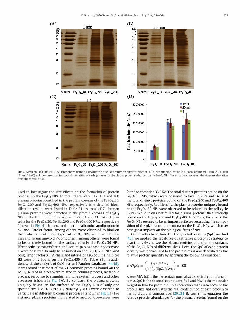
Once introduced into biological systems, proteins would com-pete for binding sites on the surfaces of NPs. After incubationin human plasma for 1 min, 30 min and 1 h, the obtainednanoparticle–protein complexes were washed at least 3 timeswith PBS to obtain the hard protein corona of Fe3O4 NPs. Thenanoparticle–protein complexes were resuspended in SDS loadingbuffer and separated using 1-D SDS-PAGE. The adsorption dynam-ics of the human plasma protein coronas on the Fe3O4 NPs areshown in Fig. 2. It was observed that the adsorption of plasmaproteins on the three different sizes of Fe3O4 NPs evolved as theincubation time went along and reached a relative equilibriumstate after 1 h of incubation in human plasma. On the other hand,the plasma proteins displayed different binding patterns depend-ing on the size of the Fe3O4 NPs, illustrating that nanoparticlesize impacted the formation of the protein corona. Additionally,the total band intensities for the plasma protein coronas exhibitedobvious differences depending on NP size (shown in Fig. 2D); i.e.,there were much more plasma proteins bound on the Fe3O4 200and Fe3O4 400 NPs than on the Fe3O4 30 NPs, which may bedue to the obvious aggregation of the Fe3O4 30 NPs, indicatingthat NP aggregation also affects the formation of protein coronas.Moreover, the optical intensity distributions of the plasma proteincoronas formed on Fe3O4 NPs of different particle sizes were fur-ther analyzed with the Image Lab program (shown in Fig. 3), whichalso confirmed the different adsorption patterns on the Fe3O4 NPsand the equilibrium state achieved after 1 h of incubation in humanplasma.
3.3. Identification and quantification of plasma proteins boundon Fe3O4 NPs with different sizes
In our study, to guarantee identification accuracy, each samplewas analyzed in at least three replicates, and only proteins detected
in at least two runs and identified by at least two unique peptidesin each analysis were considered as constituents of the proteincoronas formed on the Fe3O4 NPs. The protein coronas bound onthe Fe3O4 NPs after 1 h of incubation with human plasma were(mV) Z-Ave diameter (nm) PDI
822.4 ± 73.9 0.41 ± 0.02643.1 ± 34.6 0.38 ± 0.03571.1 ± 10.8 0.44 ± 0.06
Z. Hu et al. / Colloids and Surfaces B: Biointerfaces 121 (2014) 354–361 357
Fig. 2. Silver stained SDS-PAGE gel lanes showing the plasma protein binding profiles on different sizes of Fe3O4 NPs after incubation in human plasma for 1 min (A), 30 min( sma pf
ucpFtpNt(AtmtF1cHtiFppuspi
B) and 1 h (C) and the corresponding optical intensities of each gel lanes for the plarom the mean (n = 3).
sed to investigate the size effects on the formation of proteinoronas on the Fe3O4 NPs. In total, there were 117, 133 and 100lasma proteins identified in the protein coronas of the Fe3O4 30,e3O4 200 and Fe3O4 400 NPs, respectively (the detailed iden-ification results were listed in Table S1). A total of 71 humanlasma proteins were detected in the protein coronas of Fe3O4Ps of the three different sizes, with 22, 31 and 11 distinct pro-
eins for the Fe3O4 30, Fe3O4 200 and Fe3O4 400 NPs, respectivelyshown in Fig. 4). For example, serum albumin, apolipoprotein-I and Platelet factor, among others, were observed to bind on
he surfaces of all three types of Fe3O4 NPs, while ceruloplas-in and serum amyloid P-component, among others, were found
o be uniquely bound on the surface of only the Fe3O4 30 NPs.ibronectin, serotransferrin and serum paraoxonase/arylesterase
were observed to only be adsorbed on the Fe3O4 200 NPs, andoagulation factor XIII A chain and inter-alpha (Globulin) inhibitor2 were only bound on the Fe3O4 400 NPs (Table S1). In addi-
ion, with the analysis of GoMiner and Panther databases [44,45],t was found that most of the 71 common proteins bound on thee3O4 NPs of all sizes were related to cellular process, metabolicrocess, response to stimulus, immune system process and otherrocesses (shown in Fig. 5A). By contrast, the plasma proteins
niquely bound on the surfaces of the Fe3O4 NPs of only onepecific size (Fe3O4 30/Fe3O4 200/Fe3O4 400) were observed toarticipate in different biological processes (shown in Fig. 5B). Fornstance, plasma proteins that related to metabolic processes were
roteins adsorbed on the Fe3O4 NPs. The error bars represent the standard deviation
found to comprise 33.3% of the total distinct proteins bound on theFe3O4 30 NPs, which were observed to take up 9.5% and 16.7% ofthe total distinct proteins bound on the Fe3O4 200 and Fe3O4 400NPs, respectively. Additionally, the plasma proteins uniquely boundon the Fe3O4 30 NPs were observed to be related to the cell cycle(6.7%), while it was not found for plasma proteins that uniquelybound on the Fe3O4 200 and Fe3O4 400 NPs. Thus, the size of theFe3O4 NPs seemed to be an important factor regulating the compo-sition of the plasma protein corona on the Fe3O4 NPs, which maypose great impacts on the biological fates of NPs.
On the other hand, based on the spectral counting (SpC) method[46], we applied the label-free quantitative proteomic strategy toquantitatively analyze the plasma proteins bound on the surfacesof the Fe3O4 NPs of different sizes. Here, the SpC of each proteinidentity was normalized to the protein mass and described as therelative protein quantity by applying the following equation:
MWSpCk =(
(SpC/Mw)k∑ni=1(SpC/Mw)i
)× 100
where MWSpC is the percentage normalized spectral count for pro-tein k, SpC is the spectral count identified and Mw is the molecular
weight in kDa for protein k. This correction takes into account theprotein size and evaluates the real contribution of each protein tothe hard corona composition [20,21]. By using this equation, therelative protein abundances for the plasma proteins bound on the
358 Z. Hu et al. / Colloids and Surfaces B: Biointerfaces 121 (2014) 354–361
F inding( and 1
Fadtio
ig. 3. The optical intensities across the SDS-PAGE gel lanes for the plasma protein bG-I)) after incubation in human plasma for 1 min (A, D and G), 30 min (B, E and H)
e3O4 NPs were calculated and listed in Table S1. It was found thatlthough there were 71 common plasma proteins bound on the
ifferent sizes of Fe3O4 NPs, the relative protein abundances forhese proteins differed. For instance, serum albumin was observedn the protein corona of the Fe3O4 200 NPs with an average MWSpCf 9.01, while the average MWSpC for serum albumin in theprofiles on different sizes of Fe3O4 NPs (Fe3O4 30 (A-C)/Fe3O4 200 (D-F)/Fe3O4 400h (C, F and I).
protein coronas of the Fe3O4 30 and Fe3O4 200 NPs were 1.27 and2.50, respectively, indicating the selective binding of plasma pro-
teins on different sizes of Fe3O4 NPs. Additionally, platelet factor 4was observed in the corona of Fe3O4 30 with an average MWSpC of8.57, which accounted for 3.51% and 4.01% in the protein coronas ofthe Fe3O4 200 and Fe3O4 400 NPs, respectively. Furthermore, the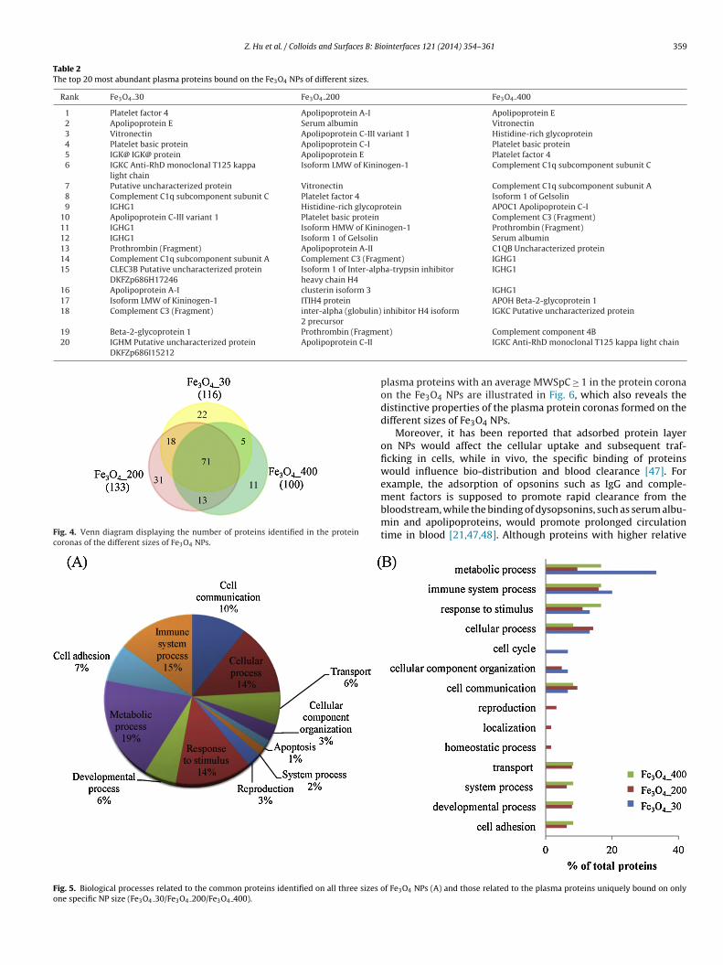
Z. Hu et al. / Colloids and Surfaces B: Biointerfaces 121 (2014) 354–361 359
Table 2The top 20 most abundant plasma proteins bound on the Fe3O4 NPs of different sizes.
Rank Fe3O4 30 Fe3O4 200 Fe3O4 400
1 Platelet factor 4 Apolipoprotein A-I Apolipoprotein E2 Apolipoprotein E Serum albumin Vitronectin3 Vitronectin Apolipoprotein C-III variant 1 Histidine-rich glycoprotein4 Platelet basic protein Apolipoprotein C-I Platelet basic protein5 IGK@ IGK@ protein Apolipoprotein E Platelet factor 46 IGKC Anti-RhD monoclonal T125 kappa
light chainIsoform LMW of Kininogen-1 Complement C1q subcomponent subunit C
7 Putative uncharacterized protein Vitronectin Complement C1q subcomponent subunit A8 Complement C1q subcomponent subunit C Platelet factor 4 Isoform 1 of Gelsolin9 IGHG1 Histidine-rich glycoprotein APOC1 Apolipoprotein C-I
10 Apolipoprotein C-III variant 1 Platelet basic protein Complement C3 (Fragment)11 IGHG1 Isoform HMW of Kininogen-1 Prothrombin (Fragment)12 IGHG1 Isoform 1 of Gelsolin Serum albumin13 Prothrombin (Fragment) Apolipoprotein A-II C1QB Uncharacterized protein14 Complement C1q subcomponent subunit A Complement C3 (Fragment) IGHG115 CLEC3B Putative uncharacterized protein
DKFZp686H17246Isoform 1 of Inter-alpha-trypsin inhibitorheavy chain H4
IGHG1
16 Apolipoprotein A-I clusterin isoform 3 IGHG117 Isoform LMW of Kininogen-1 ITIH4 protein APOH Beta-2-glycoprotein 118 Complement C3 (Fragment) inter-alpha (globulin) inhibitor H4 isoform
2 precursorIGKC Putative uncharacterized protein
19 Beta-2-glycoprotein 1 Prothrombin (Fragme20 IGHM Putative uncharacterized protein
DKFZp686I15212Apolipoprotein C-II
Fig. 4. Venn diagram displaying the number of proteins identified in the proteincoronas of the different sizes of Fe3O4 NPs.
Fig. 5. Biological processes related to the common proteins identified on all three sizes oone specific NP size (Fe3O4 30/Fe3O4 200/Fe3O4 400).
nt) Complement component 4BIGKC Anti-RhD monoclonal T125 kappa light chain
plasma proteins with an average MWSpC ≥ 1 in the protein coronaon the Fe3O4 NPs are illustrated in Fig. 6, which also reveals thedistinctive properties of the plasma protein coronas formed on thedifferent sizes of Fe3O4 NPs.
Moreover, it has been reported that adsorbed protein layeron NPs would affect the cellular uptake and subsequent traf-ficking in cells, while in vivo, the specific binding of proteinswould influence bio-distribution and blood clearance [47]. Forexample, the adsorption of opsonins such as IgG and comple-
ment factors is supposed to promote rapid clearance from thebloodstream, while the binding of dysopsonins, such as serum albu-min and apolipoproteins, would promote prolonged circulationtime in blood [21,47,48]. Although proteins with higher relativef Fe3O4 NPs (A) and those related to the plasma proteins uniquely bound on only

360 Z. Hu et al. / Colloids and Surfaces B: Biointerfaces 121 (2014) 354–361
F WSpF epres
aefNtTpippmNttNmtAptTdo
ig. 6. Relative protein abundance of proteins (normalized to molecular weight, Me3O4 NPs. As the proteins were detected in more than 2 replicates, the error bars r
bundances do not directly reflect biological impacts, proteins withxtremely low relative abundances are not expected to be criticalactors that determine the bio-distributions and biological fates ofPs [48,49]. Here, we list the top 20 most abundant plasma pro-
eins comprising the protein coronas formed on the Fe3O4 NPs inable 2 to further illustrate the different binding patterns of thelasma proteins and understand the following potential biological
mpacts. It was found that as the most abundant protein in humanlasma, serum albumin was the 2nd most abundant protein in therotein corona around the Fe3O4 200 NPs, while it was the 12thost abundant protein in the protein corona on the Fe3O4 400Ps and was not among the top 20 most abundant proteins for
he Fe3O4 30 NPs (27th). Thus, the quantitative results confirmedhe selective binding of plasma proteins to different sizes of Fe3O4Ps. Additionally, platelet factor 4, which is supposed to pro-ote rapid clearance from the bloodstream, was found to bind on
he aggregated Fe3O4 30 NPs with the highest protein abundance.polipoprotein A-I and apolipoprotein E, which are supposed toromote prolonged circulation times, were the most abundant pro-
eins bound on the Fe3O4 200 and Fe3O4 400 NPs, respectively.herefore, the different binding profiles of plasma proteins on theifferent sizes of Fe3O4 NPs may have different impacts in humanrganisms.C) satisfying the criterion that MWSpC ≥ 1 in the coronas of the different sizes ofent the standard deviation from the mean (n = 2 or n = 3).
4. Conclusions
In this study, the plasma protein coronas formed on pristineFe3O4 NPs of three different particle sizes were investigated for thefirst time. According to the results, it could be concluded that theparticle size induced different surface curvature and aggregationstate of the Fe3O4 NPs, both of which may have played impor-tant roles in the formation of plasma protein coronas on theseFe3O4 NPs; the protein compositions as well as the abundancesof the plasma proteins in the protein coronas of Fe3O4 NPs werefound to be critically affected by particle size. Moreover, the pro-teins bound on the Fe3O4 NPs of different particle sizes may posegreat influences on the biological distribution and fates of the Fe3O4NPs.
Acknowledgements
Financial supports from the National Natural ScienceFoundation of China (Nos. 21175134, 21375125), the Cre-
ative Research Group Project of National Natural ScienceFoundation of China (21321064) and the Analytical MethodInnovation Program of MOST (2012IM030900) are gratefullyacknowledged.
s B: Bi
A
f2
R
[
[
[
[
[
[
[
[
[
[
[
[
[
[
[
[
[
[
[
[
[
[
[
[
[
[
[
[
[
[
[
[
[
[
[
[
[
[
[
Z. Hu et al. / Colloids and Surface
ppendix A. Supplementary data
Supplementary data associated with this article can beound, in the online version, at http://dx.doi.org/10.1016/j.colsurfb.014.06.016.
eferences
[1] S. Laurent, D. Forge, M. Port, A. Roch, C. Robic, L.V. Elst, R.N. Muller, Magneticiron oxide nanoparticles: synthesis, stabilization, vectorization, physicochem-ical characterizations, and biological applications, Chem. Rev. 108 (2008)2064–2110.
[2] M.G. Lines, Nanomaterials for practical functional uses, J. Alloy. Compd. 449(2008) 242–245.
[3] J.A. Barreto, W. O’Malley, M. Kubeil, B. Graham, H. Stephan, L. Spiccia, Nano-materials applications in cancer imaging and therapy, Adv. Mater. 23 (2011)H18–H40.
[4] N.A. Saunders, Application of nanomaterials to arrays for infectious diseasediagnosis, Nanomedicine 6 (2011) 271–280.
[5] L. Zhang, T.J. Webster, Nanotechnology and nanomaterials promises forimproved tissue regeneration, Nano Today 4 (2009) 66–80.
[6] I. Lynch, K.A. Dawson, Protein–nanoparticle interactions, Nano Today 3 (2008)40–47.
[7] C.D. Walkey, W.C.W. Chan, Understanding and controlling the interaction ofnanomaterials with proteins in a physiological environment, Chem. Soc. Rev.41 (2012) 2780–2799.
[8] M.P. Monopoli, F.B. Bombelli, K.A. Dawson, Nanobiotechnology nanoparticlecoronas take shape, Nat. Nanotechnol. 6 (2011) 11–12.
[9] I. Lynch, A. Salvati, K.A. Dawson, Protein–nanoparticle interactions what doesthe cell see? Nat. Nanotechnol. 4 (2009) 546–547.
10] M.S. Ehrenberg, A.E. Friedman, J.N. Finkelstein, G. Oberdorster, J.L. McGrath,The influence of protein adsorption on nanoparticle association with culturedendothelial cells, Biomaterials 30 (2009) 603–610.
11] Z.J. Deng, Nanoparticle-induced unfolding of fibrinogen promotes Mac-1 recep-tor activation and inflammation, Nat. Nanotechnol. 6 (2011) 39–44.
12] W. Hu, C. Peng, M. Lv, X. Li, Y. Zhang, N. Chen, C. Fan, Q. Huang, Protein corona-mediated mitigation of cytotoxicity of graphene oxide, ACS Nano 5 (2011)3693–3700.
13] D. Dutta, S.K. Sundaram, J.G. Teeguarden, B.J. Riley, L.S. Fifield, J.M. Jacobs, S.R.Addleman, G.A. Kaysen, B.M. Moudgil, T.J. Weber, Adsorbed proteins influencethe biological activity and molecular targeting of nanomaterials, Toxicol. Sci.100 (2007) 303–315.
14] M.A. Dobrovolskaia, P. Aggarwal, J.B. Hall, S.E. McNeil, Preclinical studies tounderstand nanoparticle interaction with the immune system and its potentialeffects on nanoparticle biodistribution, Mol. Pharm. 5 (2008) 487–495.
15] A. Lesniak, F. Fenaroli, M.R. Monopoli, C. Aberg, K.A. Dawson, A. Salvati, Effectsof the presence or absence of a protein corona on silica nanoparticle uptakeand impact on cells, ACS Nano 6 (2012) 5845–5857.
16] C. Vauthier, B. Persson, P. Lindner, B. Cabane, Protein adsorption and comple-ment activation for di-block copolymer nanoparticles, Biomaterials 32 (2011)1646–1656.
17] H. Mao, W. Chen, S. Laurent, C. Thirifays, C. Burtea, F. Rezaee, M. Mahmoudi,Hard corona composition and cellular toxicities of the graphene sheets, Colloid.Surf. B 109 (2013) 212–218.
18] T. Cedervall, I. Lynch, M. Foy, T. Berggård, S.C. Donnelly, G. Cagney, S. Linse,K.A. Dawson, Detailed identification of plasma proteins adsorbed on copolymernanoparticles, Angew. Chem. Int. Ed. 46 (2007) 5754–5756.
19] T. Cedervall, I. Lynch, S. Lindman, T. Berggard, E. Thulin, H. Nilsson, K.A. Daw-son, S. Linse, Understanding the nanoparticle-protein corona using methods toquantify exchange rates and affinities of proteins for nanoparticles, Proc. Natl.Acad. Sci. U S A 104 (2007) 2050–2055.
20] S. Tenzer, D. Docter, S. Rosfa, A. Wlodarski, J. Kuharev, A. Rekik, S.K. Knauer, C.Bantz, T. Nawroth, C. Bier, J. Sirirattanapan, W. Mann, L. Treuel, R. Zellner, M.Maskos, H. Schild, R.H. Stauber, Nanoparticle size is a critical physicochemicaldeterminant of the human blood plasma corona: a comprehensive quantitativeproteomic analysis, ACS Nano 5 (2011) 7155–7167.
21] M.P. Monopoli, D. Walczyk, A. Campbell, G. Elia, I. Lynch, F.B. Bombelli, K.A.Dawson, Physical-chemical aspects of protein corona: relevance to in vitroand in vivo biological impacts of nanoparticles, J. Am. Chem. Soc. 133 (2011)2525–2534.
22] D. Walczyk, F.B. Bombelli, M.P. Monopoli, I. Lynch, K.A. Dawson, What the cellsees in bionanoscience, J. Am. Chem. Soc. 132 (2010) 5761–5768.
23] M. Lundqvist, J. Stigler, G. Elia, I. Lynch, T. Cedervall, K.A. Dawson, Nanopar-ticle size and surface properties determine the protein corona with possible
implications for biological impacts, Proc. Natl. Acad. Sci. U S A 105 (2008)14265–14270.24] C.D. Walkey, J.B. Olsen, H. Guo, A. Emili, W.C.W. Chan, Nanoparticle size and sur-face chemistry determine serum protein adsorption and macrophage uptake,J. Am. Chem. Soc. 134 (2012) 2139–2147.
[
ointerfaces 121 (2014) 354–361 361
25] Z.J. Deng, M. Liang, I. Toth, M.J. Monteiro, R.F. Minchin, Molecular interactionof poly(acrylic acid) gold nanoparticles with human fibrinogen, ACS Nano 6(2012) 8962–8969.
26] Z.J. Deng, G. Mortimer, T. Schiller, A. Musumeci, D. Martin, R.F. Minchin, Differ-ential plasma protein binding to metal oxide nanoparticles, Nanotechnology20 (2009).
27] G. Maiorano, S. Sabella, B. Sorce, V. Brunetti, M.A. Malvindi, R. Cingolani, P.P.Pompa, Effects of cell culture media on the dynamic formation of protein-nanoparticle complexes and influence on the cellular response, ACS Nano 4(2010) 7481–7491.
28] A. Lesniak, A. Campbell, M.P. Monopoli, I. Lynch, A. Salvati, K.A. Dawson, Serumheat inactivation affects protein corona composition and nanoparticle uptake,Biomaterials 31 (2010) 9511–9518.
29] G. Caracciolo, D. Pozzi, A.L. Capriotti, C. Cavaliere, P. Foglia, H. Amenitsch, A.Laganà, Evolution of the protein corona of lipid gene vectors as a function ofplasma concentration, Langmuir 27 (2011) 15048–15053.
30] A.L. Capriotti, G. Caracciolo, C. Cavaliere, P. Foglia, D. Pozzi, R. Samperi, A. Laganà,Do plasma proteins distinguish between liposomes of varying charge density?J. Proteomics 75 (2012) 1924–1932.
31] H.W. Gu, P.L. Ho, K.W.T. Tsang, L. Wang, B. Xu, Using biofunctional mag-netic nanoparticles to capture vancomycin-resistant Enterococci and othergram-positive bacteria at ultralow concentration, J. Am. Chem. Soc. 125 (2003)15702–15703.
32] S. Bucak, D.A. Jones, P.E. Laibinis, T.A. Hatton, Protein separations using colloidalmagnetic nanoparticles, Biotechnol. Progr. 19 (2003) 477–484.
33] A.S. Arbab, G.T. Yocum, H. Kalish, E.K. Jordan, S.A. Anderson, A.Y. Khakoo, E.J.Read, J.A. Frank, Efficient magnetic cell labeling with protamine sulfate com-plexed to ferumoxides for cellular MRI, Blood 104 (2004) 1217–1223.
34] S.J.H. Soenen, U. Himmelreich, N. Nuytten, M. De Cuyper, Cytotoxic effects ofiron oxide nanoparticles and implications for safety in cell labelling, Biomate-rials 32 (2011) 195–205.
35] Y.F. Zhu, T. Ikoma, N. Hanagata, S. Kaskel, Rattle-type Fe3O4@SiO2 hollow meso-porous spheres as carriers for drug delivery, Small 6 (2010) 471–478.
36] C. Alexiou, R.J. Schmid, R. Jurgons, M. Kremer, G. Wanner, C. Bergemann, E.Huenges, T. Nawroth, W. Arnold, F.G. Parak, Targeting cancer cells: magneticnanoparticles as drug carriers, Eur. Biophys. J. 35 (2006) 446–450.
37] M. Mahmoudi, M.A. Shokrgozar, S. Sardari, M.K. Moghadam, H. Vali, S. Laurent,P. Stroeve, Irreversible changes in protein conformation due to interac-tion with superparamagnetic iron oxide nanoparticles, Nanoscale 3 (2011)1127–1138.
38] L. Lartigue, C. Wilhelm, J. Servais, C. Factor, A. Dencausse, J.-C. Bacri, N. Luciani,F. Gazeau, Nanomagnetic sensing of blood plasma protein interactions withiron oxide nanoparticles: impact on macrophage uptake, ACS Nano 6 (2012)2665–2678.
39] A.A. Shemetov, I. Nabiev, A. Sukhanova, Molecular interaction of proteins andpeptides with nanoparticles, ACS Nano 6 (2012) 4585–4602.
40] H. Deng, X. Li, Q. Peng, X. Wang, J. Chen, Y. Li, Monodisperse magnetic single-crystal ferrite microspheres, Angew. Chem. Int. Ed. 44 (2005) 2782–2785.
41] H. Zhang, K.E. Burnum, M.L. Luna, B.O. Petritis, J.-S. Kim, W.-J. Qian, R.J. Moore,A. Heredia-Langner, B.-J.M. Webb-Robertson, B.D. Thrall, D.G. Camp II, R.D.Smith, J.G. Pounds, T. Liu, Quantitative proteomics analysis of adsorbed plasmaproteins classifies nanoparticles with different surface properties and size, Pro-teomics 11 (2011) 4569–4577.
42] A.A. Klammer, M.J. MacCoss, Effects of modified digestion schemes on theidentification of proteins from complex mixtures, J. Proteome Res. 5 (2006)695–700.
43] I.V. Shilov, S.L. Seymour, A.A. Patel, A. Loboda, W.H. Tang, S.P. Keating, C.L.Hunter, L.M. Nuwaysir, D.A. Schaeffer, The paragon algorithm, a next generationsearch engine that uses sequence temperature values and feature probabilitiesto identify peptides from tandem mass spectra, Mol. Cell. Proteomics 6 (2007)1638–1655.
44] B. Zeeberg, W. Feng, G. Wang, M. Wang, A. Fojo, M. Sunshine, S. Narasimhan,D. Kane, W. Reinhold, S. Lababidi, K. Bussey, J. Riss, J. Barrett, J. Weinstein,Gominer: a resource for biological interpretation of genomic and proteomicdata, Genome Biol. 4 (2003) R28.
45] P.D. Thomas, M.J. Campbell, A. Kejariwal, H. Mi, B. Karlak, R. Daverman,K. Diemer, A. Muruganujan, A. Narechania, Panther: a library of proteinfamilies and subfamilies indexed by function, Genome Res. 13 (2003)2129–2141.
46] D.H. Lundgren, S.-I. Hwang, L. Wu, D.K. Han, Role of spectral counting in quan-titative proteomics, Expert Rev. Proteomics 7 (2010) 39–53.
47] P.P. Karmali, D. Simberg, Interactions of nanoparticles with plasma proteins:implication on clearance and toxicity of drug delivery systems, Expert Opin.Drug Deliv. 8 (2011) 343–357.
48] Z.J. Deng, M. Liang, M. Monteiro, I. Toth, R.F. Minchin, Nanoparticle-inducedunfolding of fibrinogen promotes Mac-1 receptor activation and inflammation,
Nat. Nanotechnol. 6 (2011) 39–44.49] R.Y. Prasad, K. Wallace, K.M. Daniel, A.H. Tennant, R.M. Zucker, J. Strickland,K. Dreher, A.D. Kligerman, C.F. Blackman, D.M. DeMarini, Effect of treatmentmedia on the agglomeration of titanium dioxide nanoparticles: impact on geno-toxicity, cellular interaction, and cell cycle, ACS Nano 7 (2013) 1929–1942.