colony morphology, examination of stained microorganisms
TRANSCRIPT

Examination of stained
microorganisms,
Smear preparation, simple staining,
Gram staining and Acid-fast staining

Bacteria are
transparent and
colorless so they
would be invisible to
naked eye if
observed under
microscope thus
bacteria should be
stained with certain
dyes in order to
visualize bacterial cell
or their internal
structures using the
light microscope.
Why we need to stain
Bacteria???

Stains and Staining A stain is a substance that adheres to a cell, giving the cell
color. The presence of color gives the cells significant contrast
so they are much more visible. A stain is a dye consisting of a
colored organic compound in the form of salt composed of
positive and negative ion, one of these ions is responsible for
colour (a chromophore)
Staining is an auxiliary technique
used in microscopy to enhance
contrast in the microscopic image.
Stains are frequently used in
biology and medicine for
viewing.

Based on the charges:
A stain is classified in to:-
Basic stain/dyes – stain with +ve charge,
example include crystal violate,
methylene blue and safranin
Acidic stain/dyes – stain with –ve charge,
example nigrosin and India ink.
Neutral stain/dyes – stain with both charges.

Based on function of stain:
1. Simple staining – only one dye is used- differentiation among bacteria is impossible- Eg. Simple Staining.
2. Differential staining- more than one dye is used- Differentiation among bacteria is possible- Eg. Gram’s staining, Acid-fast staining.
3. Special staining – more than one dye used -Special structures are seen. Eg. Capsule staining, Spore staining
.

Principle of staining Basic stain(+ve charge)
To stain -ve charged molecules of bacteria
Mostly used because cell surface is –ve charge.
Acidic Stain(-ve charge)
To stain +ve charged molecules of bacteria.
Used to stain the bacterial capsules.

Fixation Fixation–which may itself consist of
several steps–aims to preserve the
shape of the cells or tissue involved
as much as possible. Sometimes
heat fixation is used to kill, adhere,
and alter the specimen so it will
accept stains.
Fixation Fixation–which may itself consist of
several steps–aims to preserve the
shape of the cells or tissue involved
as much as possible. Sometimes
heat fixation is used to kill, adhere,
and alter the specimen so it will
accept stains.

How do you prepare a smear?
From liquid media :
Sterilize the loop by Bunsen flame then let it cool.
Shake the specimen container (broth culture) then withdraw one or more if needed loopful from the specimen and spread it on the center of a clean slide to form a thin film of 1- 2 cm in diameter, then sterilize the loop.
Allow the smear to dry by air.
The smear fixed by passing it (3- 4) times through the Bunsen flame then allow the slide to cool before staining.

From solid media (slant or plate) :
Sterile the loop on Bunsen flame and let
it cool.
Place a loopful of clean water on the
center of a clean slide.
Re sterilize the loop, transfer a small
portion of the growth, mix it with water
thoroughly and spread the mixture
evenly on the slide to form a thin film of
1- 2 cm in diameter.
Dry and fix (as mentioned above)


Type of staining
a. Simple Staining:
This procedure uses only one basic dye e.g.
Crystal violet or Methylene blue or Safranin
to stain bacteria. The bacteria will simply take
the color of the dye.


b. Differential Staining:
These involve more than one dye
solution. The dyes may be added
in several steps according to the
procedure.

Gram Stain
Gram stain is used to stain bacteria. Bacteria stain either Gram-
positive or Gram negative on the basis of the differences in their
cell wall composition

What is the different
between Gram
positive and Gram
negative bacteria
????? Gram positive species have a thick
peptidoglycan layer and large
amount of teichoic acid and are
therefore unaffected by alcohol
decolourization and retain the
initial stain (crystal volet) giving the
organism violet apperance .
Gram negative cell wall have a single
peptidoglycan layer. The outer membrane
is damaged by alcohol decolorizer allowing
crystal violet- iodine complex to take out
and be replaced by the counter stain
(safranine) giving the organism a pink/ red
appearance.

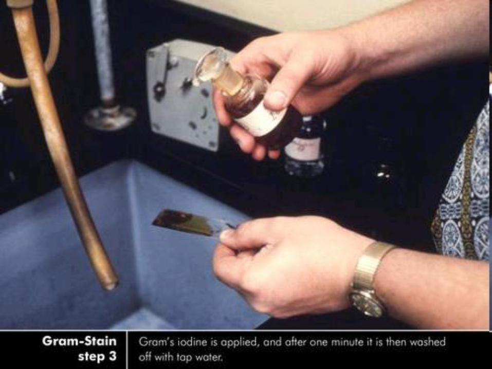
Major Steps of Gram Stain


The reports
1. If no microorganisms are seen in a smear of a
clinical specimen, report “No microorganisms seen.”
2. If microorganisms are seen describe morphology.

Color of
Gram + cells
Color of
Gram – cells
Primary stain:
Crystal violet
Mordant:
Iodine
Decolorizing agent:
Alcohol-acetone
Colorless
Counterstain:
Safranin

Gram Staining – Gram +’ve

Gram Staining – Gram -’ve


Gm+ve cocci & Gm-ve bacilli


positive bacteria-Gram
negative bacteria-Gram
Streptococcus
Staphylococcus
Lactobacillus
Bacillus Clostridium
Escherichia
Salmonella
Vibrio
Treponema

ACID FAST STAIN (ZIEHL- NEELSEN STAIN)
The acid fast stain is one
of the most medically
important stains.
Purpose:
Used in the demonstration of acid-fast bacteria
belonging to the genus 'mycobacterium', which contain
fatty acids (mycolic acid) in their cell wall.

The carbolfuchsin dye penetrates the cell wall and
stains the bacteria. The slide must be heated to melt
the mycolic acids. The mycolic acid does not allow the
acid alcohol to penetrate, so the cell resists de-
colorization and remains a bright pink or red.
Methylene
blue stain
Acid- alcohol
Carbolfuch
-sin stain
materials
the
principle

Zeihl-Neelsen Staining Procedure

Named this staining Technique