colorimetric determination of serum acid phosphatase activity using
TRANSCRIPT
J. clin. Path., 1971, 24, 493-500
Colorimetric determination of serum acid phosphataseactivity using adenosine 3'-monophosphate assubstrateGRAHAM ELLIS, ALAN BELFIELD, AND DAVID M. GOLDBERG
From the Department of Chemical Pathology, Royal Hospital, West Street, Sheffield
SYNOPSIS The hydrolysis of adenosine 3-monophosphate by serum acid phosphatase has beencoupled to the liberation of ammonia from the adenosine generated through the action of exogenousadenosine deaminase. The ammonia is measured at the end of the incubation by a modification ofthe phenol-hypochlorite reaction of Berthelot. Optimum conditions for the enzyme reaction havebeen defined. Inhibition of the Berthelot reaction by the serum used in the assay is small, and maybe compensated by a correction factor. Although the value for the control is high in relation toEhe test over the normal range, this is largely outweighed by the good sensitivity and precision ofthe method.The substrate is not significantly hydrolysed by erythrocyte acid phosphatase within the limits
encountered in haemolysed sera. Experience of the method in routine hospital diagnosis comparedfavourably with that of a standard method employing disodium phenyl phosphate as substrate. Itis suggested that activities greater than 3-1 IU/l should be further investigated and those greaterthan 3-7 IU/l should be regarded as definitely raised. The stability of human serum AcPase whenpromptly separated and held at 4°C or - 20°C was confirmed. At room temperature, acidificationto pH 6-0 greatly improved stability.
The determination of acid phosphatase (AcPase,EC 3.1.3.2, orthophosphoric monoester phospho-hydrolase) activity of human serum has beenestablished as a laboratory procedure in the diagnosisof prostatic cancer (Gutman and Gutman, 1938;Woodard, 1959), metastatic mammary cancer(Joplin and Jegatheesan, 1962; Jegatheesan andJoplin, 1962), and Gaucher's disease (Ockerman andKohlin, 1969).Many substrates have been used in this determina-
tion. These include adenosine 3'-monophosphate(3'AMP) (Fischman, Chamberlin, Cubiles, andSchmidt, 1948; Reynolds, Reynolds, and Walker,1956). This has the convenient property of beingpoorly hydrolysed by erythrocyte AcPase (Tsuboiand Hudson, 1953) and yet provides an excellentsubstrate for prostatic AcPase (Tsuboi and Hudson,1955). Its failure to gain wider acceptance relates tothe fact that hydrolysis has in the past been measuredby the rather tedious techniques of protein precipita-tion and phosphate estimation.
Recently, we have shown that hydrolysis ofReceived for publication 29 October 1970.
3'AMP can be coupled through adenosine de-aminase (ADase, EC 3.5.4.4, adenosine amino-hydrolase) to produce inosine and ammonia underconditions optimal for AcPase (Belfield and Gold-berg, 1970). The following reactions are involved:
AcPase3'AMP + H20 - Adenosine + Pi
ADaseAdenosine + H2O -* Inosine + NH3
The decrease in extinction at 265 nm consequentupon conversion of adenosine to inosine may bemeasured spectrophotometrically and provides amethod that is rapid and sensitive employing tissuepreparations with moderate AcPase activity, butcannot cope with the high absorbance of serum atthe wavelength used. An alternative possibility layin determining ammonia production utilizing thephenol-hypochlorite reaction of Berthelot as modi-fied by Chaney and Marbach (1962). This procedureis described in the present paper, which also presentssome preliminary clinical data comparing the pro-posed method with a standard technique in routinehospital diagnosis.
493
group.bmj.com on February 7, 2018 - Published by http://jcp.bmj.com/Downloaded from

494 Graham Ellis, Alan Belfield, and David M. Goldberg
8 Ammonium standard (2 mM)Materials and Methods
CLINICAL CATEGORIESBlood samples wete obtained from three groups ofsubjects.
Group AThis comprised 36 healthy members of the labora-tory staff aged 18-40 years.
Group BThis comprised 97 subjects over the age of 65attending a special general practitioner screeningclinic. All were ambulant; a few were being treatedfor chronic non-malignant disease and were wellcompensated at the time of examination.
Group CThis comprised samples of venous blood drawn from137 subjects referred for laboratory examinationbecause of prostatic symptoms or possible metastaticbone involvement.
REAGENTSAll were of highest analytical grade and all waterused was glass distilled and de-ionized.
1 0-1 M Acetate bufferpH 5-6 at 20°C.
2 Adenosine deaminaseObtained from Boehringer Corporation (London)Ltd as a suspension in 50% (v/v) glycerol (catalogueno. 15069 EAAT).
3 Acetate buffer/ADaseOne pi of neat ADase suspension was added per mlof buffer, the mixture being prepared fresh daily.
4 3'4MP (80 mM)NaOH is required to dissolve the free acid, and thefinal pH must lie between 5 and 8. This was storedat -20°C in aliquots of about 2 ml, a fresh aliquotbeing used each day.
5 3'AMP/ADaseOne ,ul of neat ADase was added per ml of substrate,the mixture being prepared fresh daily.
6 Phenol/nitroprusside
7 HypochloriteThe above two reagents were in the concentratedform described by Chaney and Marbach (1962).
TECHNIQUEFor each serum, a test and control were run, andwith each batch, standards and reagent blanks were
put through. The protocol is set out in Table L
Test Control Standard ReagentBlank
Acetate buffer/ADase (ml) 0-9Ammonia standard (M)Water (jsl)Serum (g.1) 50
Equilibrate 5 minwtes at 37°C3'AMP/ADase (OA) 50Incubate exactly 1 hour at 37°CPhenol/nitroprusside (ml) I3'AMP/ADase (OA)Hypochlorite (ml) 1
09
50
0950
50
09
100
50
Incubate at 37°C for 30 minutes and read extinction at 630 nm againstwater
Table I Protocol for determination o serum AcPaseactivity using 3'AMP as substrate
Batches were processed in such a way that the testand control for each serum were incubated for anidentical period before adding the phenol/nitroprus-side reagent.
Activity as IU/I was given by the expressionEtest -Econtroi X 33-3 x 1-04
Estandard - Ereagent blank
The derivation of the factor 104 which corrects fordepression of the Berthelot reaction by serum isdescribed later.
Results
DEVELOPMENT OF THE METHOD
Choice of bufferA pH activity curve was constructed over the range4-5-6-5 using 01 M acetate and 0.1 M citrate buffers.The pH was measured at 37°C before and afterincubation, using the Micro-Astrup apparatus(Radiometer Corporation, Copenhagen, Denmark).Optimal activity was obtained at pH 5-6 with bothbuffers (Fig. 1). Comparison of a number of buffersadjusted to 0-1 M and pH 5-6 at 37°C yielded thefollowing activities relative to acetate: citrate 0-95,succinate/borate 0-84, phosphate 0-20, citrate/phos-phate 0-16, veronal/acetate 0-68, phthalate 0-81. Thebuffers were prepared from standard tables (Geigy,1956). Varying molarity of acetate from 0-05 to0-9 M did not affect activity. Since ammoniagenerated by a very active serum produced negligiblechange in pH of 0-1 M acetate buffer even when
1
I
group.bmj.com on February 7, 2018 - Published by http://jcp.bmj.com/Downloaded from

Colorimetric determination ofserum acidphosphatase activity using adenosine 3'-monophosphate
Fig. 1 pH-activity curves for serum AcPase in 0-1 M630 acetate buffer (solid lines) and 0-1 M citrate buffer
(broken lines) using two sera of high activity.
--_,~~~~1
4.5 5-5
PH6-0 6-5
the incubation was extended to six hours, thisstrength was deemed appropriate. The addition ofNaCl in final concentrations of 0-08 to 0-32 M in-hibited AcPase activity; this was 80% of the controlat 0-32 M.
Choice of substrateThe criteria governing this choice were that thesubstrate should have a high affinity towardsprostatic AcPase aaid low affinity towards erythro-cyte AcPase. Of the monophosphates of adenosinetested, 3'AMP best fulfilled these criteria (Table II).
Substrate (4 mM) Activity as JE,. (Test - Control)
Prostate' Erythrocytes'
2'AMP 0 096 03'AMP 0-294 05'AMP 0107 0-229
Table II Relative AcPase activities ofhuman prostateand erythrocytes using various monophosphates ofadenosine as substrate150 ju of a 1: 200 (v/v) mixture of human prostatic AcPase in pooledhuman serum.250 gA of a 1: 10 water haemolysate of washed packed humanerythrocytes.
In this study purified human prostatic AcPase pre-pared according to the method of Belfield andGoldberg (1970) was dialysed until free of am-monium sulphate and 1 part added to 200 parts ofpooled human serum. The Km for 3'AMP was
determined utilizing a serum of high AcPase activityand an incubation time of 10 minutes and 11 con-centrations of substrate in the range 0-046-1-824mMfinal concentration. Statistical evaluation of the datausing a computer program (Hoy and Goldberg,1971) gave a value of 0-391 with standard error
± 0-055. The concentration in the definitive assay,being 10 times Km, sustains 91 % of theoreticalVmax.
Adenosine deaminaseThe preparation used was free of AcPase activity.Using a serum Gf high AcPase activity, it was foundthat a final concentration of 0-1 ,ul/ml of the neatADase preparation supported the maximal reactionrate over 60 minutes. This is one-ninth of the ADaseconcentration of the definite assay.
Required blanksThe recommended method allows for the ammoniaoriginally present in the serum as well as thatgenerated from sources other than AMP during theincubation, since the serum is incubated under con-ditions identical with those of the test but omittingsubstrate. It is also necessary to take account ofammonia and adenosine present as a contaminant ofAMP. Further allowance is made for generation ofadenosine from AMP during incubation at 37°C.All of the above sources of ammonia, other thanthat due to specific hydrolysis of AMP by AcPaseof serum, were fully allowed for in the controlemployed in the definitive assay.
r409g
0-6
03L
495
group.bmj.com on February 7, 2018 - Published by http://jcp.bmj.com/Downloaded from

496
Characteristics of colour reactionThe reaction gave values for AE630 linear withammonia concentration to an absorbence of 2 60.At 37°C, and with the reagents of the definitiveassay, absorbence reached a maximum within 10minutes of adding hypochlorite, and remained stablefor a further 10 minutes, thereafter decreasing at arate of 0 05 % per minute at room temperature. Noloss of ammonia took place from stoppered tubesunder the assay conditions even when incubation at370C was continued for 18 hours.The presence of protein during development of the
Berthelot reaction reduced the colour yield fromadded ammonia. This inhibition increased non-linearly with increasing amount of serum; althoughreduced in the presence of serum, the absorbance wasalways linear with ammonia added to values of 2 50.This enabled the use of a correction factor to allowfor this inhibition. Using 50 ,ul serum and the condi-tions of the enzyme assay, the percentage recoveryof ammonia obtained with 24 sera gave a mean± SD of 95 9 ± 3 1, hence the factor of 1 04 used incalculation of enzyme activity. This may be em-ployed only for determinations at pH 5-6 with 50 ,ulserum.When the colour was developed at 56°C, not only
was the yield reduced, but the colour became pro-gressively unstable as the amount of serum was in-creased. Other studies to overcome this inhibitionwere unsuccessful. They were similar to experimentsdescribed in an earlier report (Belfield, Ellis, andGoldberg, 1970).
Characteristics of enizyme reactionThe definitive procedure yielded activity propor-tional to the amount of enzyme up to LE630 of 1-10,corresponding to approximately 58 IU/litre. Thereaction, even with sera of very high activity, waslinear with time up to at least one hour. It is recom-mended that assays should be repeated using ashorter incubation time where LE630 after one hourexceeds 1-10; this maintains the validity of the cor-rection factor used in calculating activity.The within-batch precision of the method gauged
by 13 replicate assays of a serum gave a mean of501 IU/l with the coefficient of variation = 2-4%.The SD of pooled duplicates was derived accordingto Pearson (1942) from analyses on the 137 patientsin group C. The range and median value of this casematerial is given in Table IV and the SD of theduplicates was 0 8 IU/litre.
EFFECT OF HAEMOLYSISHeparinized and clotted blood were obtained froma normal individual. The latter was centrifuged toobtain the serum and the former to obtain the
Graham Ellis, Alan Belfield, and David M. Goldberg
erythrocytes which were washed twice with ice-coldisotonic saline, packed by centrifugation, andruptuied by freezing and thawing. The haemolysatewas added in varying amounts to the serum, and theAcPase activity of the mixture measured by thepresent technique as well as by the method of Gut-man and Gutman (1940) with and without form-aldehyde in a final concentration of 0 5 %. Althoughthe total AcPase as determined by the latter methodwas greatly increased by addition of haemolysate, nosignificant change in activity by the present tech-nique or in the formol-stable AcPase took place upto a haemolysate concentration of 2% (Table III).
Haemolysate 3'AMP (IL 1) Substrate for AcPase ActivityvConcentrationas Percentage o Phenyl PhenylSerumnt Volume Phosphate' Phosphate'
(Total) (Formol-stable)
0 14 2 2010 09 2 1067 1-2 5 1200 1-8 10 1
Table III Effect of haemolysis on AcPase activity using3'AMP and disodium phenyl phosphate as substrates'Determined by method of Gutman and Gutman (1940) as units per100 ml.
STABILITY OF HUMAN SERUM ACPASEThis was tested by drawing a large sample of venousblood from a patient with high activity due toadvanced prostatic cancer. The serum was separatedwithout delay and split into three major aliquots, onebeing untreated, a second being adjusted to a finalconcentration of 0-1 00 with respect to Triton X-100,and a third adjusted to a pH of approximately 6 0by adding 10 ,ul of 20% (v/v) acetic acid per millilitreserum. All three lost approximately 100/ activitydue to the initial freezing, but thereafter no signi-ficant change took place at - 20°C over a period ofsix months when fractions stored individually toavoid the need for repeated freezing and thawingwere assayed. Neither was there any significantchange in the three preparations when stored at 4°Cfor up to eight days; but at room temperature theactivity of the untreated and detergent-treated pre-parations declined steadily, whereas the activity ofthe acid-treated preparation was unchanged (Fig. 2).
OBSERVATIONS ON HUMAN SUBJECTSThe activities observed in the three groups ofsubjects studied are summarized in Table IV.Because the distribution was skewed in all groups,the data were plotted on probability paper to givethe 95-percentile range. Where, as occasionallyhappened, the value for the control exceeded that of
group.bmj.com on February 7, 2018 - Published by http://jcp.bmj.com/Downloaded from

Colorimetric determination ofserum acidphosphatase activity using adenosine 3'-monophosphate
y = 1-00 + 031x, where y and x are AcPaseactivities in Gutman units and IU/l respectively. Thestandard errors of the slopes and intercept were 0 11and 0-14 respectively. Clearly, the two methods arenot reflecting an identical enzyme activity, values forthe Gutman procedure being relatively greater atlow AcPase levels, presumably because that methodincludes a contribution from erythrocyte AcPase.An analysis of those cases in group C where a
value of 3-1 IU/I was exceeded appears in Table V.
OL
1*01
0 7
'---=&--
0 2 4 6 8
DAYS
Fig. 2 Stability ofserum AcPase activity at roomtemperature (a, top) and at 4°C (b, bottom) when un-treated (circles) and when treated with acid (triangles) ordetergent (squares).
the test, a negative activity was assigned to thesample so that the ranges take full account of theexperimental error.The upper limit for the laboratory staff (group A)
was lower than that of the elderly population (groupB) which in turn was lower than the hospital popula-tion (group C), but the median values for all threewere similar. The total AcPase activity of all patientsin group C was also determined by the method ofGutman and Gutman (1940). The upper normallimit for this laboratory is 3 units per 100 ml. Wherethis value was exceeded, the assay was repeated atthe earliest opportunity in the presence of a finalconcentration of 05% formaldehyde, a value inexcess of 2 units per 100 ml being regarded asabnormal. A statistical analysis of the AcPaseactivities, as measured by the two methods in thesame patients, yielded a correlation coefficient of0-248 (p < 0-01) and the regression equation,
Case Clinical Details Serum AcPase ActivityNo.
Gutman and PresentGutman Method(1940)' (lU/I)
1 Prostatic cancer stage II 2 3-52 Prostatic cancer stage II 2 3-73 Prostatic cancer stage III 3 (1) 4 94 Prostatic cancer stage III 5 (2) 6-95 Benign prostatic hypertrophy 2 4-161 Benign prostatic hypertrophy 2 3-37' Benign prostatic hypertrophy 4 (2) 5 581.8 Benign prostatic hypertrophy 2 7 591 Benign prostatic hypertrophy 4 (0) 3 5101 Benign prostatic hypertrophy 1 3-911' Benign prostatic hypertrophy 2 6-512 Benign prostatic hypertrophy 2 3-313 Non-prostatic bone secondaries 1 6-014 Non-prostatic bone secondaries 5 (1) 7-915 Non-prostatic bone secondaries 5 (2) 5-716 Severe burns, bone biopsy 2 4 117' Urinary tract infection 2 4-218' Diverticulitis, spinal opacity 3 (1) 7-219' Renal stone 3 (1) 3-420 Notes unavailable 1 3-4
Table V Details ofpatients from group C with serumAcPase activity > 31 IUIl by present method'No operation or biopsy'Catheter in situ at time of sample'Sample taken within two hours of rectal examination'Units per 100 ml, formol-stable activity in parenthesis.
Cases 1-4 had prostatic cancer which was intra-capsular (stage II) in two and had spread to softtissues (stage III) in two; total AcPase measured bythe Gutman procedure was raised in only one ofthese cases, and none had elevated formol-stableAcPase. Cases 5-12 had benign prostatic hyper-trophy, but surgical and pathological confirmationwas available in only three of these eight cases; totalAcPase by the Gutman procedure was raised in two
Group Number of Subjects 95-Percentile Range of Serum AcPase (IU/I)
Total Males Females Lower Limit Median Upper Limit
A 36 19 17 0-9 1-8 3-1B 97 41 56 0-7 2-1 3-7C 137 133 4 -0-4 1-2 5.1
Table IV Serum AcPase values in three groups ofhuman subjects using 3'AMP as substrate
°Or(a)
497
0.-[
group.bmj.com on February 7, 2018 - Published by http://jcp.bmj.com/Downloaded from
498
of these cases and formol-stable AcPase in none.
Cases 13-15 had non-prostatic malignant bonesecondaries; total AcPase by the Gutman procedurewas raised in two and formol-stable AcPase in none.
Cases 16-20 had miscellaneous conditions itemizedin Table V, and total AcPase by the Gutman pro-
cedure was normal in all. Seven of the patients inTable V in categories other than prostatic or bonecancer were catheterized at the time of venepuncture,or had blood taken within two hours of rectalexamination.
Five cases, presented in Table VI, had normalserum AcPase with 3'AMP as substrate but raisedactivity as determined by the Gutman procedure,although in only one case was the formol-stableactivity abnormal (4 units). One was a case of stageII prostatic cancer; noteworthy is the fact thatformol-stable AcPase in this case was normal, thesample being grossly haemolysed.
Case Clinical Details Serum AcPase ActivityNo.
Gutman and PresentGutman Method(1940)' (lU/f)
21 Prostatic cancer stage II 12 (1) 0-222 Benign prostatic hypertrophy 6 (4) 2-423 Non-prostatic bone secondaries 5 (2) 1.124 Diabetes, cervical spondylosis 4 (2) 0-8251 Acute postoperative retention 4 (1) 1-4
Table VI Details ofpatients from group C with serumAcPase activity > 3 Gutman units per 100 ml but< 31 IU/I by present method'Catheter in situ at time of sample.'Units per 100 ml, formol-stable activity in parentheses
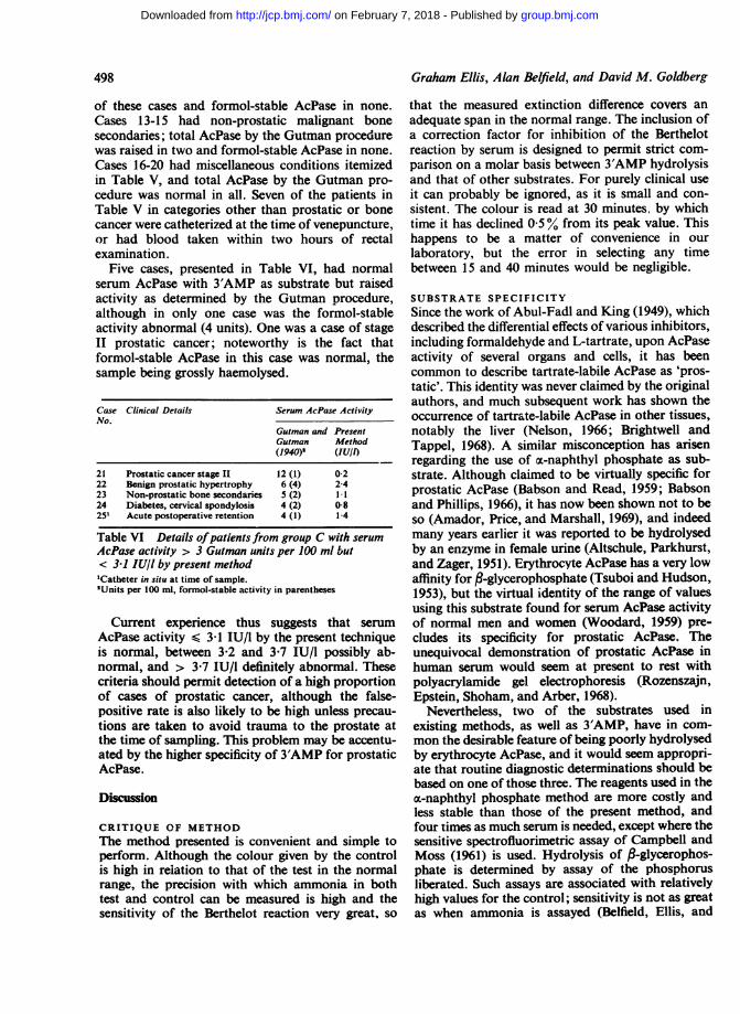
Current experience thus suggests that serumAcPase activity < 3-1 IU/I by the present techniqueis normal, between 3-2 and 3-7 IU/I possibly ab-normal, and > 3-7 IU/l definitely abnormal. Thesecriteria should permit detection of a high proportionof cases of prostatic cancer, although the false-positive rate is also likely to be high unless precau-tions are taken to avoid trauma to the prostate atthe time of sampling. This problem may be accentu-ated by the higher specificity of 3'AMP for prostaticAcPase.
Discussion
CRITIQUE OF METHODThe method presented is convenient and simple toperform. Although the colour given by the controlis high in relation to that of the test in the normalrange, the precision with which ammonia in bothtest and control can be measured is high and thesensitivity of the Berthelot reaction very great, so
Graham Ellis, Alan Belfield, and David M. Goldberg
that the measured extinction difference covers anadequate span in the normal range. The inclusion ofa correction factor for inhibition of the Berthelotreaction by serum is designed to permit strict com-parison on a molar basis between 3'AMP hydrolysisand that of other substrates. For purely clinical useit can probably be ignored, as it is small and con-sistent. The colour is read at 30 minutes, by whichtime it has declined 0 5 % from its peak value. Thishappens to be a matter of convenience in ourlaboratory, but the error in selecting any timebetween 15 and 40 minutes would be negligible.
SUBSTRATE SPECIFICITYSince the work of Abul-Fadl and King (1949), whichdescribed the differential effects of various inhibitors,including formaldehyde and L-tartrate, upon AcPaseactivity of several organs and cells, it has beencommon to describe tartrate-labile AcPase as 'pros-tatic'. This identity was never claimed by the originalauthors, and much subsequent work has shown theoccurrence of tartrate-labile AcPase in other tissues,notably the liver (Nelson, 1966; Brightwell andTappel, 1968). A similar misconception has arisenregarding the use of a-naphthyl phosphate as sub-strate. Although claimed to be virtually specific forprostatic AcPase (Babson and Read, 1959; Babsonand Phillips, 1966), it has now been shown not to beso (Amador, Price, and Marshall, 1969), and indeedmany years earlier it was reported to be hydrolysedby an enzyme in female urine (Altschule, Parkhurst,and Zager, 1951). Erythrocyte AcPase has a very lowaffinity for ,-glycerophosphate (Tsuboi and Hudson,1953), but the virtual identity of the range of valuesusing this substrate found for serum AcPase activityof normal men and women (Woodard, 1959) pre-cludes its specificity for prostatic AcPase. Theunequivocal demonstration of prostatic AcPase inhuman serum would seem at present to rest withpolyacrylamide gel electrophoresis (Rozenszajn,Epstein, Shoham, and Arber, 1968).
Nevertheless, two of the substrates used inexisting methods, as well as 3'AMP, have in com-mon the desirable feature of being poorly hydrolysedby erythrocyte AcPase, and it would seem appropri-ate that routine diagnostic determinations should bebased on one of those three. The reagents used in thex-naphthyl phosphate method are more costly andless stable than those of the present method, andfour times as much serum is needed, except where thesensitive spectrofluorimetric assay of Campbell andMoss (1961) is used. Hydrolysis of P-glycerophos-phate is determined by assay of the phosphorusliberated. Such assays are associated with relativelyhigh values for the control; sensitivity is not as greatas when ammonia is assayed (Belfield, Ellis, and
group.bmj.com on February 7, 2018 - Published by http://jcp.bmj.com/Downloaded from

Colorimetric determination ofserum acidphosphatase activity using adenosine 3'-monophosphate
Goldberg, 1970); the methods, when used in aroutine laboratory, give unsatisfactory precision(Belfield and Goldberg, 1969); and contamination isa continual problem (Tsuboi and Hudson, 1953;Belfield and Goldberg, 1971). Apart from its tech-nical difficulties, determination of tartrate-labileAcPase appears to offer no advantage over totalAcPase in the diagnosis of prostatic cancer (Murphy,Reynoso, Kenny, and Gaeta, 1969) and the case forcontinuing its use is difficult to sustain.
STABILITY OF SERUM ACPASE
This study was undertaken in view of the reportedinstability of prostatic AcPase (Tsuboi and Hudson,1955; Kilsheimer and Axelrod, 1957) and serumAcPase (Jacobson, 1960); the first two sets of authorsused Triton X-100 to stabilize the enzyme againstsurface inactivation while the latter author acidifiedthe serum to prevent pH-dependent inactivation. Itwas thought that this instability might be the causeof frequent failure to confirm high values by thetechniques of Gutman and Gutman (1940) when theywere repeated the following day, although subse-quently this was shown to be due to the poor preci-sion of the method and its susceptibility to con-tamination by trace materials adsorbed on glass-ware. The present work shows (Fig. 2) that, providedthe serum is promptly refrigerated, no other treat-ment is necessary; failing this, quite rapid inactiva-tion occurs which is prevented by acidification butnot by detergent. Once acidified, the serum may bekept several days at room temperature-certainlylong enough to permit mailing of the sample to a
central laboratory. Our findings regarding thestability of untreated serum are in good agreementwith those of an earlier study (King and Jegatheesan,1959).
CLINICAL VALUE OF THE ASSAYThe significance of raised serum AcPase activity hasbeen discussed by many authors. It has becomeestablished as a fact of laboratory medicine thatwhen carcinoma of the prostate metastasizes to boneraised and indeed sometimes very high levels ofAcPase are found in the serum of most but not byany means all patients (Gutman and Gutman, 1938;Woodard, 1959). These earlier workers on the wholeagree that raised levels occur in about half the caseswhere the metastases are to soft tissues only, andthey are uncommon when the tumour remains withinthe confines of the prostatic capsule. Most of therecent reports based on fairly large surveys supportthis consensus (Veterans Administration, CooperativeUrological Research Group, 1968; Murphy et al,1969; Prout, 1969; Amador et al, 1969; Marshalland Amador, 1969). Indeed the point has been made
by Marshall and Amador (1969) that no single casein their series was diagnosed on the basis of AcPaseactivity; in all their cases with raised values thediagnosis had already been reached by the clinicianbefore the result of the estimation was known. Thetraditional view regarding the value of AcPase deter-minations in following the progress of the disease,succinctly summarized by Woodard (1959), has alsobeen thrown in doubt (Schwartz, Greenberg, andBodansky, 1963; Prout and Brewer, 1967). It has,moreover, been shown by Mobley and Frank (1968)that anaplastic tumours neither produce nor secreteAcPase and, even in the presence of bone metastases,are usually associated with normal levels.The method presented appears to be quite sensitive
in detecting lesions before they reach the stage ofbone metastases. Against this must be set the higherincidence of elevations among patients with otherprostatic abnormalities or even apparently normalprostates, although the influence ofprostatic massageis known in some of these cases and suspected inothers. It is also very difficult to exclude a small andclinically undetectable lesion in these patients whodid not come to operation, except by careful followup. At the present there is no method of choice forAcPase determination, but the evidence of this reportsuggests that the method described is worthy ofserious consideration by those laboratories startingto develop the test, or unhappy with their currentmethod.
References
Abul-Fadi, M. A. M., and King, E. J. (1949). Properties of the acidphosphatases of erythrocytes and of the human prostate gland.Biochem. J., 45, 51-60.
Altschule, M. D., Parkhurst, B. H., and Zager, G. R. (1951). Measure-ment of acid phosphatase activity of urine. Amer. J. clin. Path.,21, 480-483.
Amador, E., Price, J. W., and Marshall, G. (1969). Serum acidx-naphthyl phosphatase activity. Amer. J. clin. Path., 51,202-206.
Babson, A. L., and Phillips, G. E. (1966). An improved acid phos-phatase procedure. Clin. chim. Acta, 13, 264-265.
Babson, A. L., and Read, P. A. (1959). A new assay for prostatic acidphosphatase in serum. Amer. J. clin. Path., 32, 88-91.
Belfield, A., Ellis, G., and Goldberg, D. M. (1970). A specific colori-metric 5'-nucleotidase assay utilizing the Berthelot reaction.Clin. Chem., 16, 396-401.
Belfield, A., and Goldberg, D. M. (1969). Activation of serum5'-nucleotidase by magnesium ions and its diagnostic applica-tions. J. clin. Path., 22, 144-151.
Belfield, A., and Goldberg, D. M. (1970). Hydrolysis of adenosinemonophosphates by acid phosphatases as measured by acontinuous spectrophotometric assay. Biochem. Med., 4, 135-148.
Belfield, A., and Goldberg, D. M. (1971). Human serum glucose-6-phosphatase activity: confirmation of its presence and lackof diagnostic value. Clin. chim. Acta, 31, 81-85.
Brightwell, R., and Tappel, A. L. (1968). Lysosomal acid pyrophos-phatase and acid phosphatase. Arch. Biochem., 124, 333-343.
Campbell, D. M., and Moss, D. W. (1961). Spectrofluorimetricdetermination of acid phosphatase activity. Clin. chi'n. Acta, 6,307-315.
Chaney, A. L., and Marbach, E. P. (1962). Modified reagents fordetermination of urea and ammonia. Clin. Chem., 8, 130-132.
Fischmann, J., Chamberlin, H. A., Cubiles, R., and Schmidt, G.
499
group.bmj.com on February 7, 2018 - Published by http://jcp.bmj.com/Downloaded from

500
(1948). Quantitative determinations of acid phosphatase in theprostate under various normal and pathological conditions:preliminary report. J. Urol., 59, 1194-1197.
Geigy, J. R. (1956). Documenta Geigy Scientific Tables, p. 105,5th ed. Geigy, S.A., Basle.
Gutman, A. B., and Gutman, E. B. (1938). An 'acid' phosphataseoccurring in the serum of patients with metastasizing car-cinoma of the prostate gland. J. clin. Invest., 17, 473-478.
Gutman, E. B., and Gutman, A. B. (1940). Estimation of 'acid'phosphatase activity of blood serum. J. biol. Chem., 136,201-209.
Hoy, T. G., and Goldberg, D. M. (1971). An Algol programme forcomputer-assisted calculation of enzyme kinetic data. Int. J.Biomed. Comput., 2, 71-77.
Jacobsson, K. 1960). The determination of tartrate-inhibited phos-phatase in serum. Scand. J. clin. Lab. Invest., 12, 367-380.
Jegatheesan, K. A., and Joplin, G. F. (1962). Correlation of serumglycolytic enzymes and acid phosphatases with sites of meta-stases in mammary carcinomatosis. Brit. med. J., 1, 831-834.
Joplin, G. F., and Jegatheesan, K. A. (1962). Serum glycolytic en-zymes and acid phosphatases in mammary carcinomatosis.Brit. med. J., 1, 827-831.
Kilsheimer, G. S., and Axelrod, B. (1957). Inhibition of prostatic acidphosphatase by x-hydroxycarboxylic acids. J. biol. Chem., 227,879-890.
King. E. J., and Jegatheesan, K. A. (1959). A method for the deter-mination of tartrate-labile, prostatic acid phosphatase inserum. J. clin. Path., 12, 85-89.
Marshall, G., and Amador, E. (1969). Diagnostic usefulness of serumacid O-glycerophosphatase activities in prostatic disease. Amer.J. clin. Path., 51, 551-554.
Mobley, T. L., and Frank, I. N. (1968). Influence of tumor grade onsurvival and on serum acid phosphatase levels in metastaticcarcinoma of the prostate. J. Urol., 99, 312-323.
Murphy, G. P., Reynoso, G., Kenny, G. M., and Gaeta, J. F. (1969).Comparison of total and prostatic fraction serum acid phos-phatase levels in patients with differentiated and undifferenti-
Graham Ellis, Alan Belfield, and David M. Goldberg
ated prostatic carcinoma. Cancer (Philad.), 23, 1309-1314.Nelson, B. D. (1966). Rat liver acid phosphatase: differences in
lysosomal and cytoplasmic forms. Proc. Soc. exp. Biol. (N. Y.),121, 998-1001.
Ockerman, P. A., and Kohlin, P. (1969). Acid hydrolases in plasmain Gaucher's disease. Clin. Chem., 15, 61-64.
Pearson, E. S. (1942). The probability integral of the range in samplesof n observations from a normal population. 1. Foreword andtables. Biometrika, 32, 301-308.
Prout, G. R., Jr. (1969). Chemical tests in the diagnosis of prostaticcarcinoma. J. Amer. med. Ass., 209, 1699-1670.
Prout, G. R., Jr., and Brewer, W. R. (1967). Response of men withadvanced prostatic arcinoma to exogenous administrationof testosterone. Cancer (Philad.), 20, 1871-1878.
Reynolds, 0. B., Reynolds, M. D., and Walker, B. S. (1956). Yeastadenylic acid as a substrate for serum acid phosphatase. Clin.Chem., 2, 117-124.
Rozenszajn, L., Epstein, Y., Shoham, D., and Arber, I. (1968). Theacid phosphatase isoenzymes in normal and pathological seraand in tissue homogenates. J. Lab. clin. Med., 72, 786-793.
Schwartz, M. K., Greenberg, E., and Bodansky, 0. (1963). Compara-tive values of phosphatase and other serum enzymes in follow-ing patients with prostatic carcinoma. Consideration ofphosphohexose isomerase, glutamic oxalacetic transaminase,isocitric dehydrogenase, and acid and alkaline phosphatases.Cancer (Philad.), 16, 583-594.
Tsuboi, K. K., and Hudson, P. B. (1953). Acid phosphatase. I.Human red cell phosphomonoesterase; general properties.Arch. Biochem., 43, 339-357.
Tsuboi, K. K., and Hudson, P. B. (1955). Acid phosphatase. III.Specific kinetic properties of highly purified human prostaticphosphomonoesterase. Arch. Biochem., 55, 191-205.
Veterans Administration, Cooperative Urological Research Group(1968). Factors in the prognosis ofcarcinoma of the prostate:a cooperative study. J. Urol., 100, 59-65.
Woodard, H. Q. (1959). The clinical significance of serum acidphosphatase. Amer. J. Med., 27, 902-910.
group.bmj.com on February 7, 2018 - Published by http://jcp.bmj.com/Downloaded from

-monophosphate as substrate′using adenosine 3
serum acid phosphatase activity Colorimetric determination of
Graham Ellis, Alan Belfield and David M. Goldberg
doi: 10.1136/jcp.24.6.4931971 24: 493-500 J Clin Pathol
http://jcp.bmj.com/content/24/6/493Updated information and services can be found at:
These include:
serviceEmail alerting
the online article. article. Sign up in the box at the top right corner of Receive free email alerts when new articles cite this
Notes
http://group.bmj.com/group/rights-licensing/permissionsTo request permissions go to:
http://journals.bmj.com/cgi/reprintformTo order reprints go to:
http://group.bmj.com/subscribe/To subscribe to BMJ go to:
group.bmj.com on February 7, 2018 - Published by http://jcp.bmj.com/Downloaded from