columnar cell lesions of the breast:
TRANSCRIPT

Columnar cell lesions of the breast:
Clinical significance and molecular background
Anoek Verschuur-Maes

Financial support was kindly provided by: Department of Pathology UMC Utrecht,
St. Antonius Hospital Nieuwegein, J.E. Jurriaanse Stichting, MRC-Holland, A. Menarini
Diagnostics, Roche Nederland, Dako Benelux, Toine en Verolique, Jo en Els
© A.H.J. Verschuur-Maes 2012
ISBN: 978-90-393-5714-9
Lay out: www.wenziD.nl | Wendy Schoneveld
Printed by: Gildeprint Drukkerijen BV, Enschede

Columnar cell lesions of the breast:
Clinical significance and molecular background
Cilindercel laesies van de borst:
Klinische significantie en moleculaire achtergrond
(met een samenvatting in het Nederlands)
Proefschrift
ter verkrijging van de graad van doctor aan de Universiteit Utrecht
op gezag van de rector magnificus, prof.dr. G.J. van der Zwaan, ingevolge het
besluit van het college voor promoties in het openbaar te verdedigen op
dinsdag 31 januari 2012 des middags te 2.30 uur
door
Anoek Huberdine Josef Maes
geboren op 14 september 1981 te Berkel en Rodenrijs

Promotor Prof.dr. P.J. van Diest
Co-promotor Dr. P.C. de Bruin

CONTENTS
Chapter 1
General Introduction 7
Chapter 2Digital mammography: more microcalcifications, more columnar cell lesions without atypiaModern Pathology 2011;24:1191-1197
13
Chapter 3Progression risk of columnar cell lesions of the breast diagnosed in core needle biopsiesInternational Journal of Cancer 2011;129:2674-2680
27
Chapter 4Columnar cell lesions on breast needle biopsies: Is surgical excision necessary? A systematic reviewAnnals of Surgery 2011, in press
43
Chapter 5Epigenetic progression of columnar cell lesions of the breast to ductal carcinoma in situ and invasive breast cancerSubmitted
61
Chapter 6Oncogene amplication in columnar cell lesions of the breast: analysis by Multiplex Ligation-dependent Probe AmplificationSubmitted
81
Chapter 7The mucinous variant of columnar cell lesions Histopathology 2011;58:847-853
97
Chapter 8Do columnar cell lesions exist in male breasts?Manuscript in preparation
111
Chapter 9Summary and future perspectives 123
Nederlandse samenvatting 135Dankwoord 142Curriculum Vitae 146List of Publications 148


General Introduction
Chapter
1

CHAPTER 1
8
Breast cancer is worldwide the most prevalent cancer in women and is the main
cause of death in women. In The Netherlands, about 14100 women are diagnosed
per year (www.ikcnet.nl). Men are generally at low risk for breast cancer, with 106
men diagnosed in our country in 2009 (www.ikcnet.nl). The incidence of breast
cancer in women is still rising, which can be related to the ageing population and
also the introduction of screening for breast cancer in 1988. Due to screening,
tumours are identified at an earlier stage and also precursor lesions are often
observed in biopsies taken for abnormalities in breasts. Precursor lesions like
atypical ductal hyperplasia (ADH), atypical lobular neoplasia and ductal carcinoma
in situ (DCIS) are frequently studied lesions, which are believed to play a role in
the development of breast cancer. DCIS is the most important precursor of breast
cancer; a clonal proliferation of abnormal cells in a duct still surrounded by a
basement membrane, whereas in invasive carcinoma abnormal breast epithelial
cells are infiltrating through the basement membrane into the surrounding stroma.
ADH is closely related to low grade ductal carcinoma in situ, both with cellular
monotony and composed of cells with low grade nuclei that are uniformly spaced.
The distinction between ADH and low grade DCIS is based on the morphology and
extent of the involvement of affected spaces, usually translating to an area of at
least 3 mm 1. Over the last decade, also columnar cell lesions (CCLs) of the breast
have gained interest as possibly premalignant lesions and evidence is provided
that CCLs may be the ‘missing link’ between normal breast tissue and ADH / DCIS
2. Many alternate names have been used to describe these lesions, like flat epithelial
atypia as is proposed in the World Health Organisation Working Group 3, ductal
intraepithelial neoplasia flat type 4, columnar cell alterations with apical snouts and
secretions with atypia 5, enlarged lobular units with columnar alteration 6, atypical
cystic lobules 7, atypical cystic ducts 8, and clinging carcinoma monomorphic type
9. The term CCL is most consistently used and seems to best describe the
lesions.
As described in the classification by Schnitt and Vincent-Salomon 10, CCLs are
cystically dilated enlarged terminal duct lobular units lined by columnar cell
epithelium with one or two cell layers (columnar cell change) or more cell layers
(columnar cell hyperplasia). The columnar cells have uniform ovoid to elongated
nuclei and there are no or inconspicuous nucleoli. Moreover, apical cytoplasmic
blebs or snouts are often present at the luminal surface. Intraluminal secretions
and microcalcifications are frequently seen and they characterize the CCLs at
mammography. The calcifications as seen on mammography are mostly small and
often clustered, amorphous or fine pleiomorphic. In columnar cell change with
atypia and columnar cell hyperplasia with atypia, cytonuclear atypia is present

9
General Introduction
showing relatively round or ovoid (instead of elongated) nuclei that are not regularly
oriented along the basement membrane. The nuclei can also be irregular, frequently
with prominent nucleoli and they show an increase in the nuclear/cytoplasmic ratio.
Mitotic figures can sporadically be found. Complex architectural patterns as seen
in ADH and low grade DCIS may not be present.
Since some CCLs diagnosed in breast core biopsies are associated with more
advanced lesions in the remaining breast, this poses difficulties for the optimal
management when found on core needle biopsies. Moreover, it is very difficult to
predict which ones are associated with an increased risk of subsequent invasive
cancer. Therefore, sometimes further surgery is performed which is overtreatment
in many cases. Also the molecular background of CCLs is not well defined and more
knowledge might help in the understanding of the lesions. Some of these matters
are studied in this thesis in order to arrive at a better understanding of CCLs, as
well as to contribute to the recommendations for the management.
OuTliNE Of THiS THESiS
Over the last years, microcalcifications on mammography are increasingly biopsied
to exclude ADH, DCIS or invasive carcinoma. Also CCLs, which tend to calcify, are
increasingly recognised as a result of the widespread use of mammography
screening. We hypothesized that the recent increase in the incidence of CCLs might
be related to the introduction of full-field digital mammography due to the higher
sensitivity for microcalcifications compared to the screen-filmed mammography.
In Chapter 2 we evaluated the incidence of CCLs between these two mammography
eras and we related this to the number of core needle biopsies taken for
microcalcifications.
Although several studies have studied the presence of more advanced lesions in
surgical excision biopsies following the diagnosis of CCL in a core needle biopsy,
the optimal management of CCLs remains to be determined. Moreover, follow-up
studies investigating the long-term progression risk of CCLs for developing DCIS
or invasive carcinoma are sparse. In Chapter 3 we explored the presence of (in
situ) carcinoma in surgical excision biopsies performed within 4 months after the
diagnosis of CCL in a core needle biopsy. Secondly, the long term progression risk
(up to 8 years) for CCLs to evolve into (in situ) carcinoma was studied. Moreover,
the current literature is reviewed in Chapter 4 in order to provide more evidence
for the best management of CCLs diagnosed in core needle biopsies.
The ‘progression model’ in which CCLs is integrated, is based on the results of

CHAPTER 1
10
molecular studies showing the close genetic relationships between CCLs and low
grade DCIS and/or low grade invasive carcinomas and studies with
immunohistochemical data showing similarities in protein expression between low
grade DCIS and ADH 10-12. Observational studies provided evidence that CCLs are
associated with DCIS, ADH, lobular neoplasia and low grade luminal type invasive
cancer as well 5,7,13-16. In breast carcinogenesis, genetic alterations like the activation
of oncogenes and inactivation of tumour suppressor genes play an important role.
In addition, epigenetic abnormalities such as hypermethylation of promoter CpG
islands (further denoted as methylation) may lead to inactivation of multiple
(tumour suppressor) genes and may contribute to cancer initiation and progression.
The term methylation describes the addition of a methyl group to the cytosine base
of DNA. In Chapter 5 we investigated methylation of 50 tumour suppressor genes
by Methylation Specific Multiplex Ligation-Dependent Probe Amplification (MS-
MLPA) in normal breast tissue, CCL with and without atypia, DCIS and invasive
carcinoma, largely comprising synchronous lesions within the same patient. With
this study we wanted to further shed light on the role of CCLs and methylation
therein in breast carcinogenesis. Moreover, we studied the copy number changes
of 17 established breast cancer related genes in the same patients by Multiplex
Ligation-Dependent Probe Amplification (MLPA). These results are presented in
Chapter 6.
Mucinous lesions of the breast are rare. Benign and premalignant mucinous lesions
of the breast have only been described over the last few decades, like a mucocele-
like tumour, mucinous usual ductal hyperplasia, mucinous atypical ductal
hyperplasia, mucinous papillary lesions, mucinous ductal carcinoma in situ and
mucinous or colloid carcinoma 17-20. In Chapter 7, we are the first to systematically
describe the incidence of mucinous CCLs. Moreover, we studied the possible
precursor role of mucinous CCLs in the mucinous pathway towards mucinous
carcinoma by reviewing the histological slides of mucinous carcinomas resections
for the presence of mucinous CCLs and compared them to ductal carcinomas.
Breast cancer in men is a rare disease, that can cause high morbidity and mortality.
In male breasts, gynaecomastia is the most common benign lesion, that does not
seem to be involved in carcinogenesis. On the other hand, in females the possible
precursor lesion CCL is a prevalent lesion. In Chapter 8 we wondered if these
lesions also exist in male breasts and if they are important in the carcinogenesis
of male breast cancer as well.
Finally, in Chapter 9, the results of the studies are once more summarized and
discussed together, including the future perspectives.

11
General Introduction
REfERENCES
1. Simpson JF, Simpson JF. Update on atypical epithelial hyperplasia and ductal carcinoma in situ. Pathology (Phila) 2009; 41:36-39.
2. Abdel-Fatah TM, Powe DG, Hodi Z et al. Morphologic and molecular evolutionary pathways of low nuclear grade invasive breast cancers and their putative precursor lesions: further evidence to support the concept of low nuclear grade breast neoplasia family. Am J Surg Pathol 2008; 32:513-523.
3. Tavassoli FA, Devilee P. WHO Classification Tumours of the Breast and Female Genital Organs, IARC, Lyon. 2003.
4. Tavassoli FA. Ductal carcinoma in situ: introduction of the concept of ductal intraepithelial neoplasia. Mod Pathol 1998; 11:140-154.
5. Fraser JL, Raza S, Chorny K et al. Columnar alteration with prominent apical snouts and secretions: a spectrum of changes frequently present in breast biopsies performed for microcalcifications. Am J Surg Pathol 1998; 22:1521-1527.
6. McLaren BK, Gobbi H, Schuyler PA et al. Immunohistochemical expression of estrogen receptor in enlarged lobular units with columnar alteration in benign breast biopsies: a nested case-control study. Am J Surg Pathol 2005; 29:105-108.
7. Oyama T, Maluf H, Koerner F. Atypical cystic lobules: an early stage in the formation of low-grade ductal carcinoma in situ. Virchows Arch 1999; 435:413-421.
8. Kusama R, Fujimori M, Matsuyama I et al. Clinicopathological characteristics of atypical cystic duct (ACD) of the breast: assessment of ACD as a precancerous lesion. Pathol Int 2000; 50:793-800.
9. Azzopardi JG, Ahmed A, Millis RR. Problems in breast pathology. Major Probl Pathol 1979; 11:i-466.
10. Schnitt SJ, Vincent-Salomon A. Columnar cell lesions of the breast. Adv Anat Pathol 2003; 10:113-124.
11. Feeley L, Quinn CM. Columnar cell lesions of the breast. Histopathology 2008; 52:11-19.12. Simpson PT, Gale T, Reis-Filho JS et al. Columnar cell lesions of the breast: the missing link in breast
cancer progression? A morphological and molecular analysis. Am J Surg Pathol 2005; 29:734-746.13. Aulmann S, Elsawaf Z, Penzel R et al. Invasive tubular carcinoma of the breast frequently is clonally
related to flat epithelial atypia and low-grade ductal carcinoma in situ. Am J Surg Pathol 2009; 33:1646-1653.
14. Goldstein NS, O’Malley BA, Goldstein NS et al. Cancerization of small ectatic ducts of the breast by ductal carcinoma in situ cells with apocrine snouts: a lesion associated with tubular carcinoma. Am J Clin Pathol 1997; 107:561-566.
15. Page DL, Simpson JF. Pathology of preinvasive and excellent-prognosis breast cancer. Curr Opin Oncol 1996; 8:462-467.
16. Rosen PP. Columnar cell hyperplasia is associated with lobular carcinoma in situ and tubular carcinoma. Am J Surg Pathol 1999; 23:1561.
17. Chinyama CN, Davies JD. Mammary mucinous lesions: congeners, prevalence and important pathological associations. Histopathology 1996; 29:533-539.
18. Farshid G, Pieterse S, King JM et al. Mucocele-like lesions of the breast: a benign cause for indeterminate or suspicious mammographic microcalcifications. Breast J 2005; 11:15-22.
19. Tan PH, Tse GM, Bay BH et al. Mucinous breast lesions: diagnostic challenges. J Clin Pathol 2008; 61:11-19.
20. Weaver MG, Abdul-Karim FW, al-Kaisi N. Mucinous lesions of the breast. A pathological continuum. Pathol Res Pract 1993; 189:873-876.


Anoek H.J. Verschuur-Maes1, Carla H. van Gils2,
Maurice A.A.J. van den Bosch3, Peter C. de Bruin4, Paul J. van Diest1
1 Department of Pathology, 2 Julius Center for Health Sciences and Primary Care,
3 Department of Radiology, University Medical Center Utrecht, The Netherlands4 Department of Pathology, St. Antonius Hospital Nieuwegein, The Netherlands
Modern Pathology, 2011; 24: 1191-1197
Digital mammography: more microcalcifications,
more columnar cell lesions without atypia
Chapter
2

CHAPTER 2
14
AbSTRACT
introduction
The incidence of columnar cell lesions (CCLs) in breast core needle biopsies since
full-field digital mammography in comparison with screen-filmed mammography
was analyzed. As tiny microcalcifications characterize CCLs at mammography, we
hypothesized that more CCLs are diagnosed since full-field digital mammography
due to its higher sensitivity for microcalcifications.
Methods
3437 breast core needle biopsies performed in three hospitals and resulting from
in total 55 159 mammographies were revised: 1424 taken in the screen-filmed
mammography and 2013 in the full-field digital mammography period. Between
the screen-filmed mammography and full-field digital mammography periods we
compared the proportion of mammographies that led to core needle biopsies, the
mammographic indication for core needle biopsies (density, microcalcifications or
both) and the proportion of CCLs with or without atypia. The CCLs were graded
according to Schnitt, and we included atypical ductal hyperplasia (ADH) arising in
the context of CCLs (ADH-CCL). Proportions were compared using Chi-square tests
and prevalence ratios adjusted for age and hospital.
Results
We found that more core needle biopsies per mammogram were taken in the full-
field digital mammography period (7.6%) compared to the screen-filmed
mammography period (5.0%, p<0.0001). Microcalcifications were more often
diagnosed with full-field digital mammography than with screen-filmed
mammography (adjusted prevalence ratio 1.14, confidence interval 95% 1.01-
1.28). Core needle biopsies from the full-field digital mammography era showed
more CCLs (10.8%) than those from the screen-filmed mammography era (4.9%;
adjusted prevalence ratio 1.93, confidence interval 95% 1.48-2.51), particularly
due to more CCLs without atypia (8.2% respectively 2.8%) while the proportion
of CCLs with atypia remained nearly constant (2.0% versus 2.6%).
Conclusion
Since the implementation of full-field digital mammography, more microcalcifications
are seen at mammography, more often resulting in core needle biopsies which
especially yields more CCLs without atypia.
15
Digital mammography: more columnar cell lesions
iNTROduCTiON
Over the decade, columnar cell lesions (CCLs) of the breast have gained interest as
possibly premalignant lesions. However, the clinical significance of CCLs is not well
known. Many alternate names have been used to describe these lesions, such as flat
epithelial atypia 1, columnar cell alterations with apical snouts and secretions with
atypia 2, enlarged lobular units with columnar alteration 3, atypical cystic lobules 4,
ductal intraepithelial neoplasia flat type 5, atypical cystic ducts 6, and clinging
carcinoma monomorphic type 7. CCLs are enlarged terminal duct lobular units lined
by columnar type epithelial cells, often with luminal secretions and tiny
microcalcifications. CCLs differ with respect to the degree of architectural and/or
cytonuclear atypia, ranging from no atypia to CCLs with atypia, towards almost low
grade ductal carcinoma in situ (DCIS) 8. Many studies have shown that there is an
association between the presence of CCLs and DCIS, atypical ductal hyperplasia
(ADH) or lobular neoplasia, and low grade luminal type invasive carcinomas like
tubular and lobular carcinomas 2,4,9-15. Moreover, protein expression as detected by
immunohistochemistry is quite similar between CCLs and ADH or DCIS grade I8,15,16.
Also molecular studies provide evidence that atypical CCLs may be the ‘missing link’
between normal breast tissue and low grade DCIS and/or low grade invasive
carcinomas9,10,15,17,18, thereby being true precursors 15,17,19. On mammography, CCLs
characteristically present as microcalcifications. The calcifications are mostly small
and often clustered, amorphous or fine pleiomorphic like in DCIS, and are therefore
often classified as BIRADS III or IV on mammography and a reason to take a core
needle biopsy to exclude ADH, DCIS, or invasive carcinoma 2,20-22. Full-field digital
mammography (further denoted as digital mammography) has increasingly been
implemented in hospitals in the last years to replace conventional screen-filmed
mammography. Digital mammography has better image quality and the practical
advantage of digital data retrieving and storage 23-26. Some studies described detection
of more lesions in patients under 50 years of age and in women with dense breasts,
others concluded that digital mammography seemed to be a valid alternative to
screen-filmed mammography with regard to diagnostic accuracy 27-32. However, some
studies have reported an increased detection rate of (tiny) microcalcifications resulting
in an increased number of core needle biopsies taken for microcalcifications 23,24,26,33,
we hypothesized that the incidence of CCLs in core needle biopsies has increased
since the implementation of full-field digital mammography. Therefore, the aim of
this study was to investigate the incidence of CCLs in core needle biopsies specimens
since the implementation of the digital mammography in comparison with the screen-
filmed mammography era, in relation to the presence of atypia in CCLs.

CHAPTER 2
16
METHOdS
Using the Dutch national pathology archiving system (PALGA), all breast core needle
biopsy specimens from women were identified from the University Medical Center
of Utrecht from January 2001 until May 2008; the St. Antonius Hospital Nieuwegein
and the Mesos Medical Center Utrecht from 2002 until 2006, in The Netherlands.
Anonymous use of redundant tissue for research purposes is part of the standard
treatment agreement with patients in our hospitals 34. During these years, there
was a switch from screen-filmed mammography to the digital mammography
(August 2003, December 2004 and November 2004, respectively). The
mammographic technique (screen-filmed mammography or digital mammography)
preceding the core needle biopsy was recorded.
Mammography
For the screen-filmed technique, Philips Mammo Diagnost MD3000 was used in
University Medical Center Utrecht and St. Antonius Hospital Nieuwegein, and
General Electric Senographe DMR Mammo unit in Mesos Medical Center Utrecht.
In all three hospitals, the Selenia™ LORAD/Hologic system was used for the digital
mammography. The core needle biopsies were performed under ultrasound (in case
of a solid mass) or stereotactic guidance (in case of a solid mass not visible with
ultrasound, or for biopsy of microcalcifications), using 14-gauge needles in the
University Medical Center Utrecht, and St. Antonius Hospital Nieuwegein. In the
Mesos Medical Center Utrecht, 16 to 18-gauge was used and sporadically 11-gauge
(with Mammotome). In case of a breast lesion consisting out of microcalcifications,
a specimen X-ray of the core needle biopsies was performed in all hospitals to
confirm presence of microcalcifications.
The mammography records were reviewed to ascertain the reason for taking the
core needle biopsies and divided into three categories: density, microcalcifications
or both. In the group of densities, also palpable masses, architectural distortions
and asymmetries were included.
Pathology
The original hematoxylin-and-eosin-stained slides from all 3437 female breast core
needle biopsies were reviewed by two experienced observers (AVM and PJvD),
blinded to the radiological findings. The biopsies were scored for presence of
invasive carcinoma, DCIS, CCLs and other lesions (including usual ductal hyperplasia,
ADH, fibroepithelial lesions, and lobular neoplasia) as most advanced lesion. These
core needle biopsies were taken in 2959 women; some women underwent more

17
Digital mammography: more columnar cell lesions
core needle biopsies at different sites of the breast or at different time points. When
women were biopsied more than once in the same session and had infiltrative
carcinoma or DCIS, the core needle biopsies were calculated as only one.
We used the scheme described by Schnitt and Vincent-Salomon for classifying
CCLs into the four following categories 8. Columnar cell change is characterized by
dilated terminal duct lobular units, lined by one or two layers of columnar type
epithelium. The cells contain elongated nuclei with inconspicuous or absent nucleoli.
Apical snouts are often present, and often intraluminal secretions and
microcalcifications are seen. Columnar cell hyperplasia has a similar appearance
as columnar cell change, but the terminal duct lobular units are now lined by more
than two stratified cell layers. In columnar cell change with atypia and columnar
cell hyperplasia with atypia, cytonuclear atypia is superimposed showing relatively
round or ovoid (instead of elongated) nuclei that are not regularly oriented along
the basement membrane. The nuclei are irregular, often with prominent nucleoli
and show an increase in the nuclear/cytoplasmic ratio. Mitotic figures may be
present. Complex architectural patterns as seen in ADH and low grade DCIS are
lacking. Columnar cell change with atypia and columnar cell hyperplasia with atypia
were grouped for further analysis (as CCL-A), as we often saw the two appearing
together.
According to usual criteria 1, lesions with enlarged ducts with complex architectural
patterns with arcades, bridging or micropapillae were considered as ADH or low-
grade DCIS, depending on the size of the lesion and the extensiveness of the
architectural complexity and regularity. ADH lesions arising in the context of a CCL
were noted (ADH-CCL), as these lesions might represent a further step in the
progression of CCLs to ADH and low grade DCIS.
Moreover, the presence of microcalcifications in the CCLs was noted in each
specimen. Since the extent of sampling is a potential confounder, we noted the
number of cores taken and the number of histological slides produced from the
core needle biopsies. The number of cores ranged between 1 and 8 and the number
of histological slides ranged from 1 to 14. There were no significant differences
between the number of cores and histological slides in the screen-filmed
mammography and full-field digital mammography periods.
Statistical analysis
Numbers of mammographies in the screen-filmed mammography and digital
mammography periods were described, as well as the proportions of core needle
biopsies after mammography. Characteristics of the women biopsied were also
described using descriptive statistics. The proportions of core needle biopsies that

CHAPTER 2
18
were done for microcalcifications only were compared between the screen-filmed
mammography and digital mammography periods, as well as the numbers of
invasive carcinomas, DCIS, CCLs and benign lesions. Among the biopsies resulting
in a CCL, we compared the proportions of CCLs with and without atypia between
the screen-filmed mammography and the digital mammography groups, considering
columnar cell change and columnar cell hyperplasia as CCLs without atypia, and
CCL-A and ADH-CCLs as CCLs with atypia. Also, we compared the proportions of
mammographies with ‘microcalcifications only’ that led to the diagnosis of CCLs
between the two periods.
Additionally, the core needle biopsy rate and the detection rate of malignant breast
tumours were calculated as the number of core needle biopsies and the number
of malignant breast tumours, respectively, divided by the total number of
mammograms taken in the screen-filmed mammography or digital mammography
periods. Then, the relative risks of obtaining a core needle biopsies and of detecting
a malignant tumour for digital mammography mammograms compared to screen-
filmed mammography mammograms were calculated, adjusted for the hospital
where the mammography was performed using a Mantel-Haenszel procedure.
Statistical differences in proportions were tested using Chi-square test. The
relationships between mammography technique (screen-filmed mammography
versus digital mammography) and the results of mammography and core needle
biopsies were also estimated with prevalence ratios and accompanying 95%
confidence intervals. Modified Poisson regression models were used to adjust the
prevalence ratios for age of the woman at examination and the hospital where she
was diagnosed 35.
All analysis were performed using SPSS version 15 (SPSS Inc., Chicago, IL, USA)
except for the modified Poisson regression analyses that were performed using the
PROC GENMOD procedure in SAS version 9.1 (SAS Institute Inc., Cary, NC, USA).
The two-tailed significance level was set at 0.05.

19
Digital mammography: more columnar cell lesions
RESulTS
The numbers of mammographies in the screen-filmed mammography and digital
mammography period are presented in Table 1. The proportion of core needle
biopsies was higher in the digital mammography (7.6%) than in the screen-filmed
mammography period (5.0%) (p<0.001). The proportions of screen-filmed and
digital mammographies were not equally distributed among the hospitals. However,
the increased risk of a core needle biopsy procedure after mammography in the
digital mammography period compared to the screen-filmed mammography period
remained increased after adjustment for hospital (relative risk crude 1.25, 95%
confidence interval 1.19-1.31 and relative risk adjusted 1.24, 95% confidence
interval 1.18-1.30).
Table 2 refers to core needle biopsies only. The age of the patients biopsied was
slightly, but statistically significantly higher in the digital mammography than in the
screen-filmed mammography period. Again, the proportions of screen-filmed
mammography and digital mammography core needle biopsies were not equally
distributed among the hospitals. The proportion of core needle biopsies taken for
only microcalcifications as abnormality at mammography, was significantly higher
in the digital mammography (28%) than in the screen-filmed mammography (21%)
period (Table 2), also after adjustment for age at examination and hospital (adjusted
prevalence ratio of ‘microcalcifications only’ for digital mammography versus screen-
filmed mammography = 1.14, 95% confidence interval 1.01-1.28) (Table 3).
CCLs were significantly more present in the digital mammography period compared
Table 1 Comparison of number of mammographies and core needle biopsy procedures between screen-filmed mammography and full-field digital mammography
# Screen-filmed mammography-based mammographies (%)
# digital mammo-graphy-based mammographies (%) Total p-value
Nr. of mammographies 28646 26513 55159
Hospital
UMCU 7435 (26.0%) 11643 (43.9%) 19078
AHN 11011 (38.4%) 7507 (28.3%) 18518
MMCU 10200 (35.6%) 7363 (27.7%) 17563
Number of core needle biopsies
1424 (5.0%) 2013 (7.6%) 3437 p<0.0001
UMCU, University Medical Center Utrecht; AHN, St. Antonius Hospital Nieuwegein; MMCU, Mesos Medical Center Utrecht

CHAPTER 2
20
Table 2 Characteristics of core needle biopsies with screen-filmed mammography and full-field digital mammography
# Core needle
biopsies after
screen-filmed
mammographies (%)
# Core needle
biopsies after
full-field digital
mammographies (%)
p-value
Age p=0.018
<40 283 (20%) 396 (20%)
40-50 360 (25%) 427 (21%)
50-60 368 (26%) 609 (30%)
60-70 215 (15%) 307 (15%)
>70 198 (14%) 272 (14%)
Hospital p<0.0001
UMCU 428 (30%) 872 (43%)
AHN 322 (21%) 540 (27%)
MMCU 674 (47%) 601 (30%)
Mammographic feature indicating core needle biopsy
p<0.0001
Density 974 (68%) 1239 (62%)
Microcalcifications 303 (21%) 557 (28%)
Density + microcalcifications 147 (10%) 217 (11%)
Result of core needle biopsy 1424 2013 p=0.003
Invasive carcinoma 513 (36%) 659 (33%)
Ductal carcinoma in situ 83 (5.8%) 112 (5.6%)
Benign 758 (53%) 1024 (51%)
Columnar cell lesion 70 (4.9%) 218 (10.8%)
Columnar cell change 29 (2.0%) 91 (4.5%)
Columnar cell hyperplasia 12 (0.8%) 74 (3.7%)
Columnar cell lesion with atypia 26 (1.8%) 37 (1.8%)
Atypical ductal hyperplasia - columnar cell lesion
3 (0.2%) 16 (0.8%)
UMCU, University Medical Center Utrecht; AHN, St. Antonius Hospital Nieuwegein; MMCU, Mesos Medical Center Utrecht
Table 3 Prevalence ratios of microcalcifications, columnar cell lesions versus other outcomes and columnar cell lesions with versus without atypia for full-field digital mammography versus screen-filmed mammography
Crude Adjusted*
PR 95% Ci PR 95% Ci
‘Microcalcifications only’ versus ‘density with or without microcalcifications’
1.30 1.15-1.47 1.14 1.01-1.28
‘CCL versus ‘other biopsy outcomes’ 2.21 1.71-2.87 1.93 1.48-2.51
‘CCL with atypia’ versus ‘CCL without atypia’**
0.59 0.41-0.84 0.59 0.41-0.84
* adjusted for age at examination and hospital in which the diagnosis was made. ** calculated among the 288 women with columnar cell lesions. PR, prevalence ratio; CI, confidence interval

21
Digital mammography: more columnar cell lesions
to the screen-filmed mammography period (10.8% vs. 4.9%, p<0.001) (Table 2),
also after adjustment for age at examination and hospital (adjusted prevalence
ratio of CCLs versus other diagnoses, for digital mammography versus screen-
filmed mammography = 1.93, 95% confidence interval 1.48 – 2.51) (Table 3). As
shown in Table 2 and 3, the proportion of CCLs without atypia increased significantly
from 2.8% in the screen-filmed mammography to 8.2% in the digital mammography
period, whereas the proportion of CCLs with atypia remained nearly constant (2.0%
versus 2.6%) (adjusted prevalence ratio of CCLs with atypia versus CCLs without
atypia for digital mammography versus screen-filmed mammography = 0.59, 95%
confidence interval 0.41-0.84).
In both the screen-filmed mammography and digital mammography periods, CCLs
were significantly more often diagnosed in biopsies taken on the basis of only
microcalcifications than in biopsies taken on the basis of density with or without
microcalcifications (42/303 = 14% vs. 28/1121 = 2.5%, p<0.001 in the screen-
filmed mammography period and 158/557 = 28% vs 60/1256 = 4.1%, p<0.001
in the digital mammography period). This relationship appeared to be stronger
in the digital mammography period than in the screen-filmed mammography
period, but the p-value for interaction was not statistically significant (p for
interaction = 0.20).
More CCLs were diagnosed with digital mammography compared to screen-filmed
mammography and relatively slightly fewer invasive carcinoma, DCIS, and benign
tumours as shown in Table 2. Per mammography, however, the detection rate of
malignant tumours (invasive carcinoma and DCIS in core needle biopsies) was
higher in the digital mammography period (771 / 26 513= 2.91%) than in the
screen-filmed mammography period (596 / 28 646= 2.08%, p<0.001). This result
remained unchanged after adjustment for hospital with relative risk for detection
of malignant tumour being 1.39 (95% confidence interval 1.24 – 1.56).
diSCuSSiON
This is the first study that systematically investigated the incidence of CCLs in
breast core needle biopsy specimens since the implementation of digital
mammography in comparison with screen-filmed mammography. We found
significantly more CCLs in the digital mammography era compared with the screen-
filmed mammography era (10.8% versus 4.9%), which was also related to a higher
number of core needle biopsies taken for microcalcifications.
First, more diagnostic procedures per mammography were performed in the digital

CHAPTER 2
22
mammography era, due to an increase of core needle biopsies (from 5.0 to 7.6%).
Other studies described an increased number of core needle biopsies taken for
abnormalities with digital mammography as well, since more abnormalities are
recognized due to the higher resolution of digital mammography and also because
the accessibility of lesions is facilitated by (particular stereotactic guided)
equipment 23,26,32.
Second, in the digital mammography period more core needle biopsies were taken
for microcalcifications found at mammography, confirmed by the adjusted prevalence
ratio of 1.14. Detecting more and smaller microcalcifications by digital mammography
due to the increased resolution resulting in more core needle biopsies (due to only
microcalcifications) has also been described by other authors 23,24,26,33,36. Moreover,
CCLs were significantly more often diagnosed in the digital mammography period
than in the screen-filmed mammography period, with a prevalence ratio of 1.93.
Consistent with our hypothesis was the increase of CCLs related to the significant
increase of core needle biopsies performed for microcalcifications.
As described before, CCLs usually present as indistinct/amorphous, round or
pleiomorphic microcalcifications that are non-branching on mammography 21,37,38.
These calcifications represent the psammomatous appearance in the terminal duct
lobular units on histology, developed from the calcium deposits in the secretory
material 8. The fact that more CCLs were diagnosed in core needle biopsies on the
basis of only microcalcifications during digital mammography suggests that a
different type of calcifications is biopsied, for instances smaller microcalcifications,
as described by some other authors as well 25,33.
Percentagewise, we found the same amount of CCLs with atypia during the digital
mammography period as the screen-filmed mammography period (1.8%) and
significantly more CCLs without atypia (8.2% resp. 2.8%). The question is whether
it is relevant to find more CCLs without atypia in core needle biopsies, since for
CCLs without atypia a wait-and-see approach is usually followed and these CCLs
are therefore regarded as clinically insignificant 8,16,39. For CCLs with atypia, most
advice a surgical excision biopsy because several large studies showed more
significant lesions in up to 33% in the subsequent resections 16,38,40-44.
Next to the finding of more CCLs without atypia, also more tumours (including
DCIS and invasive carcinoma) in core needle biopsies were diagnosed per
mammogram with digital mammography, showing that not only more irrelevant
lesions were biopsied.
So, the increased frequency of tissue sampling instigated by seeing more
microcalcifications since the use of digital mammography particularly resulted in
more benign lesions. This must have led to higher costs since digital mammography

23
Digital mammography: more columnar cell lesions
and more women encountering anxiety about the outcome of their biopsy. Therefore,
more research is needed to study the patterns of microcalcifications in relation to
the diagnosis in order to better identify harmless microcalcification clusters and
minimize the number of unnecessary tissue sampling.
In conclusion, this study showed that more CCLs in core needle biopsies are found
since the implementation of the digital mammography in comparison with screen-
filmed mammography, in particular relatively insignificant CCLs without atypia. This
seemed to be correlated with the increase of core needle biopsies taken for only
microcalcifications with digital mammography.
Acknowledgements
This work was supported by The Oncology Centre of St. Antonius Hospital Nieu-
wegein.

CHAPTER 2
24
REfERENCES
1. Tavassoli FA, Devilee P. WHO Classification Tumours of the Breast and Female Genital Organs, IARC, Lyon. 2003.
2. Fraser JL, Raza S, Chorny K et al. Columnar alteration with prominent apical snouts and secretions: a spectrum of changes frequently present in breast biopsies performed for microcalcifications. Am J Surg Pathol 1998; 22:1521-1527.
3. McLaren BK, Gobbi H, Schuyler PA et al. Immunohistochemical expression of estrogen receptor in enlarged lobular units with columnar alteration in benign breast biopsies: a nested case-control study. Am J Surg Pathol 2005; 29:105-108.
4. Oyama T, Maluf H, Koerner F. Atypical cystic lobules: an early stage in the formation of low-grade ductal carcinoma in situ. Virchows Arch 1999; 435:413-421.
5. Tavassoli FA. Ductal carcinoma in situ: introduction of the concept of ductal intraepithelial neoplasia. Mod Pathol 1998; 11:140-154.
6. Kusama R, Fujimori M, Matsuyama I et al. Clinicopathological characteristics of atypical cystic duct (ACD) of the breast: assessment of ACD as a precancerous lesion. Pathol Int 2000; 50:793-800.
7. Azzopardi JG, Ahmed A, Millis RR. Problems in breast pathology. Major Probl Pathol 1979; 11:i-466.
8. Schnitt SJ, Vincent-Salomon A. Columnar cell lesions of the breast. Adv Anat Pathol 2003; 10:113-124.
9. Aulmann S, Elsawaf Z, Penzel R et al. Invasive tubular carcinoma of the breast frequently is clonally related to flat epithelial atypia and low-grade ductal carcinoma in situ. Am J Surg Pathol 2009; 33:1646-1653.
10. Dabbs DJ, Carter G, Fudge M et al. Molecular alterations in columnar cell lesions of the breast. Mod Pathol 2006; 19:344-349.
11. Goldstein NS, O’Malley BA, Goldstein NS et al. Cancerization of small ectatic ducts of the breast by ductal carcinoma in situ cells with apocrine snouts: a lesion associated with tubular carcinoma. Am J Clin Pathol 1997; 107:561-566.
12. Page DL. Cancer risk assessment in benign breast biopsies. Hum Pathol 1986; 17:871-874.13. Page DL, Simpson JF. Pathology of preinvasive and excellent-prognosis breast cancer. Curr Opin Oncol
1996; 8:462-467.14. Rosen PP. Columnar cell hyperplasia is associated with lobular carcinoma in situ and tubular carcinoma.
Am J Surg Pathol 1999; 23:1561.15. Simpson PT, Gale T, Reis-Filho JS et al. Columnar cell lesions of the breast: the missing link in breast
cancer progression? A morphological and molecular analysis. Am J Surg Pathol 2005; 29:734-746.16. Feeley L, Quinn CM. Columnar cell lesions of the breast. Histopathology 2008; 52:11-19.17. Lee S, Medina D, Tsimelzon A et al. Alterations of gene expression in the development of early
hyperplastic precursors of breast cancer. Am J Pathol 2007; 171:252-262.18. Moinfar F, Man YG, Bratthauer GL et al. Genetic abnormalities in mammary ductal intraepithelial
neoplasia-flat type (“clinging ductal carcinoma in situ”): a simulator of normal mammary epithelium. Cancer 2000; 88:2072-2081.
19. Sinn HP. Breast cancer precursors: lessons learned from molecular genetics. J Mol Med 2009; 87:113-115.
20. Jara-Lazaro AR, Tse GM, Tan PH. Columnar cell lesions of the breast: an update and significance on core biopsy. Pathology (Phila) 2009; 41:18-27.
21. Kim MJ, Kim EK, Oh KK et al. Columnar cell lesions of the breast: mammographic and US features. Eur J Radiol 2006; 60:264-269.
22. Schnitt SJ. Columnar cell lesions of the breast: Pathological features and clinical significance. Current Diagnostic Pathology 2004; 10:193-203.
23. del Turco MR, Mantellini P, Ciatto S et al. Full-field digital versus screen-film mammography: comparative accuracy in concurrent screening cohorts. AJR Am J Roentgenol 2007; 189:860-866.
24. Fischer U, Baum F, Obenauer S et al. Comparative study in patients with microcalcifications: full-field digital mammography vs screen-film mammography. Eur Radiol 2002; 12:2679-2683.
25. Fischmann A, Siegmann KC, Wersebe A et al. Comparison of full-field digital mammography and film-screen mammography: image quality and lesion detection. Br J Radiol 2005; 78:312-315.

25
Digital mammography: more columnar cell lesions
26. Karssemeijer N, Bluekens AM, Beijerinck D et al. Breast cancer screening results 5 years after introduction of digital mammography in a population-based screening program. Radiology 2009; 253:353-358.
27. Berman CG. Recent advances in breast-specific imaging. Cancer Control 2007; 14:338-349.28. D’Orsi CJ, Newell MS. Digital mammography: clinical implementation and clinical trials. Semin
Roentgenol 2007; 42:236-242.29. Heddson B, Ronnow K, Olsson M et al. Digital versus screen-film mammography: a retrospective
comparison in a population-based screening program. Eur J Radiol 2007; 64:419-425.30. Lewin JM, Hendrick RE, D’Orsi CJ et al. Comparison of full-field digital mammography with screen-
film mammography for cancer detection: results of 4,945 paired examinations. Radiology 2001; 218:873-880.
31. Pisano ED, Gatsonis C, Hendrick E et al. Diagnostic performance of digital versus film mammography for breast-cancer screening. N Engl J Med 2005; 353:1773-1783.
32. Skaane P, Hofvind S, Skjennald A. Randomized trial of screen-film versus full-field digital mammography with soft-copy reading in population-based screening program: follow-up and final results of Oslo II study. Radiology 2007; 244:708-717.
33. Becker L, Taves D, McCurdy L et al. Stereotactic core biopsy of breast microcalcifications: comparison of film versus digital mammography, both using an add-on unit. AJR Am J Roentgenol 2001; 177:1451-1457.
34. van Diest PJ. No consent should be needed for using leftover body material for scientific purposes. BMJ 2002; 325:648-651.
35. Zou G. A modified poisson regression approach to prospective studies with binary data. Am J Epidemiol 2004; 159:702-706.
36. Pun E, Lau WF, Cassumbhoy R et al. Clinical experience of the first digital mammographic unit in Australia in its first year of use. Med J Aust 2007; 187:576-579.
37. Kunju LP, Kleer CG. Significance of flat epithelial atypia on mammotome core needle biopsy: Should it be excised? Hum Pathol 2007; 38:35-41.
38. Senetta R, Campanino PP, Mariscotti G et al. Columnar cell lesions associated with breast calcifications on vacuum-assisted core biopsies: clinical, radiographic, and histological correlations. Mod Pathol 2009; 22:762-769.
39. Jacobs TW, Connolly JL, Schnitt SJ. Nonmalignant lesions in breast core needle biopsies: to excise or not to excise? Am J Surg Pathol 2002; 26:1095-1110.
40. Chivukula M, Bhargava R, Tseng G et al. Clinicopathologic implications of “flat epithelial atypia” in core needle biopsy specimens of the breast. Am J Clin Pathol 2009; 131:802-808.
41. David N, Labbe-Devilliers C, Moreau D et al. Diagnosis of flat epithelial atypia (FEA) after stereotactic vacuum-assisted biopsy (VAB) of the breast: What is the best management: systematic surgery for all or follow-up? J Radiol 2006; 87:1671-1677.
42. Guerra-Wallace MM, Christensen WN, White RL, Jr. A retrospective study of columnar alteration with prominent apical snouts and secretions and the association with cancer. Am J Surg 2004; 188:395-398.
43. Noske A, Pahl S, Fallenberg E et al. Flat epithelial atypia is a common subtype of B3 breast lesions and is associated with noninvasive cancer but not with invasive cancer in final excision histology. Hum Pathol 2010; 41:522-527.
44. Tomasino RM, Morello V, Gullo A et al. Assessment of “grading” with Ki-67 and c-kit immunohistochemical expressions may be a helpful tool in management of patients with flat epithelial atypia (FEA) and columnar cell lesions (CCLs) on core breast biopsy. J Cell Physiol 2009; 221:343-349.

Anoek H.J. Verschuur-Maes1, Arjen J. Witkamp2, Peter C. de Bruin4,
Elsken van der Wall3, Paul J. van Diest1
1 Department of Pathology, 2 Department of Surgery and 3 Department of
Internal Medicine, University Medical Center Utrecht, The Netherlands 4 Department of Pathology, St. Antonius Ziekenhuis Nieuwegein, The Netherlands
International Journal of Cancer 2011; 129: 2674-2680
Progression risk of columnar cell lesions
of the breast diagnosed in core needle biopsies
Chapter
3

CHAPTER 3
28
AbSTRACT
introduction and Methods
Columnar cell lesions (CCLs) of the breast are recognized as putative precursor
lesions of invasive carcinoma, but their management remains controversial. We
therefore conducted a retrospective study on 311 CCLs, diagnosed in 4164 14-gauge
core needle biopsies (CNB): 221 CCLs without atypia (CCL), 69 with atypia (CCL-A)
and 21 atypical ductal hyperplasias originating in CCL (ADH-CCL). Two groups were
identified: “immediate treatment” group undergoing excision within 4 months after
the CNB diagnosis of CCL (N=52) and the “wait-and-see” group followed up to 8
years (median 3.5 years, N=259).
Results
In 7 of 31 women (22.5%, 1 CCL, 4 CCL-A, 2 ADH-CCL) that underwent immediate
surgical excision and were initially biopsied for microcalcifications, ductal carcinoma
in situ (DCIS) was present and in 2/31 women (6.5%, 1 CCL, 1 CCL-A) invasive
carcinoma. In 2/21 excisions (9.5%, 1 CCL, 1 CCL-A) initially biopsied for a density,
DCIS was present and invasive carcinoma in 5/21 excisions (23.8%, 2 CCL, 3
CCL-A). In the wait-and-see group, 9/259 women (3.5%) developed invasive
carcinoma, 6 ipsi- and 3 contralaterally. Progression risks of CCL-A and ADH-CCL
were 18% and 22%, versus 2% for CCL without atypia (p<0.001).
Conclusion
CCL-A or ADH-CCL in a CNB were associated with a high risk of DCIS/invasive
carcinoma in immediate surgical excision biopsies. The 8-years progression risks
for CCL-A and ADH-CCL were around 20%. This illustrates that an atypical CCL in
a CNB may signal the presence of concurrent lesions or development of advanced
lesions in future and may justify (‘mini’) surgical excision.

29
Progression risk of columnar cell lesions
iNTROduCTiON
Columnar cell lesions (CCLs) of the breast were probably first described by Azzopardi
as clinging carcinoma (monomorphic type) 1. Many different names have since been
used to describe these lesions, the most well known ones being flat epithelial
atypia 2, columnar cell alterations with apical snouts and secretions with atypia 3,
enlarged lobular units with columnar alteration 4, atypical cystic lobules 5, ductal
intraepithelial neoplasia flat type 6 and atypical cystic ducts 7. The term CCL is most
consistently used and seems to best describe enlarged terminal duct lobular units
(TDLU) which are lined with columnar epithelium. CCLs differ with respect to
architectural and/or cytonuclear atypia, ranging from columnar cell change with
no atypia and columnar cell hyperplasia with atypia to atypical ductal hyperplasia
(ADH) arising in a CCL, a lesion which is very close to low grade ductal carcinoma
in situ (DCIS) 8.
Over recent years, CCLs (especially those with atypia) have increasingly been
regarded as putative precursor lesions of low grade invasive carcinoma 9-11. They
may form the ‘missing link’ between normal breast tissue and low grade DCIS and/
or low grade invasive carcinomas. This ‘progression model’ is based on the results
of molecular studies showing the close genetic relationships between CCLs and low
grade DCIS and/or low grade invasive carcinomas 10,12,13, and is based on studies
with immunohistochemical data showing similarities in protein expression between
low grade DCIS and ADH 8,10,14, and on observational studies providing evidence that
CCLs are associated with DCIS, ADH, lobular neoplasia and low grade luminal type
invasive cancer 3,5,15,16. Core needle biopsies (CNB) are frequently used to evaluate
breast lesions or abnormalities at mammography, often indicated due to the presence
of microcalcifications. CCLs are usually associated with microcalcifications, and
indeed the diagnosis of CCL is often made by the pathologist within breast screening
programs. As the natural history of CCLs is not yet well known, this generates
difficulties for further clinical management. Several studies have shown invasive
carcinoma rates between 0 and 25% in the subsequent surgical excision biopsies
after a CNB diagnosis of CCL 17-22. Longer follow-up studies investigating the chance
of developing DCIS or invasive carcinoma after the diagnosis of a CCL in a CNB not
followed by excision biopsy are sparse 23-26. We therefore conducted this retrospective
study to gain more knowledge on the association between the diagnosis of CCL in
a CNB and the presence of DCIS or invasive carcinoma in immediate surgical excision
biopsies. Secondly, we calculated the risk of developing DCIS or invasive carcinoma
during a “wait-and-see” approach after a CNB diagnosis of CCL to gain more insight
into the natural course of CCLs.

CHAPTER 3
30
METHOdS
Using the national Pathology Archiving system (PALGA), all breast CNB were
retrieved from biopsies taken in female patients in the following hospitals in The
Netherlands: University Medical Centre Utrecht from January 2001 until May 2008
(except for 2006 since these slides were missing), the Rivierenland Hospital Tiel
between 2001 until 2005; the St. Antonius Hospital Nieuwegein and the Mesos
Medical Centre from 2002 until 2006. Anonymous use of redundant tissue for
research purposes is part of the standard treatment agreement with patients in
our hospitals 27.
The CNB were mostly performed using 14-gauge needles and sporadically with
11-, 16 and 18-gauge needles. The mammography records were reviewed to
ascertain the reason for taking the CNB: density (including palpable masses and
architectural distortions), microcalcifications or both.
When women were biopsied more than once in the same session in the same breast
and had the same pathology diagnosis, the CNBs were assumed to represent the
same lesion and were calculated as one. In the case of an invasive carcinoma or
DCIS in one breast and a CNB with the diagnosis CCL in the other breast, follow-up
was registered for the breast with the CCL only.
Histology
All original hematoxylin-and-eosin-stained slides (H&E-stained) from the CNB or
follow-up surgical excision biopsies or mastectomies were reviewed by two
experienced observers (AVM and PvD), blinded to previous or future findings. The
most advanced lesion in each CNB was noted and scored as CCL, DCIS, invasive
carcinoma, or “other lesion” (including usual ductal hyperplasia, non-CCL associated
ADH, fibroepithelial lesions, and lobular neoplasia). All CNB cases with DCIS,
invasive carcinoma or an “other lesion” were excluded, leaving 311 CNB with a CCL
as the most advanced lesion.
We used the scheme described by Schnitt and Vincent-Salomon for classifying CCLs
into the four following categories 8. Columnar cell change (CCC) is characterized
by dilated TDLU, lined by one or two layers of columnar type epithelium. The cells
contain elongated nuclei with inconspicuous or absent nucleoli. Apical snouts are
frequently present, and often intraluminal secretions and microcalcifications are
seen. Columnar cell hyperplasia (CCH) has a similar appearance as CCC, but the
TDLUs are now lined by more than two stratified cell layers. In CCC with atypia
(CCC-A) and CCH with atypia (CCH-A), cytonuclear atypia is superimposed showing
relatively round or ovoid (instead of elongated) nuclei that are not regularly oriented

31
Progression risk of columnar cell lesions
along the basement membrane. The nuclei are irregular, often with prominent
nucleoli and show an increase in the nuclear/cytoplasmic ratio. Mitotic figures may
rarely be present. Complex architectural patterns as seen in ADH and low grade
DCIS are lacking. CCC-A and CCH-A were grouped for further analysis (CCL-A), as
we often saw the two appearing together. ADH, DCIS and invasive carcinoma were
diagnosed according to usual criteria 2. ADH arising in the context of a CCL were
also noted (ADH-CCL), as these lesions might represent a further step in the
progression of CCLs to low grade DCIS and invasive carcinoma.
follow-up
We discerned two groups in the follow-up of patients with a CNB diagnosis of CCL:
the group with “immediate treatment” by surgical excision biopsy (SEB) of the
ipsilateral breast within 4 months, and the “wait-and-see” group with no immediate
surgery after a CNB diagnosis of CCL, that was closely followed up to 8 years
(median of 3.5 years). The decision to perform immediate surgical excision or to
follow a wait-and-see approach was taken during regular multidisciplinary meetings.
Since there was no standard protocol for treatment of CCL at the time, treatment
decisions were individualized taking into account the assumed representativeness
of the CNB (with regard to microcalcification content or lack of explanation for
densities), presence of atypia on initial diagnosis, age and (family) history. After
surgery, patients were followed again over a median of 3.9 years to detect whether
these surgical excisions had been “curative”.
Since events (DCIS or invasive carcinoma) occurring during follow-up in the “wait-
and-see” group were always indicated by new mammographic abnormalities leading
to renewed CNB and/or SEB, we supposed these events to reflect the natural course
of the originally diagnosed CCLs. For the “wait-and-see” group, the contralateral
or ipsilateral side of the event was noted. Absence of events until the last recorded
visit to the hospital or outpatient clinic was verified by searches in the local hospital
information systems and the national PALGA system. This PALGA system reveals
pathology reports from all Dutch pathology labs, making sure we would not miss
events diagnosed in other centers. For CCL cases it was also noted whether invasive
carcinoma had occurred in the past.
Statistics
In the “immediate treatment” group, associations between CCL grade in the CNB
and presence of DCIS or invasive carcinoma in the SEB were evaluated using the
chi-square test, stratified for the presence of a mammographic density or
microcalcifications. In the “wait-and-see” group, the probability of developing
CHAPTER 3
32
invasive or in situ carcinoma was estimated by plotting Kaplan-Meier progression
curves that were compared by log-rank test. In this group, the Relative Risk (RR)
of an event after an initial CNB with CCL-A or ADH-CCL (with 95% confidence
intervals (CI)) was calculated with the CCLs without atypia as reference group.
Separately, we analyzed the overall progression risk (taking events in the immediate
SEB and during follow-up together) in relation to mammographic presentation.
Statistical analysis was done with SPSS (version 15.0).
RESulTS
Patient characteristics
Of the 4164 breast CNBs in 3622 patients, 290 harboured a CCL and 21 ADH-CCLs
as the most advanced lesion. The patients ranged in age between 25 and 85 years,
with a mean age of 51.7 years for women with CCL and 55.4 years for women with
ADH-CCL. In the CCL group, the majority of the CNBs (192 cases, 66%) were
performed for microcalcifications as the only abnormality on mammography, 69
cases (24%) of CNB for densities, and 29 cases (10%) for both findings. For the
ADH-CCL group, these figures were 17 (81%), 1 (5%) and 3 (14%), respectively.
The grades of CCL in the different subgroups were as follows: 128 CCC, 93 CCH,
69 CCL-A and 21 ADH-CCL cases.
Events in the immediate treatment group
In 52 out of 311 breasts with a CNB diagnoses of CCL (N=44) or ADH-CCL (N=8),
ipsilateral SEB was performed (in 51 women). Seven SEBs showed invasive
carcinoma, and DCIS was present in 9 cases (Table 1). Most cases with invasive
carcinoma (5/7) had a mammographic density with or without microcalcifications,
whereas DCIS was typically related to microcalcifications only (7/9). The mean age
of women with an invasive carcinoma was 47 years old and with DCIS 57 years
old. The invasive cancers varied in grade: one grade I, two grade II and three grade
III cases were seen. DCIS was mainly of low (N=5) or intermediate grade (N=3)
with only one high grade case.
Events (DCIS or invasive carcinoma) were seen in 9/24 (38%) of the CCL-A cases
and in 5/20 (25%) of CCL cases without atypia. In 2 of 3 CCL cases without atypia
and 3 of 4 CCL-A cases, SEB showed an invasive carcinoma with an initial density
as abnormality at mammography. In 8 cases with ADH-CCL in the CNB initially
biopsied for microcalcifications, 2 cases had DCIS in the immediate SEB (25%). In
1 of the ADH-CCL cases, lobular neoplasia was also encountered in the SEB.

33
Progression risk of columnar cell lesions
Follow-up of these cases over a median period of 3.9 years showed a second event
in one of the carcinoma cases (bilateral DCIS and invasive carcinoma, 2%) after
2 years.
Events in the wait-and-see group
246 breasts with CCL (in 231 women) and 13 breasts with ADH-CCL were not
treated by immediate SEB but instead followed a “wait-and-see” approach. During
follow-up, 19 SEBs were performed between 5 and 51 months after the initial CNB,
revealing 9 cases of invasive ductal carcinoma (see Table 2). No pure DCIS was
observed as the most advanced lesion. Six of these invasive carcinomas occurred
on the ipsilateral side and three contralaterally. The median age of the women from
the CCL group who developed invasive carcinoma was 54.5 years at time of initial
CNB and at time of diagnosis of invasive carcinoma the median age was 56.9 years,
and in the ADH-CCL group the median age was 56.7 and 58.3 years respectively.
The invasive carcinomas were mostly of low and intermediate grade (3 and 4 cases
respectively) while 2 were of high grade.
We found that both CCL-A and ADH-CCL in the initial CNBs had significantly more
ipsilateral events (6/58) compared to CCL without atypia (0/201, p<0.001, chi-
square test).
The chance of developing an invasive carcinoma on the ipsilateral side after an
initial CNB diagnosis of CCL-A and ADH-CCL after eight years of follow-up was
similar: approximately 16%, while progression risk for CCL without atypia was very
Table 1 Pathologic findings in immediate (< 4 months) surgical excision biopsies performed in 52 breasts of 51 women with a breast core needle biopsy (CNB) of CCL
Histology in immediate surgical excision biopsy
N benign CCl AdH lobular neoplasia
dCiS invasive cancer
CCL grade in initial CNB
CCC 8 0 5 (63%) 1 (13%) 0 0 2 (25%)
CCH 12 2 (17%) 6 (50%) 1 (8%) 0 2 (17%) 1 (8%)
CCL-A 24 4 (17%) 10 (42%) 1 (4%) 0 5 (21%) 4 (17%)
ADH-CCL 8 0 3 (38%) 2 (25%) 1 (13%) * 2 (25%) 0
Total 52 6 (12%) 24 (46%) 5 (10%) 1 (2%) 9 (17%) 7 (13%)
* 1 patient had 2 excisions, one CCL with lobular neoplasia < 4 months ipsilateral, and one with invasive carcinoma > 4 months contralaterallyCCL = columnar cell lesion; ADH = atypical ductal hyperplasia; DCIS = ductal carcinoma in situ; CCC = columnar cell change; CCH = columnar cell hyperplasia; CCL-A = columnar cell lesion with atypia; ADH-CCL = ADH originating in a CCL

CHAPTER 3
34
low at 2%. The difference in progression risk between CCL with atypia versus CCL
without atypia was significant (p<0.001, logrank test). Compared to CCL without
atypia, the RR of developing invasive breast cancer for CCL-A was 19.9 (CI 95%
2.4 - 166.7, p<0.001) and for ADH-CCL 25.3 (CI 95% 2.4 - 263.7, p<0.001) for
the ipsilateral breast.
Table 2 Pathologic findings in surgical excision biopsies performed during the follow-up for 19 patients with a breast core needle biopsy (CNB) of CCL that did not undergo immediate surgical excision biopsy (wait-and-see approach)
Histology in surgical excision biopsy during follow-up
N benign CCl AdH lobular neoplasia
dCiS invasive cancer
CCL grade in initial CNB
CCC 6 1 (17%) 5 (83%) 0 0 0 0
CCH 2 0 1 (50%) 0 0 0 1 (50%)*
CCL-A 7 0 1 (14%) 1 (14%) 0 0 5 (71%)*
ADH-CCL 4 0 1 (25%) 0 0 0 3 (75%)*
Total 19 1 (5%) 8 (42%) 1 (5%) 0 0 9 (47%)
* preceded by repeat CNB showing invasive carcinoma in all casesCCL = columnar cell lesion; ADH = atypical ductal hyperplasia; DCIS = ductal carcinoma in situ; CCC = columnar cell change; CCH = columnar cell hyperplasia; CCL-A = columnar cell lesion with atypia; ADH-CCL = ADH originating in a CCL
figure 1 Progression curves for developing ipsi- or contral-ateral invasive carcinoma for patients with a core needle biopsy diagnosis of CCL under-going a wait-and-see approach
35
Progression risk of columnar cell lesions
The progression risks for CCL-A and ADH-CCL for the contralateral side were both
2%, with a RR of 4.4 (CI 95% 0.3 - 68.9, p=0.25) of developing invasive breast
cancer for CCL with atypia and a RR of 13.5 (CI 95% 0.9 - 204.8, p=0.16) for
ADH-CCL compared to CCL without atypia.
Taking ipsi- and contralateral events together (Figure 1), the chance of developing
invasive carcinoma was 18% for CCL-A and 22% for ADH-CCL, and RR of developing
invasive breast cancer for CCL-A compared to CCL without atypia was 20.2 (CI
95% 2.4 - 169.1, p< 0.001) and for ADH-CCL 35.7 (CI 95% 3.9 - 324.4, p<0.001).
Four cases had a contralateral invasive carcinoma at the same time as the CNB
diagnosed with CCL (2 CCC, 1 CCH, 1 CCL-A). Moreover, of all CCL or ADH-CCL
cases, 20 (6.4%, 7 CCC, 7 CCH, 1 CCL-A, 5 ADH-CCL) had a history of invasive
carcinoma, which on average had occurred 5.8 years before the CNB with CCL.
Eight were invasive lobular carcinomas and 12 ductal carcinomas (3 low, 5
intermediate and 3 high grade). In the follow-up, one of them developed a further
invasive carcinoma (ipsilateral to the CCL) and one DCIS (contralateral to the CCL).
Table 3 shows the histological findings during follow-up for the patients in the wait-
and-see group that did not undergo SEB during follow-up but had a CNB only. In
45% of these CNB, a CCL was again the most advanced lesion present. No DCIS
or invasive carcinoma was seen in the cases with only a CNB in the follow-up.
The diagnoses of the subsequent SEBs with a benign lesion, CCL, ADH or lobular
neoplasia in the immediate treatment and the wait-and-see groups are shown in
Tables 1 and 2. Upgrading of the initial CNB with the diagnosis CCL without atypia
to a CCL-A in the SEB during follow-up occurred in 5 cases, while in 4 cases the
atypia was not seen in the SEB. Four cases with a CCL in the initial CNB (2 with
and 2 without atypia) were upgraded to ADH-CCL in the follow-up SEB. The
remainder 214 CNB (99 CCC, 71 CCH, 35 CCL-A and 9 ADH-CCL) did not show any
event in the follow-up.
Overall progression risks in relation to mammographic presentation
In the overall group of 221 CCL without atypia (see Table 4), there were 5 events
in the immediate SEB (3 with a density and 2 with microcalcifications at
mammography) and one during follow-up (6/221, 2.7%). In the total group of 69
CCL-A, there were 9 events in the immediate SEB (4 with a density and 5 with
microcalcifications at mammography) and 5 during follow-up (14/69, 20.3%,
p<0.001 vs. CCL without atypia). In 21 ADH-CCL cases, 2 events were present in
the immediate SEB and 3 during follow-up (5/21, 23.8%, p<0.001 vs. CCL without
atypia). As shown in Table 5, mammographic densities with or without
microcalcifications leading to an initial CNB with a CCL (N=102) were most

CHAPTER 3
36
Table 3 Pathologic findings in core needle biopsies (CNB) performed during the follow-up for 27 patients with a breast core needle biopsy (CNB) of CCL that did not undergo immediate or follow-up surgical excision biopsy (wait-and-see approach)
Histology in follow-up CNb*
N benign CCC CCH CCl-A AdH-CCl
invasive cancer
CCL grade in initial CNB
CCC 15 10 (67%) 3 (20%) 1 (7%) 1 (7%) 0 0
CCH 8 3 (38%) 0 4 (50%) 1 (13%) 0 1 (50%)*
CCL-A 3 2 (67%) 0 0 1 (33%) 0 5 (71%)*
ADH-CCL 1 0 0 0 1 (100%) 0 3 (75%)*
Total 27 15 (55%) 3 (11%) 5 (19%) 4 (15%) 0 9 (47%)
* lobular neoplasia, DCIS and invasive cancer did not occur in this groupCCL = columnar cell lesion; ADH = atypical ductal hyperplasia; CCC = columnar cell change; CCH = co-lumnar cell hyperplasia; CCL-A = columnar cell lesion with atypia; ADH-CCL = ADH originating in a CCL
Table 4 Overall number of events (on immediate surgical excision biopsy (SEB) or during follow-up) according to grade of CCL diagnosed on initial breast core needle biopsy
N Number of events
Total
CCL Immediate SEB 20 5 6/221 (2.7%)
Wait-and-see 201 1
CCL-A Immediate SEB 24 9 14/69 (20.3%)*
Wait-and-see 45 5
ADH-CCL Immediate SEB 8 2 5/21 (23.8%)*
Wait-and-see 13 3
Total 311 25 25/311 (8.0%)
* significant compared with CCL without atypia (p<0.0001) CCL = columnar cell lesion; CCL-A = columnar cell lesion with atypia; ADH-CCL = ADH originating in a CCL
Table 5 Overall numbers of events (on immediate surgical excision biopsy (SEB) or during follow-up) according to mammographic presentation (microcalcifications or density) at the time of CCL diagnosis in the initial breast core needle biopsy
N Number of invasive cancer Number of dCiS Total
Immediate SEB
Wait-and-see Immediate SEB
Wait-and-see
Density (with or without microcalcifications) 102 5 3 2 0 10/102 (9.8%)
Microcalcifications 209 2 6 7 0 15/209 (7.2%)
Total 311 7 9 9 0 25/311 (8.0%)

37
Progression risk of columnar cell lesions
frequently associated with invasive cancer on immediate SEB (N=5) or during
follow-up (N=3) (total 8/102, 7.8%), whereas DCIS occurred only twice on
immediate SEB (2/102, 2.0%) (p=0.051) (see Table 5). On the other hand,
microcalcifications as only mammographic abnormality at the time of initial CCL
diagnosis on CNB (N=209) showed a trend towards more DCIS on immediate
surgical excision (7/209, 3.3%) than invasive cancer (2/209, 1.0%) (p=0.092).
Moreover, 3 CCL-A and 3 ADH-CCL cases with initially only microcalcifications on
mammography, developed a density during follow-up, and on SEB revealed an
invasive carcinoma (6/209, 2.9% ).
diSCuSSiON
We conducted this retrospective study to gain more insight into the clinical
significance of a breast CNB diagnosis of CCL or ADH-CCL. To this end, we evaluated
the pathology of “immediate” SEB following a CNB diagnosis of CCL or ADH-CCL,
and followed cases not treated by immediate SEB for up to 8 years to gain more
knowledge on the natural course of CCLs and ADH-CCL.
When CCL was diagnosed on an initial CNB, DCIS or invasive carcinoma was present
in about 30% of immediate SEB, particularly when associated with the diagnosis
of CCL-A or ADH-CCL on the original CNB (9/24 CCL-A cases, 38%, and 2/8 ADH-
CCL cases, 25%). Comparing our study to some other studies, we found a high
number of events associated with a CCL in a CNB, whereas in most other studies
a lower number of events was seen. The studies that investigated CCL-A (including
“flat epithelial atypia”) demonstrated percentages between 0 and 33% of DCIS
and/or invasive carcinoma in the subsequent SEB for CCL with atypia 17-19,28-30 and
11 to 45% for ADH-CCL 17,21,28. The fact that we included cases with densities (with
or without microcalcifications) as initial reason for taking a CNB can be one of the
reasons for the differences between studies and the higher number of events we
found, although some studies also included densities. Further, we mostly used 14-G
needles for the CNB while other studies frequently used 11-G vacuum assisted
breast biopsies which extract more tissue and therefore inherently lower the
underestimation rate. Finally, grading of CCL lesions is difficult and interobserver
variability is a well known problem 31.
We also found DCIS or invasive carcinoma in 5 out of 20 CCL cases without atypia
that were excised; three of them had a density at mammography. Possibly, presence
of densities at mammography and clinical findings may, during interdisciplinary
meetings, have led to the decision to perform a surgical excision, next to or

CHAPTER 3
38
independent of the histological diagnosis. This may have given bias to our results.
Moreover, only a small proportion of all CNB with CCL without atypia cases was
excised (19/221), showing a low number of events in the group of CCLs without
atypia (2.7%). The immediate follow-up of CCLs without atypia is hardly mentioned
in other studies, except for Senetta et al. and Guerra-Wallace et al. with 5 and 6
cases, showing only one case of DCIS in the latter study 19,29. Clearly, these numbers
of studied cases are low and further studies are required in assessing the natural
history of CCLs without atypia, but it seems that these lesions have a low
underestimate rate and long term progression risk.
In addition to the events that we found in the immediate treatment group of patients
with CCL with atypia (CCL-A/ADH-CCL), we also found a high chance of progression
to invasive carcinoma (about 20%) in the CCL cases with atypia treated by a wait-
and-see approach during a follow-up period up to 8 years (mean 3.5 years). The
progression risks for CCL-A and ADH-CCL were similar (18% for CCL-A and 22% for
ADH-CCL). Although some studies reported low numbers of events during follow-up
23,25,26, Martel et al. reported 17% events in CCL-A cases during a follow-up period
up to 10 years 22. The RRs for CCL-A and ADH-CCL in the present study (RR 20.3
and 35.7) are high in comparison with the literature where RRs of 1.7 and 2.3 have
been reported 24,32. We have no good explanation for this, except that follow-up is
relatively short and in time more events might develop in the non-atypia CCL group
that we used as reference group. E.g., we compared our data to the reference group
of the Nashville Breast Study cohort 33,34, and found that our RRs would be in the
order of 2.0 - 3.5 in this instance. Progression in the wait-and-see group was usually
ipsilateral. Also contralaterally a few invasive cancers developed after an initial CNB
diagnosis of CCL-A, but the frequency of these events was not significantly higher
than for cases without atypia, thereby likely reflecting a baseline risk.
Since in practice one needs to decide on treatment based on the initial CNB
diagnosis of CCL, we also assessed overall progression risks, taking events on
immediate SEB and during follow-up together. There were significantly more events
in CCL-A and ADH-CCL cases than in CCL cases without atypia (p<0.0001). Densities
were more often associated with invasive carcinoma than DCIS (p=0.051) and
microcalcifications showed a trend towards more DCIS (p=0.092) in the resections.
These numbers indicate that when a CNB is taken for a mammographic density
and only a CCL is diagnosed, care has to be taken that an invasive carcinoma is
not undersampled or may develop over the next years.
At the time of this study being carried out, there was no standard protocol for
treatment of CCL, so treatment was individualized based on the original CCL
diagnosis, age and (family) history of the patient. This, and the fact that CCL

39
Progression risk of columnar cell lesions
diagnosis was done by different pathologists and not very refined (many CCL cases
were simply called “flat epithelial atypia”), explains the heterogeneity of treatment
for CCL cases in this retrospective study using revised pathology diagnosis.
All in all, our data seem to underscore the importance of the finding of a CCL with
atypia in a CNB. The relatively high chance that CCL-A or ADH-CCL is associated
with a synchronous DCIS or invasive carcinoma, has led most studies to advise a
surgical excision biopsy in case of CCL with atypia in a CNB 18,20,21,28. However, there
is no consensus in the literature about this option 22,29, and alternatively a ‘mini
surgical resection’ by e.g. multiple vacuum assisted biopsies or Intact BLES
procedure 35 may be considered. The fact that we observed only one event in the
present study (in a patient with invasive carcinoma in the immediate SEB) during
a median follow-up period of 3.9 years after SEB (2%) indicates that SEB for CCL
diagnosed on CNB is usually curative on this term.
In conclusion, a CNB diagnosis of CCL-A and ADH-CCL cases is associated with a
high chance of finding DCIS or invasive carcinoma in the immediate SEB. This
illustrates that these CCLs in a CNB signal the presence of more advanced lesions
and may therefore justify an immediate SEB or ‘mini’ resection by multiple vacuum
assisted biopsies or Intact BLES procedure. The long term progression risks (for
the ipsilateral breast) for CCLs with atypia and ADH-CCL not treated by immediate
surgery were also significant (around 20%), in contrast with CCLs without atypia,
which seem to have a low long term progression risk.
Acknowledgements
This work was supported by The Oncology Centre of St. Antonius Hospital
Nieuwegein.

CHAPTER 3
40
REfERENCES
1. Azzopardi JG, Ahmed A, Millis RR. Problems in breast pathology. Major Probl Pathol 1979; 11:i-466.
2. Tavassoli FA, Devilee P. WHO Classification Tumours of the Breast and Female Genital Organs, IARC, Lyon. 2003.
3. Fraser JL, Raza S, Chorny K et al. Columnar alteration with prominent apical snouts and secretions: a spectrum of changes frequently present in breast biopsies performed for microcalcifications. Am J Surg Pathol 1998; 22:1521-1527.
4. McLaren BK, Gobbi H, Schuyler PA et al. Immunohistochemical expression of estrogen receptor in enlarged lobular units with columnar alteration in benign breast biopsies: a nested case-control study. Am J Surg Pathol 2005; 29:105-108.
5. Oyama T, Maluf H, Koerner F. Atypical cystic lobules: an early stage in the formation of low-grade ductal carcinoma in situ. Virchows Arch 1999; 435:413-421.
6. Tavassoli FA. Ductal carcinoma in situ: introduction of the concept of ductal intraepithelial neoplasia. Mod Pathol 1998; 11:140-154.
7. Kusama R, Fujimori M, Matsuyama I et al. Clinicopathological characteristics of atypical cystic duct (ACD) of the breast: assessment of ACD as a precancerous lesion. Pathol Int 2000; 50:793-800.
8. Schnitt SJ, Vincent-Salomon A. Columnar cell lesions of the breast. Adv Anat Pathol 2003; 10:113-124.
9. Lee S, Medina D, Tsimelzon A et al. Alterations of gene expression in the development of early hyperplastic precursors of breast cancer. Am J Pathol 2007; 171:252-262.
10. Simpson PT, Gale T, Reis-Filho JS et al. Columnar cell lesions of the breast: the missing link in breast cancer progression? A morphological and molecular analysis. Am J Surg Pathol 2005; 29:734-746.
11. Sinn HP. Breast cancer precursors: lessons learned from molecular genetics. J Mol Med 2009; 87:113-115.
12. Aulmann S, Elsawaf Z, Penzel R et al. Invasive tubular carcinoma of the breast frequently is clonally related to flat epithelial atypia and low-grade ductal carcinoma in situ. Am J Surg Pathol 2009; 33:1646-1653.
13. Dabbs DJ, Carter G, Fudge M et al. Molecular alterations in columnar cell lesions of the breast. Mod Pathol 2006; 19:344-349.
14. Feeley L, Quinn CM. Columnar cell lesions of the breast. Histopathology 2008; 52:11-19.15. Goldstein NS, O’Malley BA, Goldstein NS et al. Cancerization of small ectatic ducts of the breast by
ductal carcinoma in situ cells with apocrine snouts: a lesion associated with tubular carcinoma. Am J Clin Pathol 1997; 107:561-566.
16. Rosen PP. Columnar cell hyperplasia is associated with lobular carcinoma in situ and tubular carcinoma. Am J Surg Pathol 1999; 23:1561.
17. Chivukula M, Bhargava R, Tseng G et al. Clinicopathologic implications of “flat epithelial atypia” in core needle biopsy specimens of the breast. Am J Clin Pathol 2009; 131:802-808.
18. David N, Labbe-Devilliers C, Moreau D et al. Diagnosis of flat epithelial atypia (FEA) after stereotactic vacuum-assisted biopsy (VAB) of the breast: What is the best management: systematic surgery for all or follow-up? J Radiol 2006; 87:1671-1677.
19. Guerra-Wallace MM, Christensen WN, White RL, Jr. A retrospective study of columnar alteration with prominent apical snouts and secretions and the association with cancer. Am J Surg 2004; 188:395-398.
20. Ingegnoli A, d’Aloia C, Frattaruolo A et al. Flat epithelial atypia and atypical ductal hyperplasia: carcinoma underestimation rate. Breast J 2010; 16:55-59.
21. Kunju LP, Kleer CG. Significance of flat epithelial atypia on mammotome core needle biopsy: Should it be excised? Hum Pathol 2007; 38:35-41.
22. Martel M, Barron-Rodriguez P, Tolgay O, I et al. Flat DIN 1 (flat epithelial atypia) on core needle biopsy: 63 cases identified retrospectively among 1,751 core biopsies performed over an 8-year period (1992-1999). Virchows Arch 2007; 451:883-891.
23. Bijker N, Peterse JL, Duchateau L et al. Risk factors for recurrence and metastasis after breast-conserving therapy for ductal carcinoma-in-situ: analysis of European Organization for Research and Treatment of Cancer Trial 10853. J Clin Oncol 2001; 19:2263-2271.

41
Progression risk of columnar cell lesions
24. Boulos FI, Dupont WD, Simpson JF et al. Histologic associations and long-term cancer risk in columnar cell lesions of the breast: a retrospective cohort and a nested case-control study. Cancer 2008; 113:2415-2421.
25. de Mascarel I, MacGrogan G, Mathoulin-Pelissier S et al. Epithelial atypia in biopsies performed for microcalcifications. practical considerations about 2,833 serially sectioned surgical biopsies with a long follow-up. Virchows Arch 2007; 451:1-10.
26. Eusebi V, Feudale E, Foschini MP et al. Long-term follow-up of in situ carcinoma of the breast. Semin Diagn Pathol 1994; 11:223-235.
27. van Diest PJ. No consent should be needed for using leftover body material for scientific purposes. BMJ 2002; 325:648-651.
28. Noske A, Pahl S, Fallenberg E et al. Flat epithelial atypia is a common subtype of B3 breast lesions and is associated with noninvasive cancer but not with invasive cancer in final excision histology. Hum Pathol 2010; 41:522-527.
29. Senetta R, Campanino PP, Mariscotti G et al. Columnar cell lesions associated with breast calcifications on vacuum-assisted core biopsies: clinical, radiographic, and histological correlations. Mod Pathol 2009; 22:762-769.
30. Tomasino RM, Morello V, Gullo A et al. Assessment of “grading” with Ki-67 and c-kit immunohistochemical expressions may be a helpful tool in management of patients with flat epithelial atypia (FEA) and columnar cell lesions (CCLs) on core breast biopsy. J Cell Physiol 2009; 221:343-349.
31. O’Malley FP, Mohsin SK, Badve S et al. Interobserver reproducibility in the diagnosis of flat epithelial atypia of the breast. Mod Pathol 2006; 19:172-179.
32. Shaaban AM, Sloane JP, West CR et al. Histopathologic types of benign breast lesions and the risk of breast cancer: Case-control study. Am J Surg Pathol 2002; 26:421-430.
33. Dupont WD, Page DL. Risk factors for breast cancer in women with proliferative breast disease. N Engl J Med 1985; 312:146-151.
34. Page DL, Schuyler PA, Dupont WD et al. Atypical lobular hyperplasia as a unilateral predictor of breast cancer risk: a retrospective cohort study. Lancet 2003; 361:125-129.
35. Sie A, Bryan DC, Gaines V et al. Multicenter evaluation of the breast lesion excision system, a percutaneous, vacuum-assisted, intact-specimen breast biopsy device. Cancer 2006; 107:945-949.


Anoek H.J. Verschuur-Maes1, Carolien H.M. van Deurzen2,
Evelyn M. Monninkhof 3, Paul J. van Diest1
1 Department of Pathology, University Medical Center Utrecht, The Netherlands2 Department of Pathology, Erasmus Medical Center Rotterdam, The Netherlands
3 Julius Center for Health Sciences and Primary Care,
University Medical Center Utrecht, The Netherlands
Annals of Surgery 2011, in press
Columnar cell lesions on breast needle biopsies:
Is surgical excision necessary? A systematic review
Chapter
4

CHAPTER 4
44
AbSTRACT
introduction
This systematic review was conducted to provide treatment recommendations for
patients with a diagnosis of columnar cell lesions (CCLs) in a breast core needle
biopsy (CNB). CCLs are putative breast cancer precursors and are often associated
with (in situ) carcinoma in excision specimens. Although several studies reported
on the progression risk and underestimation rate of a CNB diagnosis of CCL, there
is no consensus regarding optimal clinical management in this context.
Methods
We searched Medline, Embase and Cochrane databases from 1990 to October 2010
for studies on patients with a CNB diagnosis of CCL without atypia, CCL with atypia
and atypical ductal hyperplasia associated with CCL followed by surgical excision
or clinical follow-up.
Results
Of 1759 selected articles, 24 were included in this review. The pooled underestimation
risks for (in situ) carcinoma were as follow: CCL without atypia 1.5% (95%
confidence interval (CI) 0.6-4%), CCL with atypia 9% (95% CI 5-14%), and
atypical ductal hyperplasia associated with CCL 20% (95% CI 13-28%), based on
the whole groups of patients with a CNB. Studies including CCLs with long-term
clinical follow-up showed a trend towards a limited elevated breast cancer risk.
Conclusions
Based on the (in situ) carcinoma underestimation rates of patients with a CNB
diagnosis of CCL with atypia and atypical ductal hyperplasia associated with CCL,
surgical excision should be considered. For CCL without atypia, more studies with
a long-term follow-up are required, but so far, surgical excision biopsy does not
seem to be necessary.
45
Columnar cell lesions: is surgical excision necessary?
iNTROduCTiON
Over the last years, columnar cell lesions (CCLs), which tend to calcify, are
increasingly recognised as a result of the widespread use of mammography
screening, possibly related to the introduction of full field digital mammography 1.
These lesions have gained much interest in view of their association with atypical
ductal hyperplasia (ADH), ductal carcinoma in situ (DCIS), lobular carcinoma in
situ (LCIS) and low grade invasive carcinomas in excision specimens 2-8. Since
mammography detected lesions are usually evaluated by a core needle biopsy
(CNB), there is increased interest regarding optimal further management of patients
with a CNB diagnosis of CCL.
Historically, CCLs have been described under a variety of terms, which has hampered
the assessment of their clinical significance. The characteristic histological finding
of CCLs is the presence of enlarged terminal ductolo-lobular units with dilated acini,
lined by columnar epithelial cells with apical snouts. According to Schnitt and
Vincent-Salomon 9, CCLs have been divided into columnar cell change and columnar
cell hyperplasia with or without atypia. The World Health Organisation Working
group on the Breast and Genetics of Tumours of the Breast proposed the unifying
term ‘flat epithelial atypia’ for CCLs with cytonuclear (and/or architectural) atypia 10, further denoted as CCL-A. Other frequently used terms to describe CCL-A include
ductal intraepithelial neoplasia DIN 11, clinging carcinoma in situ flat type 12,13,
columnar cell alteration with prominent apical snouts, secretions and atypia 4, blunt
duct adenosis with atypical columnar cell metaplasia 14 and atypical cystic lobules
or ducts 6,15. Complex architectural patterns by definition preclude a diagnosis of
CCL and lead to the diagnosis of ADH or DCIS. However, CCL frequently coexists
with these latter lesions, showing the same genetic and immunohistochemical
alterations 2,3,8,9,16-19. Based on these studies, CCLs (especially with atypia) are
regarded as a premalignant ‘missing link’ between normal breast tissue and low
grade carcinoma in situ and/or invasive carcinomas.
The significance and optimal management of CCLs remain to be determined. Two
issues have to be considered. The first issue is the risk that the CNB sample is not
representative for the lesion. The second concern is the long term progression risk
for CCLs to evolve into (in situ) carcinoma. A growing number of generally small
studies have assessed the underestimation rate and progression risk of CCLs
diagnosed on a CNB, with varying results. This systematic review was conducted
to give an overview of these results, in order to provide recommendations regarding
the need for direct surgical excision of these lesions.
CHAPTER 4
46
METHOdS
literature search strategy
We performed a literature search in the electronic databases of Medline, Embase
and Cochrane from January 1990 to October 2010 using the terms surgical excision
biopsy (SEB) and CCLs, including its synonyms (columnar cell change, columnar
cell hyperplasia (with or without atypia), columnar cell atypia, columnar metaplasia,
flat epithelial atypia, atypical cystic lobules, atypical cystic duct, atypical cystic type
A, clinging carcinoma (monomorphic type), senile parenchymatous hypertrophy,
blunt duct adenosis, columnar cell alteration with prominent apical snouts and
secretions, enlarged lobular units with columnar alteration, ductal intraepithelial
neoplasia 1b (of the flat monomorphic type), ductal intraepithelial neoplasia 1a,
hypersecretory hyperplasia with atypia, hyperplastic enlarged lobular units,
monomorphic epithelial proliferation, small ectatic ducts lined by atypical ductal
cells with apocrine snouts). Moreover, bibliographic lists of relevant articles were
hand-searched for additional studies. We searched from 1990 because the majority
of studies concerning the prognosis of CCLs were published after this year. Articles
written in English, German, French and Dutch were considered. The search included
all study designs. Two reviewers (A.H.J. Verschuur-Maes and C.H.M. van Deurzen)
independently evaluated titles and abstracts for relevance for the present review.
Thereafter, the full text of potentially relevant papers was reviewed. If there were
missing data, we contacted the corresponding author for additional information.
Study inclusion Criteria
To be included in this review, patients had to be diagnosed with CCL on a CNB,
followed by SEB or clinical follow-up. The lesions had to fit the definition of CCLs,
as described by Schnitt and Vincent-Salomon 9. Columnar cell hyperplasia with
atypia and columnar cell change with atypia were grouped as CCL-A as these lesions
are often associated. Columnar cell change and columnar cell hyperplasia without
atypia were grouped as CCL without atypia. Moreover, we studied atypical ductal
hyperplasia associated with CCL (ADH-CCL) diagnosed on a CNB. Patients with a
CNB diagnosis of CCL adjacent to carcinoma in situ or an invasive carcinoma were
excluded.
data extraction
Data extraction was performed independently by two reviewers (A.H.J. Verschuur-
Maes and C.H.M.van Deurzen). Standardized forms created for this study were
used for documenting the following data: number of patients with CCL diagnosed
47
Columnar cell lesions: is surgical excision necessary?
by CNB, number of patients with either direct SEB or follow-up, biopsy technique
(vacuum assisted needle biopsy, often 11-gauge, versus ’regular’ needle biopsies,
often 14-gauge), abnormality on mammography (density and/or microcalcifications),
definitive diagnosis after SEB. The definitive diagnosis was given as the total
number of events (with percentages and 95% confidence interval (CI)), defined
as DCIS or an invasive carcinoma. Other definite diagnoses not directly affecting
treatment regimen, including CCL, ADH and lobular intraepithelial neoplasia, were
also recorded. Direct surgical excision was defined as excision within 4 months
after the CNB as before 20. In patients with clinical follow-up only, the follow-up
time was recorded. The progression risk was postulated as relative risk (RR) and/
or absolute number of events (either DCIS or invasive carcinoma). Any discrepancy
between the two reviewers was dissolved by consensus.
We discerned four categories for studying the underestimation or progression risk
of CCLs diagnosed by CNB: 1. CCL without atypia followed by SEB; 2. CCL-A followed
by SEB; 3. ADH-CCL followed by SEB; and 4. CCL with long-term follow-up.
Statistical analysis
We calculated 95% CIs for the risk of having DCIS or invasive carcinoma in the
SEB within the studies by use of the Rothman spreadsheet 21. A random-effects
meta-analysis with an exact likelihood approach was used to calculate pooled risk
estimates and 95% CIs for the number of patients that underwent an SEB in each
of the four categories 22. Moreover, in order to compensate for the differences in
the numbers of patients that underwent an SEB, we also calculated the pooled
underestimation risks based on the total number of initially taken CNBs for patients
with CCL without atypia, CCL-A or ADH-CCL. The extent to which the biopsy
technique (vacuum assisted needle biopsy versus ’regular’ core needle biopsies)
explained between-study heterogeneity was explored by use of meta-regression
analysis 22,23. Two sided p-values < 0.05 were considered significant. Statistical
analyses were performed using SAS version 9.1.
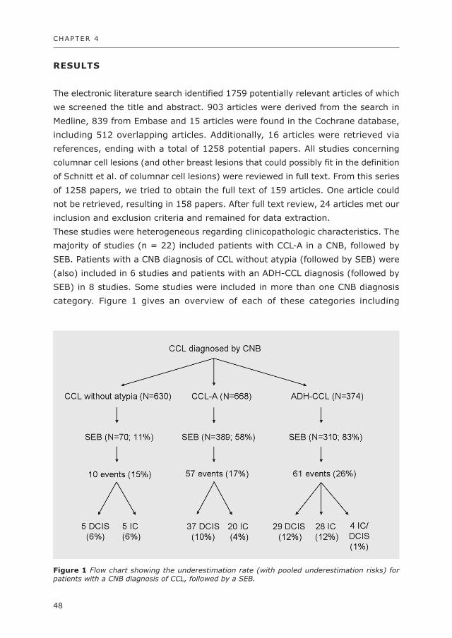
CHAPTER 4
48
RESulTS
The electronic literature search identified 1759 potentially relevant articles of which
we screened the title and abstract. 903 articles were derived from the search in
Medline, 839 from Embase and 15 articles were found in the Cochrane database,
including 512 overlapping articles. Additionally, 16 articles were retrieved via
references, ending with a total of 1258 potential papers. All studies concerning
columnar cell lesions (and other breast lesions that could possibly fit in the definition
of Schnitt et al. of columnar cell lesions) were reviewed in full text. From this series
of 1258 papers, we tried to obtain the full text of 159 articles. One article could
not be retrieved, resulting in 158 papers. After full text review, 24 articles met our
inclusion and exclusion criteria and remained for data extraction.
These studies were heterogeneous regarding clinicopathologic characteristics. The
majority of studies (n = 22) included patients with CCL-A in a CNB, followed by
SEB. Patients with a CNB diagnosis of CCL without atypia (followed by SEB) were
(also) included in 6 studies and patients with an ADH-CCL diagnosis (followed by
SEB) in 8 studies. Some studies were included in more than one CNB diagnosis
category. Figure 1 gives an overview of each of these categories including
figure 1 Flow chart showing the underestimation rate (with pooled underestimation risks) for patients with a CNB diagnosis of CCL, followed by a SEB.
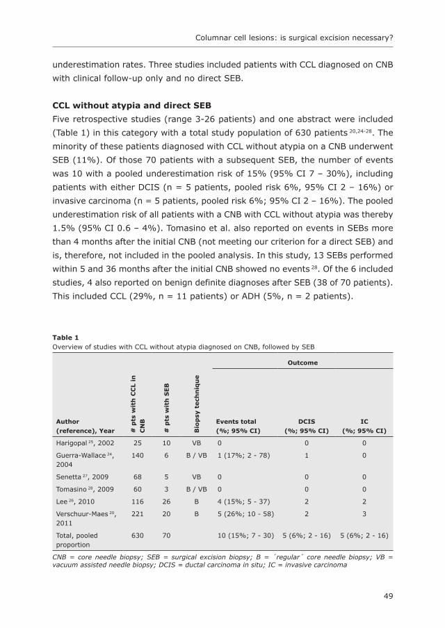
49
Columnar cell lesions: is surgical excision necessary?
underestimation rates. Three studies included patients with CCL diagnosed on CNB
with clinical follow-up only and no direct SEB.
CCl without atypia and direct SEb
Five retrospective studies (range 3-26 patients) and one abstract were included
(Table 1) in this category with a total study population of 630 patients 20,24-28. The
minority of these patients diagnosed with CCL without atypia on a CNB underwent
SEB (11%). Of those 70 patients with a subsequent SEB, the number of events
was 10 with a pooled underestimation risk of 15% (95% CI 7 – 30%), including
patients with either DCIS (n = 5 patients, pooled risk 6%, 95% CI 2 – 16%) or
invasive carcinoma (n = 5 patients, pooled risk 6%; 95% CI 2 – 16%). The pooled
underestimation risk of all patients with a CNB with CCL without atypia was thereby
1.5% (95% CI 0.6 – 4%). Tomasino et al. also reported on events in SEBs more
than 4 months after the initial CNB (not meeting our criterion for a direct SEB) and
is, therefore, not included in the pooled analysis. In this study, 13 SEBs performed
within 5 and 36 months after the initial CNB showed no events 28. Of the 6 included
studies, 4 also reported on benign definite diagnoses after SEB (38 of 70 patients).
This included CCL (29%, n = 11 patients) or ADH (5%, n = 2 patients).
Table 1 Overview of studies with CCL without atypia diagnosed on CNB, followed by SEB
Outcome
Author
(reference), Year # p
ts w
ith
CC
l in
CN
b
# p
ts w
ith
SEb
bio
psy
tech
niq
ue
Events total
(%; 95% Ci)
dCiS
(%; 95% Ci)
iC
(%; 95% Ci)
Harigopal 25, 2002 25 10 VB 0 0 0
Guerra-Wallace 24, 2004
140 6 B / VB 1 (17%; 2 - 78) 1 0
Senetta 27, 2009 68 5 VB 0 0 0
Tomasino 28, 2009 60 3 B / VB 0 0 0
Lee 26, 2010 116 26 B 4 (15%; 5 - 37) 2 2
Verschuur-Maes 20, 2011
221 20 B 5 (26%; 10 - 58) 2 3
Total, pooled proportion
630 70 10 (15%; 7 - 30) 5 (6%; 2 - 16) 5 (6%; 2 - 16)
CNB = core needle biopsy; SEB = surgical excision biopsy; B = ´regular´ core needle biopsy; VB = vacuum assisted needle biopsy; DCIS = ductal carcinoma in situ; IC = invasive carcinoma
CHAPTER 4
50
The other SEBs showed either a benign lesion, no lesion or data were missing. In
those studies restricted to vacuum assisted biopsies (n = 2 studies, 15 patients),
none had a significant lesion in the subsequent SEB.
CCl-A and direct SEb
We included 22 studies (range 1-60 patients) with a total study population of 668
patients in this category 20,24,26-44 (Table 2). The majority of these studies collected
their data retrospectively (20/22 studies). A CNB diagnosis of CCL-A was followed
by SEB in 58% of the patients. Of those 389 patients with a subsequent SEB, the
number of events was 57 with a pooled underestimation risk of 17% (95% CI 10
– 27%), including patients with DCIS (n = 37 patients, 10%; 95% CI 7 – 16%) or
invasive carcinoma (n=20 patients, 4%; 95% CI 2 – 9%). The pooled underestimation
risk of the number for all patients with a CNB with CCL-A was 9% (95% CI 5 –
14%). Tomasino et al. also reported on events in SEBs performed between 5 and
8 months after initial CNB and were, therefore, not included in the pooled analysis.
Out of 18 patients with a SEB, 2 had a definitive diagnose of DCIS and 3 patients
had invasive carcinoma 28.
Of the 22 included studies, 17 also reported on benign definite diagnoses after SEB
(337 of 389 patients). This included CCL (with atypia) (24%, n = 80 patients), lobular
intraepithelial neoplasia (6%, n = 21 patients) or ADH (14%, n = 47 patients). In
7% of the SEBs, a CCL, lobular intraepithelial neoplasia or ADH (without subdivision)
was found. The other SEBs showed a benign lesion, no lesion or data were missing.
The pooled underestimation rate in studies restricted to vacuum assisted needle
biopsies (n = 9 studies; 204 patients) was 12%, compared to 17% in studies
restricted to regular core needle biopsies (n = 9 studies; 109 patients), which was
not statistically significant different in meta-regression analysis (p=0.42).
AdH-CCl and direct SEb
Seven retrospective studies (range 2-190 patients) and one abstract were included
in this category with a total study population of 374 patients (Table 3) 20,25,26,31,32,37,39,45.
The majority of these patients (83%) underwent an SEB. Of those 310 patients
with a subsequent SEB, the number of events was 61 with a pooled underestimation
risk of 26% (95% CI 16 – 40%). This included patients with DCIS (n = 28 patients,
12%, 95% CI 7 – 19), invasive carcinoma (n = 29 patients, 12%, 95% CI 7 –
19%), or an uncategorized event (n = 4 patients). The pooled underestimation
risk of all patients with a CNB with ADH-CCL was 20% (95% CI 13 – 28%).
Of the 8 studies, 5 also reported on benign definite diagnoses after SEB (224 of 310
patients). This included CCL (with atypia) (2%, n = 5 patients), lobular intraepithelial

51
Columnar cell lesions: is surgical excision necessary?
neoplasia (11%, n = 25 patients) or ADH (35%, n = 79 patients). The other SEBs
showed either a benign lesion, no lesion or data were missing. The pooled
underestimation rate in studies restricted to vacuum assisted needle biopsies (n =
4 studies; 107 patients) was 6%, compared to 20% in studies restricted to regular
Table 2 Overview of studies with CCL-A diagnosed on CNB, followed by SEB
Outcome
Author
(reference), Year # p
ts w
ith
CC
l-A
in
CN
b
# p
ts w
ith
SEb
bio
psy
tech
-
niq
ue
No. of events
(%; 95% Ci)
dCiS
(%; 95% Ci)
iC
(%; 95% Ci)
Bibeau 29, 2003 8 3 VB 2 (67%; 13-214) 2 0
Bonnett 30, 2003 9 9 B / VB 2 (22%; 4 - 71) 2 0
Guerra-Wallace 24,
2004
39 31 B / VB 4 (13%; 4 - 31) 3 1
David 34, 2006 59 40 VB 7 (18%; 8 - 34) 3 4
Lim 41, 2006 8 5 B 1 (20%; 2 - 93) 1 0
Datrice 33, 2007 2 1 B 0 0 0
Kunju 39, 2007 14 12 B 3 (13%; 7 - 67) 1 2
Martel 42, 2007 63 5 B 0 0 0
Kumaroswamy 38,
2008
9 5 VB 2 (40%; 8 - 128) 0 2
Chivukula 31, 2009 39 35 B 5 (14%; 5 – 31) 3 2
Darvishian 32, 2009 12 12 B 2 (17%; 3 - 53) 2 0
Hayes 36, 2009 8 8 B 1 (13%; 1 – 58) 1 0
Ingegnoli 37, 2009 18 15 VB 3 (20%; 6 - 53) 1 2
Noël 43, 2010 60 20 VB 0 0 0
Noske 44, 2009 43 30 B / VB 2 (7%; 1 - 21) 2 0
Piubello 45, 2009 33 20 VB 0 0 0
Senetta 27, 2009 41 36 VB 0 0 0
Tomasino 28, 2009* 54 6 B / VB 3 (50%; 14 - 133) 3 0
Flegg 35, 2010 5 5 VB 2 (40%; 8 – 128) 1 1
Lavoué 40, 2010 60 60 VB 8 (13%; 6 - 25%) 6 2
Lee 26, 2010 15 7 B 1 (14%; 1 - 67) 1 0
Verschuur-Maes 20,
2011
69 24 B 9 (36%; 18 - 66) 5 4
Total, pooled
proportion
668 389 57 (17%; 10-27) 37 (10%; 7 - 16) 20 (4%; 2 - 9)
CCL-A = columnar cell lesion with atypia; CNB = core needle biopsy; SEB = surgical excision biopsy; B = ´regular´ core needle biopsy; VB = vacuum assisted needle biopsy; DCIS = ductal carcinoma in situ; IC = invasive carcinoma, * patients with a SEB after 4 months were excluded from analysis.

CHAPTER 4
52
core needle biopsies (n = 4 studies; 203 patients), which was not statistically
significant different in the meta-regression analysis (p=0.79).
CCl and long-term follow-up
Three studies were included in this category, including two retrospective studies
and one nested case- control study (Table 4) 20,42,46 with a total study population of
408 patients (range 13-201 patients). The results of these studies could not be
pooled because of heterogeneity. In a nested case control study, Boulos et al.
reported a RR of 1.5 (CI 95% 1.0 – 2.2) for developing invasive carcinoma after a
CNB diagnosis of CCL, compared to a RR of 3.5 (CI 95% 1.2 – 9.7) for patients
with ADH-CCL 20. Of those 103 patients diagnosed initially with CCL-A, Martel et al.
and Verschuur-Maes et al reported a total of 15 events (pooled underestimation
risk 16%, CI 95% 9% – 28%), including patients with DCIS (n=1 patient, ipsilateral)
or an invasive carcinoma (n=14 patients, 11 ipsilateral and 3 contralateral) in the
follow-up 20,42. Verschuur-Maes et al. reported a progression risk of 0.5% (95% CI
0.04 – 2.3%) for patients diagnosed with CCL without atypia and 23.1% (2 invasive
carcinomas ipsilateral and 1 contralateral, 95% CI 6.4% – 61.6%) for patients with
an initial diagnosis of ADH-CCL 20.
Table 3 Overview of studies with ADH-CCL diagnosed on CNB, followed by SEB
Outcome
Author (reference),
Year # p
ts
wit
h A
dH
-CC
l in
CN
b
# p
ts w
ith
SEb
bio
psy
tech
niq
ue
Events total
(%; 95% Ci)
dCiS
(%; 95% Ci)
iC
(%; 95% Ci)
Harigopal 25, 2002 5 5 VB 0 0 0
Kunju 39, 2007 38 31 VB 4 (11%; 4 - 31) M M
Chivukula 31, 2009 214 190 B 31 (16%; 11 - 23) 15 16
Darvishian 32, 2009 3 3 B 0 0 0
Ingegnoli 37, 2009 2 2 B 1 (50%; 5 - 233) 0 1
Piubello 45, 2009 11 10 VB 3 (30%; 8 - 80) 2 1
Lee 26, 2010 80 61 VB 20 (33%; 21 - 50) 10 10
Verschuur-Maes20, 2011 21 8 B 2 (33%; 5 -80) 2 0
Total, pooled proportion 374 310 61 (26%; 16 - 40) 29 (12%; 7 - 19) 28 (12%; 7 - 19)
ADH-CCL = columnar cell lesion associated with atypical ductal hyperplasia; CNB = core needle biopsy; SEB = surgical excision biopsy; B = ´regular´ core needle biopsy; VB = vacuum assisted needle biopsy; DCIS = ductal carcinoma in situ; IC = invasive carcinoma; M = missing data

53
Columnar cell lesions: is surgical excision necessary?
diSCuSSiON
CCLs, which are regarded as putative breast cancer precursors, are increasingly
diagnosed by CNB, as more microcalcifications are recognized by improved breast
cancer screening techniques. This systematic review was conducted to give an
overview of the current literature regarding the underestimation rate and progression
risk of patients with a CNB diagnosis of CCL, in order to provide treatment
recommendations for these patients.
The pooled underestimation risks for all patients initially diagnosed with CCL without
atypia, CCL-A or ADH-CCL were 1.5%, 9% and 20%, respectively. However, this
systematic review has several limitations, which could have biased these results.
Not all patients underwent SEB after an initial diagnosis of CCL-A (58%) or ADH-
CCL (83%) and particularly only a minority of patients with an initial diagnosis of
CCL without atypia (11%) received an SEB. This likely resulted in work-up bias,
since it is unknown whether more significant lesions were in fact left in the breast.
Based on the number of patients that underwent an SEB, the pooled underestimation
risks were 15% for CCL without atypia, 17% for patients with a CNB diagnosis of
CCL-A and 26% for ADH-CCL. These latter differences seem relatively limited, which
is interesting since several articles suggested that the underestimation rate and
Table 4 Overview of studies including CCL-A, CCL without atypia and ADH-CCL diagnosed on CNB, followed by clinical follow-up. The progression rate is postulated as relative risk (RR) or absolute number of events.
Outcome
Author
(reference),
Year Typ
e o
f
stu
dy
# p
ts w
ith
CC
l i
n C
Nb
bio
psy
tech
niq
ue
fo
llo
w-u
p
tim
e
RR
(95
% C
i)
dC
iS (
%)
iC (
%)
Martel 42,
2007
Retrospective 58 CCL-A B Mean 6.2 yrs
(range 5 m – 11 yr)
1 (2%) 9 (16%)
Boulos 46,
2008
Retrospective/
nested case
control study
47 CCL B Mean 17 year RR 1.5
(95% CI 1.0 – 2.15;)
44 ADH-CCL RR 3.5
(CI 95% 1.2–9.7)
Verschuur-
Maes20,
2011
Retrospective 45 CCL-A B Mean 3.5 yrs
(range 5 m – 8 yr)
5 (11%)
201 CCL 1 (0.5%)
13 ADH-CCL 3 (23%)
ADH-CCL = columnar cell lesion associated with atypical ductal hyperplasia; CNB = core needle biopsy; SEB = surgical excision biopsy; B = ´regular´ core needle biopsy; VB = vacuum assisted needle biopsy; DCIS = ductal carcinoma in situ; IC = invasive carcinoma; M = missing data

CHAPTER 4
54
the progression risk of CCL without atypia are probably limited. However, the
calculated underestimation rate based on the number of patients that underwent
an SEB likely resulted in an overestimated upgrade risk by selection of high risk
patients. The rates based on the whole group of patients initially diagnosed with a
CCL, therefore, seem to better reflect the actual underestimation risk (assuming
that the number of events in the group of patients without SEB is limited),
suggesting that CCL without atypia indeed gives a lower underestimation rate for
DCIS / invasive carcinoma, compared to CCL-A and ADH-CCL.
Several other factors could have affected the results of this review. The studies
included in this systematic review were generally small, heterogeneous and
retrospective. The mammographic abnormality (density versus microcalcifications)
and size are likely associated with underestimation rate but this could not be
assessed in this review because of missing and heterogeneous data. Furthermore,
the underestimation rate of finding (in situ) carcinoma after a CNB of CCL depends
on the size and number of needle biopsies and the experience of the operator 47.
In this review, we observed a trend towards a lower underestimation rate with the
use of vacuum assisted biopsies versus regular CNB for CCL-A and ADH-CCL, but
these differences were not statistically significant. Consistent with this finding,
Lavoué et al. reported on one of the largest series of CCL-A published to date and
concluded that the size of the vacuum assisted biopsies did not significantly affect
the underestimation rate after SEB 40. Previous studies concerning pure ADH,
however, showed statistically significant lower underestimation rates after vacuum
assisted biopsies compared to the smaller CNBs 48-50. The effect of the number of
biopsies with regard to underestimation rate could not be assessed in this review
because of missing data. Classification of breast cancer precursor lesions is
associated with suboptimal interobserver reproducibility 51,52, which could also have
caused heterogeneity between studies included in this review.
Follow-up studies including patients with a CNB diagnosis of CCL showed that
the progression risk to (in situ) carcinoma seems to be higher than the baseline
risk, although no firm conclusions can be drawn based on three heterogeneous
studies 20,42,46. Several other follow-up studies included patients with CCLs diagnosed
on SEB. A recent nested case control study by Kabat et al. reported a non-
significantly elevated risk for CCL-A and for multiple CCLs 53. Aroner et al. reported
a significantly increased breast cancer risk after a SEB diagnosis of CCL (OR=1.44;
95% CI 1.14-1.83) 54. However, this became non-significant after adjustment for
concurrent proliferative changes, which suggests that CCLs does not increase
breast cancer risk independently of concurrent proliferative changes in the breast.
Shaaban et al. described a slightly increased risk (p=0.058) for CCL-A compared

55
Columnar cell lesions: is surgical excision necessary?
with controls 14. Other retrospective studies analysed breast cancer risk following
CCLs diagnosed on a SEB and reported no (in situ) carcinomas after long term
clinical follow-up 13,55,56. The results of these studies support the hypothesis that
excision of CCLs might be curative by removing the breast cancer precursor.
The pooled underestimation risks of 9% and 20% for (in situ) carcinomas for all
patients initially diagnosed with CCL-A or ADH-CCL respectively, and risks of 17%
and 26% for (in situ) carcinomas diagnosed in all patients undergoing SEB seem
to support the indication for surgical excision, which is generally consistent with
conclusions from individual studies 9,50,57. Another therapeutic option might be
complete removal of all microcalcifications by vacuum assisted biopsies 34 or the
BLES procedure 58. Based on CCL-A diagnoses, several other factors such as type
and size of mammographic abnormalities and family history have to be considered
in deciding whether to proceed with SEB, in order to minimize the number of
surgical excisions without therapeutic benefit. Also prevention of these lesions or
limitation of their growth should be considered, for instance by limiting hormonal
stimulation of the ER (over)expressed in CCL and CCL-A 59-61. Another factor that
has to be considered is the finding that the majority of (in situ) carcinomas
associated with CCLs are low or intermediate grade, which suggests hormonal
receptor positivity and relatively early detection with favourable treatment options
and good prognosis. A multidisciplinary approach between clinicians, radiologist
and pathologist is essential in these cases.
In conclusion, based on the high underestimation rates of patients with a CNB
diagnosis of CCL-A and ADH-CCL, surgical excision should be considered. Based
on current literature, no definitive conclusion can be drawn regarding optimal
treatment of patients with a CNB diagnosis of CCL without atypia and more studies
are needed to establish the long-term follow-up, but for now the underestimation
and progression rates seem to be limited and surgical excision is not deemed
necessary in these cases.
Acknowledgements
We thank Dr. A.J. Witkamp for critically reading the manuscript.

CHAPTER 4
56
REfERENCES
1. Verschuur-Maes AHJ, van Gils CH, Van den Bosch MAAJ et al. Digital mammography: more microcalcifications, more columnar cell lesions without atypia. Mod Pathol 2011; 24:1191-1197.
2. Aulmann S, Elsawaf Z, Penzel R et al. Invasive tubular carcinoma of the breast frequently is clonally related to flat epithelial atypia and low-grade ductal carcinoma in situ. Am J Surg Pathol 2009; 33:1646-1653.
3. Dabbs DJ, Carter G, Fudge M et al. Molecular alterations in columnar cell lesions of the breast. Mod Pathol 2006; 19:344-349.
4. Fraser JL, Raza S, Chorny K et al. Columnar alteration with prominent apical snouts and secretions: a spectrum of changes frequently present in breast biopsies performed for microcalcifications. Am J Surg Pathol 1998; 22:1521-1527.
5. Goldstein NS, O’Malley BA, Goldstein NS et al. Cancerization of small ectatic ducts of the breast by ductal carcinoma in situ cells with apocrine snouts: a lesion associated with tubular carcinoma. Am J Clin Pathol 1997; 107:561-566.
6. Oyama T, Maluf H, Koerner F. Atypical cystic lobules: an early stage in the formation of low-grade ductal carcinoma in situ. Virchows Arch 1999; 435:413-421.
7. Rosen PP. Columnar cell hyperplasia is associated with lobular carcinoma in situ and tubular carcinoma. Am J Surg Pathol 1999; 23:1561.
8. Simpson PT, Gale T, Reis-Filho JS et al. Columnar cell lesions of the breast: the missing link in breast cancer progression? A morphological and molecular analysis. Am J Surg Pathol 2005; 29:734-746.
9. Schnitt SJ, Vincent-Salomon A. Columnar cell lesions of the breast. Adv Anat Pathol 2003; 10:113-124.
10. Tavassoli FA, Devilee P. WHO Classification Tumours of the Breast and Female Genital Organs, IARC, Lyon. 2003.
11. Tavassoli FA. Ductal carcinoma in situ: introduction of the concept of ductal intraepithelial neoplasia. Mod Pathol 1998; 11:140-154.
12. Azzopardi JG, Ahmed A, Millis RR. Problems in breast pathology. Major Probl Pathol 1979; 11:i-466.
13. Eusebi V, Feudale E, Foschini MP et al. Long-term follow-up of in situ carcinoma of the breast. Semin Diagn Pathol 1994; 11:223-235.
14. Shaaban AM, Sloane JP, West CR et al. Histopathologic types of benign breast lesions and the risk of breast cancer: Case-control study. Am J Surg Pathol 2002; 26:421-430.
15. Kusama R, Fujimori M, Matsuyama I et al. Clinicopathological characteristics of atypical cystic duct (ACD) of the breast: assessment of ACD as a precancerous lesion. Pathol Int 2000; 50:793-800.
16. Feeley L, Quinn CM. Columnar cell lesions of the breast. Histopathology 2008; 52:11-19.17. Lee S, Medina D, Tsimelzon A et al. Alterations of gene expression in the development of early
hyperplastic precursors of breast cancer. Am J Pathol 2007; 171:252-262.18. Moinfar F, Man YG, Bratthauer GL et al. Genetic abnormalities in mammary ductal intraepithelial
neoplasia-flat type (“clinging ductal carcinoma in situ”): a simulator of normal mammary epithelium. Cancer 2000; 88:2072-2081.
19. Sinn HP. Breast cancer precursors: lessons learned from molecular genetics. J Mol Med 2009; 87:113-115.
20. Verschuur-Maes AH, Witkamp AJ, De Bruin PC et al. Progression risk of columnar cell lesions of the breast diagnosed in core needle biopsies. Int J Cancer 2011 ;129:2674-2680.
21. Rothman K.J. Epidemiology. An introduction. New York, Oxford University Press. 2002.22. Hamza TH, van Houwelingen HC, Stijnen T. The binomial distribution of meta-analysis was preferred
to model within-study variability. J Clin Epidemiol 2008; 61:41-51.23. Greenland S. Quantitative methods in the review of epidemiologic literature. Epidemiol. Rev. 1987.24. Guerra-Wallace MM, Christensen WN, White RL, Jr. A retrospective study of columnar alteration
with prominent apical snouts and secretions and the association with cancer. Am J Surg 2004; 188:395-398.
25. Harigopal M, Yao DX, Hoda SA et al. Columnar change alteration diagnosed on mammotome core needle biopsy for indeterminate microcalcifications: resultss of subsequent mammograms and surgical excision. Mod Pathol 2002; 15:36A.

57
Columnar cell lesions: is surgical excision necessary?
26. Lee TY, Macintosh RF, Rayson D et al. Flat epithelial atypia on breast needle core biopsy: a retrospective study with clinical-pathological correlation. Breast J 2010; 16:377-383.
27. Senetta R, Campanino PP, Mariscotti G et al. Columnar cell lesions associated with breast calcifications on vacuum-assisted core biopsies: clinical, radiographic, and histological correlations. Mod Pathol 2009; 22:762-769.
28. Tomasino RM, Morello V, Gullo A et al. Assessment of “grading” with Ki-67 and c-kit immunohistochemical expressions may be a helpful tool in management of patients with flat epithelial atypia (FEA) and columnar cell lesions (CCLs) on core breast biopsy. J Cell Physiol 2009; 221:343-349.
29. Bibeau F, Chateau MC, Masson B et al. Management of non-palpable breast lesions with vacuum-assisted large core needle biopsies (Mammotome). Experience with 560 procedures at the Val d’Aurelle Center. Ann Pathol 2003; 23:582-592.
30. Bonnett M, Wallis T, Rossmann M et al. Histopathologic analysis of atypical lesions in image-guided core breast biopsies. Mod Pathol 2003; 16:154-160.
31. Chivukula M, Bhargava R, Tseng G et al. Clinicopathologic implications of “flat epithelial atypia” in core needle biopsy specimens of the breast. Am J Clin Pathol 2009; 131:802-808.
32. Darvishian F, Singh B, Simsir A et al. Atypia on breast core needle biopsies: reproducibility and significance. Ann Clin Lab Sci 2009; 39:270-276.
33. Datrice N, Narula N, Maggard M et al. Do breast columnar cell lesions with atypia need to be excised? Am Surg 2007; 73:984-986.
34. David N, Labbe-Devilliers C, Moreau D et al. Diagnosis of flat epithelial atypia (FEA) after stereotactic vacuum-assisted biopsy (VAB) of the breast: What is the best management: systematic surgery for all or follow-up? J Radiol 2006; 87:1671-1677.
35. Flegg KM, Flaherty JJ, Bicknell AM et al. Surgical outcomes of borderline breast lesions detected by needle biopsy in a breast screening program. World J Surg Oncol 2010; 8:78.
36. Hayes BD, O’Doherty A, Quinn CM. Correlation of needle core biopsy with excision histology in screen-detected B3 lesions: the Merrion Breast Screening Unit experience. J Clin Pathol 2009; 62:1136-1140.
37. Ingegnoli A, d’Aloia C, Frattaruolo A et al. Flat epithelial atypia and atypical ductal hyperplasia: carcinoma underestimation rate. Breast J 2010; 16:55-59.
38. Kumaroswamy V, Liston J, Shaaban AM et al. Vacuum assisted stereotactic guided mammotome biopsies in the management of screen detected microcalcifications: experience of a large breast screening centre. J Clin Pathol 2008; 61:766-769.
39. Kunju LP, Kleer CG. Significance of flat epithelial atypia on mammotome core needle biopsy: Should it be excised? Hum Pathol 2007; 38:35-41.
40. Lavoue V, Roger CM, Poilblanc M et al. Pure flat epithelial atypia (DIN 1a) on core needle biopsy: study of 60 biopsies with follow-up surgical excision. Breast Cancer Res Treat 2011; 125:121-126.
41. Lim CN, Ho BC, Bay BH et al. Nuclear morphometry in columnar cell lesions of the breast: is it useful? J Clin Pathol 2006; 59:1283-1286.
42. Martel M, Barron-Rodriguez P, Tolgay O, I et al. Flat DIN 1 (flat epithelial atypia) on core needle biopsy: 63 cases identified retrospectively among 1,751 core biopsies performed over an 8-year period (1992-1999). Virchows Arch 2007; 451:883-891.
43. Noel JC, Buxant F, Engohan-Aloghe C. Immediate surgical resection of residual microcalcifications after a diagnosis of pure flat epithelial atypia on core biopsy: A word of caution. Surg Oncol 2010; 19:243-246.
44. Noske A, Pahl S, Fallenberg E et al. Flat epithelial atypia is a common subtype of B3 breast lesions and is associated with noninvasive cancer but not with invasive cancer in final excision histology. Hum Pathol 2010; 41:522-527.
45. Piubello Q, Parisi A, Eccher A et al. Flat epithelial atypia on core needle biopsy: which is the right management? Am J Surg Pathol 2009; 33:1078-1084.
46. Boulos FI, Dupont WD, Simpson JF et al. Histologic associations and long-term cancer risk in columnar cell lesions of the breast: a retrospective cohort and a nested case-control study. Cancer 2008; 113:2415-2421.
47. Ciatto S, Houssami N, Ambrogetti D et al. Accuracy and underestimation of malignancy of breast core needle biopsy: the Florence experience of over 4000 consecutive biopsies. Breast Cancer Res Treat 2007; 101:291-297.

CHAPTER 4
58
48. Darling ML, Smith DN, Lester SC et al. Atypical ductal hyperplasia and ductal carcinoma in situ as revealed by large-core needle breast biopsy: results of surgical excision. AJR Am J Roentgenol 2000; American Journal of Roentgenology. 175:1341-1346.
49. Houssami N, Ciatto S, Ellis I et al. Underestimation of malignancy of breast core-needle biopsy: concepts and precise overall and category-specific estimates. Cancer 2007; 109:487-495.
50. Johnson NB, Collins LC. Update on percutaneous needle biopsy of nonmalignant breast lesions. Adv Anat Pathol 2009; 16:183-195.
51. Haupt B, Schwartz MR, Xu Q et al. Columnar cell lesions: a consensus study among pathology trainees. Hum Pathol 2010; 41:895-901.
52. Tan PH, Ho BC, Selvarajan S et al. Pathological diagnosis of columnar cell lesions of the breast: are there issues of reproducibility? J Clin Pathol 2005; 58:705-709.
53. Kabat GC, Jones JG, Olson N et al. A multi-center prospective cohort study of benign breast disease and risk of subsequent breast cancer. Cancer Causes Control 2010; 21:821-828.
54. Aroner SA, Collins LC, Schnitt SJ et al. Columnar cell lesions and subsequent breast cancer risk: a nested case-control study. Breast Cancer Res 2010; 12:R61.
55. Bijker N, Peterse JL, Duchateau L et al. Risk factors for recurrence and metastasis after breast-conserving therapy for ductal carcinoma-in-situ: analysis of European Organization for Research and Treatment of Cancer Trial 10853. J Clin Oncol 2001; 19:2263-2271.
56. de Mascarel I, MacGrogan G, Mathoulin-Pelissier S et al. Epithelial atypia in biopsies performed for microcalcifications. practical considerations about 2,833 serially sectioned surgical biopsies with a long follow-up. Virchows Arch 2007; 451:1-10.
57. Jacobs TW, Connolly JL, Schnitt SJ. Nonmalignant lesions in breast core needle biopsies: to excise or not to excise? Am J Surg Pathol 2002; 26:1095-1110.
58. Allen SD, Nerurkar A, la Rovere GU. The breast lesion excision system (BLES): a novel technique in the diagnostic and therapeutic management of small indeterminate breast lesions? Eur Radiol 2011 ;21:919-924.
59. Lee S, Mohsin SK, Mao S et al. Hormones, receptors, and growth in hyperplastic enlarged lobular units: Early potential precursors of breast cancer. Breast Cancer Research 2006; 8:R6.
60. Tremblay G, Deschenes J, Alpert L et al. Overexpression of estrogen receptors in columnar cell change and in unfolding breast lobules. Breast Journal 2005; 11:326-332.
61. Wood CE, Hester JM, Appt SE et al. Estrogen effects on epithelial proliferation and benign proliferative lesions in the postmenopausal primate mammary gland. Lab Invest 2008; 88:938-948.

59
Columnar cell lesions: is surgical excision necessary?


Anoek H.J. Verschuur-Maes1, Peter C. de Bruin2, Paul J. van Diest1
1 Department of Pathology, University Medical Center Utrecht, The Netherlands. 2 Department of Pathology, St. Antonius Hospital Nieuwegein, The Netherlands
Submitted
Epigenetic progression of columnar cell lesions
of the breast to ductal carcinoma in situ and invasive
breast cancer: a Methylation-specific Multiplex
Ligation-dependent Probe Amplification study
Chapter
5
CHAPTER 5
62
AbSTRACT
introduction
Promoter hypermethylation of several tumour suppressor genes often occurs during
breast carcinogenesis, but little is known about epigenetic silencing in the possible
precursor columnar cell lesion (CCL).
Methods
Promoter hypermethylation of 50 different tumour suppressor genes was assessed
in normal breast tissue (N=10), CCL (N=15), DCIS grade I originating in CCL
(N=5), and paired CCL (N=15) with DCIS (N=7) and/or invasive carcinoma (N=14)
by Methylation-specific Multiplex Ligation-dependent Probe Amplification.
Results
Increasing mean cumulative methylation levels were found from normal breast
tissue to CCL to DCIS and invasive carcinoma (p<0.001), with similar methylation
levels in DCIS and invasive carcinoma. Methylation levels and frequencies (in the
overall analysis and analysis of only the synchronous lesions) were highest for
RASSF1, CCND2, ID4, SCGB3A1 and CDH13. The methylation levels of ID4, CCND2
and CDH13 increased significantly from normal breast tissue to CCL and to DCIS/
invasive carcinoma. RASSF1, SCGB3A1 and SFRP5 had significant higher methylation
levels in CCL compared to normal breast tissue but showed no significant differences
between CCL, DCIS and invasive carcinoma. Also no difference were found between
CCLs with and without atypia, or CCLs with or without synchronous cancer.
Conclusion
Promoter hypermethylation for several established tumour suppressor genes is
already present in CCLs, underlining that promoter hypermethylation is an early
event in breast carcinogenesis. Atypia in CCL or presence of synchronous more
advanced lesions does not seem to be accompanied by higher methylation
levels.

63
Methylation and columnar cell lesions
iNTROduCTiON
The development of breast cancer is believed to be a complex multistep process
in which invasive cancer arises from normal breast epithelium via precursor
lesions like atypical ductal hyperplasia (ADH), lobular neoplasia and ductal
carcinoma in situ (DCIS) 1. In recent years, more evidence has become available
that also columnar cell lesions (CCLs) play a role in carcinogenesis of breast
cancer 2-4. These are cystically dilated enlarged terminal duct lobular units lined
by columnar cell epithelium with one or two cell layers (columnar cell change,
CCC) or more cell layers (columnar cell hyperplasia, CCH). Intraluminal secretions
and microcalcifications are often seen. When the cells show cytonuclear or
architectural atypia, the lesions are designated as columnar cell change with
atypia or columnar cell hyperplasia with atypia according to the classification by
Schnitt et al. 5. Together, the lesions with atypia are called flat epithelial atypia 6
or columnar cell lesions with atypia (CCL-A), the latter being further used in this
article. Although CCL-A lack the complex architecture, they share many overlapping
morphological and immunohistochemical features with ADH and low grade DCIS
3,5,7. Recently, a review on follow-up studies showed that CCL-A diagnosed in core
needle biopsies have invasive carcinoma and/or DCIS in the subsequent surgical
resection specimens in 9% of the cases and are recommended for excision or
removal by vacuum assisted biopsies or the BLES procedure 8. At a more
fundamental level there is also evidence, albeit sparse, that CCLs (with atypia)
show similar genetic abnormalities as the accompanying DCIS and invasive
carcinoma 3,9-11.
In the carcinogenesis of breast cancer, activation of oncogenes and inactivation of
tumour suppressor genes play an important role. In addition, epigenetic
abnormalities such as hypermethylation of promotor CpG islands (further denoted
“methylation”) are considered to be an early event during tumour development
and may lead to inactivation of tumour suppressor genes, DNA repair genes, cell
cycle regulators and transcription factors 12-17. A few studies reported on promoter
methylation in precursor lesions like ADH 18, but methylation studies of CCLs are
rare 19. A single study showed that the number of methylated genes increased
stepwise from normal breast tissue to ADH/CCL-A and to DCIS and invasive
carcinoma, suggesting that methylation is an early event in breast carcinogenesis20.
The investigated samples were however all from different patients, which provides
limitations in identifying true progression related events. In the present study we
therefore set out to investigate methylation of 50 tumour suppressor genes by
MS-MLPA in normal breast tissue, CCL with and without atypia, DCIS and invasive

CHAPTER 5
64
carcinoma, largely comprising synchronous lesions within the same patient. This
should shed further light on the role of CCLs and methylation therein in breast
carcinogenesis.
MATERiAl ANd METHOdS
Patient material
Tissue samples with only a CCL (with or without atypia), as well as cases with
abundant CCL and synchronous DCIS and/or invasive carcinoma were collected
between 1996 and 2009 at the Departments of Pathology of the University Medical
Center Utrecht and the St. Antonius Hospital Nieuwegein, The Netherlands. The
CCLs were graded according to the classification described by Schnitt and Vincent-
Salomon 5. Histologic type of the invasive carcinoma as well as grade of the invasive
and in situ cancers was assessed by two experienced observers (AVM and PvD)
according to the World Health Organization classification 6. As a refinement, we
recognized a subgroup of ductal carcinoma in situ (DCIS) grade I lesions with still
recognizable columnar cell type with apical snouts (“DCIS grade I originating from
CCL”). Normal breast tissue from 10 women undergoing mammoplasty was taken
along as controls. Anonymous use of redundant tissue for research purposes is
part of the standard treatment agreement with patients in our hospitals 21.
dNA isolation
Guided by marked H&E stained slides, the abundant CCL, DCIS and invasive
carcinoma lesions (cellularity >70%) at least 5 mm apart were harvested from serial
8 to 10 μm thick paraffin slides using a scalpel. In all other cases laser microdissection
(PALM Microlaser Technologies AG, Bernried, Germany) was used to isolate areas
of interest from the surrounding tissue. Depending on the amount of material, 50
or 100 μl direct lysis buffer (consisting of 10 mM TRIS-Hcl pH 8.3, 0.5% Tween
0.20, 1 mM EDTA) and 10 or 20 μl proteinase K (10 mg/ml, Roche, Almere, The
Netherlands) was added, and heated in a 56°C water bath for 16 hours, followed
by another proteinase K dose after one hour. The samples were then heat inactivated
by boiling for 10 minutes, and stored by 4 °C until analysis.
Methylation-specific Multiplex Ligation-dependent Probe Amplification
(MS-MlPA)
Using the isolated DNA solutions (100-240 ng), MS-MLPA analysis was performed
with the tumour suppressor gene kits ME001-C1, ME002-A1 and ME003-A1 (MRC

65
Methylation and columnar cell lesions
Holland, Amsterdam, The Netherlands), according to the manufacturers’ instructions
as described before 22-25. Blood samples from healthy volunteers without any history
of neoplastic malignancy were used as control samples and they were treated with
SssI methylase as a positive control. The reaction products were separated by
capillary electrophoresis using the 3130 DNA Analyzer (Applied Biosystems, Foster
City, CA, USA) and methylation percentages were analyzed using GeneScan Analysis
(Applied Biosystems) and Coffalyser software (version 9.4, MRC-Holland). Most
samples could be tested in duplicate.
Table 1 shows the 50 genes studied for promoter methylation. Twenty-five of the
genes had two probe sites in the different kits and one had three probe sites, which
were all analyzed separately. STK11 was present in ME002, which we excluded
from the analysis since some adaptations in the kit were carried out by MRC Holland
during the period in which we performed our tests.
Statistical analysis
Both the absolute methylation percentages as well as a cut-off value of 15% to
determine a sample as methylation positive were used in the analysis. This threshold
was chosen based on previous cell line experiments 26,27 and validated before 22-25.
The cumulative methylation index (CMI) was calculated as the sum of the
methylation percentages for all genes, as described before 28. All analysis were
carried out and shown in the tables per probe. However, in the text only the genes
(without subdivision per probe) are mentioned for sake of readability.
The chi-square test was used for comparing categorical variables. Absolute
methylation percentages between groups were compared with the Mann-Whitney
U or Kruskall-Wallis tests. Comparison of methylation percentages in the paired
CCL and invasive carcinoma cases were analyzed by a paired-samples t-test or
Wilcoxon signed rank test for normally respectively not normally distributed
populations, and by the Friedman test for the paired CCL, DCIS and invasive
carcinoma samples. For paired analysis of categorical variables, the McNemar test
was used. In the comparison of the different grades of CCL, the CCLs without atypia
(CCC and CCH) were grouped and compared with CCL-A. All tests were performed
with SPSS version 15.0, regarding two-sided p-values <0.05 as significant.

CHAPTER 5
66
Table 1 Tumour suppressor genes studied for methylation in premalignant and malignant breast lesions.
APC Adenomatous polyposis coli ID4* Inhibitor of DNA binding 4
ATM* Ataxia telangiectasia mutated IGSF4* Immunoglobulin superfamily, member 4
BCL2 B-cell lymphoma 2 MGMT* O6-methylguanine-DNA methyltransferase
BNIP3 BCL2/adenovirus E1B 19 kd-interacting protein 3
MLH1 MutL homologue 1
BRCA1* Breast cancer type 1 susceptibility protein
MSH6 MutS homolog 6
BRCA2* Breast cancer type 2 susceptibility protein
PAX5 Paired box gene 5
CACNA1A Calcium channel, voltage-depend-ent, P/Q type, alpha 1A subunit
PAX6 Paired box gene 6
CACNA1G Calcium channel, voltage-depend-ent, T type, alpha 1G subunit
PRDM2 PR domain containing 2
CASP8 Caspase 8 PTEN* Phosphatase and tensin homologue
CCND2* Cyclin D2 PYCARD N-terminal PYRIN-PAAD-DAPIN domain (PYD) and a C-terminal caspase-recruitment domain (CARD)
CD44* CD44 antigen precursor RARB** Retinoic acid receptor, b
CDH13* Cadherin 13, H-cadherin RASSF1* RAS-associated domain family 1 (isoform A)
CDKN1B Cyclin-dependent kinase inhibitor 1B (p27kip)
RB1* Retinioblastoma 1
CDKN2A* Cyclin-dependent kinase inhibitor 2A (encoding p14ARF)
RUNX3 Runt-related transcription factor 3
CDKN2B Cyclin-dependent kinase inhibitor 2B (p15)
SCGB3A1* Secretoglobulin family 3A
CHFR* Checkpoint protein with forkhead and RING finger domains
SFRP4* Secreted frizzled-related protein 4
DAPK1 Death-associated protein kinase 1 SFRP5* Secreted frizzled-related protein 5
DLC1* Deleted in liver cancer 1 TGIF Transforming Growth ß-Induced Factor
ESR1* Oestrogen receptor 1 THBS1 Thrombospondin 1
FHIT Fragile histidine triad gene TIMP3* Tissue inhibitor of metalloproteinases 3
GATA5 GATA 5 binding protein TP53 Tumour protein 53
GSTP1* Glutathione S-transferase pi TP73* Cellular tumour antigen p73
H2AFX* H2A histone family, member X TWIST1 Twist homologue 1
HIC1 Hypermethylated in cancer 1 VHL* Von Hippel–Lindau disease tumour suppressor
HLTF* Helicase-like transcription factor WT1 Wilms tumour 1
* two probe sites of the same genes in the three kits with tumour suppressor genes** three probe sites of the same gene in the three kits with tumour suppressor genes

67
Methylation and columnar cell lesions
RESulTS
baseline characteristics
Table 2 shows the number of studied lesions per category and grade of the lesions.
66 lesions in 45 patients were studied. The majority of the DCIS and invasive
cancers were of low grade. Most patients with CCL and synchronous DCIS and/or
invasive carcinoma showed atypia (4x CCC, 1x CCH and 10x CCL-A). The mean
age at time of breast surgery of women with CCL only was 53.0 years, with CCL
and DCIS 55.4 years and with CCL and invasive carcinoma 51.9 years, with an
overall mean age of 54.0 years (range 32-82 years). Mean age of the controls was
32.1 years.
Overall frequencies of promoter hypermethylation
Using a 15% methylation dosage ratio threshold, in paired analysis, the number
of methylated genes differed between normal breast (4.0 genes, 4.1 probes) on
the one hand and CCL (9.6 genes, p=0.084; 11.3 probes, p=0.057), DCIS (12.3
genes, p=0.029; 15.4 probes, p=0.007) and invasive carcinoma (11.6 genes,
p=0.029; 14.5 probes, p=0.008) on the other. In contrast, there were no significant
differences between the number of methylated genes between CCL and DCIS
(p=0.629 per genes; p=0.388 per probes), between CCL and invasive carcinoma
(p=0.629 per genes; p=0.409 per probes) and between DCIS and invasive
carcinoma (p=1.000).
Table 2 Overview of the breast lesions studied for differences in methylation of tumour suppressor genes.
Samples Patients
Pure lesions
Normal breast tissue 10
CCL 15
CCL without atypia 5
CCL-A 10
DCIS grade I originated in CCL-A 5
Synchronous lesions
CCL + DCIS + IC* 6
CCL + DCIS 1
CCL + IC** 8
* both DCIS and IC: 3x grade I, 1x grade II, 2x grade III** IC: 3x grade I, 3x grade II, 1x grade III, 1x lobular

CHAPTER 5
68
Table 3 Frequency of methylation in tumour suppressor genes in different premalignant and malignant breast lesions using a methylation threshold > 15%.
All lesions, frequencies (%) P-values *
Gen
es
Pro
be
No
rmal,
N=
10
CC
l,
N=
30
dC
iS,
N=
12
iC,
N=
14
All
CC
l-d
CiS
-iC
Nl-C
Cl
Nl-d
CiS
Nl-i
C
CC
l-d
CiS
CC
l-i
C
dC
iS-i
C
APC 1905-L01968 1 (10) 15 (50) 5 (41.7) 7 (50) 0.148 0.877 0.025 0.097 0.040 0.625 1.000 0.671
BRCA2a** 02285-L01776 0 (0) 2 (6.7) 2 (16.7) 0 (0) 0.282 0.272 0.402 0.176 - 0.319 0.340 0.125
CACNA1A*** 09055-L09224 1 (10) 2 (6.7) 1 (8.3) 6 (42.9) 0.015 0.008 0.751 0.892 0.081 0.386 0.005 0.048
CASP8 2761-L02210 0 (0) 1 (3.3) 1 (8.3) 2 (14.3) 0.422 0.415 0.559 0.350 0.212 0.492 0.179 0.636
CCND2a*** 03313-L02668 0 (0) 8 (26.7) 9 (75) 11 (78.6) 0.000 0.001 0.062 0.000 0.000 0.055 0.002 0.829
CCND2b*** 03312-L09381 1 (10) 16 (53.3) 10 (83.3) 12 (85.7) 0.001 0.061 0.013 0.001 0.000 0.071 0.049 0.867
CD44 3817-L01731 0 (0) 0 (0) 1 (8.3) 1 (7.1) 0.361 0.298 - 0.350 0.388 0.110 0.139 0.910
CD44** 04500-L02761 2 (20) 3 (10) 2 (16.7) 2 (14.3) 0.851 0.798 0.408 0.840 0.772 0.547 0.613 0.930
CDH13 7946-L07727 1 (10) 9 (30) 3 (25) 4 (28.6) 0.653 0.984 0.206 0.314 0.269 0.865 0.923 0.943
CDH13** 02257-L01742 3 (30) 15 (50) 8 (66.7) 10 (71.4) 0.111 0.221 0.271 0.087 0.024 0.327 0.100 0.568
CDKN2A 1524-L01744 0 (0) 0 (0) 2 (16.7) 2 (14.3) 0.072 0.070 - 0.156 0.212 0.022 0.034 0.792
CDKN2B 0607-L00591 1 (10) 4 (13.3) 1 (8.3) 0 (0) 0.565 0.358 0.783 0.943 0.227 0.713 0.152 0.250
ESR1** 02746-L02173 1 (10) 6 (20) 3 (25) 4 (28.6) 0.666 0.741 0.471 0.364 0.231 0.721 0.443 0.748
FHIT 2201-L01699 0 (0) 1 (3.3) 2 (16.7) 2 (14.3) 0.272 0.282 0.559 0.176 0.212 0.130 0.179 0.867
GATA5** 03752-L06199 1 (10) 5 (16.7) 3 (25) 5 (35.7) 0.321 0.301 0.609 0.364 0.123 0.534 0.120 0.471
GSTP1 1638-L01176 0 (0) 7 (23.3) 4 (33.3) 5 (35.7) 0.169 0.588 0.093 0.034 0.034 0.404 0.390 0.973
GSTP1** 02747-L02174 0 (0) 4 (13.3) 2 (16.7) 2 (14.3) 0.624 0.958 0.224 0.176 0.194 0.780 0.858 0.930
H2AFXa*** 08511-L08607 1 (10) 3 (10) 2 (16.7) 1 (7.1) 0.888 0.732 0.975 0.650 0.803 0.641 0.735 0.449
ID4a*** 04497-L03909 2 (20) 24 (80) 11 (91.7) 13 (92.9) 0.000 0.567 0.000 0.001 0.000 0.414 0.371 0.910
ID4b*** 04496-L03908 0 (0) 1 (3.3) 2 (16.7) 1 (7.1) 0.342 0.333 0.552 0.176 0.388 0.165 0.590 0.449
IGSF4** 3819-L03848 1 (10) 4 (13.3) 2 (16.7) 2 (14.3) 0.972 0.958 0.783 0.650 0.704 0.780 0.858 0.930
MGMTa** 05670-L05146 1 (10) 9 (30) 2 (16.7) 8 (57.1) 0.030 0.046 0.206 0.650 0.012 0.375 0.052 0.022
MGMTb** 13716-L15582 1 (10) 5 (16.7) 5 (41.7) 5 (35.7) 0.145 0.151 0.609 0.097 0.123 0.086 0.120 0.870
MSH6** 01250-L00798 8 (80) 21 (70) 9 (75) 12 (85.7) 0.454 0.284 0.540 0.781 0.385 0.746 0.112 0.238
PAX5** 03750-L03210 2 (20) 9 (30) 7 (58.3) 4 (28.6) 0.229 0.202 0.540 0.069 0.560 0.088 0.960 0.165
PAX6** 03749-L03209 0 (0) 2 (6.7) 3 (25) 3 (21.4) 0.140 0.191 0.402 0.089 0.103 0.097 0.123 0.910
PTEN 2203-L08261 0 (0) 2 (6.7) 0 (0) 1 (7.1) 0.665 0.648 0.402 - 0.388 0.359 0.953 0.345
RARB 10362-L10900 0 (0) 2 (6.7) 0 (0) 2 (14.3) 0.376 0.366 0.402 - 0.212 0.359 0.413 0.173
RARB*** 10362-L10900 1 (10) 2 (6.7) 2 (16.7) 1 (7.1) 0.787 0.587 0.751 0.650 0.803 0.134 0.976 0.449
RASSF1a 2248-L01734 0 (0) 24 (80) 10 (83.3) 10 (71.4) 0.000 0.733 0.000 0.000 0.000 0.804 0.527 0.473
RASSF1b 3807-L02159 3 (30) 26 (87) 11 (91.7) 10 (71.4) 0.002 0.313 0.001 0.003 0.045 0.651 0.222 0.192
RB1b** 04502-L02199 1 (10) 3 (10) 1 (8.3) 2 (14.3) 0.943 0.829 1.000 0.892 0.704 0.868 0.613 0.588
RUNX3*** 11131-L03905 2 (20) 12 (40) 6 (50) 6 (42.9) 0.525 0.878 0.224 0.145 0.242 0.773 0.927 0.716
SCGB3A1a*** 03305-L09382 1 (10) 28 (93.3) 12 (100) 11 (78.6) 0.000 0.057 0.000 0.000 0.001 0.550 0.057 0.088
SCGB3A1b*** 11132-L12956 0 (0) 8 (26.7) 6 (50) 9 (64.3) 0.006 0.059 0.062 0.009 0.001 0.238 0.021 0.462
SFRP4a*** 03744-L03204 0 (0) 0 (0) 3 (25) 1 (7.1) 0.019 0.020 - 0.089 0.388 0.004 0.145 0.248
SFRP4b*** 09147-L03205 0 (0) 4 (13.3) 2 (16.7) 1 (7.1) 0.550 0.744 0.215 0.176 0.388 0.455 0.524 0.449
SFRP5a*** 09149-L03207 0 (0) 13 (43.3) 8 (66.7) 4 (28.6) 0.010 0.150 0.010 0.001 0.064 0.143 0.307 0.052
THBS1** 01678-L17140 0 (0) 1 (3.3) 0 (0) 1 (7.1) 0.652 0.585 0.559 - 0.370 0.522 0.533 0.327
TP73** 01684-L01264 1 (10) 3 (10) 1 (8.3) 0 (0) 0.708 0.504 1.000 0.892 0.244 0.868 0.237 0.288
TWIST1*** 02080-L02886 0 (0) 1 (3.3) 2 (16.7) 2 (14.3) 0.285 0.300 0.552 0.176 0.212 0.165 0.191 0.867
WT1** 02755-L02204 2 (20) 17 (56.7) 10 (83.3) 7 (50) 0.031 0.219 0.044 0.003 0.099 0.103 0.864 0.114
* Chisquare tests** Tumour suppressor gene kit 2 (ME002-A1)*** Tumour suppressor gene kit 3 (ME003-A1)NB. The genes without asterix: Tumour suppressor gene kit 1 (ME001-C1)
69
Methylation and columnar cell lesions
The CMI differed significantly between normal breast tissue (443), CCL (612), DCIS
(791) and invasive carcinoma (781) (p<0.001) (see Table 4). In paired analysis,
CMI differed significantly between normal breast on the one hand and CCL
(p=0.012), DCIS (p<0.001) and invasive carcinoma (p<0.001) on the other,
between CCL and DCIS (p=0.016), and between CCL and invasive carcinoma
(p=0.006). In contrast, CMI between DCIS and invasive carcinoma showed no
significant difference (p=0.828). For CCL, the number of methylated genes ranged
from 1 to 20 (for probes 1 to 25), for DCIS from 7 to 23 (for probes 10 to 27) and
for invasive carcinoma from 8 to 15 (for probes 10 to 22).
Methylation patterns of individual genes in normal breast, CCl, dCiS and iC
The tumour suppressor genes with the highest number of methylated cases (using
15% as threshold for methylation) and highest quantitative levels of methylation
in means are shown in Tables 3 and 4, respectively. The tables also show the
results of the paired analyses. The genes that are not shown had overall mean
methylation percentages below 7%. The four genes with overall the highest
methylation levels in CCL, DCIS and invasive carcinoma were RASSF1, ID4, CCND2
and SCGB3A1. In addition, high percentages of cases were methylated for MSH6
and WT1 (Table 3). As shown in Table 4, CCLs showed a significantly higher mean
methylation level than normal breast tissue for ATM, BCL2, BRCA1, BRCA2,
CCND2, CDH13, CDKN2A, CHFR, DLC1, ESR1, FHIT, HLTF, ID4, IGSF4, MGMT,
RARB, RASSF1, SFRP5, SCGB3A1, VHL and in more frequently methylated cases
for APC and WT1 as well.
The genes with overall different mean methylation levels between CCL, DCIS and
IC were ID4, CCND2, CDH13, CACNA1A, FHIT and MGMT (Table 4). Higher
methylation levels in DCIS compared to CCL were found for CCND2, ID4, FHIT and
CDH13 and in invasive carcinoma compared to CCL for ID4, CACNA1A, CCND2,
CDH13 and MSH6. Invasive carcinomas showed a higher mean methylation level
than DCIS for only one gene, MGMT. DCIS grade I originating from CCL and CCL-A
were significantly different only for ID4 (p=0.032) and CCND2 (p=0.018).
No significant differences were observed between CMI of CCL with atypia versus
CCL without atypia (595 versus 619; p=0.706). Similarly, there were no differences
between CCL with or without atypia for individual genes.
Methylation in CCls with synchronous dCiS and/or invasive carcinoma
The CMI of CCL cases with synchronous DCIS and/or invasive carcinoma was similar
to the CMI of CCL cases without DCIS or invasive carcinoma (644 vs 577; p=0.727).
Tables 5 and 6 show the genes with the highest mean methylation levels of CCL
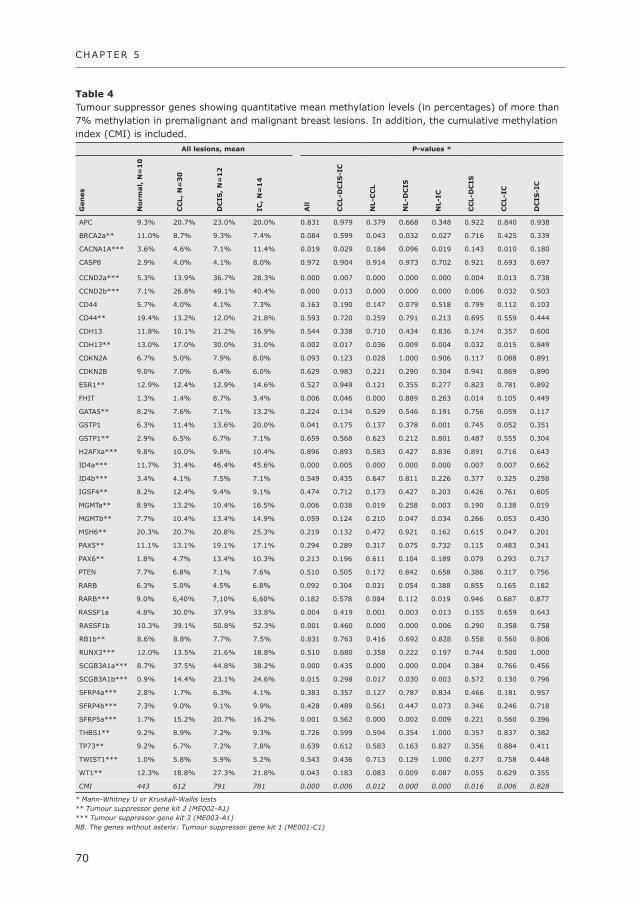
CHAPTER 5
70
Table 4 Tumour suppressor genes showing quantitative mean methylation levels (in percentages) of more than 7% methylation in premalignant and malignant breast lesions. In addition, the cumulative methylation index (CMI) is included.
All lesions, mean P-values *
Gen
es
No
rmal,
N=
10
CC
l,
N=
30
dC
iS,
N=
12
iC,
N=
14
All
CC
l-d
CiS
-iC
Nl-C
Cl
Nl-d
CiS
Nl-i
C
CC
l-d
CiS
CC
l-i
C
dC
iS-i
C
APC 9.3% 20.7% 23.0% 20.0% 0.831 0.979 0.379 0.668 0.348 0.922 0.840 0.938
BRCA2a** 11.0% 8.7% 9.3% 7.4% 0.084 0.599 0.043 0.032 0.027 0.716 0.425 0.339
CACNA1A*** 3.6% 4.6% 7.1% 11.4% 0.019 0.029 0.184 0.096 0.019 0.143 0.010 0.180
CASP8 2.9% 4.0% 4.1% 8.0% 0.972 0.904 0.914 0.973 0.702 0.921 0.693 0.697
CCND2a*** 5.3% 13.9% 36.7% 28.3% 0.000 0.007 0.000 0.000 0.000 0.004 0.013 0.738
CCND2b*** 7.1% 26.8% 49.1% 40.4% 0.000 0.013 0.000 0.000 0.000 0.006 0.032 0.503
CD44 5.7% 4.0% 4.1% 7.3% 0.163 0.190 0.147 0.079 0.518 0.799 0.112 0.103
CD44** 19.4% 13.2% 12.0% 21.8% 0.593 0.720 0.259 0.791 0.213 0.695 0.559 0.444
CDH13 11.8% 10.1% 21.2% 16.9% 0.544 0.338 0.710 0.434 0.836 0.174 0.357 0.600
CDH13** 13.0% 17.0% 30.0% 31.0% 0.002 0.017 0.036 0.009 0.004 0.032 0.015 0.849
CDKN2A 6.7% 5.0% 7.9% 8.0% 0.093 0.123 0.028 1.000 0.906 0.117 0.088 0.891
CDKN2B 9.0% 7.0% 6.4% 6.0% 0.629 0.983 0.221 0.290 0.304 0.941 0.869 0.890
ESR1** 12.9% 12.4% 12.9% 14.6% 0.527 0.949 0.121 0.355 0.277 0.823 0.781 0.892
FHIT 1.3% 1.4% 8.7% 3.4% 0.006 0.046 0.000 0.889 0.263 0.014 0.105 0.449
GATA5** 8.2% 7.6% 7.1% 13.2% 0.224 0.134 0.529 0.546 0.191 0.756 0.059 0.117
GSTP1 6.3% 11.4% 13.6% 20.0% 0.041 0.175 0.137 0.378 0.001 0.745 0.052 0.351
GSTP1** 2.9% 6.5% 6.7% 7.1% 0.659 0.568 0.623 0.212 0.801 0.487 0.555 0.304
H2AFXa*** 9.8% 10.0% 9.8% 10.4% 0.896 0.893 0.583 0.427 0.836 0.891 0.716 0.643
ID4a*** 11.7% 31.4% 46.4% 45.6% 0.000 0.005 0.000 0.000 0.000 0.007 0.007 0.662
ID4b*** 3.4% 4.1% 7.5% 7.1% 0.549 0.435 0.647 0.811 0.226 0.377 0.325 0.258
IGSF4** 8.2% 12.4% 9.4% 9.1% 0.474 0.712 0.173 0.427 0.203 0.426 0.761 0.605
MGMTa** 8.9% 13.2% 10.4% 16.5% 0.006 0.038 0.019 0.258 0.003 0.190 0.138 0.019
MGMTb** 7.7% 10.4% 13.4% 14.9% 0.059 0.124 0.210 0.047 0.034 0.266 0.053 0.430
MSH6** 20.3% 20.7% 20.8% 25.3% 0.219 0.132 0.472 0.921 0.162 0.615 0.047 0.201
PAX5** 11.1% 13.1% 19.1% 17.1% 0.294 0.289 0.317 0.075 0.732 0.115 0.483 0.341
PAX6** 1.8% 4.7% 13.4% 10.3% 0.213 0.196 0.611 0.104 0.189 0.079 0.293 0.717
PTEN 7.7% 6.8% 7.1% 7.6% 0.510 0.505 0.172 0.842 0.658 0.386 0.317 0.756
RARB 6.3% 5.0% 4.5% 6.8% 0.092 0.304 0.031 0.054 0.388 0.855 0.165 0.182
RARB*** 9.0% 6,40% 7,10% 6,60% 0.182 0.578 0.084 0.112 0.019 0.946 0.687 0.877
RASSF1a 4.8% 30.0% 37.9% 33.8% 0.004 0.419 0.001 0.003 0.013 0.155 0.659 0.643
RASSF1b 10.3% 39.1% 50.8% 52.3% 0.001 0.460 0.000 0.000 0.006 0.290 0.358 0.758
RB1b** 8.6% 8.8% 7.7% 7.5% 0.831 0.763 0.416 0.692 0.828 0.558 0.560 0.806
RUNX3*** 12.0% 13.5% 21.6% 18.8% 0.510 0.680 0.358 0.222 0.197 0.744 0.500 1.000
SCGB3A1a*** 8.7% 37.5% 44.8% 38.2% 0.000 0.435 0.000 0.000 0.004 0.384 0.766 0.456
SCGB3A1b*** 0.9% 14.4% 23.1% 24.6% 0.015 0.298 0.017 0.030 0.003 0.572 0.130 0.796
SFRP4a*** 2.8% 1.7% 6.3% 4.1% 0.383 0.357 0.127 0.787 0.834 0.466 0.181 0.957
SFRP4b*** 7.3% 9.0% 9.1% 9.9% 0.428 0.489 0.561 0.447 0.073 0.346 0.246 0.718
SFRP5a*** 1.7% 15.2% 20.7% 16.2% 0.001 0.562 0.000 0.002 0.009 0.221 0.560 0.396
THBS1** 9.2% 8.9% 7.2% 9.3% 0.726 0.599 0.594 0.354 1.000 0.357 0.837 0.382
TP73** 9.2% 6.7% 7.2% 7.8% 0.639 0.612 0.583 0.163 0.827 0.356 0.884 0.411
TWIST1*** 1.0% 5.8% 5.9% 5.2% 0.543 0.436 0.713 0.129 1.000 0.277 0.758 0.448
WT1** 12.3% 18.8% 27.3% 21.8% 0.043 0.183 0.083 0.009 0.087 0.055 0.629 0.355
CMI 443 612 791 781 0.000 0.006 0.012 0.000 0.000 0.016 0.006 0.828
* Mann-Whitney U or Kruskall-Wallis tests** Tumour suppressor gene kit 2 (ME002-A1)*** Tumour suppressor gene kit 3 (ME003-A1)NB. The genes without asterix: Tumour suppressor gene kit 1 (ME001-C1)

71
Methylation and columnar cell lesions
with synchronous invasive carcinoma and cases with synchronous CCL, DCIS and
invasive carcinoma. The same four genes (SCGB3A1, CCND2, ID4 and RASSF1)
had the highest methylation levels compared to the overall analysis.
Significant differences were found in ID4, CCND2, CDH13, GSTP1 and SCGB3A1
between CCL and invasive carcinoma (Table 5), and ID4 for CCL, DCIS and invasive
carcinoma (Table 6). The CMI of the synchronous lesions CCL, DCIS and invasive
carcinoma increased stepwise (p=0.042), just as the synchronous CCL and invasive
carcinoma cases (p=0.091).
Table 5 Quantitative mean methylation levels (in percentages) of tumour suppressor genes in 8 cases with synchronous CCL and IC.
CCl iC P-value *
APC 16% 18% 0.754
CACNA1A*** 6% 10% 0.161
CCND2a*** 11% 23% 0.050
CCND2b*** 19% 28% 0.272
CDH13 15% 13% 0.136
CDH13** 14% 26% 0.007
ESR1** 14% 16% 0.583
GATA5** 11% 15% 0.433
GSTP1 12% 21% 0.033
ID4a*** 31% 43% 0.033
MGMTa** 13% 16% 0.090
MGMTb** 12% 15% 0.119
MSH6** 29% 27% 0.093
PAX5A** 13% 13% 0.972
RASSF1a 23% 36% 0.069
RASSF1b 33% 49% 0.331
RUNX3*** 11% 18% 0.470
SCGB3A1a*** 41% 31% 0.683
SCGB3A1b*** 12% 23% 0.016
SFRP4b*** 11% 10% 0.673
SFRP5a*** 9% 18% 0.529
WT1** 25% 17% 0.969
CMI 527 689 0.091
* Wilcoxon signed Rank test** Tumour suppressor gene kit 2 (ME002-A1)*** Tumour suppressor gene kit 3 (ME003-A1)NB. The genes without asterix: Tumour suppressor gene kit 1 (ME001-C1)
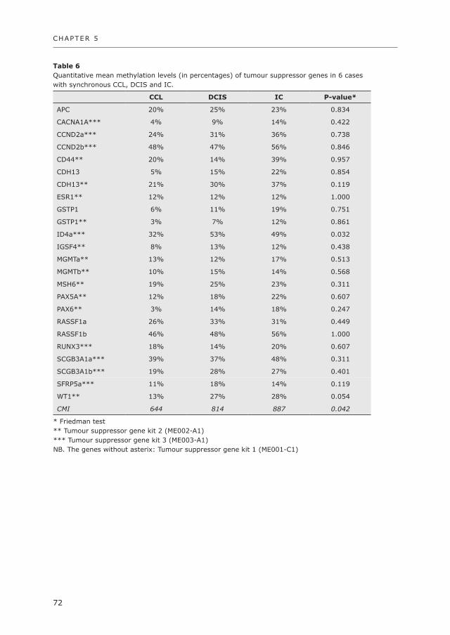
CHAPTER 5
72
Table 6 Quantitative mean methylation levels (in percentages) of tumour suppressor genes in 6 cases with synchronous CCL, DCIS and IC.
CCl dCiS iC P-value*
APC 20% 25% 23% 0.834
CACNA1A*** 4% 9% 14% 0.422
CCND2a*** 24% 31% 36% 0.738
CCND2b*** 48% 47% 56% 0.846
CD44** 20% 14% 39% 0.957
CDH13 5% 15% 22% 0.854
CDH13** 21% 30% 37% 0.119
ESR1** 12% 12% 12% 1.000
GSTP1 6% 11% 19% 0.751
GSTP1** 3% 7% 12% 0.861
ID4a*** 32% 53% 49% 0.032
IGSF4** 8% 13% 12% 0.438
MGMTa** 13% 12% 17% 0.513
MGMTb** 10% 15% 14% 0.568
MSH6** 19% 25% 23% 0.311
PAX5A** 12% 18% 22% 0.607
PAX6** 3% 14% 18% 0.247
RASSF1a 26% 33% 31% 0.449
RASSF1b 46% 48% 56% 1.000
RUNX3*** 18% 14% 20% 0.607
SCGB3A1a*** 39% 37% 48% 0.311
SCGB3A1b*** 19% 28% 27% 0.401
SFRP5a*** 11% 18% 14% 0.119
WT1** 13% 27% 28% 0.054
CMI 644 814 887 0.042
* Friedman test** Tumour suppressor gene kit 2 (ME002-A1)*** Tumour suppressor gene kit 3 (ME003-A1)NB. The genes without asterix: Tumour suppressor gene kit 1 (ME001-C1)

73
Methylation and columnar cell lesions
diSCuSSiON
The aim of this study was to investigate methylation of multiple tumour suppressor
genes in CCLs compared to normal breast tissue and (especially synchronous)
DCIS and/or invasive carcinoma by MS-MLPA, which was previously validated as
a high throughput technique showing good correlation with other methylation
detection techniques like quantitative methylation specific PCR and methylation
specific PCR 12,16,23,24. We found that CMI increased from normal breast tissue
through CCL, towards DCIS and invasive carcinoma (p<0.001), suggesting that
methylation increases stepwise in the progression of these precursor lesions to
invasive cancer. This progression in methylation from normal breast tissue, CCL-A/
ADH to DCIS and invasive carcinoma was also observed by Park et al 20, although
mostly different genes were studied and no synchronous lesions in patients were
included in their study. Also Liu et al. demonstrated that p16INK4a methylation
increased from UDH to CCL-A/ADH to low grade DCIS and high grade DCIS29.
In the first step of the progression route, CCL had significantly higher methylation
levels than normal breast tissue in the genes ATM, BCL2, BRCA1, BRCA2, CCND2,
CDH13, CDKN2A, CHFR, DLC1, ESR1, FHIT, HLTF, ID4, IGSF4, MGMT, RARB,
RASSF1, SFRP5, SCGB3A1 and VHL. This underlines that methylation may play an
important early role in the histogenesis of CCLs. In the second step, DCIS had
higher methylation levels than CCL in CCND2, ID4, FHIT and CDH13. Moreover, in
the five DCIS grade I cases originating from CCL compared to all CCL-A cases, ID4
and CCND2 showed higher methylation levels. In the third step, invasive carcinoma
had a higher mean methylation level than DCIS for MGMT, although the mean
methylation level was relatively low (16.5% for invasive carcinoma).
From all these genes, ID4, CCND2 and CDH13 seem to be the most important
tumour suppressor genes in CCL driven breast carcinogenesis, as they all had high
methylation levels in the abnormal lesions and they showed significantly increasing
methylation levels from normal breast tissue to CCL and to DCIS/invasive cancer.
Also RASSF1, SCGB3A1 and SFRP5 showed high methylation levels with significant
differences between normal breast tissue and CCL, whereas no significant increase
in methylation was observed between CCL, DCIS and invasive carcinoma. This
suggests that for these genes, the epigenetic abnormalities are already present in
CCLs, underlying the important role of CCLs in low nuclear grade breast
carcinogenesis. Moreover, in the comparison of the synchronous lesions, the same
four genes had the highest mean methylation levels (SCGB3A1, CCND2, ID4 and
RASSF1) and there were also significant differences between CCL and the more
advanced lesions.

CHAPTER 5
74
The CMI of CCL cases with synchronous DCIS and/or invasive carcinoma showed
no significant difference with the CMI of CCL cases without synchronous lesion,
just like the CMI of CCLs with and without atypia. Moreover, no differences in genes
were present. These results further suggest that methylation occurs already early
in the carcinogenesis from normal breast to CCLs without atypia and that no further
methylation occurs in more advanced CCLs.
Whereas we found methylation in CCLs without atypia, using allelic imbalance,
Dabbs et al. did not demonstrate any mutational changes in CCLs without atypia.
This suggests that mutational changes may occur rather later in CCL histogenesis
than methylation. Further studies applying other techniques like loss-of-
heterozygosity 9,11,30, comparative genomic hybridization 31, miRNA 32 and allelic
imbalance 10 have demonstrated that CCL-A exhibits increasing genetic alterations
from normal breast epithelium, to CCL-A to DCIS and invasive cancer, and that
similar chromosomal aberrations (especially 16q loss) occur over the spectrum of
ADH, low-grade DCIS and low-grade invasive ductal carcinoma, which parallels the
morphologically defined carcinogenesis. However, two studies did not give support
for CCL-A being an obligatory precursor to more advanced disease 33,34.
As shown in Table 6, the CMI of synchronous CCL, DCIS and invasive cancer showed
a stepwise increase. Interestingly, synchronous CCL/invasive cancer cases (Table
5) had a lower mean CMI in both CCL and invasive cancer than the synchronous
CCL/DCIS/invasive cancer cases, possibly related to the higher frequency of grade
III cancers in the synchronous CCL/DCIS/invasive cancer cases (see Table 2).
For the different genes, no differences in methylation were found between DCIS
and invasive cancer, except for MGMT. Moreover, the overall cumulative methylation
was more or less equal in DCIS and invasive cancer. Other studies revealed presence
of nearly similar levels of methylation in DCIS compared to invasive cancer as well,
partially using synchronous cases 20,35-37.
Of all 50 genes studied, the most important studied tumour suppressor genes
seemed to be RASSF1, ID4, CCND2, SCGB3A1, CDH13 and SFRP5. RASSF1 is
involved in DNA repair and inhibiting the accumulation of cyclin D1, thus inducing
cell cycle arrest. In this study we found methylation frequencies of 87%, 92% and
71% for CCL, DCIS and invasive cancer, respectively. These frequencies are mostly
in line or somewhat higher than other studies (49% - 82%) 12,20,35,37-45. ID4 is a
member of the inhibitor of DNA-binding family that inhibits DNA binding of basic
helix–loop–helix transcription factors. We found higher frequencies of methylation
(CCL 80%, DCIS 92% and invasive carcinoma 93%) than two previous studies (38
– 69%), underscoring the potential importance of ID4 methylation in breast
carcinogenesis 46,47. CCND2 (cyclin D2), involved in the cell cycle, was also previously

75
Methylation and columnar cell lesions
found as methylation marker for invasive carcinoma (range 11-56%) 35,43,48-50,
although we found higher frequencies of methylation (CCL 53%, DCIS 83% and
invasive carcinoma 86%). Moreover, SCGB3A1 (also known as HIN-1) was reported
to have less methylation before (range 36-74%) than in our study (CCL-A 93%,
DCIS 100% and invasive carcinoma 79%)20,35,43,51. Interestingly, Park et al. showed
large discrepancies (23% CCL-A, 34% DCIS and 36% in IC) 20. CDH13 (cadherin
13), involved in selective cell recognition and adhesion, was also found to be
methylated in breast cancer before (range 1 – 84%), just as in our study (CCL
50%, DCIS 67% and invasive carcinoma 71%) 12,41,52. And we found lower
frequencies of methylation (CCL 43%, DCIS 67% and invasive carcinoma 29%)
than in two other studies concerning the gene SFRP5 (secreted frizzled-related
protein 5) (range 71 – 73%) 53,54.
Normal breast tissue showed methylation in three or more cases for the genes
MSH6, CDH13 and RASSF1. One could argue that these methylation events may
be innocent bystanders, but in view of the significantly higher methylation levels
for CDH13 and RASSF1 in CCL this is at least not the case for the latter two genes.
The role of MSH6 methylation needs to be further established.
In conclusion, promoter hypermethylation for several established tumour suppressor
genes is already present in CCLs, underlining that promoter hypermethylation is
an early event in breast carcinogenesis. Atypia in CCL or presence of synchronous
more advanced lesions does not seem to be accompanied by higher methylation
levels.
Acknowledgements
This work was supported by The Oncology Centre of St. Antonius Hospital Nieu-
wegein.

CHAPTER 5
76
REfERENCES
1. Allred DC, Mohsin SK, Fuqua SA. Histological and biological evolution of human premalignant breast disease. Endocr Relat Cancer 2001; 8:47-61.
2. Lee S, Medina D, Tsimelzon A et al. Alterations of gene expression in the development of early hyperplastic precursors of breast cancer. Am J Pathol 2007; 171:252-262.
3. Simpson PT, Gale T, Reis-Filho JS et al. Columnar cell lesions of the breast: the missing link in breast cancer progression? A morphological and molecular analysis. Am J Surg Pathol 2005; 29:734-746.
4. Sinn HP. Breast cancer precursors: lessons learned from molecular genetics. J Mol Med 2009; 87:113-115.
5. Schnitt SJ, Vincent-Salomon A. Columnar cell lesions of the breast. Adv Anat Pathol 2003; 10:113-124.
6. Tavassoli FA, Devilee P. WHO Classification Tumours of the Breast and Female Genital Organs, IARC, Lyon. 2003.
7. Feeley L, Quinn CM. Columnar cell lesions of the breast. Histopathology 2008; 52:11-19.8. Verschuur-Maes AHJ, Van Deurzen CHM, Monninkhof EM et al. Columnar cell lesions on breast needle
biopsies: Is surgical excision necessary? A systematic review. Ann Surg 2011.9. Aulmann S, Elsawaf Z, Penzel R et al. Invasive tubular carcinoma of the breast frequently is clonally
related to flat epithelial atypia and low-grade ductal carcinoma in situ. Am J Surg Pathol 2009; 33:1646-1653.
10. Dabbs DJ, Carter G, Fudge M et al. Molecular alterations in columnar cell lesions of the breast. Mod Pathol 2006; 19:344-349.
11. Moinfar F, Man YG, Bratthauer GL et al. Genetic abnormalities in mammary ductal intraepithelial neoplasia-flat type (“clinging ductal carcinoma in situ”): a simulator of normal mammary epithelium. Cancer 2000; 88:2072-2081.
12. Buyru N, Altinisik J, Ozdemir F et al. Methylation profiles in breast cancer. Cancer Invest 2009; 27:307-312.
13. Esteller M, Silva JM, Dominguez G et al. Promoter hypermethylation and BRCA1 inactivation in sporadic breast and ovarian tumors. J Natl Cancer Inst 2000; 92:564-569.
14. Jones PA, Baylin SB. The epigenomics of cancer. Cell 2007; 128:683-692.15. Jovanovic J, Ronneberg JA, Tost J et al. The epigenetics of breast cancer. Mol Oncol 2010; 4:242-
254.16. Marzese DM, Gago FE, Vargas-Roig LM et al. Simultaneous analysis of the methylation profile of 26
cancer related regions in invasive breast carcinomas by MS-MLPA and drMS-MLPA. Mol Cell Probes 2010; 24:271-280.
17. Widschwendter M, Jones PA. DNA methylation and breast carcinogenesis. Oncogene 2002; 21:5462-5482.
18. Hoque MO, Prencipe M, Poeta ML et al. Changes in CpG islands promoter methylation patterns during ductal breast carcinoma progression. Cancer Epidemiol Biomarkers Prev 2009; 18:2694-2700.
19. Liu T, Niu Y, Feng Y et al. Methylation of CpG islands of p16(INK4a) and cyclinD1 overexpression associated with progression of intraductal proliferative lesions of the breast. Hum Pathol 2008; 39:1637-1646.
20. Park SY, Kwon HJ, Lee HE et al. Promoter CpG island hypermethylation during breast cancer progression. Virchows Arch 2011; 458:73-84.
21. van Diest PJ. No consent should be needed for using leftover body material for scientific purposes. For. BMJ 2002; 325:648-651.
22. Bol GM, Suijkerbuijk KP, Bart J et al. Methylation profiles of hereditary and sporadic ovarian cancer. Histopathology 2010; 57:363-370.
23. Seeber LM, Zweemer RP, Marchionni L et al. Methylation profiles of endometrioid and serous endometrial cancers. Endocr Relat Cancer 2010; 17:663-673.
24. Suijkerbuijk KP, Pan X, Van der Wall E et al. Comparison of different promoter methylation assays in breast cancer. Anal Cell Pathol (Amst) 2010; 33:133-141.
25. van Diest PJ, Suijkerbuijk KP, Koop EA et al. Low levels of BNIP3 promoter hypermethylation in invasive breast cancer. Anal Cell Pathol (Amst) 2010; 33:175-176.
77
Methylation and columnar cell lesions
26. Gylling A, bdel-Rahman WM, Juhola M et al. Is gastric cancer part of the tumour spectrum of hereditary non-polyposis colorectal cancer? A molecular genetic study. Gut 2007; 56:926-933.
27. Joensuu EI, bdel-Rahman WM, Ollikainen M et al. Epigenetic signatures of familial cancer are characteristic of tumor type and family category. Cancer Res 2008; 68:4597-4605.
28. Suijkerbuijk KP, Fackler MJ, Sukumar S et al. Methylation is less abundant in BRCA1-associated compared with sporadic breast cancer. Ann Oncol 2008; 19:1870-1874.
29. Liu T, Niu Y, Feng Y et al. Methylation of CpG islands of p16(INK4a) and cyclinD1 overexpression associated with progression of intraductal proliferative lesions of the breast. Hum Pathol 2008; 39:1637-1646.
30. O’Connell P, Pekkel V, Fuqua SA et al. Analysis of loss of heterozygosity in 399 premalignant breast lesions at 15 genetic loci. J Natl Cancer Inst 1998; 90:697-703.
31. Simpson PT, Gale T, Reis-Filho JS et al. Columnar cell lesions of the breast: the missing link in breast cancer progression? A morphological and molecular analysis. Am J Surg Pathol 2005; 29:734-746.
32. Qi L, Bart J, Tan LP et al. Expression of miR-21 and its targets (PTEN, PDCD4, TM1) in flat epithelial atypia of the breast in relation to ductal carcinoma in situ and invasive carcinoma. BMC Cancer 2009; 9:163.
33. Ellsworth RE, Ellsworth DL, Weyandt JD et al. Chromosomal alterations in pure nonneoplastic breast lesions: implications for breast cancer progression. Ann Surg Oncol 2010; 17:1688-1694.
34. Subramaniam MM, Chan JY, Omar MF et al. Lack of RUNX3 inactivation in columnar cell lesions of breast. Histopathology 2010; 57:555-563.
35. Fackler MJ, McVeigh M, Evron E et al. DNA methylation of RASSF1A, HIN-1, RAR-beta, Cyclin D2 and Twist in in situ and invasive lobular breast carcinoma. Int J Cancer 2003; 107:970-975.
36. Moelans CB, de Weger RA, Monsuur HN et al. Molecular differences between ductal carcinoma in situ and adjacent invasive breast carcinoma: a multiplex ligation-dependent probe amplification study. Anal Cell Pathol (Amst) 2010; 33:165-173.
37. Muggerud AA, Ronneberg JA, Warnberg F et al. Frequent aberrant DNA methylation of ABCB1, FOXC1, PPP2R2B and PTEN in ductal carcinoma in situ and early invasive breast cancer. Breast Cancer Res 2010; 12:R3.
38. Bertolo C, Guerrero D, Vicente F et al. Differences and molecular immunohistochemical parameters in the subtypes of infiltrating ductal breast cancer. Am J Clin Pathol 2008; 130:414-424.
39. Burbee DG, Forgacs E, Zochbauer-Muller S et al. Epigenetic inactivation of RASSF1A in lung and breast cancers and malignant phenotype suppression. J Natl Cancer Inst 2001; 93:691-699.
40. Chen CM, Chen HL, Hsiau TH et al. Methylation target array for rapid analysis of CpG island hypermethylation in multiple tissue genomes. Am J Pathol 2003; 163:37-45.
41. Feng W, Orlandi R, Zhao N et al. Tumor suppressor genes are frequently methylated in lymph node metastases of breast cancers. BMC Cancer 2010; 10:378.
42. Honorio S, Agathanggelou A, Schuermann M et al. Detection of RASSF1A aberrant promoter hypermethylation in sputum from chronic smokers and ductal carcinoma in situ from breast cancer patients. Oncogene 2003; 22:147-150.
43. Lee JS, Fackler MJ, Teo WW et al. Quantitative promoter hypermethylation profiles of ductal carcinoma in situ in North American and Korean women: Potential applications for diagnosis. Cancer Biol Ther 2008; 7:1398-1406.
44. Pasquali L, Bedeir A, Ringquist S et al. Quantification of CpG island methylation in progressive breast lesions from normal to invasive carcinoma. Cancer Lett 2007; 257:136-144.
45. Shinozaki M, Hoon DS, Giuliano AE et al. Distinct hypermethylation profile of primary breast cancer is associated with sentinel lymph node metastasis. Clin Cancer Res 2005; 11:2156-2162.
46. Noetzel E, Veeck J, Niederacher D et al. Promoter methylation-associated loss of ID4 expression is a marker of tumour recurrence in human breast cancer. BMC Cancer 2008; 8:154.
47. Umetani N, Mori T, Koyanagi K et al. Aberrant hypermethylation of ID4 gene promoter region increases risk of lymph node metastasis in T1 breast cancer. Oncogene 2005; 24:4721-4727.
48. Evron E, Umbricht CB, Korz D et al. Loss of cyclin D2 expression in the majority of breast cancers is associated with promoter hypermethylation. Cancer Res 2001; 61:2782-2787.
49. Li S, Rong M, Iacopetta B. DNA hypermethylation in breast cancer and its association with clinicopathological features. Cancer Lett 2006; 237:272-280.
50. Parrella P, Poeta ML, Gallo AP et al. Nonrandom distribution of aberrant promoter methylation of cancer-related genes in sporadic breast tumors. Clin Cancer Res 2004; 10:5349-5354.

CHAPTER 5
78
51. Shigematsu H, Suzuki M, Takahashi T et al. Aberrant methylation of HIN-1 (high in normal-1) is a frequent event in many human malignancies. Int J Cancer 2005; 113:600-604.
52. Toyooka KO, Toyooka S, Virmani AK et al. Loss of expression and aberrant methylation of the CDH13 (H-cadherin) gene in breast and lung carcinomas. Cancer Res 2001; 61:4556-4560.
53. Suzuki H, Toyota M, Carraway H et al. Frequent epigenetic inactivation of Wnt antagonist genes in breast cancer. Br J Cancer 2008; 98:1147-1156.
54. Veeck J, Geisler C, Noetzel E et al. Epigenetic inactivation of the secreted frizzled-related protein-5 (SFRP5) gene in human breast cancer is associated with unfavorable prognosis. Carcinogenesis 2008; 29:991-998.

79
Methylation and columnar cell lesions


Submitted
Anoek H.J. Verschuur-Maes 1, Cathy B. Moelans 1,
Peter C. de Bruin 2, Paul J. van Diest 1
1Department of Pathology, University Medical Center Utrecht,
2Department of Pathology, St. Antonius Ziekenhuis Nieuwegein, The Netherlands
Oncogene amplification in columnar cell lesions
of the breast: analysis by Multiplex
Ligation-dependent Probe Amplification
Chapter
6

CHAPTER 6
82
AbSTRACT
introduction
Columnar cell lesions (CCLs) are possible precursors of breast cancer, but little is
known about the role of several breast cancer related genes in the progression to
invasive breast cancer.
Methods
Gene copy numbers of 17 breast cancer related genes were analyzed by Multiplex
Ligation-dependent Probe Amplification (MLPA) in CCL (N=14), ductal carcinoma
in situ (DCIS) grade I originating from CCL (N=5), and paired CCL (N=15) with
DCIS (N=7) and/or invasive carcinoma (N=13). The genes included were BIRC5,
EMSY, CCND1, CCNE1, CDH1, CPD, EGFR, ESR1, FGFR1, HER2, IKBKB, MAPT,
MED1, MTDH, MYC, TOP2A and TRAF4.
Results
Overall, the frequency of gene copy number changes increased from CCL towards
DCIS and invasive carcinoma (p = 0.023). DCIS and invasive carcinoma had several
high level amplifications, whereas in CCL no high level amplifications were present.
Also fewer losses were seen in CCL. Most of the copy number alterations were
located on chromosomes 17 and 8. Also, in the cases with synchronous lesions,
CCL had fewer copy number changes than DCIS/invasive carcinoma.
Conclusion
Compared to DCIS and invasive carcinoma, there seem to be few copy number
changes in the 17 studied established cancer genes in CCL. Therefore, copy number
changes in these genes do not seem to play a major role in the genesis of CCLs,
and may rather be later events in low nuclear grade breast carcinogenesis.

83
Oncogene amplification in columnar cell lesions
iNTROduCTiON
It has widely been accepted that breast carcinogenesis is a complex multistep
process with a sequence of stages in which invasive carcinoma arises from normal
breast epithelium via precursor lesions like atypical ductal hyperplasia (ADH),
lobular neoplasia and ductal carcinoma in situ (DCIS) 1,2. In recent years, it has
been recognized that also columnar cell lesions (CCLs) play a role as a possible
precursor of (particularly low nuclear grade) breast cancer 3-5. These lesions are
increasingly diagnosed, since more biopsies are taken because of microcalcifications,
which characterize these lesions at mammography 6. CCLs are cystically dilated
enlarged terminal duct lobular units lined by columnar cell epithelium with one or
two cell layers (columnar cell change) or more cell layers (columnar cell hyperplasia).
The cells show uniform, ovoid to elongated nuclei oriented along the basement
membrane without inconspicuous nucleoli. At the intraluminal site, they often show
cytoplasmic snouts and in the lumen secretions are regularly seen, as well as
microcalcifications. According to the classification by Schnitt et al., the lesions are
designated as columnar cell change with atypia or columnar cell hyperplasia with
atypia when the cells show cytonuclear or architectural atypia 7. Together, the
lesions with atypia are called CCLs with atypia (CCL-A), also known as flat epithelial
atypia according to the World Health Organization 8. CCL-A lack the complex
architecture as is seen in ADH and low grade DCIS, although they share many
morphological and immunohistochemical features 4,7,9. In the management of CCLs,
a difference is made between CCLs with and without atypia, many recommending
for CCL-A to excise the lesion or remove all microcalcifications by vacuum assisted
biopsies or the BLES procedure 10. For CCLs without atypia, excision is so far not
deemed necessary 11.
Different genetic alterations play a role in the carcinogenesis of breast cancer, like
the inactivation of tumour suppressor genes and the activation of oncogenes.
Previous studies found a progression in the methylation levels from normal breast
epithelium, via CCLs to DCIS and invasive carcinoma, providing more evidence for
CCLs being a precursor of breast cancer 12,13. Also other studies provided evidence,
albeit sparse, that CCLs (with atypia) show similar genetic abnormalities as the
accompanying DCIS and invasive carcinoma, as illustrated by studies using
comparative genomic hybridization and loss of heterozygosity 4,14-16. However, not
much is known about genetic alterations of specific cancer related genes in CCLs
in relation to the development of breast cancer. Before, a set of genes had been
selected (and combined in a commercially available Multiplex Ligation-dependent
Probe Amplification (MLPA) kit) for showing frequent copy number changes in breast
CHAPTER 6
84
cancer and with functions related to carcinogenesis 17. The present study is the
first, to our knowledge, to investigate copy number changes of a selected set of
17 breast cancer related genes in normal breast tissue, CCLs and (largely
synchronous) DCIS and invasive carcinoma by MLPA, to unravel the role of these
genes in the progression of CCLs.
METHOdS
Patient material
From paraffin embedded breast resection specimens, tissue samples with only a
CCL (with or without atypia), as well as CCL cases with synchronous invasive
carcinoma and/or DCIS were collected between 1996 and 2009, at the Departments
Pathology of the University Medical Center Utrecht and St. Antonius Hospital
Nieuwegein, The Netherlands, as previously described 13. Normal breast tissue
from 7 women undergoing mammoplasty was taken along as control. Grading of
the CCLs was done according to the classification described by Schnitt and
Vincent-Salomon 7 as CCLs without atypia (including columnar cell change and
columnar cell hyperplasia ) and CCLs with atypia (CCL-A, including columnar cell
change with atypia and columnar cell hyperplasia with atypia). Classification and
grading of the invasive carcinoma and in situ cancers was assessed by the same
two experienced observers (AVM and PvD), according to the World Health
Organization classification 8. As a refinement, we included a subgroup of DCIS
grade I lesions with still recognizable columnar cell type with apical snouts (“DCIS
originating from CCL”). Anonymous use of redundant tissue for research purposes
is part of the standard treatment agreement with patients in our hospitals 18.
dNA isolation
Guided by marked H&E stained slides, abundant CCL, DCIS and invasive carcinoma
lesions that were apart from each other, were harvested from serial 8 to 10 μm
thick paraffin slides (deparaffinized for 10 minutes) using a scalpel (mesodissection).
In all other cases laser microdissection (PALM Microlaser Technologies AG, Bernried,
Germany) was performed on 8 to 10 μm thick paraffin slides, from which the areas
of interest were catapulted by laser pressure into a cap moistened with a drop of
mineral oil. DNA was isolated by adding 50 or 100 μl direct lysis (consisting of 10
mM TRIS-Hcl pH 8.3, 0.5% Tween 0.20, 1 mM EDTA) and 10 or 20 μl proteinase
K (10 mg/ml, Roche, Almere, The Netherlands), dependent on the amount of
material. After one hour the same amount of proteinase K was added and heated

85
Oncogene amplification in columnar cell lesions
in a 56°C water bath for another 16 hours. The samples were then heat inactivated
by boiling for 10 minutes, centrifuged and the supernatant was stored by 4 °C.
Multiplex Ligation-dependent Probe Amplification (MLPA)
According to the manufacturers’ instructions, the isolated DNA solutions (100-240
ng) were evaluated by MLPA analysis using the P078-B1 breast kit (MRC Holland,
Amsterdam, The Netherlands), as described before 17. Blood samples from four healthy
volunteers without history of malignancy were taken along as control samples.
The reaction products were separated by capillary electrophoresis using the 3130
DNA Analyzer (Applied Biosystems, Foster City, CA, USA) and gene copy numbers
were analyzed using GeneScan Analysis (Applied Biosystems) and Coffalyser
software (version 9.4, MRC-Holland). Most samples could be tested in duplicate.
For genes with more than one probe present in the kit, the means of the probe
peaks were used in the analysis. Table 1 shows the studied genes and includes
chromosomal locations of all probes. ADAM9, CDC6, PRDM14 and AURKA showed
copy number change in more than one normal breast tissue sample and were
therefore excluded from analysis, leaving reliable results for 17 genes.
Statistical analysis
Statistics were performed using SPSS statistical software. A mean value below 0.7
was defined as loss, a value between 0.7–1.3 as normal, 1.3–2.0 as gain and values
>2.0 were defined as high level amplification, as established before17,19-21. After
dichotomizing, we compared all copy number changes of each gene separately and
all genes combined between the CCL, DCIS and invasive carcinoma cases by Chi-
square test.
RESulTS
baseline characteristics
Table 2 shows the number of studied lesions per category. In total, 63 lesions were
studied in 43 patients; 14 of the patients had a CCL with synchronous DCIS and/
or invasive carcinoma. The 13 invasive carcinomas were of the ductal type except
for one lobular cancer. The majority of DCIS (4x grade I, 1x grade II, 2x grade III)
and invasive cancers (6x grade I, 4x grade II, 3x grade III) were of low/intermediate
grade and also five DCIS grade I originating from a CCL were included. Most
patients with CCL and synchronous DCIS and/or invasive carcinoma showed atypia
(4x CCC, 1x CCH and 9x CCL-A). The mean age at time of breast surgery of women

CHAPTER 6
86
with CCL only was 53.0 years, with CCL and DCIS 55.4 years and with CCL and
invasive carcinoma 50.6 years, with an overall mean age of 53.0 years (range 32-
82 years).
Table 1 Genes of the P078-B1 MLPA kit (MRC Holland, The Netherlands) studied for copy number changes in low nuclear grade breast carcinogenesis. The chromosomal position, mapview position, number of probes and transcript description is given for each gene.
Genes Chrom. Mapview Position
No. of probes
Transcript description
BIRC5 (Survivin) 17q25 17-073.722036
17-073.722396
17-073.724340
3 Adapter molecule involved in signal
transduction, cell communication and cell
survival
EMSY (C11orf30) 11q13 11–075.902087
11–075.926543
2 Transcription regulatory protein
CCND1 11q13 11–069.167779
11–069.175089
2 Cell cycle control protein involved in signal
transduction
CCNE1 19q12 19–035.000150
19–035.005214
2 Cell cycle control protein involved in signal
transduction
CDH1 16q22 16–067.328716
16–067.404826
2 Adhesion molecule associated with signal
transduction
CPD 17q11 17-025.795018 1 Carboxypeptidase involved in protein
metabolism
EGFR 7p11 07–055.191055
07–055.233957
2 Receptor tyrosine kinase involved in signal
transduction
ERBB2 17q12 17–035–118101
17–035.127183
17-035.133169
17-035.136344
4 Receptor tyrosine kinase associated with
signal transduction
ESR1 6q25 06–152.423838
06–152.457215
2 Transcription factor
FGFR1 8p12 08–038.391533
08-038.434092
2 Receptor tyrosine kinase involved in signal
transduction
IKBKB 8p11 08–042.292902
08–042.302676
2 Serine/threonine kinase associated with
signal transduction
MAPT 17q21 17-041.423085 1 Structural protein involved in cell growth
and maintenance
MED1 (PPARBP) 17q12 17-034.840858 1 Transcription regulatory protein involved in
signal transduction
MTDH 8q22 08-098.742504
08-098.788082
2 Metastasis promoting gene involved in
chemoresistence and angiogenesis
MYC 8q24 08–128.821796
08–128.822001
08–128.822151
3 Transcription factor involved in apoptosis
and cell proliferation
TOP2A 17q21 17–035.812698
17–035.816651
17–035.818297
3 DNA topoisomerase protein involved in
regulation of the topological status of DNA
TRAF4 17q11 17–024.098403 1 Adaptor molecule involved in signal trans-
duction, cell proliferation and apoptosis

87
Oncogene amplification in columnar cell lesions
Overall frequencies of copy number changes in CCl, dCiS and invasive
carcinoma
Overall, copy number changes differed significantly between CCL, DCIS and
invasive carcinoma, showing a lower number of mean copy number changes in
CCL (0.66) than in DCIS (1.33) or IC (1.46) (p = 0.023). In paired analysis, also
more copy number changes were present in invasive carcinoma than in CCL (p
= 0.009). CCL/DCIS (p = 0.51) and DCIS/invasive carcinoma (p = 0.637) also
showed differences in frequency of copy number changes, although these were
not significant. In Table 3, the copy number changes for all genes are shown.
High level amplifications were present in the genes CPD, ERBB2, MED1, TOP2A
and TRAF4, being more prevalent in invasive carcinoma (2 in 13 cases) and DCIS
(5 in 12 cases) than in CCL (0 in 29 cases) (p = 0.003). Gains were most
frequently present in EMSY, MTDH, CCND1 and MYC without difference in total
number of gains between the three types of lesions (CCL 16/29 cases, DCIS 4/13
cases, invasive carcinoma 9/12 cases; p = 0.456). Also when considering both
high and low level amplifications together, no significant differences were present
between the three types of lesions (p = 0.501). Losses were most often observed
in CDH1 and FGFR1, with in total more losses in invasive carcinoma (12/13 cases)
and DCIS (6/12 cases) than in CCL (4/29 cases) (p = 0.009). FGFR1 was the only
gene that showed a significant difference with more losses in invasive carcinoma
than in CCL and DCIS (p = 0.043). In general, CCLs with or without atypia had
comparable numbers of copy number changes (12/20 cases and 7/9 cases,
respectively, p = 0.577).
Table 2 Overview of the breast lesions studied for copy number changes in established cancer genes.
Samples Patients
Pure lesions
Normal breast tissue 10
CCL 14
CCL without atypia 4
CCL-A 10
DCIS grade I originated in CCL-A 5
Synchronous lesions
CCL + DCIS + IC 6
CCL + DCIS 1
CCL + IC 7

CHAPTER 6
88
Comparison of copy number changes in synchronous lesions
The six cases with synchronous CCL, DCIS and invasive carcinoma showed some
differences in copy number changes (N = 1, N = 8 and N = 9 changes, respectively),
as shown in Table 4. In the seven cases with synchronous CCL/invasive carcinoma
and one case with synchronous CCL/DCIS, CCL showed slightly fewer copy number
changes (N = 6) compared to the more advanced lesions as well (N = 11) (see
Table 5). In some patients, copy number changes of several genes were present
in the synchronous lesions. One patient with CCL, DCIS and invasive carcinoma
showed high level amplification of TRAF4 and CPD, gain of EMSY and loss of MAPT
in both DCIS and invasive carcinoma (without changes in the CCL). Also a gain of
CCND1 was present in the DCIS and loss of TOP2A in the CCL. The other five
patients with CCL, DCIS and invasive carcinoma showed sporadically a gain or loss
in one of the three lesions (no high level amplifications), without synchronous
Table 3 Frequencies of gains, high level amplifications and losses in 17 genes for columnar cell lesions (CCL), ductal carcinoma in situ (DCIS) and invasive carcinoma (IC), including P-values (Chisquare).
CCl, N=29 dCiS, N=12 iC, N=13
Gen
es
Ch
rom
o-
som
e
Am
plif.
Gain
lo
ss
Am
plif.
Gain
lo
ss
Am
plif.
Gain
lo
ss
P-v
alu
e*
BIRC5 17q25 0 0 0 0 1 (8.3%) 1 (8.3%) 0 0 1 (7.7%) 0.098
EMSY 11q13 0 3 (10.3%) 0 0 1 (8.3%) 0 0 1 (7.7% 0 0.956
CCND1 11q13 0 1 (3.4%) 0 0 2 (16.7%) 0 0 1 (7.7%) 0 0.339
CCNE1 19q12 0 1 (3.4%) 0 0 0 0 0 0 0 0.645
CDH1 16q22 0 0 2 (6.9%) 0 0 3 (25%) 0 1 (7.7%) 2 (15.4%) 0.209
CPD 17q11 0 2 (6.9%) 0 1 (8.3%) 0 0 1 (7.7%) 0 0 0.986
EGFR 7p11 0 0 0 0 0 0 0 0 0 -
ERBB2 17q12 0 0 0 1 (8.3%) 0 0 0 0 0 0.168
ESR1 6q25 0 1 (3.4%) 0 0 0 0 0 0 0 0.645
FGFR1 8p12 0 1 (3.4%) 0 0 0 0 0 0 3 (23.1%) 0.043
IKBKB 8p11 0 1 (3.4%) 0 0 0 0 0 0 1 (7.7%) 0.593
MAPT 17q21 0 0 0 0 0 1 (8.3%) 0 0 1 (7.7%) 0.299
MED1 17q12 0 0 0 1 (8.3%) 0 0 0 0 0 0.168
MTDH 8q22 0 2 (6.9%) 0 0 0 1 (8.3%) 0 3 (23.1%) 0 0.287
MYC 8q24 0 3 (10.3%) 0 0 0 0 0 2 (15.4%) 0 0.515
TOP2A 17q21 0 1 (3.4%) 1 (3.4%) 1 (8.3%) 0 0 0 1 (7.7%) 0 0.986
TRAF4 17q11 0 0 0 1 (8.3%) 0 0 1 (7.7%) 0 0 0.299
Total 0 16 3 5 4 6 2 9 8 0.023
* P-value calculated by adding all copy number changes and comparing CCL, DCIS and invasive carci-noma

89
Oncogene amplification in columnar cell lesions
aberrations. In the seven cases with synchronous CCL/invasive carcinoma and one
case with synchronous CCL/DCIS, one patient with CCL and DCIS (Table 5) showed
a gain of CCND1 in both lesions. In invasive carcinoma in one patient, losses of
FGFR1 and IKBKB were observed, with tendency to loss for the same genes in the
synchronous CCL (values of 0.71 and 0.72, respectively).
Co-amplifications
We also observed co-amplifications of different genes. High level co-amplification
of ERBB2, TOP2A, MED1 and gain in BIRC5 were seen in one patient with DCIS
grade I originating from CCL. These alterations were all located on chromosome
17q. As mentioned before, one patient showed co-amplification of the genes TRAF4,
CPD, EMSY, CCND1 and losses of MAPT and TOP2A, which were alterations located
on chromosomes 17q and 11q. Also, a patient with CCH (without synchronous
lesions) showed gains of EMSY, CPD and TOP2A, whereas another CCH case had
gains of CPD and ESR1. Gains of both MTDH and MYC (chromosome 8q) were seen
in two CCL cases and two invasive carcinomas. A patient with DCIS grade I
originating from CCL showed loss of both CDH1 and BIRC5, and one case with
invasive carcinoma showed loss of BIRC5 (chromosome 17q) and gain of CDH1
(chromosome 16q).
diSCuSSiON
In this study we investigated and compared copy number changes of 17 breast
cancer related genes in CCLs, DCIS and invasive breast carcinoma by MLPA, largely
comprising synchronous lesions in the same patients. Significantly more total copy
number changes in DCIS and invasive carcinoma were present than in CCL (p =
0.023) with significantly more high level amplifications and losses in DCIS / invasive
carcinoma compared to CCL.
Since more amplifications and losses were present in the more advanced lesions,
these alterations seemed to occur mostly at a later stage than CCL in the progression
from normal breast tissue, CCL(-A), ADH to DCIS and invasive cancer, which is a
well-known progression pathway for low nuclear grade carcinomas 22. High level
amplifications were not found at all in CCL. However, whereas normal breast tissue
had no gains or losses for the analyzed genes, CCL did show copy number changes,
particularly gains, although most losses (12 of 22) were based on copy number
ratios values between 0.65 and 0.70, and many gains (10 of 29) on values between
1.30 and 1.35. This means that some early alterations in these oncogenes do occur

CHAPTER 6
90
in the precursor state CCL. It is not yet clear if such gains play a role in breast
carcinogenesis, whereas this has been shown for high level amplifications 23,24.
Nevertheless, some CCL cases showed presence of gains in more than one gene
on the same chromosome, implying that these are not random. Although significantly
more high-level amplifications were present in DCIS and invasive cancer compared
to CCL, this can be related to two patients: one patient with DCIS grade I originating
from CCL with three high level amplifications (ERBB2, TOP2A, MED1) on chromosome
17, and a second patient with invasive carcinoma grade II and DCIS grade II with
two amplifications (TRAF4 and CPD) on chromosome 17 as well.
The genes with the most frequent copy number changes in CCL were MYC (10.3%)
and EMSY (10.3%), in DCIS CCND1 (16.7%) and CDH1 (25%) and in invasive
carcinoma FGFR1 (23.1%) and MTDH (23.1%), located on chromosomes 8, 11, 16
and 17. The frequencies of the copy number changes are in line with or slightly
lower compared to previous studies using this MLPA kit 17,25. Losses were most
prevalent in the genes CDH1 and FGFR1, which was reported before as well 17,
Table 4 Frequency of copy number changes (including gains, high level amplifications, and losses) of 6 cases with synchronous CCL, DCIS and invasive carcinoma.
CCl N=6 (%) dCiS N=6 (%) invasive carcinoma N=6 (%)
BIRC5 (Survivin) 0 0 0
EMSY 0 1 (16.7%) * 1 (16.7%) *
CCND1 0 1 (16.7%) * 0
CCNE1 0 0 0
CDH1 0 2 (33.3%) 2 (33.3%)
CPD 0 1 (16.7%) * 1 (16.7%) *
EGFR 0 0 0
ERBB2 0 0 0
ESR1 0 0 0
FGFR1 0 0 0
IKBKB 0 0 0
MAPT 0 1 (16.7%) * 1 (16.7%) *
MED1 (PPARBP) 0 0 0
MTDH 0 1 (16.7%) 1 (16.7%)
MYC 0 0 1 (16.7%)
TOP2A 1 (16.7%) * 0 1 (16.7%)
TRAF4 0 1 (16.7%) * 1 (16.7%) *
Total 1 8 9
* Copy number changes all present in one patient
91
Oncogene amplification in columnar cell lesions
with also significantly more losses in invasive carcinoma compared to CCL and
DCIS for FGFR1.
Overlap of the same copy number changes with CCL and synchronous DCIS or
invasive carcinoma occurred only for one CCL with DCIS. Another patient with
invasive carcinoma had losses of FGFR1 and IKBKB, with tendency to loss for the
same genes in the synchronous CCL. Synchronous DCIS and invasive carcinoma
showed the same alterations (including high level amplifications) in one other case
as mentioned above, without changes for these genes in the CCL. The other five
patients with synchronous CCL, DCIS and invasive carcinoma showed sporadically
a gain or loss in one of the three lesions, no high level amplifications, without
synchronous changes. DCIS was in this study genetically about as advanced as
invasive carcinoma as reported previously 25,35.
The genes in this kit had been selected for showing frequent copy number changes
in breast cancer and with functions related to carcinogenesis 17. The MLPA technique
has the advantage that only minute quantities of small DNA fragments are needed,
Table 5 Frequency of copy number changes (including amplification, gain or loss) of the 7 cases with synchro-nous CCL/invasive carcinoma and one case with synchronous CCL/DCIS.
CCl N=8 (%) invasive carcinoma N=7and dCiS N=1 (%)
BIRC5 (Survivin) 0 1 (12.5%)
EMSY 2 (25%) 0
CCND1 1 (12.5%) * 2 (25%)*
CCNE1 1 (12.5%) 0
CDH1 2 (25%) * 1 (12.5%)
CPD 0 0
EGFR 0 0
ERBB2 0 0
ESR1 0 0
FGFR1 0 3 (37.5%)
IKBKB 0 1 (12.5%)
MAPT 0 0
MED1 (PPARBP) 0 0
MTDH 0 2 (25%)
MYC 0 1 (12.5%)
TOP2A 0 0
TRAF4 0 0
Total 6 11
* Copy number changes present in one patient with CCL and synchronous DCIS
CHAPTER 6
92
which makes it suitable for studying paraffin embedded tissue. In earlier studies
using this kit, more carcinomas with relatively more high-grade tumours were
included, whereas we only included 3 high-grade tumours. This can be a reason
for the more frequent amplifications in DCIS and invasive ductal carcinoma of the
genes TRAF4, TOP2A, MED1, HER2, IKBKB and ESR1 in the studies by Moelans et
al. 17,25, in which it was also concluded that high level amplifications were associated
with a higher histological grade and higher mitotic index. CDH1, located on
chromosome 16q, showed relatively more losses in the present study, of which two
losses in CCL, and eight more cases (4 CCL, 2 DCIS, 2 invasive carcinoma) showed
borderline CDH1 values between 0.7 and 0.77 for CDH1. The important role of
chromosome 16q in low grade breast cancers has frequently been demonstrated
with many different genes located on this chromosome that could have an important
role in the (particularly low grade) breast carcinogenesis 26-29. Moreover, gains of
1q, 16p and 8q were displayed in low grade cancers and its precursors, whereas
high grade tumours are known to exhibit a different molecular profile 30,31.
Some patients showed amplifications in more than one gene on the same
chromosome, which may imply polysomy of chromosomal arms. Several co-
amplifications have been described before, such as EMSY and CCND1 (50 to 70%
of EMSY amplifications were also CCND1 amplified) 17,32 and HER2 and TOP2A (in
approximately 40% of HER2-amplified breast cancer patients) 24,33,34. For
chromosome 17, it has however been made clear that true polysomy of chromosome
17 is an exceedingly rare event in breast cancer 35,36.
The role of CCL in the development of low-grade breast cancer was demonstrated
before by comparative genomic hybridization or loss of heterozygosity studies,
particularly revealing recurrent loss of chromosome 16q. Also some cases of CCLs
without atypia showed this loss 37. Alterations of chromosomes 1p, 3p, 8p, 8q, 9q,
10q, 11q, 11q, 17p and 17q were reported as well 14-16,37. In these studies a high
degree of homology in allelic changes between CCL-A, low grade DCIS and invasive
carcinoma was described. Besides, by miRNA-21 in-situ hybridization and
methylation a gradual increase from normal to CCL-A, DCIS and invasive ductal
carcinoma was found 13,38. So we found significantly more losses and amplifications
in DCIS / invasive carcinoma than in CCL with only a few overlapping alterations,
although other studies provided evidence for CCLs being possible precursors of
breast cancer. This could be related to the investigated genes: many of these genes
present in this commercial kit are particularly important in high grade breast
carcinogenesis and only one gene was located on chromosome 16q, whereas many
more genes are known to be present at this chromosome 27,29. It would therefore

93
Oncogene amplification in columnar cell lesions
be of importance to develop new MLPA kits to study genes located on chromosome
16q in more detail. We do not think that ‘tumour’ percentage of the samples plays
a role since we microdissected when necessary and were previously able to obtain
good MLPA results from small lesions 25,39.
In conclusion, compared to DCIS and invasive carcinoma, there seem to be few
copy number changes in the 17 studied established cancer genes in CCL. Therefore,
copy number changes in these genes do not seem to play a role in the genesis of
CCLs, and may rather be later events in low nuclear grade breast carcinogenesis.
Acknowledgements
This work was supported by The Oncology Centre of St. Antonius Hospital
Nieuwegein.

CHAPTER 6
94
REfERENCES
1. Allred DC, Mohsin SK, Fuqua SA. Histological and biological evolution of human premalignant breast disease. Endocr Relat Cancer 2001; 8:47-61.
2. van Diest PJ. Ductal carcinoma in situ in breast carcinogenesis. J Pathol 1999; 187:383-384.3. Lee S, Medina D, Tsimelzon A et al. Alterations of gene expression in the development of early
hyperplastic precursors of breast cancer. Am J Pathol 2007; 171:252-262.4. Simpson PT, Gale T, Reis-Filho JS et al. Columnar cell lesions of the breast: the missing link in breast
cancer progression? A morphological and molecular analysis. Am J Surg Pathol 2005; 29:734-746.5. Sinn HP. Breast cancer precursors: lessons learned from molecular genetics. J Mol Med 2009; 87:113-
115.6. Verschuur-Maes AHJ, van Gils CH, Van den Bosch MAAJ et al. Digital mammography: more
microcalcifications, more columnar cell lesions without atypia. Mod Pathol 2011; 24:1191-1197.7. Schnitt SJ, Vincent-Salomon A. Columnar cell lesions of the breast. Adv Anat Pathol 2003; 10:113-
124.8. Tavassoli FA, Devilee P. WHO Classification Tumours of the Breast and Female Genital Organs, IARC,
Lyon. 2003.9. Feeley L, Quinn CM. Columnar cell lesions of the breast. Histopathology 2008; 52:11-19.10. Verschuur-Maes AHJ, Van Deurzen CHM, Monninkhof EM et al. Columnar cell lesions on breast needle
biopsies: Is surgical excision necessary? A systematic review. Ann Surg 2011.11. Verschuur-Maes AH, Witkamp AJ, De Bruin PC et al. Progression risk of columnar cell lesions of the
breast diagnosed in core needle biopsies. Int J Cancer 2011; 129:2674-2680.12. Park SY, Kwon HJ, Lee HE et al. Promoter CpG island hypermethylation during breast cancer
progression. Virchows Arch 2011; 458:73-84.13. Verschuur-Maes AHJ, De Bruin P.C., van Diest PJ. Epigenetic progression of columnar cell lesions of
the breast to ductal carcinoma in situ and invasive breast cancer. Submitted 2011.14. Aulmann S, Elsawaf Z, Penzel R et al. Invasive tubular carcinoma of the breast frequently is clonally
related to flat epithelial atypia and low-grade ductal carcinoma in situ. Am J Surg Pathol 2009; 33:1646-1653.
15. Dabbs DJ, Carter G, Fudge M et al. Molecular alterations in columnar cell lesions of the breast. Mod Pathol 2006; 19:344-349.
16. Moinfar F, Man YG, Bratthauer GL et al. Genetic abnormalities in mammary ductal intraepithelial neoplasia-flat type (“clinging ductal carcinoma in situ”): a simulator of normal mammary epithelium. Cancer 2000; 88:2072-2081.
17. Moelans CB, de Weger RA, Monsuur HN et al. Molecular profiling of invasive breast cancer by multiplex ligation-dependent probe amplification-based copy number analysis of tumor suppressor and oncogenes. Mod Pathol 2010; 23:1029-1039.
18. van Diest PJ. No consent should be needed for using leftover body material for scientific purposes. For. BMJ 2002; 325:648-651.
19. Bunyan DJ, Eccles DM, Sillibourne J et al. Dosage analysis of cancer predisposition genes by multiplex ligation-dependent probe amplification. Br J Cancer 2004; 91:1155-1159.
20. Coffa J, van de Wiel MA, Diosdado B et al. MLPAnalyzer: data analysis tool for reliable automated normalization of MLPA fragment data. Cell Oncol 2008; 30:323-335.
21. Moelans CB, de Weger RA, van Diest PJ. Multiplex ligation-dependent probe amplification to detect HER2 amplification in breast cancer: new insights in optimal cut-off value. Cell Oncol 2010; 32:311-312.
22. Abdel-Fatah TM, Powe DG, Hodi Z et al. Morphologic and molecular evolutionary pathways of low nuclear grade invasive breast cancers and their putative precursor lesions: further evidence to support the concept of low nuclear grade breast neoplasia family. Am J Surg Pathol 2008; 32:513-523.
23. Moelans CB, de Weger RA, van Blokland MT et al. HER-2/neu amplification testing in breast cancer by multiplex ligation-dependent probe amplification in comparison with immunohistochemistry and in situ hybridization. Cell Oncol 2009; 31:1-10.
24. Moelans CB, de Weger RA, van Blokland MT et al. Simultaneous detection of TOP2A and HER2 gene amplification by multiplex ligation-dependent probe amplification in breast cancer. Mod Pathol 2010; 23:62-70.

95
Oncogene amplification in columnar cell lesions
25. Moelans CB, de Weger RA, Monsuur HN et al. Molecular differences between ductal carcinoma in situ and adjacent invasive breast carcinoma: a multiplex ligation-dependent probe amplification study. Anal Cell Pathol (Amst) 2010; 33:165-173.
26. Buerger H, Simon R, Schafer KL et al. Genetic relation of lobular carcinoma in situ, ductal carcinoma in situ, and associated invasive carcinoma of the breast. Mol Pathol 2000; 53:118-121.
27. Hungermann D, Schmidt H, Natrajan R et al. Influence of whole arm loss of chromosome 16q on gene expression patterns in oestrogen receptor-positive, invasive breast cancer. J Pathol 2011; 224:517-528.
28. Korsching E, Packeisen J, Agelopoulos K et al. Cytogenetic alterations and cytokeratin expression patterns in breast cancer: integrating a new model of breast differentiation into cytogenetic pathways of breast carcinogenesis. Lab Invest 2002; 82:1525-1533.
29. Rakha EA, Green AR, Powe DG et al. Chromosome 16 tumor-suppressor genes in breast cancer. Genes Chromosomes Cancer 2006; 45:527-535.
30. Bombonati A, Sgroi DC. The molecular pathology of breast cancer progression. J Pathol 2011; 223:307-317.
31. Ma XJ, Salunga R, Tuggle JT et al. Gene expression profiles of human breast cancer progression. Proc Natl Acad Sci U S A 2003; 100:5974-5979.
32. Kirkegaard T, Nielsen KV, Jensen LB et al. Genetic alterations of CCND1 and EMSY in breast cancers. Histopathology 2008; 52:698-705.
33. Mano MS, Rosa DD, De AE et al. The 17q12-q21 amplicon: Her2 and topoisomerase-IIalpha and their importance to the biology of solid tumours. Cancer Treat Rev 2007; 33:64-77.
34. Tanner M, Isola J, Wiklund T et al. Topoisomerase IIalpha gene amplification predicts favorable treatment response to tailored and dose-escalated anthracycline-based adjuvant chemotherapy in HER-2/neu-amplified breast cancer: Scandinavian Breast Group Trial 9401. J Clin Oncol 2006; 24:2428-2436.
35. Moelans CB, de Weger RA, van Diest PJ. Absence of chromosome 17 polysomy in breast cancer: analysis by CEP17 chromogenic in situ hybridization and multiplex ligation-dependent probe amplification. Breast Cancer Res Treat 2010; 120:1-7.
36. Moelans CB, van Diest PJ. Re: How do you tell whether a breast cancer is HER2 positive? Ongoing studies keep debate in high gear. J Natl Cancer Inst 2011; 103:698-699.
37. Simpson PT, Gale T, Reis-Filho JS et al. Columnar cell lesions of the breast: the missing link in breast cancer progression? A morphological and molecular analysis. Am J Surg Pathol 2005; 29:734-746.
38. Qi L, Bart J, Tan LP et al. Expression of miR-21 and its targets (PTEN, PDCD4, TM1) in flat epithelial atypia of the breast in relation to ductal carcinoma in situ and invasive carcinoma. BMC Cancer 2009; 9:163.
39. Moelans CB, Verschuur-Maes AH, van Diest PJ. Frequent promoter hypermethylation of BRCA2, CDH13, MSH6, PAX5, PAX6 and WT1 in ductal carcinoma in situ and invasive breast cancer. J Pathol 2011; 225:222-231.

Anoek H.J. Verschuur-Maes, Paul J. van Diest
Department of Pathology, University Medical Center Utrecht, The Netherlands
Histopathology 2011;58:847-853
The mucinous variant of columnar cell lesions
Chapter
7

CHAPTER 7
98
AbSTRACT
introduction
Mucin producing columnar cell lesion (CCL) is a rare new entity of the breast. This
study describes the incidence and follow-up of mucinous CCLs.
Methods and results
In 4164 breast core needle biopsies (CNB), 291 showed a CCL and 21 atypical
ductal hyperplasias originated in a CCL (ADH-CCL). Mucin production was present
in 17 of 291 (5.8%) CCLs and 3 of 21 (14.3%) ADH-CCLs, together concerning
0.5% of all CNBs. The most common mucinous CCL pattern was a columnar cell
change without atypia and almost all were detected with microcalcifications as
abnormality at mammography. During a median follow-up of 3.1 years, no invasive
carcinoma or DCIS was detected. MUC2 was expressed in intraluminal mucin in 12
of 15 (80%) mucinous CCLs and showed cytoplasmic expression in 5 of 15 (33%)
mucinous CCLs seen in CNBs. Moreover, mucinous CCLs were significantly more
present in 46 mucinous carcinomas than in 46 ductal carcinomas (28% vs 9%).
Conclusion
Mucinous CCLs are rare lesions with an incidence of about 0.5% of breast CNB,
usually presenting with microcalcifications. Although these lesions might play a
role in the mucinous progression spectrum, the short term risk of progression to
more advanced lesions seems to be low.

99
Mucinous variant of columnar cell lesions
iNTROduCTiON
Mucinous lesions of the breast are rare. Although mucinous breast carcinomas have
been recognized since 1816 1, benign and premalignant mucinous lesions of the
breast have only been described over the last few decades. A mucocele-like tumour
was first described by Rosen in 1986 as multiple cysts with mucinous secretions lined
by flattened or cuboid epithelium with cytologically bland cells, often showing
extrusion of mucinous contents in the intervening stroma (“mucinous dissection”) 2.
Other authors published variants of mucocele-like lesions in a morphological
continuum with mucinous usual ductal hyperplasia (UDH), mucinous atypical ductal
hyperplasia (ADH), mucinous papillary lesions, mucinous ductal carcinoma in situ
(DCIS) and mucinous or colloid carcinoma 3-7.
Columnar cell lesions (CCLs) have recently drawn much attention as precursor
lesions within the low nuclear grade breast cancer spectrum 8-13 that are frequently
diagnosed in breast screening programs as they are often associated with
microcalcifications on mammography 3,4,14-16. Only two case reports have described
a mucocele like tumour and columnar cell hyperplasia occurring in a morphologic
continuum 17,18 and one paper has reported on mucin extravasation and mucocele-
like lesions accompanying CCLs 19, suggesting that the mucinous variant of CCL
may be very rare. However, to the best of our knowledge, we are the first to
systematically describe the incidence of mucin production in CCLs.
Moreover, to study the possible precursor role of mucinous CCLs in the mucinous
pathway towards mucinous carcinoma, we reviewed mucinous carcinomas for the
presence of mucinous CCLs in the vicinity of the invasive cancer and compared
them to ductal carcinomas.
The breast always produces mucins, but most of them are membrane bound. In
mucinous lesions, there is a switch from membrane bound mucin towards the
production of secreted gel forming mucins 20,21. Mucins are high molecular-weight
glycoproteins expressed by different types of epithelium and can be categorized
into two groups: membrane bound (MUC1, MUC3A, MUC3B, MUC4, MUC12, MUC13,
MUC17) and secreted or gel forming (MUC2, MUC5AC, MUC5B, MUC6) 22,23. Of all
MUC genes, MUC2 has been described to be most discriminating between mucinous
and ductal invasive carcinomas and the most frequently expressed in mucinous
carcinomas, playing a major role in the production of the extracellular mucin 21,23-26.
Besides, O’Connell et al. described MUC2 expression of cells in mucinous DCIS,
ADH and UDH in the vicinity of mucinous carcinoma as well, implying that the
altered mucin expression in these lesions precedes mucinous carcinoma 25.
Therefore, we also investigated MUC2 expression for our mucinous CCLs.

CHAPTER 7
100
METHOdS
Using the Dutch pathology archiving system (PALGA), all breast core needle biopsies
(CNB) from women were identified from the archives of the University Medical
Centre of Utrecht (UMCU) from January 2001 until May 2008 (except for 2006
because these slides were misplaced), from the Rivierenland Hospital Tiel (RHT)
between 2001 until 2005; from the Antonius Hospital Nieuwegein (AHN), and from
the Mesos Hospital Utrecht (MHU) from 2002 until 2006, in The Netherlands.
The CNBs were performed under ultrasound or stereotactic guidance, using
14-gauge needles in the UMCU, RHT, and AHN. In the MHU, 16 to 18-gauge and
sporadically 11-gauge (with Mammotome) were used. We reviewed the
mammography records to ascertain the reason for taking the CNB: density,
microcalcifications or both. In densities group, also palpable masses, architectural
distortions and asymmetries were included.
Histology
All original hematoxylin-and-eosin-stained (H&E) slides from the CNBs were
reviewed by two experienced observers (AVM and PvD) and scored for presence
of CCLs and atypical ductal hyperplasia originating in a CCL (ADH-CCL). CCLs were
classified into four categories according to Schnitt and Vincent-Salomon: columnar
cell change (CCC), columnar cell hyperplasia (CCH), CCC with atypia and CCH with
atypia 12. CCLs are variably enlarged terminal duct lobular units (TDLU), lined by
enlarged columnar cells with only one or two layers of columnar type epithelium
in CCC. The cells contain elongated nuclei, with an orientation along the basement
membrane and containing inconspicuous or absent nucleoli. Apical snouts are often
present, frequently showing intraluminal secretions and microcalcifications. CCH
has a similar appearance as CCC, but the TDLUs are lined by more than two
stratified cell layers. In CCC with atypia (CCC-A) and CCH with atypia (CCH-A),
cytonuclear atypia is superimposed showing relatively round or ovoid (instead of
elongated) not regularly orientated nuclei. The nuclei mostly have an irregular
shape, often with prominent nucleoli. According to the WHO criteria 27, lesions with
enlarged ducts with complex architectural patterns with arcades, bridging or
micropapillae were considered as ADH or low-grade DCIS, depending on the size
of the lesion and the extent of the architectural complexity and regularity. ADH
lesions arising in the context of a CCL (ADH-CCL) were noted, as these lesions
might represent a further step in the progression of CCLs to ADH and low grade
DCIS. Moreover, the presence of microcalcifications in the CCLs was noted in each
specimen.

101
Mucinous variant of columnar cell lesions
All CCLs were reviewed again to ascertain the presence of mucin in the lesion. For
the definition mucinous CCL, we used the following criteria. At first, a columnar cell
lesion has to be present as described above, comprising the largest part of the lesion.
Secondly, mucin must be seen in the centre of the TDLU, often combined with
extravasation of the mucin in the surrounding stroma (mucinous dissection) (Figure
1). A Periodic Acid-Schiff or Alcian blue staining can be used to confirm the presence
of mucin. See Figure 1, 2 and 3 for examples of mucinous CCC, mucinous CCH and
mucinous ADH-CCL. The lesion has to be differentiated from a mucocele-like tumour
that has (one layer of) flattened or cuboid lining epithelium. Other lesions in the
differential diagnosis are mucinous atypical ductal hyperplasia and mucinous DCIS,
exhibiting both the characteristic architectural and cytological atypia according to
the usual criteria 27, in combination with mucin in the cysts or ducts 3,28,29.
Mucinous CCls in mucinous versus ductal carcinomas cases
From the UMCU archive, we retrieved surgical resection specimens of 46 pure
mucinous carcinoma cases diagnosed between 1990 and 2009 and 46 consecutive
invasive ductal carcinoma cases from January 2009 onwards. The slides were
reviewed (by AVM and PvD) and scored for the presence of CCL with or without
intraluminal mucin.
immunohistochemistry
Mucinous CCLs with sufficient material left in the CNB blocks were stained with the
Bond-Max Autostainer, using the mouse monoclonal antibody MUC2 (Santa Cruz,
H-300, 1:400, Biotechnology, Santa Cruz, California, USA). Appropriate positive
and negative controls were used throughout. MUC2 was scored for the (stained)
intraluminal mucin and presence of cells with intracytoplasmic staining.
Electron microscopy
One mucinous columnar cell lesion was chosen for evaluation by electron microscopy
according to usual procedures for processing paraffin material.
follow-up
We obtained follow-up information from all cases until November 2009 by a search
in the local and national PALGA Pathology databases and hospital information systems
for the last performed mammography and last moment of follow-up. The national
PALGA searches reveal pathology reports from all Dutch pathology labs, making sure
we would not miss a more advanced lesion in the follow-up diagnosed in other centers.
The H&E slides of the subsequent surgical excision biopsies from the mucinous CCLs
CHAPTER 7
102
were reviewed by the same observers (AVM and PvD). We noted the presence of a
mucinous columnar cell lesion or other lesions in these excisions. Besides, in the
subsequent surgical excision biopsies of all other CCL cases in the initial CNBs (apart
from those with the mucinous variant), we looked for mucinous CCLs. It was noted
for which CCLs invasive carcinoma or DCIS had occurred in the past.
RESulTS
A total number of 4164 breast CNBs were reviewed. Table 1 shows the numbers
of CCLs and ADH-CCL and the abnormalities at mammography leading to these
CNBs. As summarized in Table 2, 17 mucinous CCLs of which 3 with atypia were
found in 17 women as well as 3 mucinous ADH-CCL in 2 women (overall frequency
20/4164 = 0.5%). Mucinous dissection was present in 18 out of 20 (90%) of these
lesions. All of them were associated with microcalcifications at mammography (one
combined with a density), and these microcalcifications were mostly recognized in
the CCL at histology as well. The mean age of these women was 53.9 years (range
36 to 74 years).
Luminal mucin was confirmed by Alcian blue staining (Figure 4), and PAS-D staining
showed the presence of intracytoplasmic mucin as well (Figure 5). Electron
microscopy of this part of the specimen confirmed the presence of many
intracytoplasmic vacuoles with a sharp boundary (Figure 6). Assuming these are
filled with mucin, this suggests that intracellular mucin in the columnar cells may
be the origin of the intraluminal mucin. Moreover, MUC2 was expressed in the
intraluminal mucin in 12 of 15 (80%) mucinous CCLs (Table 4, Figure 7) and showed
cytoplasmic expression in some cells in 5 of 15 (33%) mucinous CCLs seen in
CNBs.
Mucinous CCL was significantly more frequent (13/46, 28%) in mucinous carcinomas
cases than in ductal carcinoma cases (4/46, 9%) (p = 0.004) (Table 3, Figure 8).
Atypical CCLs (CCL-A or ADH-CCL, both mucinous and non-mucinous CCLs) were
more frequent in mucinous invasive carcinoma cases (14/19, 74%) compared to
ductal carcinomas (8/19, 42%) (p = 0.049).
Three women (two with mucinous CCH-A and one with mucinous CCH in the CNB)
underwent a surgical excision biopsy between 1 and 7 months. In two of the three
follow-up surgical excisions, CCH-A without mucin was found, and in one a mucinous
CCH. During the follow-up period (mean 3.1 years, range 0.5 to 7 years), no
invasive carcinoma or DCIS occurred.

103
Mucinous variant of columnar cell lesions
Apart from the subsequent excision biopsies of the mucinous CCLs in the CNBs,
68 other excision biopsies were performed of the remaining 292 CCLs and ADH-
CCLs. Of these 68, five showed a mucinous CCL in the excision (1 CCC, 2 CCH, 2
CCL-A) with in one also atypical lobular neoplasia. One subsequent invasive ductal
carcinoma of intermediate grade with intermediate grade DCIS showed a mucinous
CCL with atypia as well. In one case, invasive lobular carcinoma had preceded the
mucinous CCL four years before.
Table 1 Number of core needle biopsies (CNBs) and mammographic abnormali-ties of columnar cell lesions (CCLs) and atypical hyperplasia originating in CCL (ADH-CCLs)
Number of cases
Total number of CNB 4164
Total number of CCLs 291
Total number of ADH-CCL 21
Abnormalities at mammography (for both CCL and ADH-CCL):
Density 70
Microcalcifications 210
Both 32
Table 2 Nature of mucinous columnar cell lesions (CCL) found in a consecutive series of 4164 breast core needle biopsies and accompanying mammo-graphic abnormalities
Number of cases (%)
Number of mucinous CCLs and ADH-CCL 20 (100%)
Mucinous CCC 10 (50%)
Mucinous CCH 4 (20%)
Mucinous CCL-A 3 (15%)
Mucinous ADH-CCL 3 (15%)
Extravasation of mucin in stroma 18 (90%)
Calcification in mucinous cysts at histology 18 (90%)
Abnormalities at mammography
Density 0 (0%)
Microcalcifications 19 (95%)
Both 1 (5%)
(CCC=columnar cell change, CCH-columnar cell hyperplasia; CCL-A=CCL with atypia; ADH-CCL, atypical ductal hyperplasia originating in a CCL)
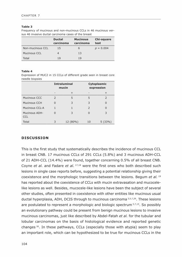
CHAPTER 7
104
diSCuSSiON
This is the first study that systematically describes the incidence of mucinous CCL
in breast CNB. 17 mucinous CCLs of 291 CCLs (5.8%) and 3 mucinous ADH-CCL
of 21 ADH-CCL (14.4%) were found, together concerning 0.5% of all breast CNB.
Coyne et al. and Fadare et al. 17,18 were the first ones who both described such
lesions in single case reports before, suggesting a potential relationship giving their
coexistence and the morphologic transitions between the lesions. Begum et al. 19
has reported about the coexistence of CCLs with mucin extravasation and mucocele-
like lesions as well. Besides, mucocele-like lesions have been the subject of several
other studies, often presented in coexistence with other entities like mucinous usual
ductal hyperplasia, ADH, DCIS through to mucinous carcinoma 3,5,7,29. These lesions
are postulated to represent a morphologic and biologic spectrum 5,7,15. So possibly
an evolutionary pathway could be present from benign mucinous lesions to invasive
mucinous carcinomas, just like described by Abdel-Fatah et al. for the tubular and
lobular carcinomas on the basis of histological evidence and reported genetic
changes 30. In these pathways, CCLs (especially those with atypia) seem to play
an important role, which can be hypothesized to be true for mucinous CCLs in the
Table 3 Frequency of mucinous and non-mucinous CCLs in 46 mucinous ver-sus 46 invasive ductal carcinoma cases of the breast
ductal carcinoma
Mucinous carcinoma
Chi-square test
Non-mucinous CCL 15 6 p = 0.004
Mucinous CCL 4 13
Total 19 19
Table 4 Expression of MUC2 in 15 CCLs of different grade seen in breast core needle biopsies
intraluminal mucin
Cytoplasmic expression
- + - +
Mucinous CCC 2 5 5 2
Mucinous CCH 0 3 3 0
Mucinous CCL-A 1 1 2 0
Mucinous ADH-CCL
0 3 0 3
Total 3 12 (80%) 10 5 (33%)
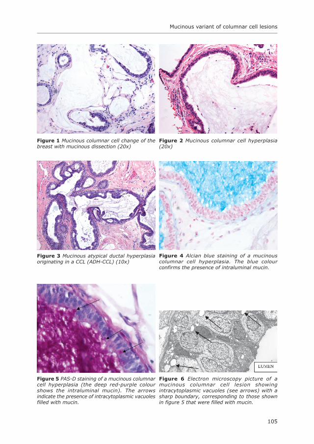
105
Mucinous variant of columnar cell lesions
figure 3 Mucinous atypical ductal hyperplasia originating in a CCL (ADH-CCL) (10x)
figure 4 Alcian blue staining of a mucinous columnar cell hyperplasia. The blue colour confirms the presence of intraluminal mucin.
figure 1 Mucinous columnar cell change of the breast with mucinous dissection (20x)
figure 2 Mucinous columnar cell hyperplasia (20x)
figure 5 PAS-D staining of a mucinous columnar cell hyperplasia (the deep red-purple colour shows the intraluminal mucin). The arrows indicate the presence of intracytoplasmic vacuoles filled with mucin.
figure 6 Electron microscopy picture of a mucinous columnar cell lesion showing intracytoplasmic vacuoles (see arrows) with a sharp boundary, corresponding to those shown in figure 5 that were filled with mucin.

CHAPTER 7
106
mucinous pathway as well. Indeed, in the study described by Begum et al. 19, DCIS
was found in a few excision biopsies with CCL and mucin extravasation in the initial
CNBs. We have not been able to demonstrate development of mucinous DCIS or
invasive mucinous cancer (or other types of DCIS or invasive cancer) from mucinous
CCL in our series. This may be related to the low number of mucinous CCL patients,
the relatively short follow-up interval and the fact that only very few mucinous
CCLs were treated by surgical excision biopsy.
We found significantly more mucinous CCLs in mucinous carcinoma cases than in
ductal carcinoma cases, confirming that mucinous CCLs indeed might be important
in the developmental pathway towards mucinous carcinoma. This is underlined by
the fact that CCLs (both mucinous and non-mucinous) were more frequently
atypical in mucinous than in ductal carcinoma cases.
MUC2 is typically expressed in the cytoplasm of tumour cells in mucinous carcinoma 21,23-26,31 and we found MUC2 expressing cells in 5 of 15 (33%) mucinous CCL cases
as well. In concordance with our finding, O’Connell et al.25 described positive cells
in mucinous DCIS, ADH and UDH surrounding mucinous carcinoma. Also “normal
epithelium” was described to show expression, but on review of the figures of
“normal epithelium” in their 1999 paper we would suggest that this in fact concerns
a columnar cell lesion. We have never seen MUC2 luminal or intracytroplasmic
staining in normal breast tissue. These data suggest that changes like MUC2
expression already occur in precursor lesions. We also found intraluminal mucin
staining in the present study, whereas other studies using other antibodies did not
show this staining pattern. This indicates that different MUC2 antibody clones can
provide different staining patterns 21,23-26,31.
figure 7 Mucinous CCL with MUC2 staining showing the intraluminal positivity of the mucin and cells with intracytoplasmic staining (20x).
figure 8 Mucinous columnar cell hyperplasia (bottom right) next to invasive mucinous carcinoma (top left, top right) (10x).

107
Mucinous variant of columnar cell lesions
We showed that the mucinous CCLs are typically biopsied for microcalcifications
as abnormality on mammography, which has been described for mucocele-like
lesions by others as well 3,4,14-16,29,32. Columnar cell lesions in general also present
by microcalcifications, which may further suggest a relationship in the origin of the
two lesions.
It is yet not fully clear what the implications are of finding a mucinous CCL in a
CNB. For mucocele-like lesions found on CNB it has been suggested that excision
should follow to exclude the presence of invasive carcinoma 33, although others
suggest that CNBs represent such lesions reliably 34,35. For columnar cell lesions
with atypia, excision biopsies are usually recommended, and for those without
atypia only follow-up is advised 11. Since we did not find any events during follow-
up, we do not suggest any other approach for mucinous CCL until further studies
better address the natural course of mucinous CCLs.
In conclusion, mucinous CCL and mucinous ADH-CCL are rare lesions occurring in
about 0.5% of breast CNB, usually presenting during the usual age range with
microcalcifications. These lesions might play a role in the mucinous progression
spectrum, which is underlined by the high incidence of mucinous CCLs in mucinous
carcinoma cases. Although further studies are needed to more extensively explore
the natural course of these lesions, the short term progression risk seems to be
low.

CHAPTER 7
108
REfERENCES
1. Geschickter CF. Gelatinous mammary cancer. Ann Surg 1938; 108:321-346.2. Rosen PP. Mucocele-like tumors of the breast. Am J Surg Pathol 1986; 10:464-469.3. Chinyama CN, Davies JD. Mammary mucinous lesions: congeners, prevalence and important
pathological associations. Histopathology 1996; 29:533-539.4. Farshid G, Pieterse S, King JM et al. Mucocele-like lesions of the breast: a benign cause for
indeterminate or suspicious mammographic microcalcifications. Breast J 2005; 11:15-22.5. Ro JY, Sneige N, Sahin AA et al. Mucocelelike tumor of the breast associated with atypical ductal
hyperplasia or mucinous carcinoma. A clinicopathologic study of seven cases. Arch Pathol Lab Med 1991; 115:137-140.
6. Tan PH, Tse GM, Bay BH et al. Mucinous breast lesions: diagnostic challenges. J Clin Pathol 2008; 61:11-19.
7. Weaver MG, Abdul-Karim FW, al-Kaisi N. Mucinous lesions of the breast. A pathological continuum. Pathol Res Pract 1993; 189:873-876.
8. Aulmann S, Elsawaf Z, Penzel R et al. Invasive tubular carcinoma of the breast frequently is clonally related to flat epithelial atypia and low-grade ductal carcinoma in situ. Am J Surg Pathol 2009; 33:1646-1653.
9. Dabbs DJ, Carter G, Fudge M et al. Molecular alterations in columnar cell lesions of the breast. Mod Pathol 2006; 19:344-349.
10. Dessauvagie BF, Zhao W, Heel-Miller KA et al. Characterization of columnar cell lesions of the breast: immunophenotypic analysis of columnar alteration of lobules with prominent apical snouts and secretions. Hum Pathol 2007; 38:284-292.
11. Feeley L, Quinn CM. Columnar cell lesions of the breast. Histopathology 2008; 52:11-19.12. Schnitt SJ, Vincent-Salomon A. Columnar cell lesions of the breast. Adv Anat Pathol 2003; 10:113-
124.13. Simpson PT, Gale T, Reis-Filho JS et al. Columnar cell lesions of the breast: the missing link in breast
cancer progression? A morphological and molecular analysis. Am J Surg Pathol 2005; 29:734-746.14. Kim JY, Han BK, Choe YH et al. Benign and malignant mucocele-like tumors of the breast:
mammographic and sonographic appearances. AJR Am J Roentgenol 2005; 185:1310-1316.15. Leibman AJ, Staeger CN, Charney DA. Mucocelelike lesions of the breast: mammographic findings
with pathologic correlation. AJR Am J Roentgenol 2006; 186:1356-1360.16. Tanaka K, Komoike Y, Egawa C et al. Indeterminate calcification and clustered cystic lesions are
strongly predictive of the presence of mucocele-like tumor of the breast: a report of six cases. Breast Cancer 2009; 16:77-82.
17. Coyne JD. Columnar cell hyperplasia with intraluminal crystalloids and features of a mucocoele-like lesion. Histopathology 2004; 44:401-403.
18. Fadare O, Mariappan MR. Mucocele-like tumor and columnar cell hyperplasia of the breast occurring in a morphologic continuum. J Med Case Reports 2008; 2:138.
19. Begum SM, Jara-Lazaro AR, Thike AA et al. Mucin extravasation in breast core biopsies--clinical significance and outcome correlation. Histopathology 2009; 55:609-617.
20. Kim YS, Gum JR, Jr. Diversity of mucin genes, structure, function, and expression. Gastroenterology 1995; 109:999-1001.
21. Matsukita S, Nomoto M, Kitajima S et al. Expression of mucins (MUC1, MUC2, MUC5AC and MUC6) in mucinous carcinoma of the breast: comparison with invasive ductal carcinoma. Histopathology 2003; 42:26-36.
22. Diaz LK, Wiley EL, Morrow M. Expression of epithelial mucins Muc1, Muc2, and Muc3 in ductal carcinoma in situ of the breast. Breast J 2001; 7:40-45.
23. Rakha EA, Boyce RW, bd El-Rehim D et al. Expression of mucins (MUC1, MUC2, MUC3, MUC4, MUC5AC and MUC6) and their prognostic significance in human breast cancer. Mod Pathol 2005; 18:1295-1304.
24. Adsay NV, Merati K, Nassar H et al. Pathogenesis of colloid (pure mucinous) carcinoma of exocrine organs: Coupling of gel-forming mucin (MUC2) production with altered cell polarity and abnormal cell-stroma interaction may be the key factor in the morphogenesis and indolent behavior of colloid carcinoma in the breast and pancreas. Am J Surg Pathol 2003; 27:571-578.

109
Mucinous variant of columnar cell lesions
25. O’Connell JT, Shao ZM, Drori E et al. Altered mucin expression is a field change that accompanies mucinous (colloid) breast carcinoma histogenesis. Hum Pathol 1998; 29:1517-1523.
26. Chu JS, Chang KJ. Mucin expression in mucinous carcinoma and other invasive carcinomas of the breast. Cancer Lett 1999; 142:121-127.
27. Tavassoli FA, Devilee P. WHO Classification Tumours of the Breast and Female Genital Organs, IARC, Lyon. 2003.
28. Carder PJ, Murphy CE, Liston JC. Surgical excision is warranted following a core biopsy diagnosis of mucocoele-like lesion of the breast. Histopathology 2004; 45:148-154.
29. Hamele-Bena D, Cranor ML, Rosen PP. Mammary mucocele-like lesions. Benign and malignant. Am J Surg Pathol 1996; 20:1081-1085.
30. Abdel-Fatah TM, Powe DG, Hodi Z et al. Morphologic and molecular evolutionary pathways of low nuclear grade invasive breast cancers and their putative precursor lesions: further evidence to support the concept of low nuclear grade breast neoplasia family. Am J Surg Pathol 2008; 32:513-523.
31. Yonezawa S, Sato E. Expression of mucin antigens in human cancers and its relationship with malignancy potential. Pathol Int 1997; 47:813-830.
32. Glazebrook K, Reynolds C. Original report. Mucocele-like tumors of the breast: mammographic and sonographic appearances. AJR Am J Roentgenol 2003; 180:949-954.
33. Jacobs TW, Connolly JL, Schnitt SJ. Nonmalignant lesions in breast core needle biopsies: to excise or not to excise? Am J Surg Pathol 2002; 26:1095-1110.
34. Renshaw AA, Cartagena N, Schenkman RH et al. Atypical ductal hyperplasia in breast core needle biopsies. Correlation of size of the lesion, complete removal of the lesion, and the incidence of carcinoma in follow-up biopsies. Am J Clin Pathol 2001; 116:92-96.
35. Wang J, Simsir A, Mercado C et al. Can core biopsy reliably diagnose mucinous lesions of the breast? Am J Clin Pathol 2007; 127:124-127.


Anoek H.J. Verschuur-Maes 1, Robert Kornegoor 1, Peter C. de Bruin 2,
Joost J. Oudejans 3, Paul J. van Diest 1
Departments of Pathology, 1 University Medical Center Utrecht, 2 St. Antonius
Ziekenhuis Nieuwegein, 3 Diakonessenhuis Utrecht, The Netherlands
Manuscript in preparation
Do columnar cell lesions exist in male breasts?
Chapter
8
CHAPTER 8
112
AbSTRACT
introduction
In females, columnar cell lesions (CCLs) are putative precursor lesions of breast
cancer. No data are yet available on the role of CCL in male breast carcinogenesis.
This study was performed to evaluate the presence of CCLs in male breast
specimens.
Methods
We reviewed 89 surgical resections from 88 patients with breast cancer, 20
gynaecomastia and 5 normal breast specimens from autopsies for the presence of
CCL, and immunohistochemistry for cytokeratin (CK) 5/6, CK14 and estrogen
receptor (ER) was performed.
Results
The majority of invasive carcinomas were of ductal type (88%) and two cases were
of the lobular type. In 19 of 89 resections (2 DCIS and 17 invasive carcinoma),
ducts were found with cells with snouts on the luminal border but lacking typical
columnar cell lesion morphology. None of these ducts showed intraluminal secretions
or microcalcifications and no unfolding terminal duct lobular units were recognized.
We mostly found three layered ductal epithelium characteristic for gynaecomastia.
Immunohistochemically, the myoepithelial layer and intermediate luminal layer
showed expression of CK5/6 and CK14 and was ER negative, while the intermediate
luminal layer was negative for CK5/6 and CK14 but strongly positive for ER.
Conclusion
We found no lesions with convincing CCL morphology in the periphery of invasive
male breast cancer, in gynaecomastia or normal male breast specimens. It therefore
seems that CCL does not exist in the male breast, denying that it plays a role in
male breast carcinogenesis.
113
Do columnar cell lesions exist in male breasts?
iNTROduCTiON
Breast cancer in men is a rare disease, representing less than 1% of all malignancies1.
Despite its low prevalence, the disease can cause high morbidity and mortality,
often showing a worse outcome than female breast cancer patients due to a delay
in diagnosis and a more advanced stage at presentation. Since male breast tissue
is predominantly rudimentary, it usually does not form lobules unless exposed to
high concentrations of estrogen 2 like in transsexuals 3. In this context, the most
common histological type is invasive ductal carcinoma while lobular carcinoma is
very rare 2 and the most frequent molecular type is Luminal A 4. The difference in
biology in male breast cancer compared with female breast cancer has been
demonstrated by immunohistochemical analysis, microRNA expression profiling,
gene expression and DNA ploidy analysis before 5-8. In spite of the differences found,
knowledge and treatment modalities have so far largely been extrapolated from
female breast cancer 2.
In females, different precursor lesions are known to play a role in the breast
carcinogenesis. Lobular neoplasia, columnar cell lesions (CCLs) and atypical ductal
hyperplasia (ADH) / low grade ductal carcinoma in situ (DCIS) seem to be of
significance in the low grade (often estrogen receptor (ER) positive) breast
neoplasias 9-11. CCLs are characterized by dilatation of terminal duct lobular units,
lined by columnar cells showing prominent apical cytoplasmic snouts, intraluminal
secretion and often microcalcifications, with varying degrees of nuclear atypia and
architectural complexity 12. In recent years, more evidence has been provided that
CCLs share overlapping morphological, immunohistochemical and molecular
features with ADH and low grade DCIS 9,13. CCLs with atypia are often found near
invasive or in situ breast cancer. As reported in our recent systematic review, CCL-A
diagnosed in core needle biopsies showed presence of cancer in 9.4% of the
subsequent excisions 14 and a progression risk to invasive cancer of about 20%
within 7 years if unexcised 15, probably necessitating surgical resection or removal
of the lesion by BLES/Intakt 14.
In male breasts, gynaecomastia is the most common benign lesion. The incidence
of gynaecomastia in association with male breast cancer was noted before with a
range of 3 to 37% 16-18. Gynaecomastia and invasive breast cancer coexist in the
high estrogen level environment as seen in Klinefelter patients 19. A few studies
reported on ADH in male patients 20,21. No studies have yet described CCLs in male
breasts as a putative precursor of male breast cancer that is also very often ER
positive. Male breast tissue is fairly rare apart from resections for gynaecomastia
and breast cancer, so we therefore scrutinized male breast tissue in breast cancer
CHAPTER 8
114
resection and gynaecomastia specimens for CCLs to unravel whether they play a
role in male breast carcinogenesis.
METHOdS
Patients
All male breast cancer resections (including invasive carcinoma and DCIS) were
retrieved from the archives of three hospitals in The Netherlands (University Medical
Center Utrecht, St. Antonius Hospital Nieuwegein and Diakonessenhuis Utrecht)
between 1986 and 2010. In addition, 20 gynaecomastia cases were taken from
the archive of the University Medical Center Utrecht, and 5 clinically normal male
breasts were sampled at autopsy. Use of anonymous or left over material for
scientific purposes is part of the standard treatment contract with patients in The
Netherlands 22.
Histopathology
All original hematoxylin-and-eosin-stained (H&E) slides from the 89 surgical excision
biopsies for breast cancer and the 20 gynaecomastia resection specimens were
reviewed by two experienced observers (AVM and PvD). The invasive carcinomas
and DCIS were graded according to usual criteria 23. Moreover, all slides were
scrutinized for gynaecomastia or CCL-like lesions using established criteria. As
described in the classification of Schnitt et al. 12, CCLs are cystically enlarged
terminal duct lobular units lined by one or several layers of regular columnar type
epithelium with nuclei oriented perpendicular along the basement membrane. Apical
cytoplasmic blebs or snouts are usually present at the luminal site, and there may
be mucus formation 24. As intraluminal secretions or microcalcifications are often
present in female CCLs, attention was paid to these features as well. In
gynaecomastia, the florid type, small to large duct-like structures with intraductal
epithelial proliferations are present with both epithelial and sometimes myoepithelial
hyperplasia. The epithelial proliferation is usually of flat or fine micropapillary type,
but can be cribriform and atypical as well. Apocrine metaplasia can be seen. The
cellular periductal stroma is characteristic for gynaecomastia and often shows areas
of pseudoangiomatous hyperplasia, or periductal fibrosis or stromal hyalinization.
The quiescent type is characterized by slightly increased numbers of ducts with no
or slight expansion of epithelium and no active expansion of the collagen-rich
periductal stroma, thereby being a rather clinical entity which is histologically
difficult to distinguish from normal male breast 25,26.

115
Do columnar cell lesions exist in male breasts?
immunohistochemistry
Immunohistochemistry was performed on 4 mm thick paraffin slides from selected
cases where male breast ducts vaguely resembled CCL. After deparaffinization and
rehydration, antigen retrieval was performed at boiling temperature in EDTA buffer
pH 9 for cytokeratin 5/6 (CK5/6). After a cooling down period of at least 30 minutes,
the slides were incubated with the primary antibody for CK5/6 (D5/16 B4, 1:100,
Dako, Glostrup, Denmark) for 60 minutes at room temperature. This was followed
by secondary antibody (Powervision, ImmunoVision Technologies, Brisbane, CA,
USA; ready to use, 30 min). The slides were developed with diaminobenzidine for
10 minutes followed by haematoxylin counterstaining. In between the different
steps, the sections were washed in PBS. Finally, slides were dehydrated in alcohol
and xylene and coverslipped. For cytokeratin 14 (CK14) and estrogen receptor (ER),
the slides were stained with the Bond Max Autostainer using the mouse monoclonal
antibodies for CK14 (LL002, 1:400, Neomarkers, Fremont, CA, USA) and ER (1D5,
1:80, Dako), according to the manufacturer’s instructions. Appropriate positive and
negative controls were used throughout. Interpretation of the immunohistochemical
slides was done by two experienced observers (AVM and PvD).
RESulTS
Table 1 shows the characteristics of the 89 resections in 88 male patients (one
patient had bilateral invasive carcinoma). There were four pure DCIS cases, and
in 26 of 85 invasive carcinoma cases, concomitant DCIS was present. The mean
age of patients with invasive carcinoma was 65.2 years and with DCIS (without
invasive carcinoma) 53 years (age range 19 - 90 years). In the majority of cases,
the invasive carcinomas were of ductal type (88%); only two cases were of the
lobular type. Three invasive carcinoma cases showed (macro)calcifications in or
in the stroma near the tumour. In the four cases with only DCIS, no calcifications
were found. Most of the slides from the resections for invasive carcinoma showed
no or just a few small normal primitive male ducts with a single layer of small
luminal cells surrounded by a single myoepithelial layer, as was also found in the
normal breast samples from autopsy (Figure 1). In 19 of 89 resections (2 DCIS
and 17 invasive carcinoma), ducts were found showing cells with snouts on the
luminal border but lacking typical columnar cell lesion morphology, that were
subjected to immunohistochemical stainings. None of these ducts showed
intraluminal secretions or microcalcifications and no unfolding terminal duct lobular
units were recognized. With thorough examination of the ducts on the H&E stained

CHAPTER 8
116
slides, we mostly found three layers (Figure 2), as described for gynaecomastia
by Kornegoor et al. 27. The myoepithelium forms the outer layer. The intermediate
luminal layer is composed of cuboid to columnar, vertically orientated cells with
ovoid regular nuclei. The inner luminal layer is formed by more horizontally
orientated smaller cells with small round to oval nuclei. Immunohistochemically,
the myoepithelial layer and inner luminal layer showed expression of CK5/6 and
CK14 while ER is negative. The intermediate luminal layer is negative for CK5/6
and CK14 but strongly positive for ER (Figure 2). Some of the ducts were also
surrounded by oedematous cellular stroma, characteristic for gynaecomastia. In
the 20 gynaecomastia cases we found similar gynaecomastia type ducts, also
lacking the typical columnar cell lesion morphology. Three of the 19 carcinoma
cases showed single ducts with monotonous columnar-like cells in the intermediate
luminal border with a hyperplastic aspect and regular ovoid nuclei (Figure 3), and
some snouts at the luminal side. CK5/6 and CK14 were clonally negative in this
intermediate layer (Figure 3). No inner luminal layer was seen in either H&E or
immunohistochemistry slides in these ducts. In CK14 immunohistochemistry, the
inner luminal layer in gynaecomastia was occasionally negative, while CK5/6 was
expressed (Figure 4).
Table 1 Patient characteristics of male breast cancer patients
No. of patients
No. of resections 89 *
DCIS 4
DCIS I 2 (50%)
DCIS II 2 (50%)
Invasive carcinoma 85 *
Grade 1 20 (24%)
Grade 2 36 (42%)
Grade 3 29 (34%) *
Type of invasive carcinoma
Ductal 75 (88%)
Encapsulated papillary 3 (4%)
Mucinous 2 (2%)
Lobular 2 (2%)
Neuro-endocrine 1 (1%)
Adenoid cystic 1 (1%)
Inverted micropapillary 1 (1%)
* of which 1 bilateral invasive carcinoma

117
Do columnar cell lesions exist in male breasts?
figure 1 Normal male breast. A. Haematoxylin and eosin staining. B. ER staining. C. CK5/6 staining. D. CK14 staining. (13x)
figure 2 Gynaecomastia in between invasive male breast cancer, showing the characteristic three cell layers. The outer myoepithelial layer and inner luminal layer express CK5/6 and CK14, while the intermediate luminal layer expresses only ER. A. Haematoxylin and eosin staining. B. ER staining. C. CK5/6 staining. D. CK14 staining. (15x)

CHAPTER 8
118
diSCuSSiON
In this study, we investigated whether CCLs, as recognized in female breasts, are
also present in male breasts to further unravel the putative role in the biology of
male breast cancer. Since CCLs are often seen near carcinoma, we reviewed a
relatively large series of male breast cancers, gynaecomastia specimens, being the
most abundant source of male breast tissue, as well as normal male breast tissue
harvested at autopsy.
We found no lesions with convincing CCL morphology. There were no enlarged
terminal duct lobular units with cystically dilated acini, and we saw no secrete or
figure 3 Clonally negative proliferation in the in the male breast. A. Haematoxylin and eosin staining. B. CK5/6 staining. C. CK14 staining.
figure 4 Gynaecomastia with expression of CK5/6 in the inner luminal layer (A) while these cells are negative for CK14 (B).

119
Do columnar cell lesions exist in male breasts?
microcalcifications, all characteristic features of female CCL 12,28. We only found
some single ducts lined by epithelium with more columnar type cells with vertically
oriented nuclei, sometimes slightly hyperplastic, and with apical snouts. However,
also in gynaecomastia (florid type) such ducts were seen as has been recognized
before 29. Further, these ducts immunohistochemically showed the three layers that
are typical of gynaecomastia: CK5/6 and CK14 positive and ER negative outer
myoepithelial and inner luminal layer, while the intermediate layer is ER positive
and CK5/6 and CK14 negative 27.
Microcalcifications in male breast cancer are far less common than in female breast
cancer 2. These microcalcifications are described to be pleiomorphic and punctate,
patterns that are generally considered benign in women, which may be associated
with cancer in men 18. Moreover, in female CCL, the typical microcalcifications seem
to be related to production of the bone matrix protein osteopontin (present in
intraluminal secretory material, necrotic debris and histiocytes), which is present
in milk produced in the female breast 30-32, which is obviously lacking in the male
breast unless exposed to estrogens.
Whereas we think CCL does not exist in male breasts and are not a precursor of
male breast cancer, ADH has been described in male breasts before 20,21. In these
studies, epithelial atypia was described in gynaecomastia cases, with cribriform
hyperplasia and abnormal nuclear polarization. In female breasts, CCL and ADH
are closely related, ADH regarded as a step further in the progression towards low
grade DCIS 9,33,34.
As shown before, we also found that gynaecomastia was often present next to
carcinoma 17,18. So far, no evidence is present that gynaecomastia has a precursor
role in male breast cancer, which is also underlined by the polyclonal CK5/6 and
CK14 patterns. However, in the slides with male breast cancer, we found a few
ducts that showed an epithelial proliferation that was CK5/6 clonally negative. The
significance of these ducts is yet unknown, but they deserve to be further studied
as a possible precursor of male breast cancer.
In CK14 immunohistochemistry, the inner luminal layer in gynaecomastia was
occasionally negative, while CK5/6 was expressed. Therefore, care has to be taken
not to misinterpret CK14 negativity in the inner luminal layer in gynaecomastia as
a sign of a clonal proliferation. CK5/6 is to be preferred here for this reason.
In conclusion, it seems that CCL does not exist in the male breast, denying that it
plays a role in male breast carcinogenesis.

CHAPTER 8
120
REfERENCES
1. Sasco AJ, Lowenfels AB, Pasker-de JP. Review article: epidemiology of male breast cancer. A meta-analysis of published case-control studies and discussion of selected aetiological factors. Int J Cancer 1993; 53:538-549.
2. Fentiman IS, Fourquet A, Hortobagyi GN. Male breast cancer. Lancet 2006; 367:595-604.3. Kanhai RC, Hage JJ, van Diest PJ et al. Short-term and long-term histologic effects of castration and
estrogen treatment on breast tissue of 14 male-to-female transsexuals in comparison with two chemically castrated men. Am J Surg Pathol 2000; 24:74-80.
4. Kornegoor R, Verschuur-Maes AHJ, Buerger H et al. Molecular subtyping of male breast cancer by immunohistochemistry. Mod Pathol 2011; In press.
5. Andre S, Pinto AE, Laranjeira C et al. Male and female breast cancer--differences in DNA ploidy, p21 and p53 expression reinforce the possibility of distinct pathways of oncogenesis. Pathobiology 2007; 74:323-327.
6. Callari M, Cappelletti V, De CL et al. Gene expression analysis reveals a different transcriptomic landscape in female and male breast cancer. Breast Cancer Res Treat 2010; 127:601-610.
7. Fassan M, Baffa R, Palazzo JP et al. MicroRNA expression profiling of male breast cancer. Breast Cancer Res 2009; 11:R58.
8. Ge Y, Sneige N, Eltorky MA et al. Immunohistochemical characterization of subtypes of male breast carcinoma. Breast Cancer Res 2009; 11:R28.
9. Abdel-Fatah TM, Powe DG, Hodi Z et al. Morphologic and molecular evolutionary pathways of low nuclear grade invasive breast cancers and their putative precursor lesions: further evidence to support the concept of low nuclear grade breast neoplasia family. Am J Surg Pathol 2008; 32:513-523.
10. Aulmann S, Elsawaf Z, Penzel R et al. Invasive tubular carcinoma of the breast frequently is clonally related to flat epithelial atypia and low-grade ductal carcinoma in situ. Am J Surg Pathol 2009; 33:1646-1653.
11. van Diest PJ. Ductal carcinoma in situ in breast carcinogenesis. J Pathol 1999; 187:383-384.12. Schnitt SJ, Vincent-Salomon A. Columnar cell lesions of the breast. Adv Anat Pathol 2003; 10:113-
124.13. Sudarshan M, Meguerditchian AN, Mesurolle B et al. Flat epithelial atypia of the breast: characteristics
and behaviors. Am J Surg 2011; 201:245-250.14. Verschuur-Maes AHJ, Van Deurzen CHM, Monninkhof EM et al. Columnar cell lesions on breast needle
biopsies: Is surgical excision necessary? A systematic review. Ann Surg 2011.15. Verschuur-Maes AH, Witkamp AJ, De Bruin PC et al. Progression risk of columnar cell lesions of the
breast diagnosed in core needle biopsies. Int J Cancer 2011; 129:2674-2680.16. Crichlow RW. Breast cancer in men. Semin Oncol 1974; 1:145-152.17. Joshi MG, Lee AK, Loda M et al. Male breast carcinoma: an evaluation of prognostic factors contributing
to a poorer outcome. Cancer 1996; 77:490-498.18. Mathew J, Perkins GH, Stephens T et al. Primary breast cancer in men: clinical, imaging, and
pathologic findings in 57 patients. AJR Am J Roentgenol 2008; 191:1631-1639.19. Bembo SA, Carlson HE. Gynecomastia: its features, and when and how to treat it. Cleve Clin J Med
2004; 71:511-517.20. Hamady ZZ, Carder PJ, Brennan TG. Atypical ductal hyperplasia in male breast tissue with
gynaecomastia. Histopathology 2005; 47:111-112.21. Prasad V, King M, McLeay W et al. Bilateral atypical ductal hyperplasia, an incidental finding in
gynaecomastia--case report and literature review. Breast 2005; 14:317-321.22. van Diest PJ. No consent should be needed for using leftover body material for scientific purposes.
BMJ 2002; 325:648-651.23. Tavassoli FA, Devilee P. WHO Classification Tumours of the Breast and Female Genital Organs, IARC,
Lyon. 2003.24. Verschuur-Maes AH, van Diest PJ. The mucinous variant of columnar cell lesions. Histopathology
2011; 58:847-853.25. Braunstein GD. Clinical practice. Gynecomastia. N Engl J Med 2007; 357:1229-1237.26. Williams MJ. Gynecomastia. Its incidence, recognition and host characterization in 447 autopsy cases.
Am J Med 1963; 34:103-112.

121
Do columnar cell lesions exist in male breasts?
27. Kornegoor R, Verschuur-Maes AHJ, Buerger H et al. The three layers in gynecomastia. American Journal of Surgical Pathology 2011.
28. Verschuur-Maes AHJ, van Gils CH, Van den Bosch MAAJ et al. Digital mammography: more microcalcifications, more columnar cell lesions without atypia. Mod Pathol 2011; 24:1191-1197.
29. Rosen PP. Rosen’s Breast Pathology. Philadelphia: Lippincott Williams & Wilkins; 2009.30. Baker R, Rogers KD, Shepherd N et al. New relationships between breast microcalcifications and
cancer. Br J Cancer 2010; 103:1034-1039.31. Haka AS, Shafer-Peltier KE, Fitzmaurice M et al. Identifying microcalcifications in benign and malignant
breast lesions by probing differences in their chemical composition using Raman spectroscopy. Cancer Res 2002; 62:5375-5380.
32. Oyama T, Sano T, Hikino T et al. Microcalcifications of breast cancer and atypical cystic lobules associated with infiltration of foam cells expressing osteopontin. Virchows Arch 2002; 440:267-273.
33. Bombonati A, Sgroi DC. The molecular pathology of breast cancer progression. J Pathol 2011; 223:307-317.
34. Verschuur-Maes AHJ, De Bruin PC, van Diest PJ. Epigenetic progression of columnar cell lesions of the breast to ductal carcinoma in situ and invasive breast cancer. Submitted 2011.


Summary and future perspectives
Chapter
9

CHAPTER 9
124
Breast carcinogenesis is a complex process in which invasive cancer arises from
normal breast epithelium via precursor lesions like atypical ductal hyperplasia (ADH),
atypical lobular neoplasia and ductal carcinoma in situ (DCIS). In the last decade,
also columnar cell lesions (CCLs) have been identified to play a possible role in the
progression to, particularly, low nuclear grade invasive breast cancer. Although
different classifications and names for these lesions have been used over the last
decades, of which flat epithelial atypia is the most well known, we used the
classification described by Schnitt and Vincent-Salomon 1 in our studies.
In this thesis the clinical significance as well as the molecular background of CCLs
were studied to contribute to the understanding of the pathogenesis of these lesions,
and to provide recommendations for the management of CCLs. The results of the
different studies as well as the future perspectives are discussed in this chapter.
Nowadays, CCLs are increasingly encountered in breast core needle biopsies, for
unknown reasons so far. In radiology, techniques for imaging breast lesions are
improving all the time, like with the introduction of full-field digital mammography
some years ago, replacing screen-filmed mammography. The former technique has
a better image quality and the practical advantage of digital data retrieving and
storage. Due to its higher resolution, it also has a higher sensitivity for the
characterization of microcalcifications 2, but whether specificity has been maintained
is the question. Since CCLs are characterized by small microcalcifications on
mammography, we studied the incidence of CCLs in 3437 breast core needle biopsy
specimens since the implementation of full-field digital mammography in comparison
with screen-filmed mammography and we related this to the number of biopsies
taken for microcalcifications, as described in Chapter 2. More diagnostic procedures
per mammography were performed in the full-field digital mammography era, due
to an increase of core needle biopsies (from 5.0 to 7.6%). We found significantly
more CCLs in the full-field digital mammography era compared with the screen-
filmed mammography era (10.8% versus 4.9%), which was also related to a higher
number of core needle biopsies taken for microcalcifications (adjusted prevalence
ratio 1.14, confidence interval 95% 1.01-1.28). Percentagewise, we found the same
amount of columnar cell lesions with atypia (CCL-A) during the full-field digital
mammography period as the screen-filmed mammography period (1.8%) and
significantly more columnar cell lesions without atypia (8.2% and 2.8%,
respectively).
Since we questioned whether it is relevant to find (more) CCLs without atypia in
core needle biopsies and since previous studies were not consistent about the
125
Summary and future perspectives
treatment recommendations for CCLs with atypia, we investigated the progression
risk of CCLs. The results are described in Chapter 3. We also included a subgroup
atypical ductal hyperplasia originated from a CCL (ADH-CCL), as these lesions
might represent a further step in the progression of CCLs to ADH and low grade
DCIS. Two groups were identified: “immediate treatment” group undergoing
excision within 4 months after the core needle biopsy diagnosis of CCL and the
“wait-and-see” group followed up to 8 years. When CCL with atypia (CCL-A or
ADH-CCL) was diagnosed on an initial core needle biopsy, DCIS or invasive
carcinoma was present in about 30% of surgical excisions performed within four
months. Including the “wait-and-see” group, the overall number of tumours were
20.3% for CCL-A and 23.8% for ADH-CCL. For CCL without atypia, DCIS or invasive
carcinoma was present in 25% of the excisions performed within four months,
however, including the events in the “wait-and-see”, in the whole group of 221
CCLs without atypia, events were present in only 2.7%. Also the indication for the
core needle biopsy was important. Densities were more often associated with
invasive carcinoma than with DCIS (p=0.051) and microcalcifications were slightly
related to more DCIS (p=0.092) in the resections. We concluded that an atypical
CCL in a core needle biopsy may signal the presence of concurrent lesions or
development of advanced lesions in future and may justify (‘mini’) surgical
excision.
In order to provide more evidence based treatment recommendations, we reviewed
the recent literature in a systematic review in Chapter 4. We found pooled
underestimation risks for all patients initially diagnosed with CCL without atypia,
CCL-A or ADH-CCL of 1.5%, 9% and 20%, respectively. However, not all patients
underwent surgical excision biopsy after initial diagnosis. Based on the number of
patients that underwent surgical excision within four months, the pooled
underestimation risks were 15% for CCL without atypia, 17% for CCL-A and 26%
for ADH-CCL. The rates based on the whole group of patients initially diagnosed
with a CCL, seemed to better reflect the actual underestimation risk, assuming
that the number of events in the group of patients without surgical excision biopsy
was limited. Follow-up studies including patients with a core needle biopsy diagnosis
of CCL showed that the progression risk to (in situ) carcinoma seems to be higher
than the baseline risk, although no firm conclusions could be drawn based on the
three included heterogeneous studies, of which one study was our study as
described in the former chapter. The relatively high underestimation risks for (in
situ) carcinomas for all patients initially diagnosed with CCL-A or ADH-CCL, seem
to support the indication for surgical excision or complete removal of all

CHAPTER 9
126
microcalcifications by vacuum assisted biopsies 3 or the BLES procedure 4. For CCLs
without atypia, more studies are needed to establish the long-term follow-up, but
for now the underestimation and progression rates seem to be limited and surgical
excision is not deemed necessary in these cases.
Different studies applying molecular techniques like loss-of-heterozygosity 5-7,
comparative genomic hybridization 8, miRNA 9 and allelic imbalance 10 have
demonstrated that CCL-A exhibits increasing genetic alterations from normal breast
epithelium to CCL-A to DCIS and invasive cancer. Similar chromosomal aberrations
(especially 16q loss) occur over the spectrum of CCL, ADH, low-grade DCIS and
low-grade invasive ductal carcinoma, which parallels the morphologically defined
carcinogenesis. During tumour development, in addition to the activation of
oncogenes and inactivation of tumour suppressor genes, DNA promoter
hypermethylation (further denoted “methylation”) is considered to be an early
event. Methylation of gene promoters may lead to inactivation of multiple (tumour
suppressor) genes and may contribute to cancer initiation and progression. Since
methylation plays an early role in tumour development, it may be a promising
biomarker for the early detection of malignancy.
In Chapter 5 we studied the methylation status of 50 tumour suppressor genes
by Methylation-Specific Multiplex Ligation-dependent Probe Amplification (MS-
MLPA) in normal breast tissue, CCL, DCIS and invasive carcinoma. Recently, Park
et al. also investigated MS-MLPA in these lesions 11, however, we investigated a
group of CCLs largely comprising synchronous lesions within the same patient. We
show that the cumulative methylation index (calculated as the sum of the
methylation percentage for all genes studied 12) increased significantly from normal
breast tissue through CCL, towards DCIS and invasive carcinoma, suggesting that
methylation increases stepwise in the progression to invasive cancer via CCL, which
is consistent with recent literature 11,13. CCL with atypia compared to CCL without
atypia, or presence of synchronous more advanced lesions was not accompanied
by higher mean methylation levels. The following genes showed significantly higher
methylation levels in CCL than normal breast tissue: ATM, BCL2, BRCA1, BRCA2,
CCND2, CDH13, CDKN2A, CHFR, DLC1, ESR1, FHIT, HLTF, ID4, IGSF4, MGMT,
RARB, RASSF1, SFRP5, SCGB3A1 and VHL. In the second step, the next genes
showed higher methylation levels in DCIS than in CCL: CCND2, ID4, FHIT and
CDH13. In the third step, invasive carcinoma had a higher mean methylation level
than DCIS for MGMT, although the mean methylation level was relatively low
(16.5% for invasive carcinoma). Particularly the first step underlines that
methylation may play an important early role in the histogenesis of CCLs. From all

127
Summary and future perspectives
these genes, RASSF1, SCGB3A1 and SFRP5 showed already high methylation levels
in CCL, whereas no significant increase in methylation was observed between CCL,
DCIS and invasive carcinoma. Moreover, ID4, CCND2 and CDH13 seem to be
important tumour suppressor genes in CCL driven breast carcinogenesis, as they
all had high methylation levels in the abnormal lesions and they showed significantly
increasing methylation levels from normal breast tissue to CCL and to DCIS/invasive
cancer. This suggests that for these genes, the epigenetic abnormalities are already
present in CCLs, underlying the important role of CCLs in low nuclear grade breast
carcinogenesis.
In Chapter 6 we investigated and compared copy number changes of 17 breast
cancer related genes by MLPA in the same cases as in the former study. More total
copy number changes in DCIS and invasive carcinoma were present than in CCL (p
= 0.023) with significantly more high level amplifications and losses in DCIS /
invasive carcinoma compared to CCL. High level amplifications were not found at
all in CCL. Moreover, overlap of the same copy number changes with CCL and
synchronous DCIS or invasive carcinoma occurred only for a few cases. So it seems
that these alterations occur mostly at a later stage than CCL in the progression from
normal breast tissue, CCL(-A), ADH to DCIS and invasive cancer, which is a well-
known progression pathway for low nuclear grade carcinomas 14. Unlike our results,
some other studies reported a certain homology of allelic changes. This might be
related to the investigated genes, since the genes in this studied kit are previously
reported to be particularly important in high grade breast carcinogenesis 15,16.
And only one gene was located on chromosome 16q, whereas many more genes
are known at this chromosome, that might be of influence in the development of
CCL driven breast carcinogenesis.
In Chapter 7 we are the first to systematically describe the incidence of mucinous
CCL in breast core needle biopsies, although some single case-reports described
this variant of CCL before 17,18. The mucinous variant of CCL was present in 5.8%
of the 291 CCLs and 14.4% of the 21 ADH-CCL, together concerning 0.5% of all
4164 studied breast core needle biopsies. In our small numbers of mucinous CCLs
with a relatively short follow-up interval (3.1 years), we have not been able to
demonstrate development of mucinous DCIS or invasive mucinous cancer (or other
types of DCIS or invasive cancer). However, we also studied the histological slides
of 46 mucinous carcinoma and 46 ductal carcinoma cases and found significantly
more mucinous CCLs in mucinous carcinoma than in ductal carcinoma cases. This
supports a possible role of mucinous CCLs in the developmental pathway towards

CHAPTER 9
128
mucinous carcinoma. Besides, MUC2, that is typically expressed in the cytoplasm
of tumour cells in mucinous carcinoma 19-21, was expressed in 33% of the mucinous
CCL cases. These results also suggested that changes like MUC2 expression already
occur in precursor lesions. Although further studies are needed to more extensively
explore the natural course of these lesions, the short term progression risk seems
to be low and for now we advise to follow the treatment recommendations of the
general CCLs.
In Chapter 8 we investigated whether CCLs are also present in male breasts to
unravel whether they play a role in male breast carcinogenesis. We reviewed a
relatively large series of 89 male breast cancers, 15 gynaecomastia specimens as
well as 5 normal male breast tissues harvested at autopsy. No lesions with
convincing CCL morphology were found in the invasive carcinoma cases, since there
were no enlarged terminal duct lobular units with cystically dilated acini, and we
saw no secrete or microcalcifications in the ducts. In 18 of the resections, we only
found single ducts with a three layered aspect, confirmed by immunohistochemistry.
This has been described to be characteristic for gynaecomastia in an article in
preparation 22. The gynaecomastia specimens and normal male breast tissues were
also devoid of CCL. So, it seems that CCL does not exist in the male breast and
that there is no role for CCLs in male breast carcinogenesis.
fuTuRE PERSPECTivES
A better understanding of breast carcinogenesis can help to improve the early
detection of malignancy or important precursor lesions, the treatment and in the
end the survival of patients. However, care has to be taken that no unnecessary
diagnostic procedures and surgical interventions are performed, since this leads
to higher costs and more women encountering anxiety about the outcome of the
procedures. The results of this thesis suggest that CCLs are increasingly diagnosed
since the introduction of full-field digital mammography due to the increased
number of core needle biopsies taken for microcalcifications. Particularly more CCLs
without atypia are recognized; lesions that are not recommended for excision,
since, so far, they seem to have a low underestimation rate and progression risk.
In order to minimize the number of unnecessary procedures (like core needle
biopsies) as well, future studies may help to identify the harmless microcalcifications
associated with CCLs without atypia. Recently, a study investigated the radiologic
presentation of CCLs retrospectively 23, showing that CCLs most often appear on

129
Summary and future perspectives
mammography as amorphous microcalcifications in a cluster. Nevertheless, it would
be of value to learn and retrain radiologists to identify the patterns of harmless
microcalcifications associated with CCLs without atypia, compared to CCLs with
atypia or DCIS / invasive carcinoma. Such criteria could help to prevent unnecessary
biopsies.
Although CCLs are best recognized as microcalcifications at mammography, in
future other modern imaging techniques (like optical imaging, electrical impedance
imaging, tomosynthesis (3D mammography)) could be tested to visualize CCLs
with malignant potential. Also the calcium carbonate content of microcalcifications
in benign, in situ and invasive breast tissues was studied before 24,25. A decrease
in carbonate content following a continuum from benign to DCIS and to invasive
carcinoma was found, with also significant differences between low and high grade
cancer. Since CCLs are often characterized by microcalcifications and might progress
to more important lesions, it would be interesting to study the carbonate content
of microcalcifications in CCLs. Possibly a distinction can be made between the more
‘benign CCLs’ and the CCLs that are prone to become DCIS / invasive cancer.
The results of our follow-up study and systematic review have led to another
approach for CCLs with atypia in our hospital; nowadays a surgical excision biopsy
is performed or a larger biopsy (using vacuum assisted biopsies or BLES) is taken
for patients diagnosed with a CCL with atypia on a core needle biopsy. In other
hospitals worldwide, this approach was already part of the daily practice, but this
was not widely implemented in The Netherlands yet. To monitor the effects of our
change of management, a further study (including all recent needle biopsies with
CCL with atypia and the following excisions or large biopsies) may be helpful to
evaluate our change of management. Besides, for all patients that did not receive
a surgical excision and were followed up to 8 years with a mean of 3.5 years
(Chapter 3), a longer follow-up after some years would be advisable to further
determine the long-term progression risk. To ultimately study the progression risk
of CCLs, a clinical trial may be helpful where CCLs (with or without atypia) are
randomized over follow-up or surgical excision. Also for CCLs without atypia, a
better and more evidence based advise could be given then. Moreover, some recent
studies (including a few recent studies not integrated in our systematic review)
investigated the value of performing surgical excisions after the diagnosis of pure
CCL on large vacuum assisted biopsies 26-31. Since in some studies cancer was found
in the subsequent excisions and in others not, different recommendations were
given. However, overall it seems that fewer invasive or in situ carcinomas are found
when vacuum assisted biopsies are used. Also other techniques like the BLES
CHAPTER 9
130
procedure extract more breast tissue and might therefore decrease the
underestimation risk 4,32. More studies are however needed to investigate the results
of these new techniques before new recommendations can be given.
Another important aspect in daily practice of pathologists is the use of
immunohistochemistry. Since nowadays a different approach and management is
present for CCLs with atypia, it would be interesting to know if different stainings
can help to make the distinction of CCL with and without atypia, or to compare
CCLs with synchronous DCIS / invasive carcinoma to CCLs without more advanced
lesions. We already made a start with this study, by performing the following
stainings on 80 CCLs cases (in surgical excisions) largely with synchronous DCIS
and/or invasive carcinoma: BCL2, BRST2, CAIX, CD117, CK5/6, CK14, CK19, Cyclin
D1, EGFR, ER, ER beta, EZH2, Glut-1, HER2, HIF1-alpha, MIB-1, p53, PR and PR
beta. We hope to find biomarkers that can help to distinguish CCLs with a benign
behaviour from those with a more malignant behaviour. Other markers that might
be important in breast carcinogenesis would also be of interest to study in CCLs,
like GATA3 33,34, FOXA1 35, androgen receptor 36,37, Claudins 38, HoxA5 39, ALDH1 40,41,
and HuR 42-44.
As we have shown, methylation of the genes RASSF1, SCGB3A1, SFRP5, ID4,
CCND2 and CDH13 seem to play a role in the development of CCL and thereafter
(in situ) carcinoma. More studies are needed to confirm the role of these genes
in the development of breast cancer and to study other possible genes that might
be important in the carcinogenesis. Since methylation as epigenetic event is a
reversible change of DNA, various demethylating agents have recently been
developed to target these processes, like 5-Azacitidine. The first studies are
emerging on treating patients with invasive cancer, but the agents seem to give
various side-effects like fatigue and bone marrow suppression 45-47. Therefore, more
improvement of these kind of agents is needed before implementation can take
place on patients with mild behaviour of the disease. In future, treating patients
with methylated CCL with a demethylating agent can possibly prevent progression
to malignancy, especially when we can demonstrate that methylation status predicts
progression towards malignancy in a follow-up study.
Besides, we aimed to elucidate chromosomal copy number changes in 17 selected
breast cancer related genes (in a commercially available kit) in CCLs, DCIS and
invasive carcinoma by MLPA. These genes did not show a progression in
amplifications and losses from CCL to cancer, which might be related to the studied
genes or the low number of studied cases. At the moment, a MLPA kit containing
genes located at chromosome 16q is in preparation. It would be very interesting

131
Summary and future perspectives
to study CCLs using this new kit, as is shown that CCLs often contain aberrations
on chromosome 16q, just as low nuclear grade breast cancers 8,15,48. Whether it is
worthwhile to study the predictive value of copy number changes in these genes
in a follow-up study, is up for discussion. And since somatic mutations in
mitochondrial DNA may play a role in the progression of breast cancer 49, this would
be of interest to study in CCLs as well.

CHAPTER 9
132
REfERENCES
1. Schnitt SJ, Vincent-Salomon A. Columnar cell lesions of the breast. Adv Anat Pathol 2003; 10:113-124.2. Fischer U, Baum F, Obenauer S et al. Comparative study in patients with microcalcifications: full-field
digital mammography vs screen-film mammography. Eur Radiol 2002; 12:2679-2683.3. David N, Labbe-Devilliers C, Moreau D et al. Diagnosis of flat epithelial atypia (FEA) after stereotactic
vacuum-assisted biopsy (VAB) of the breast: What is the best management: systematic surgery for all or follow-up? J Radiol 2006; 87:1671-1677.
4. Allen SD, Nerurkar A, la Rovere GU. The breast lesion excision system (BLES): a novel technique in the diagnostic and therapeutic management of small indeterminate breast lesions? Eur Radiol 2011; 21:919-924.
5. Aulmann S, Elsawaf Z, Penzel R et al. Invasive tubular carcinoma of the breast frequently is clonally related to flat epithelial atypia and low-grade ductal carcinoma in situ. Am J Surg Pathol 2009; 33:1646-1653.
6. Moinfar F, Man YG, Bratthauer GL et al. Genetic abnormalities in mammary ductal intraepithelial neoplasia-flat type (“clinging ductal carcinoma in situ”): a simulator of normal mammary epithelium. Cancer 2000; 88:2072-2081.
7. O’Connell P, Pekkel V, Fuqua SA et al. Analysis of loss of heterozygosity in 399 premalignant breast lesions at 15 genetic loci. J Natl Cancer Inst 1998; 90:697-703.
8. Simpson PT, Gale T, Reis-Filho JS et al. Columnar cell lesions of the breast: the missing link in breast cancer progression? A morphological and molecular analysis. Am J Surg Pathol 2005; 29:734-746.
9. Qi L, Bart J, Tan LP et al. Expression of miR-21 and its targets (PTEN, PDCD4, TM1) in flat epithelial atypia of the breast in relation to ductal carcinoma in situ and invasive carcinoma. BMC Cancer 2009; 9:163.
10. Dabbs DJ, Carter G, Fudge M et al. Molecular alterations in columnar cell lesions of the breast. Mod Pathol 2006; 19:344-349.
11. Park SY, Kwon HJ, Lee HE et al. Promoter CpG island hypermethylation during breast cancer progression. Virchows Arch 2011; 458:73-84.
12. Suijkerbuijk KP, Fackler MJ, Sukumar S et al. Methylation is less abundant in BRCA1-associated compared with sporadic breast cancer. Ann Oncol 2008; 19:1870-1874.
13. Liu T, Niu Y, Feng Y et al. Methylation of CpG islands of p16(INK4a) and cyclinD1 overexpression associated with progression of intraductal proliferative lesions of the breast. Hum Pathol 2008; 39:1637-1646.
14. Abdel-Fatah TM, Powe DG, Hodi Z et al. Morphologic and molecular evolutionary pathways of low nuclear grade invasive breast cancers and their putative precursor lesions: further evidence to support the concept of low nuclear grade breast neoplasia family. Am J Surg Pathol 2008; 32:513-523.
15. Hungermann D, Schmidt H, Natrajan R et al. Influence of whole arm loss of chromosome 16q on gene expression patterns in oestrogen receptor-positive, invasive breast cancer. J Pathol 2011; 224:517-528.
16. Rakha EA, Green AR, Powe DG et al. Chromosome 16 tumor-suppressor genes in breast cancer. Genes Chromosomes Cancer 2006; 45:527-535.
17. Coyne JD. Columnar cell hyperplasia with intraluminal crystalloids and features of a mucocoele-like lesion. Histopathology 2004; 44:401-403.
18. Fadare O, Mariappan MR. Mucocele-like tumor and columnar cell hyperplasia of the breast occurring in a morphologic continuum. J Med Case Reports 2008; 2:138.
19. Adsay NV, Merati K, Nassar H et al. Pathogenesis of colloid (pure mucinous) carcinoma of exocrine organs: Coupling of gel-forming mucin (MUC2) production with altered cell polarity and abnormal cell-stroma interaction may be the key factor in the morphogenesis and indolent behavior of colloid carcinoma in the breast and pancreas. Am J Surg Pathol 2003; 27:571-578.
20. Matsukita S, Nomoto M, Kitajima S et al. Expression of mucins (MUC1, MUC2, MUC5AC and MUC6) in mucinous carcinoma of the breast: comparison with invasive ductal carcinoma. Histopathology 2003; 42:26-36.
21. Rakha EA, Boyce RW, bd El-Rehim D et al. Expression of mucins (MUC1, MUC2, MUC3, MUC4, MUC5AC and MUC6) and their prognostic significance in human breast cancer. Mod Pathol 2005; 18:1295-1304.
133
Summary and future perspectives
22. Kornegoor R, Verschuur-Maes AHJ, Buerger H et al. The three layers in gynecomastia. American Journal of Surgical Pathology 2011.
23. Solorzano S, Mesurolle B, Omeroglu A et al. Flat epithelial atypia of the breast: pathological-radiological correlation. AJR Am J Roentgenol 2011; 197:740-746.
24. Baker R, Rogers KD, Shepherd N et al. New relationships between breast microcalcifications and cancer. Br J Cancer 2010; 103:1034-1039.
25. Haka AS, Shafer-Peltier KE, Fitzmaurice M et al. Identifying microcalcifications in benign and malignant breast lesions by probing differences in their chemical composition using Raman spectroscopy. Cancer Res 2002; 62:5375-5380.
26. de Mascarel I, Brouste V, sad-Syed M et al. All atypia diagnosed at stereotactic vacuum-assisted breast biopsy do not need surgical excision. Mod Pathol 2011; 24:1198-1206.
27. Lavoue V, Roger CM, Poilblanc M et al. Pure flat epithelial atypia (DIN 1a) on core needle biopsy: study of 60 biopsies with follow-up surgical excision. Breast Cancer Res Treat 2011; 125:121-126.
28. Noel JC, Buxant F, Engohan-Aloghe C. Immediate surgical resection of residual microcalcifications after a diagnosis of pure flat epithelial atypia on core biopsy: A word of caution. Surg Oncol 2010; 19:243-246.
29. Piubello Q, Parisi A, Eccher A et al. Flat epithelial atypia on core needle biopsy: which is the right management? Am J Surg Pathol 2009; 33:1078-1084.
30. Rajan S, Sharma N, Dall BJ et al. What is the significance of flat epithelial atypia and what are the management implications? J Clin Pathol 2011; 64:1001-1004.
31. Senetta R, Campanino PP, Mariscotti G et al. Columnar cell lesions associated with breast calcifications on vacuum-assisted core biopsies: clinical, radiographic, and histological correlations. Mod Pathol 2009; 22:762-769.
32. Sie A, Bryan DC, Gaines V et al. Multicenter evaluation of the breast lesion excision system, a percutaneous, vacuum-assisted, intact-specimen breast biopsy device. Cancer 2006; 107:945-949.
33. Chou J, Provot S, Werb Z. GATA3 in development and cancer differentiation: cells GATA have it! J Cell Physiol 2010; 222:42-49.
34. Yoon NK, Maresh EL, Shen D et al. Higher levels of GATA3 predict better survival in women with breast cancer. Hum Pathol 2010; 41:1794-1801.
35. Mehta RJ, Jain RK, Leung S et al. FOXA1 is an independent prognostic marker for ER-positive breast cancer. Breast Cancer Res Treat 2011.
36. Castellano I, Allia E, Accortanzo V et al. Androgen receptor expression is a significant prognostic factor in estrogen receptor positive breast cancers. Breast Cancer Res Treat 2010; 124:607-617.
37. Niemeier LA, Dabbs DJ, Beriwal S et al. Androgen receptor in breast cancer: expression in estrogen receptor-positive tumors and in estrogen receptor-negative tumors with apocrine differentiation. Mod Pathol 2010; 23:205-212.
38. Tokes AM, Kulka J, Paku S et al. Claudin-1, -3 and -4 proteins and mRNA expression in benign and malignant breast lesions: a research study. Breast Cancer Res 2005; 7:R296-R305.
39. Henderson GS, van Diest PJ, Burger H et al. Expression pattern of a homeotic gene, HOXA5, in normal breast and in breast tumors. Cell Oncol 2006; 28:305-313.
40. Heerma van V, Van der Groep P, Bart J et al. Expression of the stem cell marker ALDH1 in the normal breast of BRCA1 mutation carriers. Breast Cancer Res Treat 2010; 123:611-612.
41. Heerma van V, Van der Groep P, Bart J et al. Expression of the stem cell marker ALDH1 in BRCA1 related breast cancer. Cell Oncol (Dordr ) 2011; 34:3-10.
42. Heinonen M, Bono P, Narko K et al. Cytoplasmic HuR expression is a prognostic factor in invasive ductal breast carcinoma. Cancer Res 2005; 65:2157-2161.
43. Heinonen M, Hemmes A, Salmenkivi K et al. Role of RNA binding protein HuR in ductal carcinoma in situ of the breast. J Pathol 2011; 224:529-539.
44. Yuan Z, Sanders AJ, Ye L et al. Prognostic value of the human antigen R (HuR) in human breast cancer: high level predicts a favourable prognosis. Anticancer Res 2011; 31:303-310.
45. Fu S, Hu W, Iyer R et al. Phase 1b-2a study to reverse platinum resistance through use of a hypomethylating agent, azacitidine, in patients with platinum-resistant or platinum-refractory epithelial ovarian cancer. Cancer 2011; 117:1661-1669.
46. Maurillo L, Venditti A, Spagnoli A et al. Azacitidine for the treatment of patients with acute myeloid leukemia : Report of 82 patients enrolled in an italian compassionate program. Cancer 2011.

CHAPTER 9
134
47. Mirza S, Sharma G, Pandya P et al. Demethylating agent 5-aza-2-deoxycytidine enhances susceptibility of breast cancer cells to anticancer agents. Mol Cell Biochem 2010; 342:101-109.
48. Stacher E, Boldt V, Leibl S et al. Chromosomal aberrations as detected by array comparative genomic hybridization in early low-grade intraepithelial neoplasias of the breast. Histopathology 2011; 59:549-555.
49. Tseng LM, Yin PH, Yang CW et al. Somatic mutations of the mitochondrial genome in human breast cancers. Genes Chromosomes Cancer 2011; 50:800-811.

Nederlandse samenvatting
Dankwoord
Curriculum Vitae
List of Publications

136
Borstkanker is wereldwijd de meest voorkomende soort kanker en de belangrijkste
oorzaak voor overlijden bij vrouwen ten gevolge van kanker. In Nederland wordt
borstkanker jaarlijks bij ongeveer 14.000 vrouwen gediagnosticeerd. Mannen
hebben over het algemeen een veel lagere kans op het krijgen van borstkanker:
106 mannen in 2009 in Nederland. Het aantal vrouwen dat deze ziekte krijgt stijgt
nog steeds. Sinds de invoering van de landelijke screening worden tumoren echter
eerder gevonden, en ook worden voorloperafwijkingen van borstkanker vaak
gediagnosticeerd op naaldbiopten die worden genomen vanwege afwijkingen in
borsten. De ontwikkeling van borstkanker is een complex proces waarbij de tumor
ontstaat vanuit normaal borstweefsel (epitheel), via voorloperafwijkingen zoals
atypische ductale hyperplasie (ADH), atypische lobulaire neoplasie en ductal
carcinoma in situ (DCIS). In de laatste jaren, zijn ook cilindercel laesies (CCLs)
geïdentificeerd als mogelijke voorloperafwijkingen van met name de minder
agressieve soort borstkanker. CCLs zijn verwijde klierbuisjes van de borst die aan
de binnenzijde worden bekleed door cilindrisch gevormde cellen. Deze zijn een- of
tweelagig (cilindercel metaplasie) of meerlagig (cilindercel hyperplasie). Daarnaast
kunnen de kernen regelmatig zijn (zonder atypie) of erg onregelmatig en atypisch
(met atypie). Vaak wordt centaal in de buizen vocht en/of kleine kalkspatjes
(microcalcificaties) aangetroffen.
In dit proefschrift zijn het klinische belang en de moleculaire achtergrond van CCLs
onderzocht om een beter begrip van de afwijkingen te krijgen en te komen tot
goed onderbouwde adviezen voor de beste behandeling.
De laatste jaren worden CCLs steeds vaker gediagnosticeerd op naaldbiopten van
de borst, vooralsnog was dat zonder duidelijke reden. In de radiologie worden
technieken voor het in beeld brengen van borstafwijkingen steeds beter. Zo heeft
een paar jaar geleden in de meeste ziekenhuizen digitale mammografie de plaats
in genomen van analoge mammografie. De digitale mammografie heeft het voordeel
dat het een hogere resolutie heeft en betere beeldkwaliteit, zodat met name kleine
afwijkingen en afwijkingen met microcalcificaties beter opgespoord kunnen worden.
Net als ADH en DCIS, worden ook CCLs op een mammogram vaak herkend als
microcalcificaties. In Hoofdstuk 2 hebben we de incidentie van CCLs in 3437
naaldbiopten van de borst bestudeerd en vergeleken tussen de twee soorten
mammografie: de digitale versus de analoge mammografie. We hebben het aantal
CCLs gerelateerd aan het aantal biopten dat genomen was vanwege microcalcificaties.
Ten eerste bleken meer biopten genomen te zijn in het digitale mammografie
tijdperk ten opzichte van het analoge mammografie tijdperk. Ook vonden we
significant meer CCLs in de biopten (10,8% versus 4,9%) en dit was gerelateerd

137
aan het hogere aantal naaldbiopten dat verricht was vanwege microcalcificaties.
Procentueel vonden we hetzelfde aantal CCLs met atypie in de digitale mammografie
periode ten opzichte van de analoge mammografie periode (1,8%) en significant
meer CCLs zonder atypie (respectievelijk 8,2% en 2,8%).
Aangezien we ons afvroegen of het relevant is om meer CCLs zonder atypie te
vinden en aangezien eerdere studies geen consistente behandelingsadviezen gaven
voor CCLs, verrichtten we een studie om het risico dat CCLs tot kanker leiden te
onderzoeken (Hoofdstuk 3). We includeerden ook een subgroep atypische ductale
hyperplasie voortgekomen uit een CCL (ADH-CCL), dat een verder stap is in de
progressieroute van CCL naar ADH en naar laaggradig DCIS. Twee groepen werden
gemaakt: de “directe behandeling” groep die chirurgische resectie binnen 4 maanden
onderging na de diagnose van een CCL op een naaldbiopt, en de “wait-and-see”
groep die een follow-up had tot 8 jaar. Als CCL met atypie (inclusief ADH-CCL) was
gediagnosticeerd, was DCIS of borstkanker aanwezig in 30% van de chirurgische
resecties verricht binnen 4 maanden. Met het meenemen van de “wait-and-see”
groep in de analyse, was het totaal aantal tumoren 20,3% voor CCL met atypie en
23,8% voor ADH-CCL. Voor CCLs zonder atypie was DCIS of borstkanker aanwezig
in 25% van de excisies verricht binnen 4 maanden en inclusief de “wait-and-see”
groep slechts in 2,7%. We concludeerden dat een CCL met atypie gediagnosticeerd
in een naaldbiopt de aanwezigheid van een gelijktijdige ergere afwijking kan
signaleren of dat er een grotere kans is dat later een ergere afwijking kan ontstaan
vanuit een CCL. Daarom adviseren we nu chirugische resectie of ‘mini’ resectie met
behulp van extra grote biopten voor CCLs met atypie.
Om een beter ‘evidence based’ advies te kunnen geven voor de behandeling van
CCLs, voerden we een systematisch review uit, waarvan de resultaten staan
beschreven in Hoofdstuk 4. Het gepoolde risico voor patiënten gediagnosticeerd
met CCL zonder atypia, CCL met atypie en ADH-CCL was respectievelijk 1,5%, 9%
en 20%. Deze aantallen waren op basis van alle patiënten, ook diegene die geen
resectie onderging, ervan uitgaande dat het aantal afwijkingen in de groep patiënten
zonder resectie beperkt was. Vanwege de relatief hoge risico’s op tumor voor alle
patiënten gediagnosticeerd met CCL met atypie of ADH-CCL, adviseren we
chirurgische resectie. Om niet overbodig te opereren, zou complete verwijdering
van alle microcalcificaties door grote biopten (met een vrij nieuwe methode) een
alternatief zijn, alhoewel meer onderzoek naar de resultaten hiervan gewenst is.
Voor CCLs zonder atypie suggereren we voorlopig alleen follow-up te laten
plaatsvinden, aangezien de kans op latere tumorvorming beperkt lijkt te zijn.
Nederlandse samenvatting
138
Het DNA van de mens bevat ongeveer 20.000 genen, die allemaal een eigen functie
hebben. Het ontstaan van kanker kan op verschillende manieren gebeuren, zoals
de inactivering van tumor suppressor genen (genen die het ontstaan van kanker
tegengaan) en het activeren van oncogenen (genen die de groei van kankercellen
stimuleren). Ook epigenetische veranderingen (omkeerbare erfelijke veranderingen
in de genfunctie zonder wijziging in de volgorde van de basenparen) kunnen een
rol spelen bij het ontstaan en de groei van kanker. Methylering is hier een voorbeeld
van, waarbij een methylgroep aan de cytosine-base wordt toegevoegd. Als het gen
gemethyleerd is, kan het zijn normale eiwit niet produceren en daarom zijn functie
niet uitoefenen. Zo kunnen tumor suppressor genen uitgeschakeld worden. Omdat
methylering een rol lijkt te spelen in de vroege ontwikkeling van tumor, kan het
een veelbelovende biomarker zijn voor de vroege opsporing van kanker.
In Hoofdstuk 5 hebben we de methylatie status van 50 tumor suppressor genen
onderzocht door middel van Methylation-Specific Multiplex Ligation-dependent
Probe Amplification (MS-MLPA) in normaal borst weefsel, CCL, DCIS en borstkanker.
Een deel hiervan hadden gelijktijdig afwijkingen zoals CCL, DCIS en borstkanker
of CCL en alleen borstkanker. We vonden dat de cumulatieve methylatie index
(berekend door de methylatie waarden voor alle genen bij elkaar op te tellen)
significant toenam van normaal borst weefsel, naar CCL, naar DCIS en borstkanker.
Dit suggereert dat methylatie stapsgewijs toeneemt in the progressie naar kanker
waarbij CCLs een rol lijken te spelen. CCLs zonder atypie hadden vergelijkbare
methylatie waarden als CCLs met atypie, net als de CCLs die wel of niet gelijktijdig
kanker hadden. Ook eerder verrichte studies, hebben (door middel van andere
technieken) aangetoond dat CCLs met atypie genetische veranderingen hebben
die toenemen van normaal borstweefsel via CCL met atypie, ADH naar DCIS en
borstkanker. In deze ontwikkeling staan vooral afwijkingen in chromosoom 16q op
de voorgrond.
In Hoofdstuk 6 vergeleken we in dezelfde groep patiënten ‘copy number changes’
van 17 borstkanker gerelateerde genen, dat wil zeggen het ontbreken (deletie) of
het teveel aanwezig zijn (amplificatie) van bepaalde gedeeltes DNA op een
chromosoom. We vonden meer copy number changes in DCIS en borstkanker dan
in CCL. Sterke amplificaties werden niet gevonden in CCL; DCIS en borstkanker
hadden significant meer sterke amplificaties. Daarnaast was slechts in een paar
patiënten overlap aanwezig met afwijkingen tussen CCL, DCIS en borstkanker. Het
lijkt er daarom op dat dit soort veranderingen pas in een later stadium van de
progressie naar kanker optreden dan in CCL. Ook kan het panel van genen ermee

139
Nederlandse samenvatting
te maken hebben. In deze commerciële kit werden genen onderzocht die in andere
studies veel veranderingen hadden aangetoond bij borstkanker, met name bij
kanker die een slechtere prognose had, terwijl in deze studie met name het type
kanker werd onderzocht met een betere prognose.
In Hoofdstuk 7 beschreven we hoe vaak de slijmvormde variant van CCLs
voorkomt in naaldbiopten van de borst. In 5,8% van de 291 CCLs en in 14,4% van
de 21 ADH-CCLs vonden we deze afwijkingen. Dit was in totaal 0,5% van alle 4164
bestudeerde borst naaldbiopten. In het kleine aantal biopten met een relatief korte
follow-up interval (3,1 jaar) hebben we geen ontwikkeling tot slijmvormend DCIS
of slijmvormend borstkanker aangetroffen. Daarnaast hebben we microscopisch
46 slijmvormende kankers en 46 ‘gewone’ kankers (ductale carcinomen) bekeken.
We vonden significant meer slijmvormende CCLs bij de slijmvormende kankers dan
in de ‘gewone’ borstkankers. Het MUC2 eiwit, dat wordt gemaakt in de cellen van
slijmvormende borstkankers, was ook aanwezig in 33% van de slijmvormende
CCLs. Dit alles lijkt erop te duiden dat de slijmvormende CCLs een mogelijke
voorloperafwijking van de slijmvormende borstkanker zou kunnen zijn, hoewel
meer studies nodig zijn om dit te bevestigen en de kans op het krijgen van borst
kanker na deze afwijkingen te bestuderen.
In Hoofdstuk 8 bestudeerden we of CCLs ook aanwezig zijn in mannelijke borsten
om na te gaan of deze afwijkingen ook in de ontwikkeling van borstkanker bij mannen
een rol spelen. We hebben hiervoor een relatief groot aantal van 89 mannelijke
borstkankers bekeken, 20 gevallen van gynaecomastie (meest voorkomende
goedaardige afwijking bij de man met een toename van klierweefsel) en 5 keer
normaal mannelijk borstweefsel verkregen via autopsie. We hebben geen afwijkingen
met overtuigend CCL aspect gevonden. In 18 van de resecties vonden we wel
aanwezigheid van gynaecomastie naast de kanker. Ook in de gynaecomastie
preparaten en normaal mannelijk borstweefsel waren geen klierbuizen aanwezig die
op CCL leken. Dus het lijkt erop dat CCLs niet voorkomen in mannelijk borstweefsel
en deze dus geen rol spelen in de ontwikkeling van borstkanker bij mannen.
TOEkOMSTPERSPECTiEvEN
Zoals beschreven was in dit proefschrift, worden CCLs steeds vaker gediagnosticeerd.
Met name is dit het geval sinds de invoering van digitale mammografie vanwege
de toename van het biopteren van microcalcificaties die zijn gezien op het
140
mammogram. Met name CCLs zonder atypie worden meer gevonden; de afwijkingen
waarbij chirurgische resectie niet wordt geadviseerd vanwege de lage kans op het
missen van belangrijkere afwijkingen in de omgeving van de CCL en vanwege de
lage kans op progressie tot kanker. Het zou goed zijn het aantal onnodige naaldbiopt
procedures terug te dringen, zodat de kosten beperkt kunnen worden en minder
vrouwen angst hoeven te hebben over de uitslag van het biopt. Daarom zou een
toekomstige studie naar het herkennen van de microcalcificaties die gerelateerd
zijn aan de CCLs zonder atypie van waarde zijn om de soort microcalcificaties te
vergelijken met die gerelateerd aan CCLs met atypie, DCIS of borstkanker. Zodoende
kunnen radiologen worden getraind om niet onnodig een biopt te nemen. Hoewel
CCLs het beste kunnen worden herkend door microcalcificaties op een mammogram,
zouden in de toekomst wellicht ook andere beeldvormende technieken kunnen
worden toegepast om CCLs (met kwaadaardige potentie) te kunnen herkennen.
De calcium carbonaat hoeveelheid in microcalcificaties is eerder onderzocht in
goedaardige afwijkingen, DCIS en borstkanker, waarbij een continue afname in
carbonaat hoeveelheid werd gevonden van goedaardige afwijking, naar DCIS en
naar borstkanker. Aangezien CCLs ook worden gekarakteriseerd door
microcalcificaties en eventueel progressie kunnen vertonen naar DCIS of
borstkanker, zou het interessant zijn om de carbonaat hoeveelheid in
microcalcificaties in CCLs te bestuderen. Mogelijk kan dan ook een onderscheid
gemaakt worden tussen de meer ‘goedaardige’ CCLs en die CCLs met een hogere
kans op kanker.
De resultaten van onze follow-up studie en systematische review hebben in ons
ziekenhuis geleid tot een verandering van aanpak: chirurgische resecties worden
verricht voor CCLs met atypie, of grotere biopten (vacuum geassisteerde biopten
of BLES procedure) worden genomen. Het zou goed zijn een vervolgstudie te
verrichten om het effect van deze behandeling te evalueren, en een langere follow-
up zou ook goed zijn om de kans op progressie bij CCLs beter te kunnen bepalen.
Tenslotte zou een grote klinische studie interessant zijn waarbij CCLs (met of zonder
atypie) worden gerandomiseerd in een groep met alleen follow-up en een groep
van CCLs gevolgd door chirurgische resectie. Ook voor CCLs zonder atypie zou dan
een beter advies kunnen worden gegeven. Bovendien zou het goed zijn om het
effect te bestuderen van het wegbiopteren van alle microcalcificaties door middel
van het nemen van grotere biopten danwel gewone naaldbiopten.
Een ander belangrijk aspect in de dagelijkse praktijk van pathologen, is het gebruik
van immuunhistochemische kleuringen. Aangezien voor CCLs met atypie een andere
behandeling volgt, zou het interessant zijn om te weten of bepaalde kleuringen
een onderscheid kunnen maken in de CCLs met en zonder atypie. We hebben al

141
Nederlandse samenvatting
een start gemaakt met deze studie, door het verrichten van kleuringen op 80 CCL
cases in resectie preparaten, waarbij in een deel ook DCIS en/of borstkanker
aanwezig was. We hopen hiermee kleuringen te vinden die ons kunnen helpen om
te kunnen differentiëren tussen CCLs met een goed versus kwaadaardig gedrag.
Zoals we hebben getoond, speelt de methylatie van een aantal genen mogelijk een
rol in de ontwikkeling van CCLs en daarna DCIS of borstkanker. Meer studies zijn
nodig om deze rol te bevestigen en om ook andere genen te vinden die mogelijk
van belang zijn. Aangezien methylatie een omkeerbare verandering van het DNA
is, zijn recent verschillende medicijnen uitgevonden die deze processen kunnen
beïnvloeden, zoals 5-Azacitidine. Deze medicijnen blijken verschillende bijwerkingen
te geven, zoals erge vermoeidheid en beenmerg onderdrukking. Daarom is
verbetering van deze producten nodig, voordat de afwijkingen met mild gedrag
van een ziekte (zoals CCLs) kunnen worden behandeld. Mogelijk kunnen in de
toekomst wel de CCLs met de potentie op borstkanker worden behandeld met een
‘demethylerend’ medicijn, zodat uitgroei tot borstkanker zou kunnen worden
voorkomen.
Daarnaast hadden we gehoopt ook copy number afwijkingen in CCL te vinden,
gerelateerd aan DCIS en borstkanker. Dit werd niet gevonden, maar mogelijk houdt
dit verband met de 17 geselecteerde genen (in de toen voor handen zijnde kit) en
het lage aantal bestudeerde casus. Op het moment wordt een MLPA kit gemaakt
met verschillende genen gelocaliseerd op chromosoom 16q. Het zou erg interessant
zijn om CCLs met deze nieuwe kit te bestuderen, aangezien eerder is aangetoond
dat vooral afwijkingen op dit chromosoom deel bij CCLs (en het bijbehorende type
DCIS en borstkanker) een rol spelen.

Nederlandse samenvatting
Dankwoord
Curriculum Vitae
List of Publications
143
Dankwoord
Met dit proefschrift sluit ik een boeiende en intensieve periode van onderzoek af.
In deze tijd heb ik samengewerkt met verschillende mensen, waarbij ik een aantal
mensen speciaal wil noemen en bedanken.
Prof.dr. P.J. van Diest, beste Paul. Ongeveer 3,5 jaar geleden vertelde ik je dat ik
interesse had om onderzoek te gaan doen. “Weet je het heel zeker?” Ja, ik wist
het zeker. En ik heb er geen ogenblik spijt van gehad. Jouw enthousiasme was
altijd erg stimulerend en aanstekelijk. Ondanks dat je steeds erg druk was, kon je
toch tijd vrij maken om coupes te kijken. Zelfs in de weekenden beoordeelde je
mijn artikelen. ‘Check even je mail’, soms op de gekste tijden, betekende vaak
goed nieuws. Ik ben erg blij geweest met jou als promotor, dank je wel!
Dr. P.C. de Bruin, beste Peter. Tijdens een leerzaam jaar perifere opleiding in het
St. Antonius Ziekenhuis Nieuwegein werd mijn enthousiasme voor wetenschappelijk
onderzoek door jou verder aangewakkerd. Er bleek een mogelijkheid te zijn voor
een beurs, waardoor tijd voor promotie-onderzoek beschikbaar kwam. Bedankt
voor je steun bij mijn studie en medebeoordeling van de artikelen. Ook dr. C.A.
Seldenrijk, beste Kees, dank je voor het aanvragen van de subsidie en je
betrokkenheid bij mijn onderzoek.
Ook de pathologen van het UMC Utrecht wil ik graag bedanken. Prof.dr. J.G. van
den Tweel, bedankt voor het in mij gestelde vertrouwen om mij toe te laten tot de
opleiding pathologie in het UMC Utrecht.
Collega’s, arts-assistenten, een groep leuke getalenteerde mensen die voor
hetzelfde vak gaan! Met veel genoegen mag ik hier deel van uitmaken. Robert, een
goede samenwerking met de mannen mamma’s heeft ook een mooi artikel van
gynaecomastie opgeleverd. Mirthe, mijn opvolgster, super dat je je ook gaat
verdiepen in de cilindercel laesies. En oud-collega Carolien, vanuit Rotterdam heb
je ook een flinke bijdrage geleverd aan het review, bedankt voor de fijne
samenwerking.
De onderzoekers van de mamma / HIF – werkgroep, het was leerzaam om bij jullie
besprekingen te zijn en bedankt voor jullie nuttige tips en commentaar op mijn
onderzoek. Petra van der Groep, je hebt me wegwijs gemaakt in de verschillende
technieken op het lab in een gezellige sfeer. Cathy, je gaf me zinvolle adviezen op
het gebied van (MS)-MLPA. Willy, heel veel dank voor je secretariële hulp voor mijn
onderzoek. Medewerkers van het histo-, immuno- en moleculaire lab, coupes

144
snijden, immuunhistochemische kleuren, (MS-)MLPA, bedankt dat ik bij jullie
terecht kon met mijn vragen. En in het bijzonder Aad en Willem, die al die duizenden
coupes voor me opzochten en weer opborgen, in één woord geweldig.
De leden van de leescommissie wil ik bedanken voor het lezen van het manuscript
en het voeren van de oppositie.
De AntoniusMesosGroep wil ik bedanken voor het toekennen van de onderzoeks-
beurs.
Lieve vrienden en vriendinnen, Marjolein, Margreet, Linda S-vd G, Maaike, Tom,
Linda v L en natuurlijk meiden van OVM, Barbara, Cora, Janneke v I-D, Janneke
V, Jolien en Susanne, de vriendschap met jullie geeft me steeds weer energie!
Tot mijn paranimfen wil ik enkele woorden apart richten.
Marleen, ik ben blij met onze waardevolle vriendschap. Super dat je altijd zo
betrokken en vol interesse bent. Ik heb goede herinneringen aan heel wat jaren
avondjes uit, vakanties, etentjes, dat er nog maar vele mogen volgen! Fijn dat je
naast me wilt staan!
Amélie, ook wij hebben sinds je komst naar Utrecht samen veel ondernomen. We
delen de passie voor onderzoek en pathologie en dat schept een band! Over een
paar maanden volgt ook jouw promotie, succes met de laatste loodjes!
Familie Verschuur, Bob, Loes, Petra, Opa Verschuur bedankt voor jullie belangstelling
in mijn onderzoek. Het is heerlijk om in jullie familie zo opgenomen te zijn!
En mijn geweldige familie, wat ben ik blij met jullie! Timo en Linda, Martine en
Edwin, super om zo’n lieve (schoon)broers en (schoon)zussen te hebben! We
hebben samen veel plezier en we weten dat we altijd op elkaar kunnen terugvallen,
dat geeft een goed gevoel! En mijn lieve neefjes en nichtje Koen, Noah, Kick en
Isa, jullie maken het in de familie nog leuker!
Lieve pa en ma, wat moest ik zonder jullie?! Ontzettend bedankt voor jullie
onvoorwaardelijke steun en liefde van jongs af aan, alles wat jullie me hebben
geleerd en meegegeven. Ook de laatste jaren zijn jullie weer zo belangrijk voor
mij, jullie zijn fantastisch!

145
En natuurlijk mijn liefste mannen. Michiel, we hebben een tijd met veel veranderingen
achter de rug met trouwen, nieuw huis, promotie-onderzoek en een kind. Dank je
voor je niet aflatende steun, ook en vooral in de laatste tijd rond de afronding van
mijn proefschrift. Ik koester de fijne momenten samen, ik hou van je! Thomas,
wat ben ik ontzettend blij met jou, je maakt me erg gelukkig!
Dankwoord

Nederlandse samenvatting
Dankwoord
Curriculum Vitae
List of Publications

147
Anoek Huberdine Josef Verschuur-Maes is geboren op 14 september 1981 in Berkel
en Rodenrijs. In 1993 verhuisde zij naar Vlijmen en bezocht het Stedelijk Gymnasium
te ’s-Hertogenbosch. Haar gymnasium diploma behaalde zij in 1999. Vervolgens
studeerde ze Geneeskunde in Maastricht en legde in 2005 het artsexamen af.
Tijdens haar studie verrichtte ze op de afdeling Pathologie van het Academische
Ziekenhuis Maastricht wetenschappelijk onderzoek naar de rol van TGF-β bij
nierziekten in muismodellen. Ook deed ze een wetenschapsstage van vier maanden
op de afdeling Gynaecologie en Obstetrie van het Royal Hospital for Women in
Sydney, Australië. Na afronding van de studie Geneeskunde werkte ze van oktober
2005 tot en met augustus 2006 in het Diakonessenhuis Utrecht en Zeist op de
afdeling Interne Geneeskunde. Aansluitend startte ze in september 2006 met de
opleiding Pathologie in het Universitair Medisch Centrum Utrecht (opleider prof.dr.
J.G. van den Tweel). Voor het perifere deel van de opleiding was ze in 2008
werkzaam in het St. Antonius Ziekenhuis Nieuwegein (opleider dr. C.A. Seldenrijk).
In september 2008 is ze haar promotieonderzoek gestart onder begeleiding van
prof.dr. P.J. van Diest. Haar opleiding tot specialist heeft ze gedurende een jaar
onderbroken om dit onderzoek te verrichten met een beurs van het Oncologiecentrum
van de AntoniusMesosgroep. Nu haar promotie is afgesloten, vervolgt Anoek haar
opleiding tot patholoog.
Curriculum Vitae

CHAPTER 9
148
Nederlandse samenvatting
Dankwoord
Curriculum Vitae
List of Publications

149
verschuur-Maes AH, Van Diest PJ. The mucinous variant of columnar cell
lesions. Histopathology 2011;58:847-853
verschuur-Maes AHJ, Witkamp AJ, De Bruin PC, Van der Wall E, Van Diest PJ.
Progression risk of columnar cell lesions of the breast diagnosed in core needle
biopsies.
International Journal of Cancer 2011;129:2674-2680
verschuur-Maes AHJ, Van Gils CH, Van den Bosch MAAJ, De Bruin PC,
Van Diest PJ.
Digital mammography: more microcalcifications, more columnar cell lesions
without atypia.
Modern Pathology 2011;24:1191-1197
verschuur-Maes AHJ, Van Deurzen CHM, Monninkhof EM, Van Diest PJ.
Columnar cell lesions on breast needle biopsies: Is surgical excision necessary?
A systematic review.
Annals of Surgery 2011, in press
verschuur-Maes AHJ, De Bruin PC, Van Diest PJ.
Epigenetic progression of columnar cell lesions of the breast to ductal carcinoma
in situ and invasive breast cancer.
Submitted
verschuur-Maes AHJ, Moelans CB, De Bruin PC, Van Diest PJ.
Oncogene amplification in columnar cell lesions of the breast: analysis by
Multiplex Ligation-dependent Probe Amplification.
Submitted
Moelans CB, De Weger RA, Monsuur HN, Maes AH, Van Diest PJ.
Molecular differences between ductal carcinoma in situ and adjacent invasive
breast carcinoma: a multiplex ligation-dependent probe amplification study.
Analytical Cellular Pathology 2010;33:165-173
Moelans CB, verschuur-Maes AHJ, Van Diest PJ.
Frequent promoter hypermethylation of BRCA2, CDH13, MSH6, PAX5, PAX6, and
WT1 in ductal carcinoma in situ and invasive breast cancer.
Journal of Pathology 2011;225:222-231
List of Publications

150
Kornegoor R, verschuur-Maes AHJ, Buerger H, Hogenes MCH, De Bruin PC,
Oudejans JJ, Van der Groep P, Hinrichs B, Van Diest PJ.
Molecular subtyping of male breast cancer by immunohistochemistry.
Modern Pathology 2011, in press
Kornegoor R, verschuur-Maes AHJ, Buerger H, Boecker W, van Diest PJ.
The three layers in gynecomastia.
American Journal of Surgical Pathology 2011, accepted