combination of sb431542, chir9901, and bpv as a novel
TRANSCRIPT

RESEARCH Open Access
Combination of SB431542, Chir9901, andBpv as a novel supplement in the cultureof umbilical cord blood hematopoieticstem cellsMorteza Zarrabi1,2†, Elaheh Afzal2†, Mohammad Hassan Asghari3 and Marzieh Ebrahimi1*
Abstract
Background: Small molecule compounds have been well recognized for their promising power in the generation,expansion, and maintenance of embryonic or adult stem cells. The aim of this study was to identify a novel combinationof small molecules in order to optimize the ex vivo expansion of umbilical cord blood-derived CD34+ cells.
Methods: Considering the most important signaling pathways involved in the self-renewal of hematopoietic stem cells,CB-CD34+ cells were expanded with cytokines in the presence of seven small molecules including SB, PD, Chir, Bpv, Pur,Pμ, and NAM. The eliminativism approach was used to find the best combination of selected small molecules for effectiveex vivo expansion of CD34+ cell. In each step, proliferation, self-renewal, and clonogenic potential of the expanded cellsas well as expression of some hematopoietic stem cell-related genes were studied. Finally, the engraftment potential ofexpanded cells was also examined by the mouse intra-uterine transplantation model.
Results: Our data shows that the simultaneous use of SB431542 (TGF-β inhibitor), Chir9901 (GSK3 inhibitor), and Bpv(PTEN inhibitor) resulted in a 50-fold increase in the number of CD34+CD38− cells. This was further reflected inapproximately 3 times the increase in the clonogenic potential of the small molecule cocktail-expanded cells. These cells,also, showed a 1.5-fold higher engraftment potential in the peripheral blood of the NMRI model of in uterotransplantation. These results are in total conformity with the upregulation of HOXB4, GATA2, and CD34 marker gene aswell as the CXCR4 homing gene.
Conclusion: Taken together, our findings introduce a novel combination of small molecules to improve the yield ofexisting protocols used in the expansion of hematopoietic stem cells.
Keywords: Cord blood, Hematopoietic stem cells, Small molecules, Ex vivo expansion
© The Author(s). 2020 Open Access This article is licensed under a Creative Commons Attribution 4.0 International License,which permits use, sharing, adaptation, distribution and reproduction in any medium or format, as long as you giveappropriate credit to the original author(s) and the source, provide a link to the Creative Commons licence, and indicate ifchanges were made. The images or other third party material in this article are included in the article's Creative Commonslicence, unless indicated otherwise in a credit line to the material. If material is not included in the article's Creative Commonslicence and your intended use is not permitted by statutory regulation or exceeds the permitted use, you will need to obtainpermission directly from the copyright holder. To view a copy of this licence, visit http://creativecommons.org/licenses/by/4.0/.The Creative Commons Public Domain Dedication waiver (http://creativecommons.org/publicdomain/zero/1.0/) applies to thedata made available in this article, unless otherwise stated in a credit line to the data.
* Correspondence: [email protected]†Morteza Zarrabi and Elaheh Afzal contributed equally to this work.1Department of Stem Cells and Developmental Biology, Cell ScienceResearch Center, Royan Institute for Stem Cell Biology and Technology, ACECR, P.O. Box, Tehran 19395-4644, IranFull list of author information is available at the end of the article
Zarrabi et al. Stem Cell Research & Therapy (2020) 11:474 https://doi.org/10.1186/s13287-020-01945-8

IntroductionThe umbilical cord blood (UCB) as one of the mostvaluable and convenient sources of hematopoietic stemcells (HSCs) has a great potential for the treatment ofvarious hematological, non-hematological disorders, andcancers [1–4]. However, the limited number of HSCs ina UCB unit has limited its use to the young patients. Inthis regard, ex vivo expansion is one of the main solutionsproposed to acquire a sufficient number of HSCs [5, 6].Therefore, in recent years, many efforts have been made toidentify the factors affecting the self-renewal of the umbilicalcord CD34+ cells as well as the more primitivehematopoietic stem and progenitors, CD34+CD38− cells [7].The use of small molecules in the field of
hematopoietic stem cell research has grown rapidly inrecent years, as they are good tools for controlling thevariety of cellular processes [8]. There are different ap-proaches to select small molecules in HSC expansion;induction of self-renewal [9], inhibition of lineage com-mitment differentiation [10], and inhibition of HSCapoptosis [11, 12]. In the present study, we hypothesizedthat the best expansion is achieved when the prolifera-tion, survival, and self-renewal pathways are induced,while the apoptosis and differentiation pathways areinhibited, simultaneously. Therefore, through data min-ing, a limited set of seven small molecules were selectedwhich are as follows:
� SB431542 (SB) and Purmorphamin that respectivelyregulate TGFβ and SHh pathways and are associatedwith the proliferation of HSCs [13, 14].
� PD0325901 (PD) and Chir9901 (Chir) that regulateWnt/β-catenin and ERK pathways and playimportant role in HSCs differentiation [15–18].
� Bisperoxovanadium (Bpv) and Pifithrin-μ (Pμ) thatare associated with the pathways related to HSCsurvival like Akt and P53 [19, 20].
� Nicotinamide that facilitates the transcriptionalepigenetic changes of chromatin [21].
The main question was whether a cocktail of thesesmall molecules along with SCF, TPO, and Flt3L, thecommon cytokines which are basically used in theculture media of hematopoietic stem cells [22], couldimprove the self-renewal and transplantation potentialof ex vivo expanded cells. To find the best combination,the eliminative approach was used, in which the compo-nents of a system are removed one by one; then, theinteraction between the other components is investi-gated and the system is re-constructed. Here, we reportthat a cocktail consisting of SB, Chir, and Bpv is effectivein promoting the cord blood hematopoietic stem cellproliferation while their stemness and in vivo engraft-ment potential maintained.
MethodsEthical approvalAll the experiments in this study were reviewed andapproved by the Research Ethics Committee of theRoyan Institute and were conducted in accordancewith the ethical principles and the national normsand standards for conducting the Medical Research inIran (IR.ACECR.ROYAN.REC.1398.189).
Isolation of CD34+ cellsA schematic illustration of the procedure was shown inSupplementary Fig. 1. Umbilical cord blood (UCB) sam-ples were obtained from the Royan Cord Blood Bank.The collection of UCB was performed with the informedconsent of the mother. Mononuclear cells were isolatedusing hydroxyethyl starch (Grifols, Spain) followed byLymphoprepTM (Stem cell Technology Inc.) density-gradient centrifugation. To isolate CD34+ cell, immuno-magnetic selection kit (Miltenyi Biotec, Germany) wasused. Highly purified (> 90%) CD34+ cells were con-firmed by flow cytometry (Partec PAS system, USA) andthen prepared to expand in different culture condition.
MTS assayTo determine the maximum tolerated dose of smallmolecules, the MTS assay was performed. At first, theinitial concentration of small molecules was selectedbased on the previous studies (Supplementary Table 1).Two-point lower and two-higher concentrations wereselected for cytotoxic assay. Briefly, cells were seededinto 96-well plates at a density of 1.0 × 10⁴ cells/well indifferent concentrations of small molecules for 48 h.Control cells received an equal amount of 10% FBS-IMDM medium without any small molecule. Then,100 μL of MTS (promega) was subsequently added toeach well and then incubated in the dark at 37 °C for atleast 1 h. The absorbance was measured at 490 nm. Allgroups were normalized to the same control group, andsignificant data was calculated using one-way ANOVA.All data were collected from five independent experiments.
Ex vivo expansionUmbilical cord blood CD34+ cells were cultured for 10days in the serum-free StemSpan™ medium (Stem CellTechnology Inc.) supplemented with 100 ng/mL stemcell factor (SCF), 100 ng/mL Fms-related tyrosine kinase3 ligand (Flt3-L), and 50 ng/mL thrombopoietin (TPO),all from R&D. Seven small molecules: SB (10 μM), Bpv(5 μM), NAM (2.5 μM), Pur (4 μM)PD (0.25 μM), Chir(0.37 μM), and Pμ (2.5 μM) were added to the media.CD34+ cells treated just with cytokines served as apositive control. The cells were maintained at 37 °C in ahumidified atmosphere containing 5% of CO2 and pas-saged every 3 days. Total nuclear cells were enumerated
Zarrabi et al. Stem Cell Research & Therapy (2020) 11:474 Page 2 of 9

by trypan blue, and cellular expansion fold was calcu-lated based on the initial inputs.
Immunophenotyping of expanded cellsCells were collected and stained with an anti-humanCD34 monoclonal antibody conjugated to phycoerythrin(PE; BD Pharmingen™) and an anti-human CD38 mono-clonal antibody conjugated to allophycocyanin (PerCP-Cy™5.5, BD Pharmingen™), together or separately. Theappropriate isotype control antibodies were used forsetting the Partec PAS system. At least 104 events wereacquired and data was analyzed using FlowMax software.
Colony-forming assayColony-forming units (CFUs) were generated by seeding300 expanded cells into 1.1ml methylcellulose media(H4434, Stem Cell Technologies, Canada) diluted withIMDM + 2% FBS at a ratio of 1/10. The colonies includingburst-forming unit-erythroid (BFU-E), CFU granulocyte-macrophage (CFU-GM), and CFU granulocyte-erythrocyte-macrophage-megakaryocyte (CFUs-GEMM) were scoredbased on their morphology on day 14–16 at ×4 magnifica-tion under an inverted microscope. All experiments weredone as duplicates; all colonies were counted by an expert-ise in hematological colony counts and a mean of at leastthree independent experiments was reported.
RNA extraction and qPCRTotal RNA was isolated using QIAzol lysis reagent. Theintegrity and quality of RNA samples were checkedusing a Nano Drop (ND-1000) spectrophotometer. Onemicrogram of the total RNA was subjected to reversetranscription using oligo-dT and PrimeScriptTM 1st-strand cDNA kit (Takara, Japan). Transcript levels weredetermined using the SYBR Green master mix and Cor-bett Rotor-Gene 6000. The GAPDH-normalized transcriptdata are shown as relative expression levels in the smallmolecules cocktail compared to the corresponding level ina positive control group. The primer sequences for qRT-PCR are listed in Supplementary Table 2.
Animals and xeno-transplantation studyXeno-transplantation was done as reported previouslyby our group [23]. Briefly, on embryonic days E11.5–E13.5, each NMRI embryo injected intraperitoneally with2–3 × 10⁴ fresh CD34+ cells or their entire progeny fol-lowing 10 days of expansion. To repopulate CD34+ cells,newborn mice were treated with human hematopoieticgrowth factors (interleukin 3 (IL-3) 4 ng/g, SCF (4 ng/g),and granulocyte colony-stimulating factor (G-CSF) 50ng/g), beginning at 3 weeks of age. Then, the percentageof human CD45 cells (as a marker of human chimerism)in the peripheral blood of recipients was assessedmonthly up to 4 months post birth. After staining the
peripheral blood with anti-human CD45, at least 105
cells were analyzed on a Partec system. Engraftment isdefined as the detection of 0.2% or more human CD45cells.
Statistical analysisAll the data were presented as mean ± SD of at leastthree different biological replicates. One way ANOVAwas used to analyze the MTS assay data and the two-tailed Student’s t test was used for statistical comparisonsbetween the groups. P < 0.05 was considered a statisticallysignificant difference.
ResultsOptimization of small molecule doses for HSC expansionThe proper concentration of selected small moleculeswhich was not cytotoxic for CD34+ cells was determinedusing MTS assay (Fig. 1). In consistent with the otherstudies, CD34+ cells cultivated in SB (10 μM), Bpv(5 μM), NAM (2.5 μM), and Pur (4 μM) were viable.However, predetermined concentrations of PD (1 μM),Chir (3 μM), and Pμ (10 μM) were toxic for UCB-HSCs.Therefore, lower concentrations of PD (0.25 μM), Chir(0.37 μM), and Pμ (2.5 μM) were added to the culturemedium.
SB, Chir, and Bpv are sufficient for ex vivo expansion ofUCB-CD34+ cellsWe next did some serial experiments (SupplementaryFig. 1). In the first round of experiments, isolated UCB-CD34+ cells were cultured in the presence of cytokines(SCF, TPO, and Flt3L) and selected small molecules. Inthe other groups, small molecules were deleted one byone from the pool of 7 SMs. Although, individual re-moval of SB, Chir, Bpv, Pur, NAM, and Pμ did not makesignificant differences in total nuclear cell (TNC) num-ber compared to the 7SM group, removal of PD yieldedan increased total number of mononuclear cells (Fig. 2a).The precise effect of PD on ex vivo expansion of CD34+
cells has been discussed before [16]. An additional roundof small molecules removal showed that the deletion ofNAM and Pur from the cocktail increased the foldexpansion of TNCs and CD34+ cells. Furthermore, thegroups lacking NAM and Pur had a higher colony-forming potential, especially CFU-GM, compared toother groups containing small molecules (Fig. 2b). In thenext round, by removing Pμ, the number of CD34+ cells,CFU-GM, and CFU-GEMM colonies was increasedsignificantly compared to the PC group (Fig. 2c). In thefinal round, removal of SB, Chir, or Bpv reduced theexpansion of CD34+CD38− cells and abolished theformation of CFU-GM and CFU-GEMM colonies, show-ing that these are essential for CD34+ cell expansion(Fig. 2d). Although, there was no significant difference
Zarrabi et al. Stem Cell Research & Therapy (2020) 11:474 Page 3 of 9

between the 3SMCs and the positive control in terms ofTNC expansion, removal of Bpv slightly increased theTNC fold expansion compared to the 3SM group (118 to140). Moreover, the exclusion of each of the remainingthree SMs (SB, Chir, or Bpv) had a dramatic negativeimpact on the expansion CD34+CD38− cells. Expansionwith these three SMs (SB, Chir, and Bpv) produced a 2.7-fold increase in the number of CD34+CD38− cells relativeto a positive control (17 vs. 47). Finally, a CFU assay wasperformed to determine if the optimal SM cocktail actu-ally promotes the expansion of hUCB-HPCs. As shown inFig. 2d, the number of total CFUs increased more than 3-fold when CD34+ cells was expanded in the presence ofSB, Chir, and Bpv for 10 days compared to the positivecontrol. The expanded cells generated significantly moreBFU and CFU-GM than the positive control (p < 0.01).However, the number of GEMMs in the SM group wasslightly greater than that of the PC, but the difference wasnot statistically significant (p > 0.05).
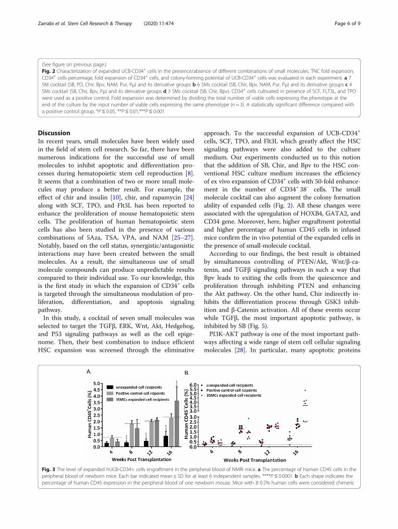
The ability of 3SM cocktail to enhance the short-termengraftment potential of ex vivo expanded CD34+ cells inthe in utero transplanted NMRI miceIn order to evaluate the in vivo functional capability ofthe expanded CD34+ cells, we used in utero transplant-ation model [23]. We transplanted 30–50 × 103 freshlyisolated hUCB-CD34+ cells or the cells harvested fromthe cultures with the same number of input hUCB-CD34+ cells in the presence or absence of SM cocktailinto NMRI mouse embryos, E11.5-E13.5. Two weeksafter birth, born mice were treated with humanhematopoietic growth factors SCF (4 ng/g), IL-3 (4 ng/g),
and G-CSF (50 ng/g) for 1 week. As shown in Fig. 3, bytreatment with the human hematopoietic factor, thehCD45+ chimerism was distinctly increased comparedwith initial values, 4 and 8 weeks post transplantation.Sixteen weeks after transplantation, the average humancell engraftment in the peripheral blood of the micetransplanted with freshly isolated hUCB CD34+ cells wasabout 1%, while the percentage of CD45+ cells in 3SMsand positive control transplanted mice was 9 times and3.4 times (3.6 ± 1 and 3.2 ± 0.3) respectively, comparedto the unexpanded cell recipients (Fig. 3). In the otherwords, the ex vivo expansion of hUCB CD34+ cells withSM cocktail resulted in 1.5 fold increase in human cellengraftment compared to the positive control.
Ability of the optimal SM cocktail to modulate the cellsignaling pathwaysSubsequently, RT-qPCR was performed in order todetermine the expression of typical genes involved inHSC stemness. The result shows that the relative expres-sion of the two major genes involved in the proliferationand self-renewal of HSCs, including HOXB4 and GATA2as well as the HSC-specific marker, CD34, have signifi-cantly increased in the presence of 3SM cocktail afternormalization to the level of the PC group. Furthermore,the expression of the CXCR4 gene involved in the migrationand transplantation of HSCs has increased dramatically inthe presence of 3SM cocktail. The expression of other genesassociated with self-renewal, such as ABCG2, Notch, andBmi1, does not show a significant difference between thegroups (Fig. 4).
Fig. 1 Dose finding and optimization for small molecules (SB, Bpv, NAM, Pur, PD, Chir, Pμ). In each graph, the middle column corresponds to thereference concentration of the small molecules based on the literatures. Cell viability was measured by MTS assay post 48 h incubation with smallmolecules. The negative control in each group was used for the normalization of data. Bars indicated as mean ± SD at least five independentreplicates. *P ≤ 0.05, **P≤ 0.01, ***P≤ 0.001, ****P ≤ 0.0001
Zarrabi et al. Stem Cell Research & Therapy (2020) 11:474 Page 4 of 9

Fig. 2 (See legend on next page.)
Zarrabi et al. Stem Cell Research & Therapy (2020) 11:474 Page 5 of 9
DiscussionIn recent years, small molecules have been widely usedin the field of stem cell research. So far, there have beennumerous indications for the successful use of smallmolecules to inhibit apoptotic and differentiation pro-cesses during hematopoietic stem cell reproduction [8].It seems that a combination of two or more small mole-cules may produce a better result. For example, theeffect of chir and insulin [10], chir, and rapamycin [24]along with SCF, TPO, and Flt3L has been reported toenhance the proliferation of mouse hematopoietic stemcells. The proliferation of human hematopoietic stemcells has also been studied in the presence of variouscombinations of 5Aza, TSA, VPA, and NAM [25–27].Notably, based on the cell status, synergistic/antagonisticinteractions may have been created between the smallmolecules. As a result, the simultaneous use of smallmolecule compounds can produce unpredictable resultscompared to their individual use. To our knowledge, thisis the first study in which the expansion of CD34+ cellsis targeted through the simultaneous modulation of pro-liferation, differentiation, and apoptosis signalingpathway.In this study, a cocktail of seven small molecules was
selected to target the TGFβ, ERK, Wnt, Akt, Hedgehog,and P53 signaling pathways as well as the cell epige-nome. Then, their best combination to induce efficientHSC expansion was screened through the eliminative
approach. To the successful expansion of UCB-CD34+
cells, SCF, TPO, and Flt3L which greatly affect the HSCsignaling pathways were also added to the culturemedium. Our experiments conducted us to this notionthat the addition of SB, Chir, and Bpv to the HSC con-ventional HSC culture medium increases the efficiencyof ex vivo expansion of CD34+ cells with 50-fold enhance-ment in the number of CD34+ 38− cells. The smallmolecule cocktail can also augment the colony formationability of expanded cells (Fig. 2). All these changes wereassociated with the upregulation of HOXB4, GATA2, andCD34 gene. Moreover, here, higher engraftment potentialand higher percentage of human CD45 cells in infusedmice confirm the in vivo potential of the expanded cells inthe presence of small-molecule cocktail.According to our findings, the best result is obtained
by simultaneous controlling of PTEN/Akt, Wnt/β-ca-tenin, and TGFβ signaling pathways in such a way thatBpv leads to exiting the cells from the quiescence andproliferation through inhibiting PTEN and enhancingthe Akt pathway. On the other hand, Chir indirectly in-hibits the differentiation process through GSK3 inhib-ition and β-Catenin activation. All of these events occurwhile TGFβ, the most important apoptotic pathway, isinhibited by SB (Fig. 5).PI3K-AKT pathway is one of the most important path-
ways affecting a wide range of stem cell cellular signalingmolecules [28]. In particular, many apoptotic proteins
(See figure on previous page.)Fig. 2 Characterization of expanded UCB-CD34+ cells in the presence/absence of different combinations of small molecules. TNC fold expansion,CD34+ cells percentage, fold expansion of CD34+ cells, and colony-forming potential of UCB-CD34+ cells was evaluated in each experiment. a 7SM cocktail (SB, PD, Chir, Bpv, NAM, Pur, Pμ) and its derivative groups b 6 SMs cocktail (SB, Chir, Bpv, NAM, Pur, Pμ) and its derivative groups c 4SMs cocktail (SB, Chir, Bpv, Pμ) and its derivative groups d 3 SMs cocktail (SB, Chir, Bpv). CD34+ cells cultivated in presence of SCF, FLT3L, and TPOwere used as a positive control. Fold expansion was determined by dividing the total number of viable cells expressing the phenotype at theend of the culture by the input number of viable cells expressing the same phenotype (n = 3). A statistically significant difference compared witha positive control group, *P≤ 0.05, **P ≤ 0.01,***P ≤ 0.001
Fig. 3 The level of expanded hUCB-CD34+ cells engraftment in the peripheral blood of NMRI mice. a The percentage of human CD45 cells in theperipheral blood of newborn mice. Each bar indicated mean ± SD for at least 6 independent samples. ****P ≤ 0.0001. b Each shape indicates thepercentage of human CD45 expression in the peripheral blood of one newborn mouse. Mice with ≥ 0·2% human cells were considered chimeric
Zarrabi et al. Stem Cell Research & Therapy (2020) 11:474 Page 6 of 9

such as Bim and Bcl-2 can be inactivated by the path-way. AKT, also, inhibits certain cell cycle inhibitors suchas P21 and P27 and activates Cyclin D, which in turnleads to exit from G0 and entry into the cell cycle [29].Furthermore, Akt facilitates the migration of HSCs andtheir binding to the bone marrow stromal cells throughinduction of integrin expression [30, 31]. PTEN is atumor suppressor protein that inhibits the PI3K-AKTpathway. Actually, inhibition of PTEN leads to increasedsurvival, proliferation, self-renewal, and incomplete
differentiation potential of embryonic stem cells [32]and also in vitro proliferation of HSCs [10].Wnt pathway not only plays a critical role in the devel-
opment of embryonic stem cells [33], but also in the pro-liferation and differentiation of adult stem cells includingHSCs [34, 35]. The major effects of Wnt are appliedthrough β-catenin which can increase the self-renewal andproliferation of HSCs, even independently of the Wntpathway [36, 37]. According to previous studies, the accu-mulation of the β-catenin, following GSK3 inactivation,
Fig. 4 Treatment by SB, Chir, and Bpv modifies the gene expression of UCB-CD34+ cells. Bars represent the mean fold-changes of geneexpression in the 3 SM-expanded cells relative to the positive control group detected by quantitative real-time PCR (n = 3), *P≤ 0.05, **P ≤ 0.01,***P ≤ 0.001 vs. positive control
Fig. 5 The molecular mechanisms which through them SB, Chir, and Bpv modulate proliferation, differentiation, and survival of hematopoietic stem cells
Zarrabi et al. Stem Cell Research & Therapy (2020) 11:474 Page 7 of 9
facilitates the maintenance of the pluripotency state ofembryonic and adult stem cells [38, 39].TGFβ is one of the major negative regulators of HSC
proliferation [40]. The pathway, specifically, inhibits cellcycle progression through the induction of P57 expres-sion, which in turn leads to CyclinD-Cdk4/6 andCyclinE-Cdk2 inactivation. P38MAPK is also a down-stream molecule of the TGFβ pathway which its inhib-ition results in decreased in vitro apoptosis and aging ofHSCs [40]. JNK is another downstream target of TGFβwhich activates some apoptotic factors such as Bcl2 andBad. Therefore, inhibition of the TGFβ pathway not onlyleads to P57, P38MAPK, and JNK inhibition which isassociated with cell cycle promotion, but also inhibitsthe apoptotic pathways [41–43].Altogether, a cocktail of SB431542, Chir99021, and
Bpv, which respectively inhibits the TGFβ differentiationpathway and activates the Wnt and Akt pathways, canbe used to improve the conventional protocol of HSCexpansion.
Supplementary informationSupplementary information accompanies this paper at https://doi.org/10.1186/s13287-020-01945-8.
Additional file 1: Figure S1. Schematic illustration of procedure to findthe best combination of small molecules to expand UCB-HSCs. Table S1.Initial concentration of small molecules based on previous studies andtheir proper concentration based on MTS assay. Table S2. List of primersequences used in the present study.
AbbreviationsUCB: Umbilical cord blood; HSCs: Hematopoietic stem cells; SMs: Smallmolecules; SB: SB431542; Pur: Purmorphamin; PD: PD0325901; Chit: Chir9901;Bpv: Bisperoxovanadium; Pμ: Pifithrin-μ; NAM: Nicotinamide; CFU: Colony-forming units; BFU-E: Burst-forming unit-erythroid; CFU-GM: CFU granulocyte-macrophage; CFU-GEMM: CFU granulocyte-erythrocyte-macrophage-megakaryocyte
AcknowledgementsThe authors would like to thank Zahra Pour-Safavi for her technical support inflowcytometery experiment and Azam Dalman for preparing the handmadeglass pipettes. The authors declare no conflict of interest in this study.
Authors’ contributionsM.Z. and E.A. performed all in vitro experiments, analyzed the data, andwrote the manuscript. M.H.A. performed in vivo experiment and analyzedthe in vivo data. M.E. contributed to the concept and design, financialsupport, and final approval of the manuscript. The authors read andapproved the final manuscript.
FundingThis study has been funded by the Royan Institute (code: 91000597) andgranted partly by the Royan Stem Cell Technology Company, Iran.
Availability of data and materialsAll data generated or analyzed during this study are included in thispublished article and in supplementary figures.
Ethics approval and consent to participateAll the experiments in this study were reviewed and approved by theResearch Ethics Committee of the Royan Institute and were conducted in
accordance with the ethical principles and the national norms and standardsfor conducting the Medical Research in Iran (IR.ACECR.ROYAN.REC.1398.189).
Consent for publicationNot applicable.
Competing interestsThe authors declare that they have no competing interests.
Author details1Department of Stem Cells and Developmental Biology, Cell ScienceResearch Center, Royan Institute for Stem Cell Biology and Technology, ACECR, P.O. Box, Tehran 19395-4644, Iran. 2Royan Stem Cell Technology Company,Cord Blood Bank, Tehran, Iran. 3Animal Core Facility, ReproductiveBiomedicine Research Center, Royan Institute for Animal Biotechnology,ACECR, Tehran, Iran.
Received: 27 June 2020 Accepted: 20 September 2020
References1. Roura S, Pujal JM, Galvez-Monton C, Bayes-Genis A. The role and potential
of umbilical cord blood in an era of new therapies: a review. Stem Cell ResTher. 2015;6:123.
2. Ilic D, Miere C, Lazic E. Umbilical cord blood stem cells: clinical trials in non-hematological disorders. Br Med Bull. 2012;102:43–57.
3. Cany J, Dolstra H, Shah N. Umbilical cord blood-derived cellular products forcancer immunotherapy. Cytotherapy. 2015;17(6):739–48.
4. Balassa K, Rocha V. Anticancer cellular immunotherapies derived fromumbilical cord blood. Expert Opin Biol Ther. 2018;18(2):121–34.
5. Delaney C, Ratajczak MZ, Laughlin MJ. Strategies to enhance umbilical cordblood stem cell engraftment in adult patients. Expert Rev Hematol. 2010;3(3):273–83.
6. Derakhshani M, Abbaszadeh H, Movassaghpour AA, Mehdizadeh A,Ebrahimi-Warkiani M, Yousefi M. Strategies for elevating hematopoietic stemcells expansion and engraftment capacity. Life Sci. 2019;232:116598.
7. Oh IH, Lau A, Eaves CJ. During ontogeny primitive (CD34(+)CD38(−))hematopoietic cells show altered expression of a subset of genesassociated with early cytokine and differentiation responses of their adultcounterparts. Blood. 2000;96(13):4160–8.
8. Zarrabi M, Afzal E, Ebrahimi M. Manipulation of hematopoietic stem cell fateby small molecule compounds. Stem Cells Dev. 2018;27(17):1175–90.
9. Gao Y, Yang P, Shen H, Yu H, Song X, Zhang L, et al. Small-moleculeinhibitors targeting INK4 protein p18(INK4C) enhance ex vivo expansion ofhaematopoietic stem cells. Nat Commun. 2015;6:6328.
10. Perry JM, He XC, Sugimura R, Grindley JC, Haug JS, Ding S, et al.Cooperation between both Wnt/{beta}-catenin and PTEN/PI3K/Akt signalingpromotes primitive hematopoietic stem cell self-renewal and expansion.Genes Dev. 2011;25(18):1928–42.
11. Sangeetha VM, Kale VP, Limaye LS. Expansion of cord blood CD34 cells inpresence of zVADfmk and zLLYfmk improved their in vitro functionality andin vivo engraftment in NOD/SCID mouse. PLoS One. 2010;5(8):e12221.
12. Zou J, Zou P, Wang J, Li L, Wang Y, Zhou D, et al. Inhibition of p38 MAPKactivity promotes ex vivo expansion of human cord blood hematopoieticstem cells. Ann Hematol. 2012;91(6):813–23.
13. Yamazaki S, Iwama A, Takayanagi S, Eto K, Ema H, Nakauchi H. TGF-beta as acandidate bone marrow niche signal to induce hematopoietic stem cellhibernation. Blood. 2009;113(6):1250–6.
14. Guo J, Wang S-Y, Zhu X-F, Li S-T, Lin F-L, Li X-M, et al. Effect of Shh and BM-MSC synergism on the proliferation of hematopoietic stem cells. ZhongguoShi Yan Xue Ye Xue Za Zhi. 2018;26(5):1523–30.
15. Kabiri Z, Numata A, Kawasaki A, Edison TDG, Virshup DM. Wnts aredispensable for differentiation and self-renewal of adult murinehematopoietic stem cells. Blood. 2015;126(9):1086–94.
16. Zarrabi M, Afzal E, Asghari MH, Mohammad M, Es HA, Ebrahimi M. Inhibitionof MEK/ERK signalling pathway promotes erythroid differentiation andreduces HSCs engraftment in ex vivo expanded haematopoietic stem cells.J Cell Mol Med. 2018;22(3):1464–74.
17. Ruiz-Herguido C, Guiu J, D'Altri T, Inglés-Esteve J, Dzierzak E, Espinosa L,et al. Hematopoietic stem cell development requires transient Wnt/β-catenin activity. J Exp Med. 2012;209(8):1457–68.
Zarrabi et al. Stem Cell Research & Therapy (2020) 11:474 Page 8 of 9

18. Miranda MB, McGuire TF, Johnson DE. Importance of MEK-1/-2 signaling inmonocytic and granulocytic differentiation of myeloid cell lines. Leukemia.2002;16(4):683–92.
19. Ghosh J, Kapur R. Regulation of hematopoietic stem cell self-renewal andleukemia maintenance by the PI3K-mTORC1 pathway. Curr Stem Cell Rep.2016;2(4):368–78.
20. Liu Y, Elf SE, Miyata Y, Sashida G, Liu Y, Huang G, et al. p53 regulateshematopoietic stem cell quiescence. Cell Stem Cell. 2009;4(1):37–48.
21. Denu JM. Vitamin B3 and sirtuin function. Trends Biochem Sci. 2005;30(9):479–83.
22. Suzuki T, Yokoyama Y, Kumano K, Takanashi M, Kozuma S, Takato T, et al.Highly efficient ex vivo expansion of human hematopoietic stem cells usingDelta1-Fc chimeric protein. Stem cells (Dayton, Ohio). 2006;24(11):2456–65.
23. Zarrabi M, Afzal E, Asghari MH, Ebrahimi M. Assessment of short-termengraftment potential of ex vivo expanded hematopoietic stem cellsusing normal fetal mouse in utero transplantation model. Cell J. 2019;21(3):259–67.
24. Huang J, Nguyen-McCarty M, Hexner EO, Danet-Desnoyers G, Klein PS.Maintenance of hematopoietic stem cells through regulation of Wnt andmTOR pathways. Nat Med. 2012;18(12):1778–85.
25. Mahmud N, Petro B, Baluchamy S, Li X, Taioli S, Lavelle D, et al. Differentialeffects of epigenetic modifiers on the expansion and maintenance ofhuman cord blood stem/progenitor cells. Biol Blood Marrow Transplant.2014;20(4):480–9.
26. Araki H, Mahmud N, Milhem M, Nunez R, Xu M, Beam CA, et al. Expansionof human umbilical cord blood SCID-repopulating cells using chromatin-modifying agents. Exp Hematol. 2006;34(2):140–9.
27. Araki H, Yoshinaga K, Boccuni P, Zhao Y, Hoffman R, Mahmud N. Chromatin-modifying agents permit human hematopoietic stem cells to undergomultiple cell divisions while retaining their repopulating potential. Blood.2007;109(8):3570–8.
28. Yu JS, Cui W. Proliferation, survival and metabolism: the role of PI3K/AKT/mTOR signalling in pluripotency and cell fate determination. Development(Cambridge, England). 2016;143(17):3050–60.
29. Chang F, Lee JT, Navolanic PM, Steelman LS, Shelton JG, Blalock WL, et al.Involvement of PI3K/Akt pathway in cell cycle progression, apoptosis, andneoplastic transformation: a target for cancer chemotherapy. Leukemia.2003;17(3):590–603.
30. Los M, Maddika S, Erb B, Schulze-Osthoff K. Switching Akt: from survivalsignaling to deadly response. BioEssays. 2009;31(5):492–5.
31. Buitenhuis M. The role of PI3K/protein kinase B (PKB/c-akt) in migration andhoming of hematopoietic stem and progenitor cells. Curr Opin Hematol.2011;18(4):226–30.
32. Jung CJ, Iyengar S, Blahnik KR, Jiang JX, Tahimic C, Torok NJ, et al. HumanESC self-renewal promoting microRNAs induce epithelial-mesenchymaltransition in hepatocytes by controlling the PTEN and TGFbeta tumorsuppressor signaling pathways. Mol Cancer Res. 2012;10(7):979–91.
33. Merrill BJ. Wnt pathway regulation of embryonic stem cell self-renewal.Cold Spring Harb Perspect Biol. 2012;4(9):a007971.
34. Richter J, Traver D, Willert K. The role of Wnt signaling in hematopoieticstem cell development. Crit Rev Biochem Mol Biol. 2017;52(4):414–24.
35. Cain CJ, Manilay JO. Hematopoietic stem cell fate decisions are regulated byWnt antagonists: comparisons and current controversies. Exp Hematol.2013;41(1):3–16.
36. Baba Y, Yokota T, Spits H, Garrett KP, Hayashi S, Kincade PW.Constitutively active beta-catenin promotes expansion of multipotenthematopoietic progenitors in culture. J Immunol (Baltimore, Md : 1950).2006;177(4):2294–303.
37. Nemeth MJ, Mak KK, Yang Y, Bodine DM. beta-Catenin expression in thebone marrow microenvironment is required for long-term maintenance ofprimitive hematopoietic cells. Stem cells (Dayton, Ohio). 2009;27(5):1109–19.
38. Yu SJ, Kim HJ, Lee ES, Park CG, Cho SJ, Jeon SH. Beta-catenin accumulationis associated with increased expression of Nanog protein and predictsmaintenance of MSC self-renewal. Cell Transplant. 2017;26(2):365–77.
39. Sanchez-Ripoll Y, Bone HK, Owen T, Guedes AM, Abranches E,Kumpfmueller B, et al. Glycogen synthase kinase-3 inhibition enhancestranslation of pluripotency-associated transcription factors to contribute tomaintenance of mouse embryonic stem cell self-renewal. PLoS One. 2013;8(4):e60148.
40. Blank U, Karlsson S. TGF-beta signaling in the control of hematopoietic stemcells. Blood. 2015;125(23):3542–50.
41. Wang C, Tang X, Sun X, Miao Z, Lv Y, Yang Y, et al. TGFbeta inhibitionenhances the generation of hematopoietic progenitors from human ES cell-derived hemogenic endothelial cells using a stepwise strategy. Cell Res.2012;22(1):194–207.
42. Zhang Y, Alexander PB, Wang XF. TGF-beta family signaling in the controlof cell proliferation and survival. Cold Spring Harb Perspect Biol. 2017;9(4):a022145.
43. Hinge A, Filippi MD. Deconstructing the complexity of TGFbeta signaling inhematopoietic stem cells: quiescence and beyond. Curr Stem Cell Rep.2016;2(4):388–97.
Publisher’s NoteSpringer Nature remains neutral with regard to jurisdictional claims inpublished maps and institutional affiliations.
Zarrabi et al. Stem Cell Research & Therapy (2020) 11:474 Page 9 of 9