combined rotation scarf and akin osteotomies for hallux valgus: a
TRANSCRIPT
RESEARCH Open Access
Combined rotation scarf and Akin osteotomiesfor hallux valgus: a patient focussed 9 year followup of 50 patientsTimothy E Kilmartin1,2,3*, Claire O’Kane1
Abstract
Background: The Cochrane review of hallux valgus surgery has disputed the scientific validity of hallux valgusresearch. Scoring systems and surrogate measures such as x-ray angles are commonly reported at just one yearpost operatively but these are of dubious relevance to the patient. In this study we extended the follow up to aminimum of 8 years and sought to address patient specific concerns with hallux valgus surgery. The long termfollow up also allowed a comprehensive review of the complications associated with the combined rotation scarfand Akin osteotomies.
Methods: Between 1996 and 1999, 101 patients underwent rotation scarf and Akin osteotomies for the treatmentof hallux valgus. All patients were contacted and asked to participate in this study. 50 female participants wereavailable allowing review of 73 procedures. The average follow up was over 9 years and the average age at thetime of surgery was 57. The participants were physically examined and interviewed.
Results: Post-operatively, in 86% of the participants there were no footwear restrictions. Stiffness of the firstmetatarsophalangeal joint was reported in 8% (6 feet); 10% were unhappy with the cosmetic appearance of theirfeet, 3 feet had hallux varus, and 2 feet had recurrent hallux valgus. There were no foot-related activity restrictionsin 92% of the group. Metatarsalgia occurred in 4% (3 feet). 96% were better than before surgery and 88% werecompletely satisfied with their post-operative result. Hallux varus was the greatest single cause of dissatisfaction.The most common adverse event in the study was internal fixation irritation. Hallux valgus surgery is not withoutrisk and these findings could be useful in the informed consent process.
Conclusions: When combined the rotation scarf and Akin osteotomies are an effective treatment for hallux valgusthat achieves good long-term correction with a low incidence of recurrence, footwear restriction or metatarsalgia.The nature of the osteotomies allows early return to normal shoes and activity without the need for postoperativeimmobilisation in a plaster cast.
IntroductionThe Cochrane review of hallux valgus surgery has dis-puted the scientific validity of hallux valgus research [1].The review reported that although many studies wereavailable on the surgical management of the condition,final outcome measures were most frequently measuredat one year with just a few trials maintaining follow upfor 3 years. Scoring systems and surrogate measuressuch as x-ray measurements were commonly used but
these were considered of dubious relevance to thepatient if they did not address their main concerns. Inall the literature considered by the Cochrane review, justone study asked the patients if they were better thanbefore surgery [2]. The review recommended that futureresearch should include patient focussed outcomes andfollow up periods of at least 5 to 10 years.In reviewing hallux valgus surgical outcomes it is
notable that a high proportion of patients, 25-33%,remain dissatisfied at final follow up [1]. Schneider andKnahr reviewed the expectations of both patients andsurgeons in hallux valgus surgery [3]. Two hundredpatients were interviewed and their principal concern
* Correspondence: [email protected] Private Clinic, Hillsborough, Co Down, Northern Ireland BT266AE, UK
Kilmartin and O’Kane Journal of Foot and Ankle Research 2010, 3:2http://www.jfootankleres.com/content/3/1/2
JOURNAL OF FOOTAND ANKLE RESEARCH
© 2010 Kilmartin and O’Kane; licensee BioMed Central Ltd. This is an Open Access article distributed under the terms of the CreativeCommons Attribution License (http://creativecommons.org/licenses/by/2.0), which permits unrestricted use, distribution, andreproduction in any medium, provided the original work is properly cited.
was relief of foot pain when wearing a conventionalshoe. Importantly, the patients hoped that surgerywould restore unlimited pain free walking, whereasalignment and cosmesis of the hallux was considered oflittle importance by either surgeons or patients. Whenthe surgeons were interviewed (186 surgeons of the Ger-man Austrian Orthopaedic Foot Surgery Society), theirprimary concern was also pain and shoe fitting issuesbut in addition restoring adequate range of motion tothe first MTP joint and relieving metatarsalgia.Common complications specific to hallux valgus sur-
gery include recurrence of deformity, first metatarsopha-langeal (MTP) joint stiffness and transfer metatarsalgia[4]. With the exception of recurrence, it is unlikely thatany of these known postoperative complications will beof automatic concern to the patient prior to surgery.Their occurrence could, however, explain the high levelsof postoperative dissatisfaction even when hallux valgusangles and first MTP joint pain have improved with sur-gery [1]. While many previous studies have focussed onx-ray outcomes, the prevalence of these specific compli-cations provides a more patient focussed measure of theoutcome of a particular procedure and will help sur-geons prepare the patients for informed consent.The scarf osteotomy was first developed in 1926 by
Meyer but never achieved widespread use due to inade-quate fixation techniques [5]. Weil popularised the tech-nique after describing an effective fixation techniqueusing two AO screws [6,7]. The advantages of the tech-nique included: rigid compression of large areas of boneto bone contact providing a good environment for pri-mary bone healing and early return to normal weightbearing activities and range of motion exercises prevent-ing joint stiffness and oedema [8]. The scarf osteotomyalso avoided the complication of metatarsus elevatusassociated with more proximal metatarsal osteotomies[9], allowed accurate correction of the intermetatarsalangle and could be modified to allow the metatarsal tobe shortened or lengthened, and plantarly or dorsallydisplaced if required [10].The scarf osteotomy has been extensively reviewed in
recent literature [10-19]. To date the scarf has generallybeen used to correct moderate hallux valgus in the pre-sence of intermetatarsal angles of less than 15 degrees,the limiting factor being that if the inferior fragment istransposed too far laterally, fixation cannot be obtainedand there will be insufficient bone to bone contact toproduce stable union of the osteotomy. Thus the scarfosteotomy may not be indicated in the treatment ofsevere hallux valgus with high intermetatarsal angles.This is frustrating for the foot surgeon as all the advan-tages of the scarf osteotomy cannot be applied topatients with more severe deformity. In view of the lim-itations of the scarf osteotomy, Duke modified the
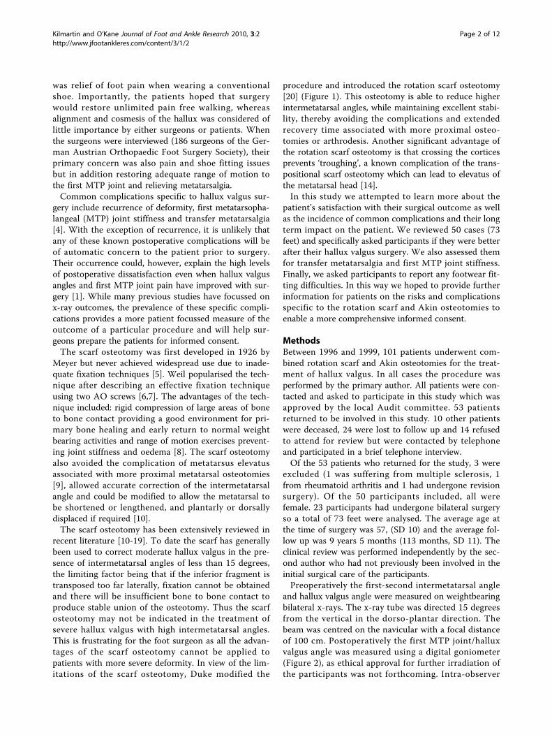
procedure and introduced the rotation scarf osteotomy[20] (Figure 1). This osteotomy is able to reduce higherintermetatarsal angles, while maintaining excellent stabi-lity, thereby avoiding the complications and extendedrecovery time associated with more proximal osteo-tomies or arthrodesis. Another significant advantage ofthe rotation scarf osteotomy is that crossing the corticesprevents ‘troughing’, a known complication of the trans-positional scarf osteotomy which can lead to elevatus ofthe metatarsal head [14].In this study we attempted to learn more about the
patient’s satisfaction with their surgical outcome as wellas the incidence of common complications and their longterm impact on the patient. We reviewed 50 cases (73feet) and specifically asked participants if they were betterafter their hallux valgus surgery. We also assessed themfor transfer metatarsalgia and first MTP joint stiffness.Finally, we asked participants to report any footwear fit-ting difficulties. In this way we hoped to provide furtherinformation for patients on the risks and complicationsspecific to the rotation scarf and Akin osteotomies toenable a more comprehensive informed consent.
MethodsBetween 1996 and 1999, 101 patients underwent com-bined rotation scarf and Akin osteotomies for the treat-ment of hallux valgus. In all cases the procedure wasperformed by the primary author. All patients were con-tacted and asked to participate in this study which wasapproved by the local Audit committee. 53 patientsreturned to be involved in this study. 10 other patientswere deceased, 24 were lost to follow up and 14 refusedto attend for review but were contacted by telephoneand participated in a brief telephone interview.Of the 53 patients who returned for the study, 3 were
excluded (1 was suffering from multiple sclerosis, 1from rheumatoid arthritis and 1 had undergone revisionsurgery). Of the 50 participants included, all werefemale. 23 participants had undergone bilateral surgeryso a total of 73 feet were analysed. The average age atthe time of surgery was 57, (SD 10) and the average fol-low up was 9 years 5 months (113 months, SD 11). Theclinical review was performed independently by the sec-ond author who had not previously been involved in theinitial surgical care of the participants.Preoperatively the first-second intermetatarsal angle
and hallux valgus angle were measured on weightbearingbilateral x-rays. The x-ray tube was directed 15 degreesfrom the vertical in the dorso-plantar direction. Thebeam was centred on the navicular with a focal distanceof 100 cm. Postoperatively the first MTP joint/halluxvalgus angle was measured using a digital goniometer(Figure 2), as ethical approval for further irradiation ofthe participants was not forthcoming. Intra-observer
Kilmartin and O’Kane Journal of Foot and Ankle Research 2010, 3:2http://www.jfootankleres.com/content/3/1/2
Page 2 of 12
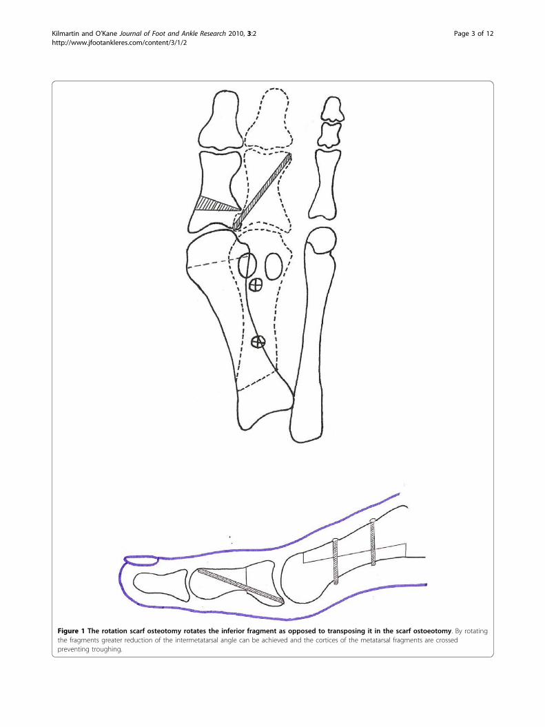
Figure 1 The rotation scarf osteotomy rotates the inferior fragment as opposed to transposing it in the scarf ostoeotomy. By rotatingthe fragments greater reduction of the intermetatarsal angle can be achieved and the cortices of the metatarsal fragments are crossedpreventing troughing.
Kilmartin and O’Kane Journal of Foot and Ankle Research 2010, 3:2http://www.jfootankleres.com/content/3/1/2
Page 3 of 12
repeatability of the goniometer had previously beenestablished [21]. A good correlation (r = 0.63) between x-ray measurement and goniometric measurement has pre-viously been found [22]. The range of dorsiflexion andplantarflexion of the MTP joint was also assessed usingthe digital goniometer (Figures 3 and 4).All the participants were then interviewed and asked if
they were completely satisfied, satisfied with reservationsor dissatisfied with the results of their surgery. Restric-tions with footwear, or any activity restrictions becauseof their feet were recorded. The participants were askedif there was any pain or stiffness in the first MTP joint.Any pain or tenderness of the lesser MTP joints was
also recorded. Finally, the participants were asked if theywere happy with the appearance of their post surgicalfoot and would they be happy to undergo surgery undersimilar circumstances in the future.A number of adjunctive procedures were performed.
In 22 feet a second toe proximal interphalangeal joint(PIPJ) arthroplasty was performed and in 9 feet PIPJarthroplasties of other toes were performed. 4 feetunderwent a Weil osteotomy of the second metatarsaland 3 feet had neuroma excision from the third inter-metatarsal space. With the exception of 1 participantwho underwent an adjunctive second joint fusion, allparticipants were encouraged to return to lace-up or
Figure 2 Goniometric measurement of the hallux valgus angle using a digital goniometer (available from Nova Instruments, MillHouse, Newgatestreet Road, Goffs Oak, Herts. EN7 5RX).
Kilmartin and O’Kane Journal of Foot and Ankle Research 2010, 3:2http://www.jfootankleres.com/content/3/1/2
Page 4 of 12

running shoes at 2 weeks postoperatively. Between 4and 6 weeks off work and sport was recommended.
Surgical techniqueThe procedure was performed in all cases under localanaesthetic ankle block on a day case basis. An ankletourniquet was applied and a medial plantar skin inci-sion running from the interphalangeal joint of the halluxto the base of the first metatarsal was made. This was
deepened to the capsule ensuring adequate haemostasis.The capsular incision was made as a double semi-ellipti-cal incision and the ellipse of tissue excised.A beaver blade was introduced into the joint capsule
between the metatarsal head and the sesamoid appara-tus and the adductor hallucis tendon and lateral sesa-moid ligament were released from their respectiveinsertions in the metatarsal head and proximal phalanx.The medial eminence of the first metatarsal was
Figure 3 With the resting non-weightbearing position being considered the zero degree angle, the passive hallux dorsiflexion rangeof motion was measured using the digital goniometer.
Figure 4 Passive hallux plantarflexion range of motion was measured from the resting position which was considered zero degrees.
Kilmartin and O’Kane Journal of Foot and Ankle Research 2010, 3:2http://www.jfootankleres.com/content/3/1/2
Page 5 of 12

resected at the sagittal groove. A guide wire was placedjust proximal to the metatarsal head articular surfaceand just inferior to the first metatarsal dorsal cortex.The guide wire was directed plantarly in the direction ofthe plantar surface of the third metatarsal head but per-pendicular to the long axis of the second metatarsal(Figure 5). An osteotomy guide was placed on the guidewire and a power saw was then used to make the hori-zontal cut along the metatarsal shaft extending fromjust proximal to the articular surface of the metatarsalhead to the basal tuberosity. The distal cut was madeparallel with the guide wire and the proximal cut atapproximately a 45° angle from medial proximal to lat-eral distal in order to allow the rotation to occur (Figure1). While it is possible to shorten the metatarsal byangling the distal cut in a proximal lateral direction, thiswas avoided as we consider any loss of first metatarsallength a predisposition to transfer metatarsalgia. Thelateral capsule was then released and the inferior frag-ment rotated toward the second metatarsal to reducethe intermetatarsal angle. The degree of rotationrequired was established pre operatively by measuringthe intermetatarsal angle on x-ray. We aimed to reducethe intermetatarsal angle to 7°. One mm of rotationequals 1° of correction which could be measured by theamount of overhanging bone of the superior fragmentonce the metatarsal head was rotated. The bone frag-ments were held with a scarf clamp and fixed with two2.0 cortical screws using AO technique (Figures 6 and7). The overhanging edges of bone were then removedfrom the medial side of the metatarsal shaft. An Akinclosing wedge osteotomy of the proximal phalanx wasperformed on all cases. The Akin osteotomy was fixatedusing a single 1.2 mm threaded k-wire (Figure 7).The capsule was then closed using 2-0 vicryl, figure of
8 sutures. The hallux was held in a plantarflexed posi-tion as the capsule was closed [10]. As an ellipse of cap-sule had previously been excised, closing the capsulepulled the sesamoids into a corrected position under thefirst metatarsal head. Tension on the capsular sutureswas increased to further draw the hallux into correctionif necessary, though we believe that soft tissue correc-tion is largely temporary and correction should beachieved almost exclusively with the osteotomies. Skinwas then closed using 5-0 vicryl subcuticular sutures.Postoperatively all but one patient who underwent a
simultaneous second metatarso-cuneiform joint fusionwore a surgical shoe and used crutches for two weeks.After two weeks, dressings were removed and the parti-cipants were encouraged to wear lace up or runningshoes and begin returning to normal activities. The par-ticipants were advised to perform range of motion exer-cises against the resistance of a powerband. In particularflexion exercises were encouraged to restore flexor
power to the hallux. (Figure 8). We also advised the par-ticipants to walk through the hallux on gait. These mea-sures, we believe, may contribute to reducing the risk oftransfer metatarsalgia.
ResultsPatient reported outcomesIn the 50 participants (73 feet) available for follow up88% of the group (44 participants), were completelysatisfied, 8% (4 participants) were satisfied with reserva-tions and 4% (2 participants) were dissatisfied (Table 1).96% (48 participants) were better than before surgeryand 4% (2 participants) were no better. All but one ofthe study group indicated that they would be happy toundergo surgery again under similar circumstances. 90%of cases (66 feet) were happy with the cosmetic appear-ance. 10% (7 feet) were unhappy with the cosmeticappearance, 3 had hallux varus and 2 had recurrent hal-lux valgus. 2 participants felt their feet were still toowide.There were no activity restrictions in 92% of the group
(46 participants). Walking distance was restricted to lessthan 3 miles in 2 participants. 1 participant felt shecould no longer do yoga because of first MTP joint stiff-ness and 1 participant had developed midfoot arthritiswhich was causing activity restriction due to pain. In94% of the group (69 feet), there was no metatarsalgia.Metatarsalgia occurred post operatively in 4% of thegroup (3 feet), all of these had hallux varus. 1 partici-pant had metatarsalgia prior to surgery and this was stillpresent postoperatively.
Footwear issuesIn 86% of the sample (63 feet) there were no footwearrestrictions. High heels could not be accommodated in14% (10 feet). This restriction was attributable to sur-gery in 7% of the sample (5 feet) where there was post-operative first MTP joint stiffness. In one other caseinternal fixation irritation was restricting the use ofcourt style shoes. Hallux varus, which had developedpostoperatively, was causing footwear problems to 1participant, and metatarsalgia, which had developedpostoperatively, was restricting the use of thin-soledfashion shoes in 1 case. Two participants had developedhammer toe deformities of the 2nd digit that restrictedshoes.
Joint alignment, range of motion and painPreoperatively the mean hallux valgus angle measuredon weight bearing bilateral x-rays was 37 degrees (SD7). The mean first-second intermetatarsal angle was 16degrees (SD 3). At final follow-up the goniometric mea-surement of the first MTP joint/hallux valgus angle was10 degrees (SD 6). The mean dorsiflexion at the first
Kilmartin and O’Kane Journal of Foot and Ankle Research 2010, 3:2http://www.jfootankleres.com/content/3/1/2
Page 6 of 12

MTP joint was 54 degrees (SD 14.6) and the mean plan-tarflexion 15 degrees (SD 8) - normal ranges arereported to be 65 to 90 degrees dorsiflexion and 15 to20 degrees plantarflexion [23-25].Hallux valgus recurrence with first MTP joint/hallux
valgus angles in excess of 15 degrees was noted in 8% ofthe sample (6 feet). In 2 participants the hallux valgusangle was 22 degrees and in 4 participants it was 20
degrees. Hallux varus occurred in 4% (3 feet). Postopera-tive soft tissue infection managed with oral antibioticsoccurred in 4% of the sample (3 feet). 1 participantrequired revision surgery for hallux varus and 25% ofthe sample (18 feet) required removal of the distal meta-tarsal screw.No stiffness of the first MTP joint was reported by 92
percent of the sample (67 feet). First MTP joint stiffness
Figure 5 Placement of the guide wire to achieve plantar displacement of the metatarsal head with the osteotomy.
Figure 6 Rotation scarf and Akin osteotomies pre-operative x-ray.
Kilmartin and O’Kane Journal of Foot and Ankle Research 2010, 3:2http://www.jfootankleres.com/content/3/1/2
Page 7 of 12
occurred in 8% (6 feet) and in 5 feet this caused foot-wear restrictions. In this subset, the mean dorsiflexionwas 46 degrees (SD 19, range 22 to 74 degrees) and themean plantarflexion was 10 degrees (SD 1.6, range 0 to10 degrees). In 94% of the group (69 feet), there was nofirst MTP joint pain. First MTP joint pain was presentin 3% of the group (two feet) and in both cases therewas hallux varus. In 2 other feet there was occasionaljoint pain.None of the 14 participants contacted by telephone
had required revision surgery at other facilities. All werehappy with the outcome of their surgery. No furtherinformation was gathered from these telephoneinterviews.
DiscussionIn the original cohort of 101 patients undergoing thecombined rotation scarf and Akin osteotomies 98% werefemale. All 50 participants that returned for assessmentrelated to this study were female. The higher incidenceof symptomatic hallux valgus in females is well docu-mented [26,27], but there is far less consideration ofwhat drives female patients to undergo surgery andwhat their expectations of surgery are [3]. Hallux valgusis often caused by shoe fitting issues wherein many ofthe symptoms are caused by footwear irritation and theexpectations of surgery are a return to a wide range of
shoe styles which previously have been difficult [3]. Inthis context, hallux valgus surgery could be seen as ahigh risk intervention because although it may alloweasier footwear accommodation, it carries the possibilityof rendering the foot painful due to the specific compli-cations of first MTP joint pain and stiffness and transfermetatarsalgia. Recurrence of hallux valgus is also a dis-appointing outcome for many patients [28], becauseonce again it recreates the shoe fitting problems.Foot surgeons may find it difficult to accept the possi-
bility that they could be performing hallux valgus cor-rection for cosmetic reasons but female interest infashionable, high-heeled footwear is high. In this seriesof participants we believe we only performed surgerywhen conservative measures failed to alleviate symptomsor when participants could not accommodate their footin conventional shoes, or when the hallux was so mala-ligned that it was beginning to underide the second toeand deform previously normal structures within thefoot. On the basis of Schneider and Khnar’s study [3],we recognise the importance of footwear postoperativelyand fixed on this as a patient focussed outcome.At an average of 9.5 years after their operation, 86% of
the sample were unrestricted in their footwear choice inthat they could wear high heels. Patients that can wearhigh heeled shoes comfortably are unlikely to be suffer-ing from painful first MTP joint stiffness or from
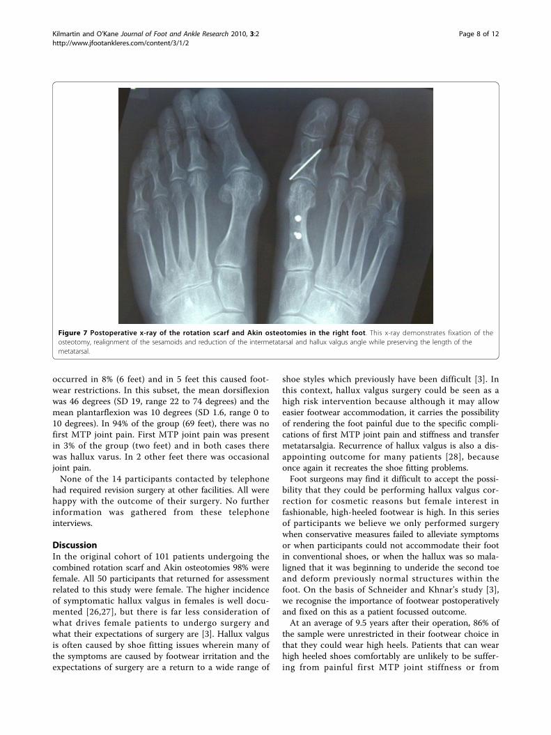
Figure 7 Postoperative x-ray of the rotation scarf and Akin osteotomies in the right foot. This x-ray demonstrates fixation of theosteotomy, realignment of the sesamoids and reduction of the intermetatarsal and hallux valgus angle while preserving the length of themetatarsal.
Kilmartin and O’Kane Journal of Foot and Ankle Research 2010, 3:2http://www.jfootankleres.com/content/3/1/2
Page 8 of 12

transfer metatarsalgia. In this way the ability to wear arange of shoes is also an indication of foot function. Inthis sample just 4% were found to be suffering fromtransfer metatarsalgia, but 8% were aware of first MTPjoint stiffness and in 6% there was joint pain.The management of transfer metatarsalgia and first
MTP joint stiffness following hallux valgus correctionhas received little attention in the literature and is cer-tainly an area with much potential for further investi-gation. We sought to prevent both problems by askingpatients to mobilise and strengthen the first MTP jointimmediately postoperatively with simple flexions of thefirst MTP joint. At two weeks postoperatively weasked patients to use a powerband (rubber band exer-ciser) to perform plantarflexion and dorsiflexion exer-cise of the first MTP joint against the resistance of thepowerband. We also advised patients to propel throughthe first MTP joint and hallux on gait so as to avoidguarding the first MTP joint. If the hallux cannot beplantarflexed, propulsion power from the hallux isreduced and we believe the patient is more likely topropel from the lesser MTP joints, which then becomebruised, inflamed and painful. Intraoperatively wealways attempted to maintain the length of the firstmetatarsal and displace the metatarsal head in a plan-tarly direction as part of the rotation scarf osteotomy.This again, we believe, may minimise the possibility oftransfer metatarsalgia.
Recurrence of hallux valgus occurred in 8% of the par-ticipants in this study. This is a disappointing outcomeas it means the patient is once more at risk of develop-ing the whole range of symptoms associated with halluxvalgus. However, cases of recurrent hallux valgus wereconsidered mild as a maximum hallux valgus angle of22 degrees was observed. This is close to the normalreported range of 15 degrees or less [29].Hallux varus developed in just 3 feet but at interview
these participants appeared more unhappy with theiroutcome than any other participant in the study. Weconsider hallux varus a significant though rare complica-tion leading to progressive joint degeneration and pain,metatarsalgia and footwear fitting problems. Its real sig-nificance lies in the degree of dissatisfaction it createswith patients often presenting with multiple symptoms.Hallux varus occurs when the tibial sesamoid is posi-tioned medial to the first metatarsal head [30-32]. In therotation scarf and Akin osteotomies hallux varus may bea consequence of excessive reduction of the intermeta-tarsal angle by the metatarsal osteotomy. Alternatively,excessive mobilisation of the sesamoids followingdetachment of the fibular sesamoid suspensory ligament,especially when combined with release of the adductorhallucis tendon, will risk hallux varus. Over tighteningthe medial capsule during deep closure will compoundthis effect by pulling the tibial sesamoid medial to themetatarsal groove. An excessively aggressive Akin
Figure 8 Post operative flexion exercises using a powerband. The patient is asked to repeatedly plantarflex the hallux while increasing theresistance of the powerband.
Kilmartin and O’Kane Journal of Foot and Ankle Research 2010, 3:2http://www.jfootankleres.com/content/3/1/2
Page 9 of 12
osteotomy will also pull the hallux into varus. Of allthese potential causes of hallux varus, the Akin osteot-omy is the easiest to assess intraoperatively and certainlyif the hallux appeared in varus after performing theAkin osteotomy, the wedge of bone would be re-insertedand the osteotomy fixed. The position of the tibial sesa-moid was also assessed intraoperatively and if it was notsitting directly inferior to the medial sesamoid groove,the rotation of the inferior fragment would be reducedbefore internal fixation was performed. Over tighteningof the medial capsule will pull the hallux into varus asthe capsule is sutured. Sutures can be removed at thispoint, a smaller bite of the capsule taken and less ten-sion applied to the suture.Clearly, in the three cases of hallux varus in this study
one or all of these predisposing factors continued tomalalign the MTP joint. This complication, however,must be considered alongside the relatively low inci-dence of hallux valgus recurrence, which we believe is aconsequence of the ability of the rotation scarf and Akinosteotomies to address all components of the hallux val-gus deformity. In particular, we believe addressing theposition of the hallux with the Akin osteotomy is vitalto ensure that the hallux lies parallel but not abuttingthe second toe. Pressure of the hallux against the secondtoe will cause the proximal phalanx to act like a wedgedriving the first metatarsal once more into varus [33].The place for the Akin osteotomy in combination with
first metatarsal osteotomy is increasingly acknowledgedin the literature [10,34,35]. Traditionally, however, hal-lux valgus repair involved osteotomy of the first meta-tarsal only. The position of the hallux improved as a
consequence of reducing the metatarsus primus varus,realigning the sesamoids, and crucially, shortening thefirst metatarsal, which relaxed the soft tissue contrac-tions around the MTP joint and in effect, allowed thehallux to ‘spring’ straight [3]. In contrast, the rotationscarf osteotomy used in this study did not shorten thefirst metatarsal and hence the hallux position wasaddressed separately by the Akin osteotomy. The Akinosteotomy allows a very deliberate and controllable cor-rection of the hallux position and its use in combinationwith the rotation scarf probably explains why recurrenceof hallux valgus, an important cause of patient dissatis-faction in most hallux valgus surgery studies and a uni-versal finding in one long-term follow up study of theMitchell osteotomy [28], occurred in just 6 feet in thisstudy of 73 hallux valgus corrections.The most common adverse event in the study was
internal fixation irritation. One quarter of the partici-pants required removal of the distal screw from themetatarsal shaft due to footwear irritation. In most casesthe participants found that the distal screw was irritatedby the proximal edge of the toe box in court style shoes.Currently, the distal screw is now countersunk moreaggressively and placed as proximal on the metatarsalshaft as possible to achieve the greatest depth of soft tis-sue coverage and reduce proximity to the shoe toe box.In this study we evaluated the long-term outcomes of
the rotation scarf and Akin osteotomies to treat partici-pants with severe hallux valgus associated withhigh intermetatarsal angles usually in excess of 15degrees [36]. Normally in these circumstances moreproximal osteotomies or indeed fusions of the first
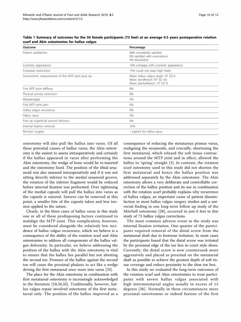
Table 1 Summary of outcomes for the 50 female participants (73 feet) at an average 9.5 years postoperative rotationscarf and Akin osteotomies for hallux valgus
Outcome Percentage
Patient satisfaction 88% completely satisfied8% satisfied with reservations4% dissatisfied
Cosmetic appearance 10% unhappy with cosmetic appearance
Footwear restrictions 14% could not wear high heels
Goniometric measurement of first MTP joint post op Mean Hallux valgus angle 10° SD 6Mean dorsiflexion 54° SD 4.6Mean plantarflexion 15° SD 8
First MTP joint stiffness 8%
Physical activity restriction 8%
Metatarsalgia 6%
First MTP joint pain 6%
Hallux valgus recurrence 8%
Hallux varus 4%
Post op superficial wound infection 4%
Internal fixation removal 25%
Revision surgery 1 patient for hallux varus
Kilmartin and O’Kane Journal of Foot and Ankle Research 2010, 3:2http://www.jfootankleres.com/content/3/1/2
Page 10 of 12

metatarsocuneiform joint are recommended [36]. Theadvantages of the rotation scarf and Akin osteotomiesover the scarf osteotomy alone or more proximal meta-tarsal osteotomies or fusions include:(i) Accurate correction of the intermetatarsal angle, 1
mm of lateral transposition of the metatarsal headequals 1° of intermetatarsal angle correction allowingintraoperative evaluation of the correction [37].(ii) Large areas of bone to bone contact reducing the
risk of non union.(iii) High stability of the osteotomy with the inferior
and superior fragments of the metatarsal being pushedtogether by weight bearing forces rather than being dis-tracted apart by those forces, which may happen in theLapidus first metatarsocuneiform fusion or oblique clos-ing wedge osteotomy procedures. The 2 points of fixa-tion also contributes to quick return to normalweightbearing activities at 2 weeks postoperatively, thusdecreasing the risk of complications associated with pro-longed immobilisation of the postoperative patient.(iv) Rotation of the inferior fragment also allows
greater correction of the intermetatarsal angle than canbe achieved with the scarf osteotomy, where the inferiorfragment can be transposed no more than 50% of thewidth of the superior fragment.(v) Troughing of the fragments is an important risk
with the transposition scarf [14]. Because the rotationscarf osteotomy causes the cortical shell of the dorsalfragment to form an ‘X’ shape with the plantar fragment(Figure 1), troughing is prevented.The results of this study need to be viewed in light of
a number of limitations. While this study sought toestablish participants’ views of their hallux valgus sur-gery outcome it could have been strengthened by theuse of validated patient-reported outcome measures,such as the Manchester Oxford Foot Questionnaire [27].Unfortunately, this become available after most partici-pants received their initial surgery and it cannot bevalidly applied retrospectively.Clearly, the postoperative goniometric measurement of
first MTP joint angles rather than the x-ray measure-ment does weaken this study. However, ethical require-ments in the future may prevent x-rays being used as anoutcome measure in postoperative outcome studies. Thevalidity of first MTP joint/hallux valgus angle measure-ments using the digital goniometer has been evaluatedin a study of 77 childrens’ feet with hallux valgus, whounderwent weightbearing x-ray and charting of the firstMTP joint angle followed by digital goniometric mea-surement [22]. The correlation between the two meth-ods was good (r = 0.63). The mean angle charted on x-ray was 20 degrees (SD 4) and the mean angle measuredwith the goniometer was 19 degrees (SD 4). This was,however, a statistically significant difference (p < 0.05),
with the goniometer generating on average a 1° smallervalue (SD 3.5), the 95% confidence interval being 0.4 to2 degrees. This very small difference in measurementdoes support the use of the digital goniometer and wewould recommend it for similar outcome studies whereuse of x-rays are prohibited by an ethical committee.Severe hallux valgus is a syndrome that involves the
whole forefoot [36]. While it is not surprising that manyadjunctive forefoot procedures were performed in thisgroup with marked hallux valgus deformity, the addi-tional procedures could potentially have confounded ourattempts to measure the outcome of the hallux valgussurgery only. In all cases, however, the participants wereasked to focus exclusively on the surgery to their greattoe in the responses they provided.The rotation scarf and Akin osteotomies are an effec-
tive treatment for hallux valgus. It achieves good longterm correction with a low incidence of recurrence,joint stiffness, or metatarsalgia. Hallux varus was thesingle most important cause of patient dissatisfaction.The nature of the osteotomy allows the surgeon flexibil-ity to correct a range of positional abnormalities withinthe first ray while allowing early return to normal shoesand activity without the need for postoperative immobi-lisation in a plaster cast.
AcknowledgementsAndrew MacFarlane FCPodS for line drawing Figure 1.
Author details1Hillsborough Private Clinic, Hillsborough, Co Down, Northern Ireland BT266AE, UK. 2North West Independent Hospital, Ballykelly, Co Derry, NorthernIreland BT49 9HS, UK. 3Department of Podiatric Surgery, Derbyshire CountyPrimary Care Trust, Ilkeston Hospital, Heanor Road, Ilkeston Derbyshire DE78LN, UK.
Authors’ contributionsTEK performed all the surgery, assisted in design of the study, analysed andsummarized the data and drafted the manuscript. CO designed the study,reviewed all the participants, collected the data and assisted in data analysis.Both authors read and approved the final manuscript.
Competing interestsThe authors declare that they have no competing interests.
Received: 21 May 2009Accepted: 15 February 2010 Published: 15 February 2010
References1. Ferrari J, Higgins JP, Prior TD: Interventions for treating hallux valgus
(abductovalgus) and bunions (Cochrane Review). Cochrane Database ofSystematic Reviews 2004, , 1: CD000964.
2. Torkki M, Malmivaara A, Seitsalo S, Hoikka V, Laippala P: Surgery vs orthosisvs watchful waiting for hallux valgus: a randomised controlled trial. J AmMed Assoc 2001, 285(19):2474-2480.
3. Schneider W, Knahr K: Surgery for hallux valgus. The expectations ofpatients and surgeons. Int Orthop 2001, 25:382-385.
4. Kilmartin TE: Critical Review: The surgical management of Hallux valgus.Br J Podiatry 2006, 9:4-25.
5. O’Kane C, Kilmartin TE: The rotation Scarf and Akin osteotomy for thecorrection of severe hallux valgus. Foot 2002, 12:203-212.
Kilmartin and O’Kane Journal of Foot and Ankle Research 2010, 3:2http://www.jfootankleres.com/content/3/1/2
Page 11 of 12
6. Borelli AH, Weil LS: Modified scarf bunionectomy: our experience in morethan 1000 cases. J Foot Ankle Surg 1991, 30:609.
7. Weil LS: Scarf osteotomy for correction of hallux valgus:historicalperspective, surgical technique and results. Foot Ankle Clin 2000, 5:559-80.
8. Zygmunt KH, Gudas CJ, Laros GS: Z-bunionectomy with internal screwfixation. J Am Podiatr Med Assoc 1989, 79:322-9.
9. Schubert HJM, Reilly CH, Gudas CJ: The closing wedge osteotomy: acritical analysis of first metatarsal elevation. J Am Podiatr Med Assoc 1984,74:13-24.
10. Jones S, Al Hussainy HA, Ali F, Betts RP, Flowers MJ: Scarf osteotomy forhallux valgus. A prospective clinical and pedobarographic study. J BoneJoint Surgery 2004, 86-B:830-6.
11. Malviya A, Makwana N, Laing P: Scarf osteotomy for hallux valgus - is anAkin osteotomy necessary. Foot Ankle Surg 2007, 13:177-181.
12. Deenik AR, Pilot P, Barndt SE, van Mameren H, Geesink RGT: Scarf versuschevron osteotomy in hallux valgus: A randomized control trial of 96patients. Foot Ankle Int 2007, 28:537-541.
13. Lipscombe S, Molloy A, Sirikonda S: Scarf osteotomy for the correction ofhallux valgus: Midterm clinical outcome. J Foot Ankle Surg 2008,47:273-277.
14. Coetzee JC: Scarf osteotomy for hallux valgus repair: The dark side. FootAnkle Int 2003, 24:29-33.
15. Berg RP, Kelder W, Olsthoorn PGM, Poil RG: Scarf and Weil osteotomies forcorrection of rheumatoid forefoot deformities: A review of 20 cases. FootAnkle Surg 2007, 13:35-40.
16. Coetzee JC, Rippstein P: Surgical strategies: scarf osteotomy for halluxvalgus. Foot Ankle Int 2007, 28:529-535.
17. Kristen KH, Berger C, Stelzig S, Thalhammer E, Posch M: The scarfosteotomy for correction of hallux valgus deformities. Foot Ankle Int2002, 23:221-229.
18. Perugia D, Basile A, Gensini A, Stopponi M, Simmonibus AU: The scarfosteotomy for severe hallux valgus. Int Orthop 2003, 27:103-106.
19. Crevoisier X, Mouhsine E, Ortolano V, Udin B, Dutoit M: The scarfosteotomy for the treatment of hallux valgus deformity: a review of 84cases. Foot Ankle Int 2001, 22:970-976.
20. Duke HF: Rotational scarf (Z) osteotomy bunionectomy for correction ofhigh intermetatarsal angles. J Am Podiatr Med Assoc 1992, 82:352-60.
21. Kilmartin TE, Bishop A: Hallux abductus measurement. Repeatability trialsof a clinical measuring instrument. Chiropodist 1988, 43:185-187.
22. Kilmartin TE: Phd Thesis: The orthotic treatment of juvenile hallux valgus.Nottingham University, Department of Orthopaedic and Accident Surgery1994, 108-110.
23. Bojsen-Moller F, Lamorex L: Significance of free dorsiflexion of the toes inwalking. Acta Orthop Scand 1979, 50:471-479.
24. Loudon J, Bell S, Johnston J: Clinical orthopaedic assessment. Ilinois,Human Kinetics 1998, 186.
25. Martin DE, Pontious J: Evaluation of hallux abducto valgus in McGlamry,ED; Banks, AS; Downey, MS: Comprehensive textbook of foot surgery.Baltimore, Williams Wilkins 1992, 483.
26. Dawson J, Coffey J, Doll H: A patient based questionnaire to assessoutcomes of foot surgery: Validation in the context of surgery for halluxvalgus. Qual Life Res 2006, 15:1211-1222.
27. Dawson J, Doll H, Coffey J: Responsiveness and minimally importantchange for the Manchester-Oxford foot questionnaire compared withAOFAS and SF-36 assessments following surgery for hallux valgus.Osteoarthritis Cartilage 2007, 15:918-931.
28. Merkel KD, Katoh Y, Johnson EW, Chao EYS: Mitchell osteotomy for halluxvalgus: Long term follow up and gait analysis. Foot Ankle 1983, 3:189-196.
29. Kilmartin TE, Barrington RL: Metatarsus primus varus - A statistical study. JBone Joint Surg 1991, 73-B:937-940.
30. Donley BG: Acquired hallux varus. Foot Ankle Int 1997, 18:586-592.31. Edelman RD: Iatrogenically induced hallux varus. Clin Podiatr Med Surg
1991, 8:367-382.32. Tourne Y, Saragaglia D, Picard F, De Sousa B, Montbarbon E, Charbel AA:
Iatrogenic hallux varus surgical procedure: a study of 14 cases. FootAnkle Int 1995, 16:457-463.
33. Snijders CJ, Snijders JGN, Phillipens MGM: Biomechanics of hallux valgusand spread foot. Foot Ankle 1986, 7:26-39.
34. Dereymaeker G: Scarf osteotomy for correction of hallux valgus surgicaltechnique and results as compared to distal chevron osteotomy. FootAnkle Clinics 2000, 5:513-524.
35. Perugia D, Basile A, Gensini A, Stopponi M, Simeonibus A: The scarfosteotomy for severe hallux valgus. International Orthop 2003, 27:103-106.
36. Coughlin MJ, Mann RA: Hallux Valgus. Surgery of the Foot and Ankle StLouis: Mosby IncCoughlin MJ, Mann RA, Saltzman CL , 8 2007, 231-233.
37. Sarrafian SK: A method for predicting the degree of functional correctionof the metatarsus primus varus with a distal lateral displacementosteotomy in hallux valgus. Foot Ankle 1985, 5:322-329.
doi:10.1186/1757-1146-3-2Cite this article as: Kilmartin and O’Kane: Combined rotation scarf andAkin osteotomies for hallux valgus: a patient focussed 9 year follow upof 50 patients. Journal of Foot and Ankle Research 2010 3:2.
Submit your next manuscript to BioMed Centraland take full advantage of:
• Convenient online submission
• Thorough peer review
• No space constraints or color figure charges
• Immediate publication on acceptance
• Inclusion in PubMed, CAS, Scopus and Google Scholar
• Research which is freely available for redistribution
Submit your manuscript at www.biomedcentral.com/submit
Kilmartin and O’Kane Journal of Foot and Ankle Research 2010, 3:2http://www.jfootankleres.com/content/3/1/2
Page 12 of 12