combinedfunctional immunochemical analysis of normal · pdf filepoint of interface for both...
TRANSCRIPT
Combined Functional and Immunochemical Analysis ofNormal and Abnormal Human Factor X
DARYLS. FAIR, EDWARDF. PLOW, and THOMASS. EDGINGTON,Department ofMolecular Immunology, The Research Institute of Scripps Clinic, La Jolla,California 92037
A B S T RA C T Human Factor X was isolated fromCohn fraction III and characterized by polyacrylamidegel electrophoresis, amino acid composition, and iso-electric focusing. Two molecular forms with biologicalactivity were observed at isoelectric points of 4.8 and5.0. Antisera generated to Factor X was monospecificand used to establish an equilibrium competitiveinhibition radioimmunoassay. This assay was specificfor human Factor X and did not cross-react withhuman prothrombin or bovine Factor X within thesensitivity range of 6-300 ng Factor X antigen/ml. Themean concentration of Factor X based on the antigenwas 11.9 ,ug0ml, whereas concentration values based oncoagulant activity was 7.8 ,ug/ml. This 30% differencein measurement appears to result from the presence ofa subpopulation of Factor X molecules devoid ofcoagulant activity. The radioimmunoassay was used toqualitatively and quantitatively compare purified FactorX to plasmic Factor X obtained from normal, warfarin-treated, acquired Factor X-deficient, and congenital-deficient patients. In all but one case, the Factor Xpresent in these plasmas was immunochemicallyidentical to the purified Factor X and permittedprecise quantitation of these abnormal Factor Xmolecules. Factor X procoagulant activity was analyzedrelative to Factor X antigen and the specific activitieswere used to characterize normal and abnormalFactor X molecules. Reduced Factor X activity inplasmas from warfarin-treated and acquired Factor X-deficient patients was attributed to both decreases inFactor X antigen and decreased function of theFactor X molecules. Congenitally deficient patients,in general, showed a reduction in Factor X antigenin parallel with Factor X procoagulant activities
This is publication 1688 of the Research Institute, and waspresented in part at the meeting of the American Society ofBiological Chemists 4-8 June 1978 in Atlanta, Ga.
Dr. Plow is an Established Investigator of the AmericanHeart Association.
Received for publication 19 January 1979 and in revisedform 14 May 1979.
884
resulting from comparable decreases in specificbiological activity of the molecules.
INTRODUCTION
Plasma Factor X occupies a pivotal position in theblood coagulation scheme and, as such, its activationand function are of central importance in the studyof thrombotic and hemostatic diseases. Factor X is thepoint of interface for both the intrinsic and extrinsicprocoagulant pathways and is the substrate zymogenfor these proteolytic activation complexes. The activeenzyme, Factor Xa, initiates the late stage of bloodcoagulation by cleavage of prothrombin to formthrombin (1). Although bovine Factor X has beenextensively investigated, isolation of Factor X fromhuman plasma has only recently been described (2-6).Human Factor X, a glycoprotein, has an estimatedmol wt of 59,000 with 15% carbohydrate and consistsof a disulfide-linked 42,000-mol wt heavy chain and17,000-mol wt light chain (2). The activation of FactorX and subsequent proteolytic cleavages of prothrombinis supported by phospholipid membranes and calcium.These events are mediated through the gammacarboxylated glutamic acid residues located on theamino terminal of the light chain of this vitaminK-dependent protein (1).
Most clinical studies of Factor X have been limitedto biological clotting assays which utilize congenitalFactor X-deficient plasma or bovine plasma artificiallydepleted of Factor X. In a few studies antisera withspecificity to Factor X have been used to immuno-logically estimate the presence of Factor X protein(7-15). Whereas qualitative assessment has employedimmunoelectrophoresis (9, 11, 12), quantitation ofFactor X antigen has been attempted by antibodyneutralization (7, 8, 10), and radioimmunoassay(14, 15). The latter method has been described forbovine (14) and canine (15) but not human Factor X.
Factor X molecules known or presumed to beabnormal have been described in congenital and in
J. Clin. Invest. C The American Society for Clinical Investigation, Inc. - 0021-9738/79/10/0884/11 $1.00Volume 64 October 1979 884 -894
aquired deficiencies. Congenital Factor X deficiencyhas been characterized as an incompletely recessiveautosomal trait (11, 16); and unrelated individualsafflicted with this disorder presumably represent avariety of abnormalities at different points in themolecule giving rise to a spectrum of biologicalactivities and variable concentrations of plasmaFactor X (7). Acquired Factor X deficiency independentof vitamin K deficiency is a rare coagulation defectin which the majority of cases described have beenassociated with amyloidosis (17-19). RadiolabeledFactor X injected into these patients was rapidly clearedfrom the plasma (18) and shown to bind more readilyto amyloid fibrils isolated from one patient with thiscoagulation disorder (19). Although the Factor X of thepatients was assumed to be normal, this has not beeninvestigated. Finally, individuals undergoing anti-coagulant therapy with pheninidione or warfarin havebeen reported to have a decrease in Factor X coagulantactivity relative to Factor X antigen (8, 12). Thisdecrease in the ratio of activity to antigen couldreflect the synthesis of biologically inactive moleculeslacking the posttranslational carboxylation of specificglutamic acid residues implicated in the calcium-dependent interaction of Factor X with phospholipid (1).
In the present investigation, we describe theisolation of highly purified human Factor X fromCohn fraction III, the development of a radio-immunoassay specific for the intact molecule, and thequantitation of Factor X antigen and activity inplasma from normal, Factor X-deficient and warfarin-treated patients.
METHODSMaterials. Acrylamide, N,N'-methylenebisacrylamide, hy-
droxylapatite, and sodium dodecyl sulfate (SDS)l wereobtained from Bio-Rad Laboratories, Richmond, Calif.Bovine Factor VII -X-deficient plasma, Russell's viper venom,rabbit brain cephalin, disodium EDTA, Coomassie BrilliantBlue R, Coomassie Blue G-250, Sigma 7-9, E-aminocaproicacid, heparin sodium salt grade I (150 USP U/mg),morpholinoethanesulfonic acid, poly-L-lysine (type I-B) andp-aminobenzamidine hydrochloride were purchased fromSigma Chemical Co., St. Louis, Mo. Barium chloride,ammonium sulfate, sodium citrate, chloramine T, and sodiummetabisulfite were obtained from J. T. Baker Chemical Co.,Phillipsburg, N. J. Sephadex G-25, G-50, G-100, andSepharose 2B were bought from Pharmacia Fine Chemicals,Div. of Pharmacia Inc., N. J. Kaolin was a product ofFisher Scientific Co., Pittsburgh, Pa. Cyclohexylmorpholino-ethylearbodiimide and O-methylisourea were obtained fromAldrich Chemical Co., Milwaukee, Wisc. Benzamidine andphenylmethylsulfonyl fluoride were purchased from Calbio-chem-Behring Corp., American Hoechst Corp., San Diego,Calif. Sodium azide was a product of Mallinckrodt Inc., St.Louis, Mo. DEAE-cellulose (DEAE-52) was obtained from
1 Abbreviations used in this paper: r, correlation coefficient;RVV, Russell's viper venom; SDS, sodium dodecyl sulfate.
Whatman Inc., Clifton, N. J. 125I-Na (50 mCi/ml) was purchasedfrom Amersham Corp., Arlington Heights, Ill. Trasylol was aproduct of FBA Pharmaceuticals, New York. Brain thrombo-plastin was obtained from Ortho Pharmaceutical Corp.,Raritan, N. J. Nobel agar was a product of Difco Laboratories,Detroit, Mich. Ampholytes were purchased from LKBInstruments, Inc., Rockville, Md. Human Factor X-deficientplasma was obtained from George King Biomedical, Inc., Over-land Park, Kans. Benzamidine-Sepharose 2B was constructed bythe method of Schmer (20) as modified by DiScipio et al. (2).Homoarginine-Sepharose 2B was prepared by the proceduresof Kimmel (21) and DiScipio et al. (2).
Plasma samples. Normal plasma samples and plasma frompatients were collected in citrate-phosphate-dextrose or 2.9%sodium citrate. Many of the Factor X-deficient plasmas werethe generous gifts of Doctors H. L. Nossel, B. Furie, J. B.Graham, A. L. Bloom, S. Lemkin, J. Baker, and Y. Sultan. Someof these plasmas arrived in a lyophilized state which underappropriate conditions we had shown did not affect theprocoagulant activity or antigenic structure of Factor X.
Coagulation tests. Assays for Factor X during isolationemployed the use of Russell's viper venom (RW) accordingto the method of Bachmann et al. (22). In addition to thismethod, normal, warfarin-treated and Factor X-deficientplasma were assayed for extrinsic activation by the one-stageprothrombin time using human Factor X-deficient plasma andfor intrinsic activation by the kaolin-cephalin clotting timeemploying Factor X-deficient human plasma as the substrateplasma (23). All values represent the mean of triplicatedeterminations and have been normalized to a pool of 26normal donors.
Isolation of human Factor X. All isolation procedureswere carried out at 4°C to minimize activation, and Factor Xactivity and antigen (antisera kindly provided by Dr. D.Aronson) were monitored. Several of the steps weremodified from previously described procedures (2, 3).1 kg of Cohn fraction III (generously supplied by Dr. M.Hrinda of Armour Pharmaceutical Company, Phoenix, Ariz.)was extracted in 15 liters of 0.01 M Tris-HCl, pH 9.0,0.025 M sodium citrate, 0.15 M sodium chloride, 2 mMbenzamidine, 15,000 U heparin for 1 h. The solution wasclarified by centrifugation at 1,500g for 20 min. To thesupernate was added drop-wise 1.05 liters of 1 M bariumchloride, and after mixing for 15 min the precipitate wascollected by centrifugation at 1,500 g for 20 min. Proteinsabsorbed to the barium citrate pellet were washed twice withwater and then eluted into 3 liters of 0.05 M morpholino-ethanesulfonic acid-HCl (pH 5.85) containing 0.2 M sodiumcitrate, 0.01 Mbenzamidine, 3,000 U heparin overnight. Aftercentrifugation at 10,000 g for 20 min, solid ammoniumsulfate was added to the pooled supemates to 40% saturationand mixed for 30 min. After centrifugation the resultingsupernate was adjusted to 70% saturation with solidammonium sulfate, mixed for 45 min, and then centrifugedat 10,000 g for 20 min. The pellet was dissolved in a minimalvolume of 0.02 MTris-NaOH, pH 5.85, 0.1 M EDTA, 2 mMbenzamidine, 0.02% sodium azide, and then dialyzed over-night against 1 liter of this buffer. After dialysis against1 liter of 0.02 M Tris-H3PO4 (pH 5.85) containing 2 mMbenzamidine, 0.02% sodium azide with two changes, theprotein was applied to a DEAE-cellulose column (5 x 28 cm)equilibrated in this buffer. The protein was eluted over48 h with a linear gradient from 0.10 to 0.35 M NaCl in theinitial buffer at 55 ml/h. After concentration and dialysisagainst 1 liter of 0. 15 Mpotassium phosphate pH 6.8 containing1 mMbenzamidine, 0.02% sodium azide with one change,the protein solution was applied to a hydroxylapatite-Sephadex G-25 (1:1 vol/vol) column (2.5 x 45 cm) equilibrated in
Measurement of Factor X 885
this same buffer. Factor X was eluted from the column over48 h with a linear gradient from 0.15 to 0.30 M potassiumphosphate, pH 6.8, containing 1 mMbenzamidine, 0.02%sodium azide at 16 ml/h. After concentration and dialysisagainst 0.05 M morpholinoethanesulfonic acid-Tris pH 5.85containing 2 mMbenzamidine, 0.02% sodium azide, theprotein was applied to a benzamidine-Sepharose column(1.6 x 23 cm) equilibrated in this buffer, and eluted over24 h using a linear gradient from 0.1 to 0.4 MI NaCl at17 ml/h. The Factor X-containing peak was chromatographedon a homoarginine-Sepharose column (1.6 x 20 cm) equili-brated in 0.02 Mmorpholinoethanesulfonic acid-Tris pH 5.85,0.02% sodium azide, and a 24-h linear gradient from 0.2 to2 M NaCl in this buffer was developed at 14 ml/h. Afterconcentrating, the Factor X protein was chromatographed onSephadex G-100 (2.5 x 90 cm) equilibrated 0.01 Mpotassiumphosphate, pH 6.0 containing 0.14 NaCl, 0.02% sodiumazide, at 14 ml/h. The Factor X eluting from this columnshowed a constant specific activity over the protein profileand was concentrated and stored at -70°C. Some samplesalso contained 10 mMbenzamidine.
Most Factor X protein determinations were based on E280 nm= 11.6 (2). The concentration of standard Factor X wasestablished from the amino acid composition.
Gel electrophoresis. Analytical polyacrylamide gels werecarried ouit at alkaline pH according to Davis (24) andSDS-polyacrylamide gel electrophoresis was employed atneutral pH (25) and at alkaline pH (26). Protein bands weredeveloped with freshly prepared Coomassie Brilliant Blue Raccording to the procedure of Weber and Osborn (25).Isoelectric focusing was performed with a pH gradientbetween 3 and 8 as described by Righetti and Drysdale (27)and stained with Coomassie Blue G-250 according to theprocedure of Reisner et al. (28). Molecular weight standardsused in the SDS-polyacrylamide gel electrophoresis were theAa-, B,8-, and y-chain of fibrinogen (63,500, 56,000, and47,000, respectively), bovine serum albumin (68,000),ovalbumin (43,000), chymotrypsinogen (25,000), and myo-globin (17,000).
Amino acid compositions. Samples were hydrolyzed for24 h in 6.0 N HCI in evacuated tubes at 1 10°C. Analyses wereperformed on a Beckman 121 M automated amino acidanalyzer (Beckman Instruments, Inc., Fullerton, Calif.) usingthe two-column procedure. Values represent the mean ofduplicate determinations, and a mol wt of 50,355 was adaptedfor the protein portion of human Factor X as determined byDiScipio et al (2) for purposes of' calculation.
Antisera. Two New Zealand white female rabbits wereinjected with 39 ,ug of purified Factor X emulsified inFreund's complete adjuvant at multiple subcutaneous sites.The animals were boosted with 20 ,ug of protein emulsifiedin Freund's incomplete adjuvant every 3 wk. After the thirdboost, antisera were judged to be of relative high titerand employed in these studies.
Antibody neutralization. These studies utilized eithernormal rabbit serum or rabbit antiserum to human Factor Xwhich had been absorbed with barium sulfate (100 mg/ml)and heat inactivated (56°C for 40 min). The sera werediluted in 0.025 M barbital pH 7.4 containing 0.125 MNaCland 0.1% sodium azide. To 1 ml of a b/oth dilution ofnormal human plasma was added 1.0 ml of' diluted antiserumor normal serum. After incubation for 24 h at 40C the solutionwas tested for Factor X activity.
Immunoelectrophoresis was carried out on 1 x 3-inch slidesoverlayered with 3 ml of 1% Nobel agar in 0.04 M barbital,pH 8.2. Electrophoresis was carried out at 5 mA/slide for105 min. After addition of antiserum to the trough, thereaction was developed at room temperature for 24 h.
Radioiodination. Factor X was labeled with '251-Na by amodification of the chloramine T procedure (29). Factor X(20 ,Ag) was mixed with 0.5 M potassium phosphate pH 7.2to a final volume of 100 gl. To this was added 10 y1 of1251-Na (50 mCi/ml) and 10 Al of' chloramine T in phosphate-buffered saline (3()0 ,tg/ml) and reacted for 2 min withconstant agitation. Sodium metabisulfite in phosphate-bufferedsaline (10 ,ul of a 300-,g/ml solution) was added to terminatelabeling. After mixing for 30 s, 100 al of 2% potassiumiodide and 100 Al 1%lbovine serum albumin in phosphate-buffered saline were added. lodinated Factor X wasisolated by chromatography on a 12-ml column of SephadexG-50 equilibrated with 1% bovine serum albumin, 0.1%sodium azide in phosphate-buffered saline. The radiolabeledFactorX was 94-97% precipitable by 10% trichloroacetic acid.
Radioimmunoassay. The equilibrium competitive inhibi-tion radioimmunoassay for Factor X was carried out with afour compartment procedure. To: (a) 0.5 ml of' labeledFactor X (0.1 or 0.05 nM) was added, (b) 0.5 ml of' buffer,dilution of competing antigen or plasma, and (c) 0.5 ml ofdiltuted rabbit anti-Factor X antiserum. After 18 h at 4°C,(d) 0.5 ml of goat anti-rabbit IgG in slight antibody excess wasadded. After incubation for 6 h at 4°C, the immunoprecipitatewas sedimented at 2,500 rpm f'or 20 min anid 1.0 ml of thestupernate was collected, counted, and the binding wascalculated. Statistical analysis of' the data was handledaccording to Rodbard (30). The coefficient of variation withinassays was 1-4% and between assays 2.9-10.6%.
The buffer utilized in the assay and for all dilutions was0.042 M borate buffer, pH 8.3 containing 0.025 NI NaCl,normal rabbit serum (1:50), 2 mMbenzamidine, 100 U/mlTrasylol and 1 mNMphenvlmethylsulfonyl fluoride. All serawere heat inactivated.
RESULTS
HumanFactor X of high purity was isolated from Cohnfraction III by the sequence summarized in Table I.From 1 kg of Cohn fraction III paste 528 g of proteinwas solubilized. A loss of 50% of Factor X occurredupon elution from barium citrate, but subsequent stepsgave high yields with a final recovery of 20% of thesolubilized Factor X at nearly 6,000-fold purificationbased on activity and antigen assays. The Factor X wasdevoid of Xa (<0.03%) by coagulant assay lackingRVV, and the specific activity was 128 U/mg, ingood agreement with specific activities reported byothers (2, 6).
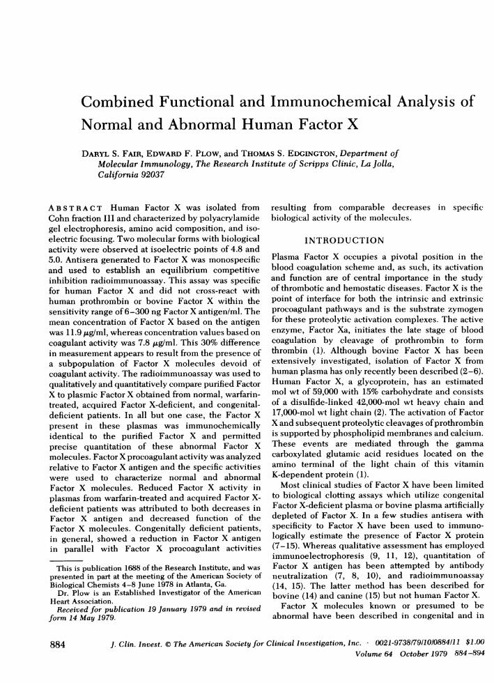
The isolated Factor X was devoid of Xa or otherplasma proteins as determined by polyacrylamide gelin SDS and disc gel electrophoresis (Fig. 1). Onunreduced SDS gels only a single band was observedmigrating with an apparent mol wt of 64,000. Uponreduction two polypeptide chains were detected chainswith estimated mol wt of 46,000 and 17,000, respectively.The observed molecular weights were similar to valuesdescribed for Factor X isolated from plasma (2, 6).The amino acid composition of human Factor X(Table II) was comparable to that reported by DiScipioet al. (2).
Isoelectric focusing of Factor X in ampholytecontaining gels w\ith a pH 3-8 linear gradient resolved
886 D. S. Fair, E. F. Plow, and T. S. Edgington
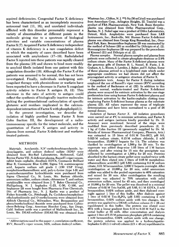
TABLE IPurification of Human Factor X from Cohn Fraction IIl
Purification step Volume Total protein* Total activityt sp act Recovery Purification
ml mg U Ulmg %
Tris-citrate extraction 15,000 528,000 11,460 0.022 100.0Barium citrate elution 7,990 12,784 5,800 0.454 50.6 20.9Ammonium sulfate (40-70%) 192 4,493 4,904 1.09 42.8 50.2DEAE-cellulose 46 3,487 3,926 1.13 34.3 52.1Hydroxylapatite 19.4 366 3,111 8.5 27.1 392Benzamidine-Sepharose 87 168 2,945 17.5 25.7 806Homoarginine-Sepharose 60.9 36.8 2,573 69.9 22.5 3,221Sephadex G-100 16.9 18.6 2,383 128.0 20.8 5,897
* Protein concentrations were determined by absorbance at 280 nmand assumed El2nm of 11.6 (2).t A unit of Factor X is defined as the activity present in 1 ml of pooled normal human plasma.
two discrete bands with pls of 4.8 and 5.0 (Fig. 1).When gels were sliced and the protein eluted,Factor X coagulant activity was coincident with bothprotein bands, suggesting that human Factor X existsin two distinct charge forms similar to that observed forthe bovine molecule (31, 32).
Rabbit antisera to Factor X were monospecific byimmunoelectrophoresis, producing only a singleprecipitin arc with the purified molecule as well aswith barium citrate concentrates of normal humanplasma. Further, 1 ml of these antiserum had thecapacity to completely neutralize the Factor Xcoagulant activity of 22 ml of normal human plasma.
S
a
A B C 0
FIGURE 1 PAGEanalysis of purified human Factor X. (A)Migration of 8 ,g of Factor X in Laemmli (26) gels containing10%acrylamide, 0.1% SDS (B) Migration of 16 jAg of Factor Xreduced with 2-mercaptoethanol in 10% acrylamide, 0.1%SDS containing gels (C) Migration of 16 ,ug Factor X inalkaline disc gel (pH 8.9) containing 6% arylamide. (D)Migration of 16 itg of Factor X in isoelectric focusinggels with a pH gradient from 3 to 8. The protein bandscoincide with pH 4.8 and 5.0. The anode is to the bottom inall the gel systems.
Neutralization of Factor X activity in plasmas ofpatients on warfarin therapy was also observed. Normalrabbit serum had no effect on Factor X activitypresent in these plasmas.
Radioiodinated Factor X was analyzed by SDS-polyacrylamide gel electrophoresis with and withoutreduction (Fig. 2). Bands of 1251 were coincident onlywith the stained protein bands observed for Factor X,indicating that the 1251-ligand was structurally intactand that radiolabeled contaminants of differentmolecular weights were not present. Antigenic equiv-alence of labeled Factor X was analyzed by com-paring the binding by antibody of four differentmixtures of labeled and unlabeled Factor X at constanttotal concentration. The binding curves superimposedindicating that the affinity of the antibody for theFactor X was not altered by radioiodination. The
TABLE IIAmino Acid Composition of Human Factor X
Amino acid Residue No. Residue No.*
Lys 31.6 31.1His 7.8 8.9Arg 23.2 22.1Asx 48.8 45.8Thr 33.2 35.3Ser 22.8 26.7Glx 64.8 61.5Pro 18.8 19.6Gly 40.4 38.8Ala 27.2 26.2Val 20.1 21.1Met 6.4 6.3Ile 15.6 15.6Leu 29.0 29.4Tyr 12.2 12.8Phe 20.2 20.9
* Data from Di Scipio et al. (2).
Measurement of Factor X 887

50,
40-
30-
20
x
4-
I
Competing Plasma llotilo
Unreduced
24-Reduced
18-
12 0
6-
10l 20 30 4.0 50 60Slice (mm)
FIGURE 2 Electrophoretic migration of iodinated humanFactor X on 10% acrylamide gels in SDS in the absence(top) and presence (bottom) of 2-mercaptoethanol. Theanode and bottom of the gels are to the right.
10 pM 0.1 nM I nM 10mnM 0.111M
Computing Protein
FIGURE 4 Equilibrium competitive inhibition of the bindingof 1251-Factor X (0.1 nM added) to anti-human Factor X(5,000-1 dilution) by purified human Factor X (x), purifiedhuman prothrombin (A), normal human plasma pool (0), andnormal bovine plasma (0).
authenticity of the labeled Facto]substantiated since analyses of nonradFactor X behaved in a competitiveimmunoassay identical to the unrA representative antibody binding pFig. 3. At high antibody concentratlabeled ligand was bound. The percebound decreased linearly with serial tof antibody and nonspecific bindingFor competitive inhibition assays anto bind 60% of the '251-Factor X was
The equilibrium competitive iimmunoassay was used to comparewith normal and abnormal plasmaspecificity of the radioimmunoass,supported by: (a) complete inhibi
100
so
- 60
Wn 40
20
I10'- 3 10i4Antibojy Dilution
FIGURE 3 Binding of '251-Factor X byanti-Factor X antigen. The ligand wasconcentration.
Factor X and normal pooled plasma; (b) parallel slopesr X was further of inhibition by Factor X and normal pooled plasma;lioactive iodinated (c) absence of any inhibition by normal bovine plasma;inhibition radio- and (d) the absence of inhibition by purified pro-
nodified protein. thrombin at a concentration at which Factor X producesrofile is shown in complete inhibition. Slight inhibition by prothrombintion >95% of the was observed at a 1,000-fold molar excess andnt of 1251-Factor X presumably represents contamination (0.1%) withthreefold dilutions Factor X. Either these antisera do not contain antibodieswas insignificant. or their affinity is too low for structurally homologoustiserum sufficient sequences of human prothrombin and bovine Factor Xused. to exhibit significant binding. The range of sensitivity
inhibition radio- was from 6 to 300 ng Factor X/ml.purified Factor X Accuracy of measurement of Factor X in plasma wasa Factor X. The estimated by addition of serially increasing concen-ay (Fig. 4) was trations of purified Factor X to normal pooled humanition by purified plasma. Using purified human Factor X as the standard:
(a) the concentration of Factor X measured in plasmacorresponded linearly to the amount of Factor X addedto plasma and (b) the Factor X concentration resultingfrom extrapolation of the linear regression to zeroagreed with the assay value for the intrinsic plasmaFactor X of 11.9 jig/ml. These results indicated ananalytic recovery of the amount of Factor X added toplasma and accurate quantitation of protein by radio-immunoassay.
The difference in concentration of Factor X inplasma by immunochemical (11.9 ,ug/ml) as contrasted
10%5 l c6 to coagulant (7.8 ,g/ml) assays is significant andreproducible. This prompted us to verify this observation
dilutions of rabbit by adsorption of Factor X to increasing amounts ofadded at 0.1 nM barium citrate. The absorption of Factor X coagulant
activity and Factor X antigen from plasma is shown in
888 D. S. Fair, E. F. Plow, and T. S. Edgington
M
m
.8
in914
ae
10z
Cmptitor Ceuectratie
80- \
^ 60-
° 40-
a 20
O-0 10 20 50 100 200
Barium Chloride Added I, 1/m1 Plasma)
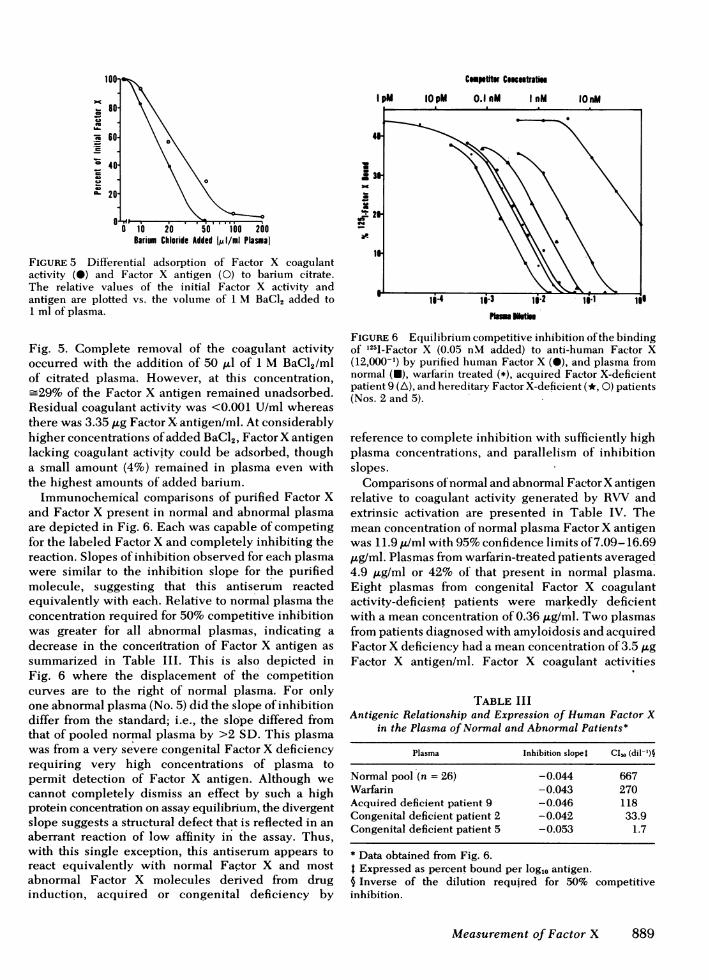
FIGURE 5 Differential adsorption of Factor X coagulantactivity (0) and Factor X antigen (0) to barium citrate.The relative values of the initial Factor X activity andantigen are plotted vs. the volume of 1 M BaCl, added to1 ml of plasma.
Fig. 5. Complete removal of the coagulant activityoccurred with the addition of 50 ,ul of 1 M BaCI2/mlof citrated plasma. However, at this concentration,=29% of the Factor X antigen remained unadsorbed.
Residual coagulant activity was <0.001 U/ml whereasthere was 3.35 ,ug Factor X antigen/ml. At considerablyhigher concentrations of added BaCl2, Factor X antigenlacking coagulant activity could be adsorbed, thougha small amount (4%) remained in plasma even withthe highest amounts of added barium.
Immunochemical comparisons of purified Factor Xand Factor X present in normal and abnormal plasmaare depicted in Fig. 6. Each was capable of competingfor the labeled Factor X and completely inhibiting thereaction. Slopes of inhibition observed for each plasmawere similar to the inhibition slope for the purifiedmolecule, suggesting that this antiserum reactedequivalently with each. Relative to normal plasma theconcentration required for 50% competitive inhibitionwas greater for all abnormal plasmas, indicating adecrease in the concenltration of Factor X antigen assummarized in Table III. This is also depicted inFig. 6 where the displacement of the competitioncurves are to the right of normal plasma. For onlyone abnormal plasma (No. 5) did the slope of inhibitiondiffer from the standard; i.e., the slope differed fromthat of pooled normal plasma by >2 SD. This plasmawas from a very severe congenital Factor X deficiencyrequiring very high concentrations of plasma topermit detection of Factor X antigen. Although wecannot completely dismiss an effect by such a highprotein concentration on assay equilibrium, the divergentslope suggests a structural defect that is reflected in anaberrant reaction of low affinity in the assay. Thus,with this single exception, this antiserum appears toreact equivalently with normal Factor X and mostabnormal Factor X molecules derived from druginductiQn, acquired or congenital deficiency by
'W
m
M
FIGURE 6 Equilibrium competitive inhibition of the bindingof 1251-Factor X (0.05 nM added) to anti-human Factor X(12,000-1) by purified human Factor X (0), and plasma fromnormal (U), warfarin treated (*), acquired Factor X-deficientpatient 9 (A), and hereditary Factor X-deficient (*, 0) patients(Nos. 2 and 5).
reference to complete inhibition with sufficiently highplasma concentrations, and parallelism of inhibitionslopes.
Comparisons of normal and abnormal FactorX antigenrelative to coagulant activity generated by RXWVandextrinsic activation are presented in Table IV. Themean concentration of normal plasma Factor X antigenwas 11.9 ,u/ml with 95%confidence limits of 7.09-16.69,ug/ml. Plasmas from warfarin-treated patients averaged4.9 ,ug/ml or 42% of that present in normal plasma.Eight plasmas from congenital Factor X coagulantactivity-deficient patients were markedly deficientwith a mean concentration of 0.36 ,ugtml. Two plasmasfrom patients diagnosed with amyloidosis and acquiredFactor X deficiency had a mean concentration of 3.5 p.gFactor X antigen/ml. Factor X coagulant activities
TABLE IIIAntigenic Relationship and Expression of Human Factor X
in the Plasma of Normal and Abnormal Patients*
Plasma Inhibition slopet CI,s (dil-')§
Normal pool (n = 26) -0.044 667Warfarin -0.043 270Acquired deficient patient 9 -0.046 118Congenital deficient patient 2 -0.042 33.9Congenital deficient patient 5 -0.053 1.7
* Data obtained from Fig. 6.t Expressed as percent bound per log,, antigen.§ Inverse of the dilution required for 50% competitiveinhibition.
Measurement of Factor X 889
11
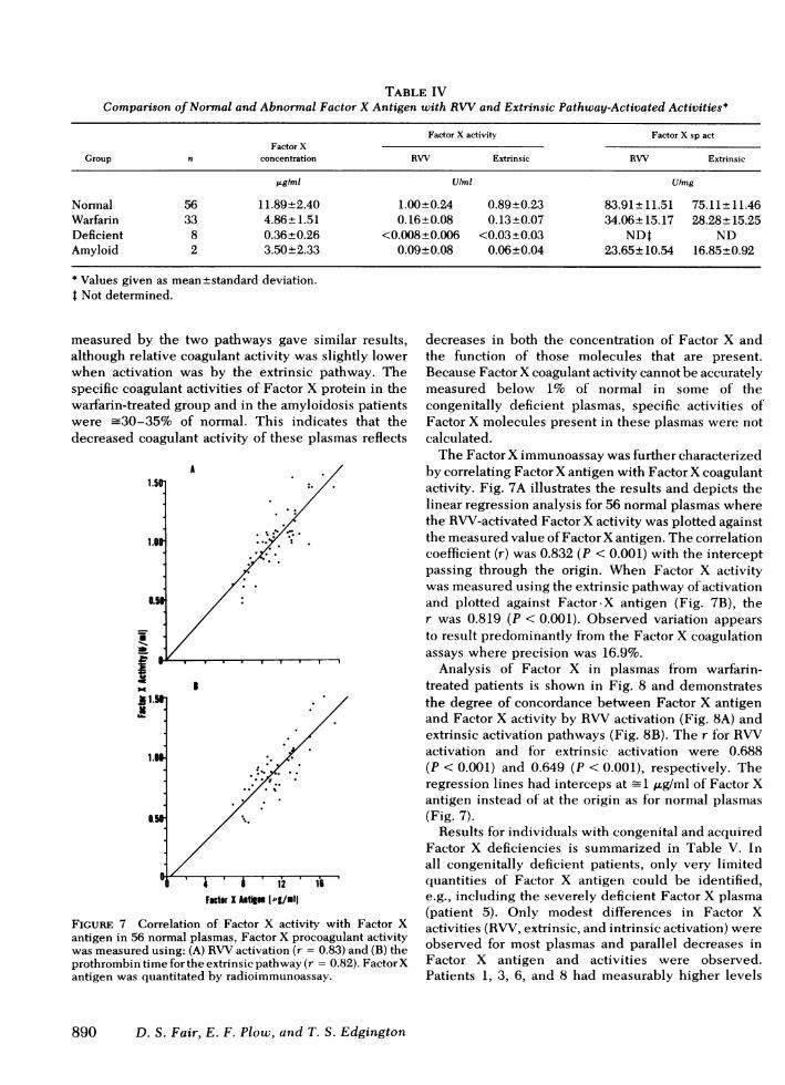
TABLE IVComparison of Normal and Abnormal Factor X Antigen with RVVand Extrinsic Pathway-Activated Activities*
Factor X activity Factor X sp actFactor X
Group n concentration RW Extrinsic RW Extrinsic
jgIml U/mi Ulmg
Normal 56 11.89+2.40 1.00+0.24 0.89+0.23 83.91±+ 11.51 75.11±+ 11.46Warfarin 33 4.86±1.51 0.16±0.08 0.13±0.07 34.06±15.17 28.28±15.25Deficient 8 0.36±0.26 <0.008±0.006 <0.03±0.03 NDt NDAmyloid 2 3.50±2.33 0.09±0.08 0.06±0.04 23.65±10.54 16.85±0.92
* Values given as mean±standard deviation.t Not determined.
measured by the two pathways gave similar results,although relative coagulant activity was slightly lowerwhen activation was by the extrinsic pathway. Thespecific coagulant activities of Factor X protein in thewarfarin-treated group and in the amyloidosis patientswere -30-35% of normal. This indicates that thedecreased coagulant activity of these plasmas reflects
A
p1.5
1.I
Ii5
I
It. 4 A
tI A 1u 216/Facto X Alltign [,-g/ml
FIGURE 7 Correlation of Factor X activity with Factor Xantigen in 56 normal plasmas, Factor X procoagulant activitywas measured using: (A) RWactivation (r = 0.83) and (B) theprothrombin time for the extrinsic pathway (r = 0.82). Factor Xantigen was quantitated by radioimmunoassay.
decreases in both the concentration of Factor X andthe function of those molecules that are present.Because Factor X coagulant activity cannot be accuratelymeasured below 1% of normal in some of thecongenitally deficient plasmas, specific activities ofFactor X molecules present in these plasmas were notcalculated.
The Factor X immunoassay was further characterizedby correlating Factor X antigen with Factor X coagulantactivity. Fig. 7A illustrates the results and depicts thelinear regression analysis for 56 normal plasmas wherethe RVV-activated Factor X activity was plotted againstthe measured value of Factor X antigen. The correlationcoefficient (r) was 0.832 (P < 0.001) with the interceptpassing through the origin. When Factor X activitywas measured using the extrinsic pathway of activationand plotted against Factor.X antigen (Fig. 7B), ther was 0.819 (P < 0.001). Observed variation appearsto result predominantly from the Factor X coagulationassays where precision was 16.9%.
Analysis of Factor X in plasmas from warfarin-treated patients is shown in Fig. 8 and demonstratesthe degree of concordance between Factor X antigenand Factor X activity by RVVactivation (Fig. 8A) andextrinsic activation pathways (Fig. 8B). The r for RVVactivation and for extrinsic activation were 0.688(P < 0.001) and 0.649 (P < 0.001), respectively. Theregression lines had interceps at _1 ,ug/ml of Factor Xantigen instead of at the origin as for normal plasmas(Fig. 7).
Results for individuals with congenital and acquiredFactor X deficiencies is summarized in Table V. Inall congenitally deficient patients, only very limitedquantities of Factor X antigen could be identified,e.g., including the severely deficient Factor X plasma(patient 5). Only modest differences in Factor Xactivities (RVV, extrinsic, and intrinsic activation) wereobserved for most plasmas and parallel decreases inFactor X antigen and activities were observed.Patients 1, 3, 6, and 8 had measurably higher levels
890 D. S. Fair, E. F. Plow, and T. S. Edgington

Facter X Atig lpg/mll
FIGURE 8 Correlation of Factor X activity with Factor Xantigen in 33 plasmas from patients undergoing warfarintherapy. Factor X procoagulant activity was determined byactivation by: (A) RW(r = 0.69) and (B) the prothrombintime for the extrinsic pathway (r = 0.65). Factor X antigen wasquantitated by radioimmunoassay.
of Factor X activity by extrinsic activation relative toRWor intrinsic activation pathways. Also, Factor Xantigen was higher in patient 8 than other deficientcases, i.e., _8% of normal. Because this individualhad clinical hemorrhagic problems requiring severaltransfusions, the Factor X antigen levels may not beaccurately correlated with Factor X activity.
In the plasma of two patients with acquired deficiencyand amyloidosis, Factor X coagulant activity wasdiminished relative to Factor X antigen, suggesting asignificant functional abnormality which was relativelygreater than that observed for congenitally deficientplasmas. Mixing experiments did not demonstrate acoagulation inhibitor in these plasmas, and we concludeon the basis of current evidence that these may alsobe a defective Factor X molecule in this type ofamyloidosis.
DISCUSSION
Isolation of the proteins of the coagulation pathwayshas made possible not only biochemical characterizationbut also immunochemical analyses of concentration,molecular heterogeneity, and specific functional activityof these proteins in plasma (33-39) as well as specific
assays of the activated enzyme (40) and proteolyticdegradation products (41, 42). The molecular biologyof Factor X is inordinately complex as evidenced by itsmultiple interactions; and precise analyses of molecularmass are required for studies of the functional biologyof this molecule in thrombotic and hemostatic diseases.The present study has addressed the quantitativeanalysis of Factor X protein in plasma by referenceto its antigenic properties and by correlation withFactor X coagulant activity.
Cohn fraction III can be used as a readily availablesource for the isolation of highly purified Factor Xrequired for immunochemical standardization andanalyses. The physicochemical characteristics are inreasonable agreement with other reports (2-6),indicating an estimated mol wt of 64,000 for theintact molecule and 46,000 and 17,000, respectivelyfor the heavy and light chains. The 8.5% higherestimate of mass than that of DiScipio et al. (2) mayreflect the aberrantly higher molecular weight estimatesthat occur with SDS acrylamide gel analysis ofglycoproteins (43). Of interest and first described hereis the observation of two forms of human Factor Xthat were resolved by isoelectric focusing. These
TABLE VFactor X Antigen and Activities in Plasmas from Congenital
and Acquired Factor X-Deficient Patients
Factor X activityt
Kaolin-cephalin
Factor X Prothrombin clottingGroup antigen RVV time time
Jag/im* U/mi U/mi s
Congenitaldeficient
1 0.28 0.01 0.05 3102 0.30 <0.01 0.01 3183 0.56 0.01 0.09 2624 0.33 <0.01 <0.01 4015 0.03 <0.01 <0.01 3846 0.25 0.01 0.07 3527 0.24 0.02 <0.01 3288 0.89 0.01 0.03 270
Acquireddeficient
9 1.85 0.03 0.03 21510 5.15 0.16 0.09 157
Normal 11.89 1.00 0.89 112
* Determined by radioimmunoassay.t Activities were measured by activation with RVV, theextrinsic pathway using the prothrombin time, and theintrinsic pathway using the kaolin-cephalin clotting time.
Measurement of Factor X 891

species may be analogous to the two forms of bovineFactor X (31, 32).
Our radioimmunoassay for human Factor X wascharacterized by fulfilling the requirements forspecificitv, conservation of the native protein con-formation of the iodinated ligand, and demonstratinganalytical recovery of Factor X added back to plasma.Specificity of the assay was indicated by the completeinhibition of the assay by both purified Factor X andnormal human plasma, and the inability of purifiedhuman prothrombin and normal bovine plasma toinhibit the reaction. The range of assay was 6-300 ngFactor X antigen/ml, and the precision was indicatedby a coefficient of variation of 1-4% within assaysand 2.9-10.6% between assays.
Determination of Factor X concentration by directimmunochemical techniques gave higher values(11.9 ,ug/ml) than those indirectly measured usingcoagulant assays (7.8 ,ug/ml). Our results on thedifferential removal of Factor X coagulant activity andFactor X antigen by barium citrate suggests theexistence of a subpopulation of inactive or nonactivatableFactor X molecules with low affinity for bariumcitrate present in normal plasma. The radioimmuno-assays showed no cross-reaction with other plasmaproteins and very low levels of Factor X antigen weremeasured in plasmas of congenitally Factor X-deficientpatients indicating that the concentration of Factor Xantigen describes the total Factor X (active andnonactive) present in normal plasma.
The immunochemical analysis of normal as well asabnormal plasma Factor X indicated that these proteinsappeared antigenically identical because they werecapable of complete inhibition and the inhibitionslopes were similar. Antigenic identity observed betweenpurified Factor X and normal human plasma wasexpected, but has only been reported by far lesscritical and discriminating immunodiffusion tech-niques (9, 12, 13). The finding that Factor X presentin the plasma of patients on warfarin therapy appearedimmunochemically identical with normal Factor Xagrees with previous reports for bovine Factor X anddecarboxy Factor X (13), and implied for humanFactor X by immunodiffusion (9) and antibody neutrali-zation (8, 12). In those studies on the bovine molecules(13) and in the present study on human Factor X,antisera were generated in rabbits using highlypurified and well-characterized Factor X, mitigatingthe possible contamination by other plasma proteins.
Seven of eight congenital and both acquired Factor X-deficient plasma appeared antigenically indistinguishablefrom normal as judged by completely inhibiting theradioimmunoassay with similar inhibition slopes asseen with purified Factor X and normal plasma.Only one patient (No. 5) showed a divergent inhibition
slope indicating possible antigenic modulation. Becausewe were unable to completely inhibit the reaction withneat plasma, we could not determine whether or notthis Factor X was missing antigenic sites, containedall the sites but exposure was hindered, or sites weresterically altered to produce a decreased affinity for theantibody. All deficient plasmas in this study showed asignificant decrease in Factor X antigen as well as allFactor X coagulant activities. This differed from thestudies of Denson et al. (7) where distinct differenceswere observed in either the level of antigen or in oneof the three Factor X activities as measured by RVV,extrinsic, or intrinsic activation. Differences in Factor Xantigen concentration might reflect the specificity ofthe antisera generated by these workers or the relativeinsensitivity of antibody neutralization or both. TheFactor X radioimmunoassay offers a more objectiveimmunochemical and highly versatile method toobtaining plasma concentrations of this moleculeindependent of activity. Differences noted among thedeficient plasmas when Factor X was activated bythree different pathways are less well understood. Ourdata would suggest that a similar genetic defect occurredamong these patients with conservation of antigenicstructure. These events appeared to influence both theconcentration of Factor X and the activation of thismolecule by the three pathways. However, Girolamiet al. (11) have described an isolated population withinwhich occurs an abnormal Factor X, Factor X Friuli,which is very poorly and slowly activated by theextrinsic and intrinsic pathways although it is activatedby RVVin a normal fashion.
Studies on other coagulation proteins, especiallyFactors VIII and IX, indicated that for most plasmasdeficient in activity there were severe decreases inantigen levels (35-38). However, in some cases,significant levels of antigen were observed in plasmasdeficient in activity (35, 38). These data would suggesta genetic heterogeneity among the clotting proteins.The presence of high levels of Factor X antigen inplasma deficient in Factor X activity was observed inthe acquired Factor X-deficient plasma and might wellbe detected in congenitally deficient patients with alarger sampling pool.
Factor X antigen in the plasmas of warfarin-treatedpatients was decreased to -42% and the Factor Xactivity reduced to 13-16% of that seen for normals.This indicated a two- to threefold reduction in specificactivity of warfarin X. Antibody neutralization experi-ments have suggested the presence of higher Factor Xlevels in similar patients relative to their levels ofFactor X activity (8, 9). The Factor X radioimmuno-assay not only has confirmed this finding but hasquantitated the reduction in concentration to abouthalf that of normal plasma. The reduced levels of
892 D. S. Fair, E. F. Plow, and T. S. Edgington
Factor X coagulant activity are probably the result ofthe blocking effect warfarin has on the post translationalvitamin K-dependent carboxylation of Factor X (44),and the decreased concentration of this molecule mayreflect increased catabolic rate or decreased secretionof the abnormal protein. Similar findings have beenreported for Factor IX (35, 45). The regression linesfrom plot of activity vs. antigen determinations inthese patients (Fig. 8) was seen to intersect at 1 ,ug/mlof Factor X. This might suggest the presence of acompetitive inhibitor of Factor X which would interferewith the activation of normal Factor X moleculespresent in plasma. Similar findings support the kineticstudies of Hemker and Muller (46) who reported thatthe protein induced by vitamin K absence inhibitionshowed a rate-limiting step at Factor X and the inhibitionappeared to be competitive in nature.
In the two patients with amyloidosis and acquiredFactor X deficiency, we observed only a modestdecrease of Factor X antigen but markedly depressedFactor X activity indicative of a decrease in the specificactivity of the Factor X present in their plasmas.Removal of endogenous Factor X of these patients aswell as exogenous normal Factor X from plasmaappears to be mediated by the amyloid fibrils depositedthroughout the vasculature (18, 19). Immunologicidentity between the amyloid-associated Factor Xprotein and normal Factor X are consistent with thisnotion. This suggests a subset of abnormal Factor Xmolecules whose expression is enhanced in thesepatients by the removal of normal Factor X molecules.However, the activity defect observed for Factor X inthese patients may be a secondary effect and this willrequire further investigation.
ACKNOWLEDGMENTS
The authors wish to thank Dr. D. Aronson for the gift of asample of anti-Factor X antiserum; Doctors J. Graham,Y. Sultan, A. Bloom, B. Furie, S. Lemkin, H. L. Nossel,J. Baker, and Ms. L. De la Pointe for the generous provisionof abnormal plasmas; Dr. M. Hrinda, Armour PharmaceuticalCompany for Cohn fraction III; V. Byers for excellenttechnical assistance; and M. Gortmaker and S. Garland forpreparation of the manuscript.
This work was supported by research grant HL-16411 andtraining grant HL-07195 from the U. S. Public Health Service.
REFERENCES
1. Suttie, J. W., and C. M. Jackson. 1977. Prothrombinstructure, activation, and biosynthesis. Physiol. Rev.57: 1-70.
2. DiScipio, R. G., M. A. Hermodsen, S. G. Yates, andE. W. Davie. 1977. A comparison of human prothrombin,Factor IX (Christmas Factor), Factor X (Stuart Factor),and protein S. Biochemistry. 16: 698-706.
3. Aronson, D. L., A. J. Mustafa, and J. F. Mushinski. 1969.Purification of human Factor X and comparison of peptide
maps of human Factor X and prothrombin. Biochim.Biophys. Acta. 188: 25-30.
4. Kosow, D. P. 1976. Purification and activation of humanFactor X: cooperative effect of Ca++ on the activationreaction. Thromb. Res. 9: 565-573.
5. Vician, L., and G. H. Tishkoff. 1976. Purification ofhuman blood clotting Factor X by blue dextran agaroseaffinity chromatography. Biochim. Biophys. Acta. 434:199-208.
6. Miletich, J. P., C. M. Jackson, and P. W. Majerus. 1978.Properties of the Factor Xa binding site on humanplatelets. J. Biol. Chem. 253: 6908-6916.
7. Denson, K. W. E., A. Lurie, F. DeCataldo, and P. M.Mannucci. 1970. The Factor X defect: recognition ofabnormal forms of FactorX. Br.J. Haematol. 18:317-327.
8. Denson, K. W. E. 1971. The levels of Factors II, VII, IXand X by antibody neutralization techniques in theplasma of patients receiving phenindione therapy. Br. J.Haematol. 20: 643-648.
9. Prydz, H., and A. Gladhaug. 1971. Factor X. Immunologicalstudies. Thromb. Diath. Haemorrh. 25: 157-165.
10. Chodosh, B. T., S. S. Shapiro, and D. L. Aronson. 1969.Immunologic investigation of two patients with congenitalFactor X deficiency. Clin. Res. 17: 599. (Abstr.)
11. Girolami, A., A. Brunetti, G. Bareggi, and G. Cella. 1974.Abnormal Factor X (Factor X Friuli) coagulationdisorder. The heterozygote population. Acta Haematol.(Basel). 51: 40-50.
12. Gaudemack, G., A. G. Berre, B. Osterud, and H. Prydz.1974. Immunological studies on the blood coagulationFactor X and its warfarin-induced precursor. Thromb.Diath. Haemorrh. 31: 40-51.
13. Lindhout, M. J., B. H. M. Kop-Klaassen, J. M. M. Kop,and H. C. Hemker. 1978. Purification and properties ofthe phenprocoumon-induced decarboxyfactor X frombovine plasma. A comparison to normal Factor X.Biochem. Biophys. Acta. 533: 302-317.
14. Davis, A. D., R. F. Murphy, and D. T. Elmore. 1974.A radioimmunoassay of bovine Factor X. Biochem. Soc.Trans. 2: 738-739.
15. Arai, H., and Y. Takeda. 1977. Properties and radio-immunoassay of canine Factor X. Thromb. Res. 11: 57-66.
16. Graham, J. B., E. M. Barrow, and C. Hougie. 1957. Stuartclotting defect. II. Genetic aspects of a "new" hemorrhagicstate. J. Clin. Invest. 36: 497-503.
17. Krause, J. R. 1977. Acquired Factor X deficiency andamyloidosis. Am. J. Clin. Pathol. 67: 170-173.
18. Furie, B., E. Greene, and B. C. Furie. 1977. Syndrome ofacquired Factor X deficiency and systemic amyloidosis.In vivo studies of the metabolic fate of Factor X. N. Engl.
J. Med. 297: 81-85.19. Triplett, D. A., N. U. Bang, C. S. Harms, M. D. Benson, and
J. P. Miletich. 1977. Mechanisms of acquired Factor Xdeficiency in primary amyloidosis. Blood. 50: 285. (Abstr.)
20. Schmer, G. 1972. The purification of bovine thrombin byaffinity chromatography on benzamidine-agarose. Hoppe-Seyler's Z. Physiol. Chem. 353: 810-814.
21. Kimmel, J. R. 1969. Guanidination of proteins. MethodsEnzymol. 11: 584-589.
22. Bachmann, F., F. Duckert, and F. Koller. 1958. TheStuart-Prower factor assay and its clinical significance.Thromb. Diath. Haemorrh. 2: 24-38.
23. Denson, K. W. E., and R. Biggs. 1976. Laboratorydiagnosis, tests of clotting function and their standardiza-tion. In Human Blood Coagulation, Haemostasis andThrombosis. R. Biggs, editor. Blackwell ScientificPublications, Ltd., Oxford. 2nd edition. 310-364.
Measurement of Factor X 893

24. Davis, B. J. 1964. Disc electrophoresis. II. Method andapplication to human serum proteins.Ann. N. Y. Acad. Sci.121: 404-427.
25. Weber, K., and M. Osborn. 1969. The reliability ofmolecular weight determinations by dodecyl sulfate-polyacrylamide gel electrophoresis. J. Biol. Chem. 244:4406-4412.
26. Laemmli, U. K. 1970. Cleavage of structural proteinsduring the assembly of' the heat of bacteriophage T4.Nature (Lond.). 227: 680-685.
27. Righetti, P. G., and J. W. Drysdale. 1976. In IsoelectricFocusing. North Holland Publishing Co., Amsterdam.440-447.
28. Reisner, A. H., P. Nemes, and C. Bucholtz. 1975. Theuse of Coomassie brilliant blue G250 perchloric acidsolution for staining in electrophoresis and isoelectricfocusing on polyacrylamide gels. Atnal. Biochern. 64:509-516.
29. McConahey, P. J., and F. J. Dixon. 1966. A method oftrace iodination of proteins for immunologic studies. Int.Arch. Allergy Appl. Immunol. 29: 185-189.
30. Rodbard, D. 1974. Statistical quality control and routinedata processing for radioimmunoassays and immuno-radiometric assays. Clin. Chem. 20: 1255-1270.
31. Jackson, C. M., and D. J. Hanahan. 1968. Studies onbovine Factor X. II. Observations on some alterations inzone electrophoretic and chromatographic behavioroccurring during purification. Biochemnistry. 7: 4506-4517.
32. Fujikawa, K., M. E. Legaz, and E. W. Davie. 1972.Bovine Factors X, and X2 (Stuart Factor), isolationand characterization. Biochemistry. 11: 4882-4891.
33. Zimmerman, T. S., J. Roberts, and T. S. Edgington. 1975.Factor VIII-related antigen: multiple molecular forms inhuman plasma. Proc. Natl. Acad. Sci. U. S. A. 72:5121-5125.
34. Ruggeri, Z. M., P. M. Mannucci, S. L. Jeffcoate, andG. I. C. Ingram. 1976. Immunoradiometric assay ofFactor VIII related antigen, with observations in 32 patientswith von Willebrand's disease. Br. J. Haematol. 33:221-232.
35. Thompson, A. R. 1977. Factor IX antigen by radioimmuno-assay. Abnormal Factor IX protein in patients on warfarin
therapy and with hemophilia B. J. Clin. Invest. 59:900-910.
36. Yang, H. C. 1978. Immunologic studies of' Factor IX(Christmas Factor) II. Immunoradiometric assay of FactorIX antigen. Br. J. Haematol. 39: 215-224.
37. Peake, J. R., and A. L. Bloom. 1978. Immunoradiometricassay of procoagulant Factor VIII antigen in plasma andseruim and its reduction in haemophilia. Preliminarystudies on aduilt and fetal blood. Lancet. I: 473-475.
38. Lazarchick, J., and L. W. Hoyer. 1978. Immunoradio-metric measurement of the Factor VIll procoagulantantigeni. J. Clin. Invest. 62: 1048-1052.
39. Lox, C. D., G. H. Strohm, and J. J. Corrigan, Jr. 1978.Radioimmunoassay of human prothrombin-the quanti-tation of plasma Factor II antigen. Am. J. Hematol. 4:261-267.
40. Shuman, M. A., and P. W. Majerus. 1976. The measurementof thrombin in clotting blood by radioimmunoassay.
J. Clin. Invest. 58: 1249-1258.41. Plow, E. F., C. Hougie, and T. S. Edgington. 1971.
Neoantigenic expressions engendered by plasmin cleavageof fibrinogen. J. Immntunol. 107: 1496-1500.
42. Plow, E. F., and T. S. Edgington. 1973. Immunobiology offibrinogen. Emergence of neoantigenic expressionsduring physiologic cleavage in vitro and in vivo. J. Clin.Invest. 52: 273-282.
43. Segrest, J. P., and R. L. Jackson. 1972. NMolecular weightdeterminations of glycoproteins by polyacrylamide gelelectrophoresis in sodium dodecyl sulfate. MethodsEnzymol. 28: 54-63.
44. Jackson, C. M., and J. W. Suttie. 1977. Recent develop-ments in understanding the mechanism of vitamin K andvitamin K-antagonist drug action and the consequencesof vitamin K action in blood coagulation. Prog. Hematol.10: 333-359.
45. Orstavik, K. H., and K. Laake. 1978. Factor IX inwarfarin treated patients. Thromb. Res. 13: 207-218.
46. Hemker, H. C., and A. D. Muller. 1968. Kinetic aspects ofthe interaction of blood-clotting enzymes. VI. Localizationof the site of' blood coagulation inhibition by the proteininduced by vitamin K absence (PIVKA). Thromb. Diath.Haemorrh. 20: 78-87.
894 D. S. Fair, E. F. Plow, and T. S. Edgington