commentariesdm5migu4zj3pb.cloudfront.net/manuscripts/21000/21402/jci0421402.pdf · commentaries the...
TRANSCRIPT
commentaries
The Journal of Clinical Investigation http://www.jci.org Volume 113 Number 7 April 2004 957
Burlington, Vermont 05405, USA. Phone:(802) 656-2500; Fax: (802) 656-4523; E-mail: [email protected].
1. Lifton, R.P., Gharavi, A.G., and Geller, D.S. 2001.Molecular mechanisms of human hypertension.Cell. 104:545–556.
2. Huang, P.L., et al. 1995. Hypertension in mice lack-ing the gene for endothelial nitric oxide synthase.Nature. 377:196–197.
3. Shesely, E.G., et al. 1996. Elevated blood pressuresin mice lacking endothelial nitric oxide synthase.Proc. Natl. Acad. Sci. U. S. A. 93:13176–13181.
4. Pfeifer, A., et al. 1998. Defective smooth muscle reg-ulation in cGMP kinase I-deficient mice. EMBO J.17:3045–3051.
5. Taylor, M.S., et al. 2003. Altered expression of small-conductance Ca2+-activated K+ (SK3) channelsmodulates arterial tone and blood pressure. Circ.Res. 93:124–131.
6. Brenner, R., et al. 2000. Vasoregulation by the β1subunit of the calcium-activated potassium chan-nel. Nature. 407:870–876.
7. Pluger, S., et al. 2000. Mice with disrupted BK chan-nel β1 subunit gene feature abnormal Ca2+
spark/STOC coupling and elevated blood pressure.Circ. Res. 87:E53–E60.
8. Gollasch, M., et al. 2002. The BK channel β1 sub-unit gene is associated with human baroreflex andblood pressure regulation. J. Hypertens. 20:927–933.
9. Knaus, H.G., et al. 1994. Primary sequence andimmunological characterization of β-subunit ofhigh conductance Ca2+-activated K+ channel fromsmooth muscle. J. Biol. Chem. 269:17274–17278.
10. Jiang, Z., Wallner, M., Meera, P., and Toro, L. 1999.
Human and rodent MaxiK channel β-subunitgenes: cloning and characterization. Genomics.55:57–67.
11. Cox, D.H., and Aldrich, R.W. 2000. Role of the β1subunit in large-conductance Ca2+-activated K+
channel gating energetics: mechanisms of enhancedCa2+ sensitivity. J. Gen. Physiol. 116:411–432.
12. Nelson, M.T., et al. 1995. Relaxation of arterialsmooth muscle by calcium sparks. Science.270:633–637.
13. Perez, G.J., Bonev, A.D., Patlak, J.B., and Nelson,M.T. 1999. Functional coupling of ryanodine recep-tors to KCa channels in smooth muscle cells fromrat cerebral arteries. J. Gen. Physiol. 113:229–238.
14. Jaggar, J.H., Porter, V.A., Lederer, W.J., and Nelson,M.T. 2000. Calcium sparks in smooth muscle. Am.J. Physiol. 278:C235–C256.
15. Zhuge, R., Fogarty, K.E., Tuft, R.A., and Walsh, J.V.,Jr. 2002. Spontaneous transient outward currentsarise from microdomains where BK channels areexposed to a mean Ca2+ concentration on the orderof 10 µM during a Ca2+ spark. J. Gen. Physiol.120:15–27.
16. Knot, H.J., Standen, N.B., and Nelson, M.T. 1998.Ryanodine receptors regulate arterial diameter andwall [Ca2+] in cerebral arteries of rat via Ca2+-depen-dent K+ channels. J. Physiol. 508:211–221.
17. Standen, N.B., and Quayle, J.M. 1998. K+ channelmodulation in arterial smooth muscle. Acta Physiol.Scand. 164:549–557.
18. Alioua, A., et al. 1998. The large conductance, volt-age-dependent, and calcium-sensitive K+ channel,Hslo, is a target of cGMP-dependent proteinkinase phosphorylation in vivo. J. Biol. Chem.273:32950–32956.
19. Hofmann, F., Ammendola, A., and Schlossmann, J.
2000. Rising behind NO: cGMP-dependent proteinkinases. J. Cell Sci. 113:1671–1676.
20. McManus, O.B., et al. 1995. Functional role of theβ subunit of high conductance calcium-activatedpotassium channels. Neuron. 14:645–650.
21. Meera, P., Wallner, M., Jiang, Z., and Toro, L. 1996.A calcium switch for the functional couplingbetween α (hslo) and β subunits (KV,Ca beta) ofmaxi K channels. FEBS Lett. 382:84–88.
22. Cox, D.H., Cui, J., and Aldrich, R.W. 1997. Alloster-ic gating of a large conductance Ca-activated K+
channel. J. Gen. Physiol. 110:257–281.23. Amberg, G.C., Bonev, A.D., Rossow, C.F., Nelson,
M.T., and Santana, L.F. 2003. Modulation of themolecular composition of large conductance, Ca2+
activated K+ channels in vascular smooth muscleduring hypertension. J. Clin. Invest. 112:717–724.doi:10.1172/JCI200318684.
24. Amberg, G.C., and Santana, L.F. 2003. Downregu-lation of the BK channel β1 subunit in genetichypertension. Circ. Res. 93:965–971.
25. Valverde, M.A., et al. 1999. Acute activation of Maxi-K channels (hSlo) by estradiol binding to the β sub-unit. Science. 285:1929–1931.
26. Kotlikoff, M., and Hall, I. 2003. Hypertension: βtesting. J. Clin. Invest. 112:654–656. doi:10.1172/JCI200319580.
27. Fernández-Fernández, J.M., et al. 2004. Gain-of-function mutation in the KCNMB1 potassiumchannel subunit is associated with low prevalenceof diastolic hypertension. J. Clin. Invest.113:1032–1039. doi:10.1172/JCI200420347.
28. Horrigan, F.T., and Aldrich, R.W. 2003. Couplingbetween voltage sensor activation, Ca2+ binding andchannel opening in large conductance (BK) potas-sium channels. J. Gen. Physiol. 120:267–305.
Functional obstruction: the renal pelvis rulesCathy Mendelsohn
Departments of Urology and Pathology, Columbia University College of Physicians and Surgeons, New York, New York, USA.
Failure in the peristaltic mechanism that conducts urine from the kidney tothe bladder can lead to hydronephrosis, a common birth defect associated withobstructive nephropathy. New animal models reveal molecular pathwaysimportant for peristalsis and point to the central role of the renal pelvis inurine transport (see the related article beginning on page 1051).
Hydronephrosis, enlargement of a kidneyas a result of urine collection in the renalpelvis or calyces, is present in about 1% ofnewborns and can lead to obstructivenephropathy (1, 2). Often caused by staticanatomic occlusion (e.g., by stones) or byfailure of the peristaltic mechanism, theunderlying genetic and cellular defects atplay in obstructive nephropathy are notwell understood. Urine is transported outof the papilla by a peristaltic process to the
Nonstandard abbreviations used: cytoplasmic nucle-ar factor of activated T cells (NFATc).
Conflict of interest: The author has declared that noconflict of interest exists.
Citation for this article: J. Clin. Invest. 113:957–959 (2004).doi:10.1172/JCI200421402.
pelvis and ureters and then is stored in thebladder. Peristalsis is initiated in the renalpelvis and is propagated along the urinarytract by smooth muscle cells in the uretercoat. Hydronephrosis is associated with anumber of congenital abnormalitiesincluding vesico-ureteral reflux andhydroureter, which can be caused by physi-cal obstruction. Despite their differentappearances, these malformations mostlikely stem from a common defect: failureof ureters to join the bladder properly (Fig-ure 1A) (3). Ectopically terminating ureterscan join the bladder outside the normalposition in the trigone or can join the sexducts or urethra or end blindly. However, inmany congenital cases of hydronephrosisor hydroureter, no physical obstruction can
be demonstrated (Figure 1B). The cause ofthese conditions is thought to be abnor-malities in the smooth muscle of the uri-nary outflow tract (renal pelvis, ureters, orbladder) or impaired peristalsis. An elegantstudy by Chang et al. in this issue of the JCIdescribes a new mouse model of obstructivenephropathy in which the gene encodingthe calcineurin B type1 isoform (Cnb1) hasbeen deleted from mesenchyme lining theurinary outflow tract (4). These animalsdevelop hydronephrosis due to impairedpyeloureteral peristalsis, most likely causedby a failure in the outgrowth of the renalpelvis. This new study points to the crucialrole of the renal pelvis as a regulator of peri-stalsis in the urinary outflow tract.
Effective urine transport depends on formation of proper connectionsbetween the kidney and uretersDevelopment of the metanephric kidney isinitiated by the ureteric bud, an epithelialsprout that forms at the base of the Wolf-

commentaries
958 The Journal of Clinical Investigation http://www.jci.org Volume 113 Number 7 April 2004
fian ducts, paired epithelial tubes thatregress in females and persist in males,forming a part of the genital tract. Theureteric bud invades the kidney blastemaand makes contact with the metanephricmesenchyme. Once this occurs, signalsfrom the ureteric bud induce themetanephric mesenchyme to differentiateinto epithelial components of the nephron,and signals from the metanephric mes-enchyme induce the ureteric bud to under-go successive rounds of branching mor-phogenesis, forming the renal collecting
duct system. The portion of the uretericbud that lies outside the kidney differenti-ates into the ureters, which are musculartubes that conduct urine from the kidneyto the bladder. The ureters are initially con-nected to the bladder via the Wolffianducts, but prior to sexual differentiation,the ureter orifices undergo a maturationprocess, separating from the Wolffianducts and establishing direct connectionswith the bladder. Expansion and down-ward growth of the Wolffian ducts into theprimitive bladder helps induce ureter sep-
aration and, by an unknown mechanism,the now separated ureter orifice insertsinto the base of the bladder (5). Distalureters joining the bladder abnormallyoften lead to obstructed urine flow, amajor cause of hydronephrosis and relateddisorders. A number of mouse models havebeen developed that display hydronephro-sis linked to ectopic ureter orifices, includ-ing Gdf11 mutants (6), Bmp4 heterozygousmutants (7), Foxc1/Foxc2 mutants (8),angiotensin type 2 receptor mutants (9),and Rarab2– mutants (10).
Efficient fluid transport through the urinary outflow tractdepends on peristalsisThe metanephric kidney becomes activeduring prenatal life, but removal ofnitrogenous waste prior to birth is mediat-ed by the placenta; thus, the volume ofurine produced by the fetal kidney is rela-tively low. After birth, removal of nitroge-nous waste shifts from the placenta to theneonatal kidney, generating an enormousincrease in urine production. Once thisoccurs, urine must be efficiently removedfrom the kidney to avoid damage due topressure buildup and toxicity. The renalpelvis is central to this process. The renalpelvis is surrounded by a thin layer ofsmooth muscle that forms around therenal calyces and papilla during the firstweeks of life, connecting to the ureter at theureteropelvic junction. Once a bolus ofurine collects, the renal pelvis contracts,moving the urine out of the kidney into theureters. The ureter coat contains smooth
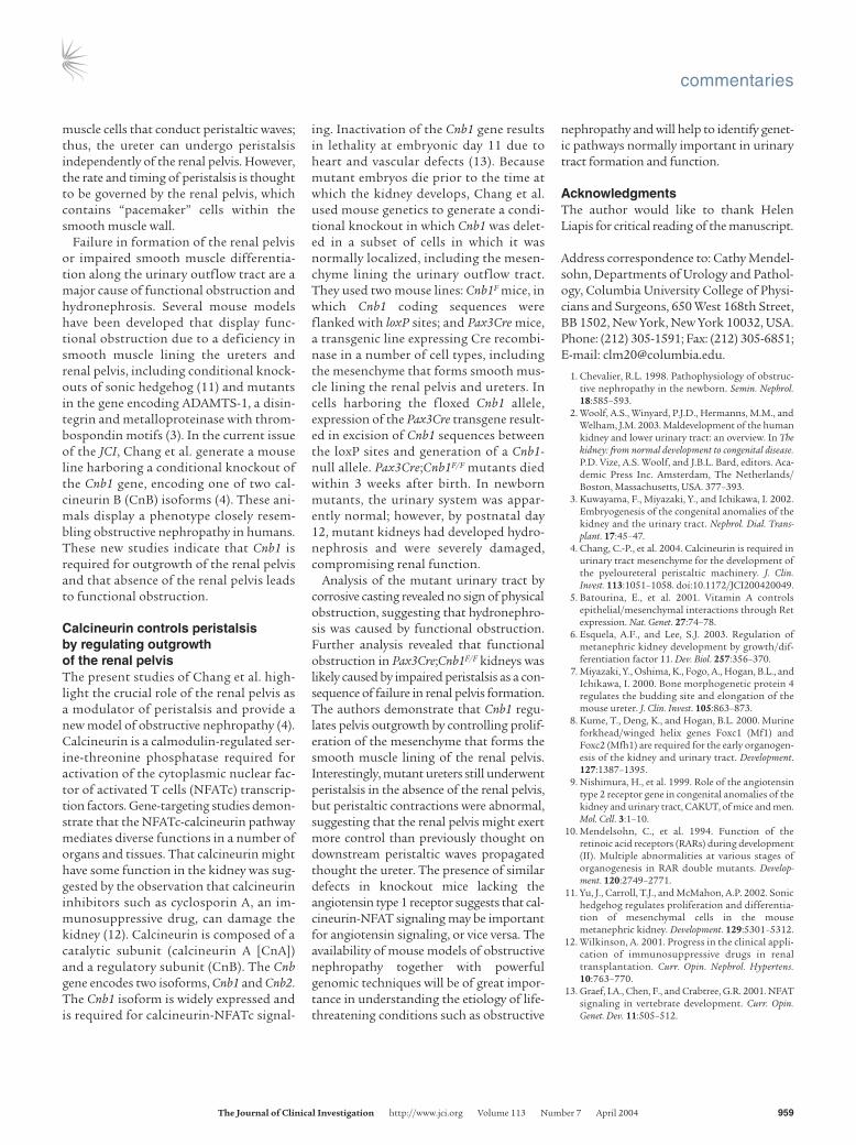
Figure 1A schematic showing different types ofobstruction that can cause hydronephrosis. (A)Top, examples of physical obstruction: ectopi-cally terminating ureter in a single (top) orduplicated (middle) collecting duct system. Inboth cases the ureter joins the urinary tractoutside the normal integration site in thetrigone. In the example showing a duplicatedsystem, one ureter joins normally; the other,abnormally. Bottom, uteropelvic junction (UPJ)stenosis or atresia causing physical blockageat the ureteropelvic junction. (B) Examples offunctional obstruction. Top, primary megau-reter caused by impaired peristalsis or defec-tive differentiation of smooth muscle in theureter coat. Bottom, UPJ abnormalities causedby failure in outgrowth or function of the renalpelvis. On the left, yellow filled arrowheadsdesignate the normal structure; the abnormalstructure on the right is designated by greenfilled arrowheads.
commentaries
The Journal of Clinical Investigation http://www.jci.org Volume 113 Number 7 April 2004 959
muscle cells that conduct peristaltic waves;thus, the ureter can undergo peristalsisindependently of the renal pelvis. However,the rate and timing of peristalsis is thoughtto be governed by the renal pelvis, whichcontains “pacemaker” cells within thesmooth muscle wall.
Failure in formation of the renal pelvisor impaired smooth muscle differentia-tion along the urinary outflow tract are amajor cause of functional obstruction andhydronephrosis. Several mouse modelshave been developed that display func-tional obstruction due to a deficiency insmooth muscle lining the ureters andrenal pelvis, including conditional knock-outs of sonic hedgehog (11) and mutantsin the gene encoding ADAMTS-1, a disin-tegrin and metalloproteinase with throm-bospondin motifs (3). In the current issueof the JCI, Chang et al. generate a mouseline harboring a conditional knockout ofthe Cnb1 gene, encoding one of two cal-cineurin B (CnB) isoforms (4). These ani-mals display a phenotype closely resem-bling obstructive nephropathy in humans.These new studies indicate that Cnb1 isrequired for outgrowth of the renal pelvisand that absence of the renal pelvis leadsto functional obstruction.
Calcineurin controls peristalsis by regulating outgrowth of the renal pelvisThe present studies of Chang et al. high-light the crucial role of the renal pelvis asa modulator of peristalsis and provide anew model of obstructive nephropathy (4).Calcineurin is a calmodulin-regulated ser-ine-threonine phosphatase required foractivation of the cytoplasmic nuclear fac-tor of activated T cells (NFATc) transcrip-tion factors. Gene-targeting studies demon-strate that the NFATc-calcineurin pathwaymediates diverse functions in a number oforgans and tissues. That calcineurin mighthave some function in the kidney was sug-gested by the observation that calcineurininhibitors such as cyclosporin A, an im-munosuppressive drug, can damage thekidney (12). Calcineurin is composed of acatalytic subunit (calcineurin A [CnA])and a regulatory subunit (CnB). The Cnbgene encodes two isoforms, Cnb1 and Cnb2.The Cnb1 isoform is widely expressed andis required for calcineurin-NFATc signal-
ing. Inactivation of the Cnb1 gene resultsin lethality at embryonic day 11 due toheart and vascular defects (13). Becausemutant embryos die prior to the time atwhich the kidney develops, Chang et al.used mouse genetics to generate a condi-tional knockout in which Cnb1 was delet-ed in a subset of cells in which it was normally localized, including the mesen-chyme lining the urinary outflow tract.They used two mouse lines: Cnb1F mice, inwhich Cnb1 coding sequences wereflanked with loxP sites; and Pax3Cre mice,a transgenic line expressing Cre recombi-nase in a number of cell types, includingthe mesenchyme that forms smooth mus-cle lining the renal pelvis and ureters. Incells harboring the floxed Cnb1 allele,expression of the Pax3Cre transgene result-ed in excision of Cnb1 sequences betweenthe loxP sites and generation of a Cnb1-null allele. Pax3Cre;Cnb1F/F mutants diedwithin 3 weeks after birth. In newbornmutants, the urinary system was appar-ently normal; however, by postnatal day12, mutant kidneys had developed hydro-nephrosis and were severely damaged,compromising renal function.
Analysis of the mutant urinary tract bycorrosive casting revealed no sign of physicalobstruction, suggesting that hydronephro-sis was caused by functional obstruction. Further analysis revealed that functionalobstruction in Pax3Cre;Cnb1F/F kidneys waslikely caused by impaired peristalsis as a con-sequence of failure in renal pelvis formation.The authors demonstrate that Cnb1 regu-lates pelvis outgrowth by controlling prolif-eration of the mesenchyme that forms thesmooth muscle lining of the renal pelvis.Interestingly, mutant ureters still underwentperistalsis in the absence of the renal pelvis,but peristaltic contractions were abnormal,suggesting that the renal pelvis might exertmore control than previously thought ondownstream peristaltic waves propagatedthought the ureter. The presence of similardefects in knockout mice lacking theangiotensin type 1 receptor suggests that cal-cineurin-NFAT signaling may be importantfor angiotensin signaling, or vice versa. Theavailability of mouse models of obstructivenephropathy together with powerfulgenomic techniques will be of great impor-tance in understanding the etiology of life-threatening conditions such as obstructive
nephropathy and will help to identify genet-ic pathways normally important in urinarytract formation and function.
AcknowledgmentsThe author would like to thank HelenLiapis for critical reading of the manuscript.
Address correspondence to: Cathy Mendel-sohn, Departments of Urology and Pathol-ogy, Columbia University College of Physi-cians and Surgeons, 650 West 168th Street,BB 1502, New York, New York 10032, USA.Phone: (212) 305-1591; Fax: (212) 305-6851;E-mail: [email protected].
1. Chevalier, R.L. 1998. Pathophysiology of obstruc-tive nephropathy in the newborn. Semin. Nephrol.18:585–593.
2. Woolf, A.S., Winyard, P.J.D., Hermanns, M.M., andWelham, J.M. 2003. Maldevelopment of the humankidney and lower urinary tract: an overview. In Thekidney: from normal development to congenital disease.P.D. Vize, A.S. Woolf, and J.B.L. Bard, editors. Aca-demic Press Inc. Amsterdam, The Netherlands/Boston, Massachusetts, USA. 377–393.
3. Kuwayama, F., Miyazaki, Y., and Ichikawa, I. 2002.Embryogenesis of the congenital anomalies of thekidney and the urinary tract. Nephrol. Dial. Trans-plant. 17:45–47.
4. Chang, C.-P., et al. 2004. Calcineurin is required inurinary tract mesenchyme for the development ofthe pyeloureteral peristaltic machinery. J. Clin.Invest. 113:1051–1058. doi:10.1172/JCI200420049.
5. Batourina, E., et al. 2001. Vitamin A controlsepithelial/mesenchymal interactions through Retexpression. Nat. Genet. 27:74–78.
6. Esquela, A.F., and Lee, S.J. 2003. Regulation ofmetanephric kidney development by growth/dif-ferentiation factor 11. Dev. Biol. 257:356–370.
7. Miyazaki, Y., Oshima, K., Fogo, A., Hogan, B.L., andIchikawa, I. 2000. Bone morphogenetic protein 4regulates the budding site and elongation of themouse ureter. J. Clin. Invest. 105:863–873.
8. Kume, T., Deng, K., and Hogan, B.L. 2000. Murineforkhead/winged helix genes Foxc1 (Mf1) andFoxc2 (Mfh1) are required for the early organogen-esis of the kidney and urinary tract. Development.127:1387–1395.
9. Nishimura, H., et al. 1999. Role of the angiotensintype 2 receptor gene in congenital anomalies of thekidney and urinary tract, CAKUT, of mice and men.Mol. Cell. 3:1–10.
10. Mendelsohn, C., et al. 1994. Function of theretinoic acid receptors (RARs) during development(II). Multiple abnormalities at various stages oforganogenesis in RAR double mutants. Develop-ment. 120:2749–2771.
11. Yu, J., Carroll, T.J., and McMahon, A.P. 2002. Sonichedgehog regulates proliferation and differentia-tion of mesenchymal cells in the mousemetanephric kidney. Development. 129:5301–5312.
12. Wilkinson, A. 2001. Progress in the clinical appli-cation of immunosuppressive drugs in renaltransplantation. Curr. Opin. Nephrol. Hypertens.10:763–770.
13. Graef, I.A., Chen, F., and Crabtree, G.R. 2001. NFATsignaling in vertebrate development. Curr. Opin.Genet. Dev. 11:505–512.