common elements in interleukin 4 and insulin signaling
TRANSCRIPT

Dartmouth College Dartmouth College
Dartmouth Digital Commons Dartmouth Digital Commons
Open Dartmouth: Peer-reviewed articles by Dartmouth faculty Faculty Work
5-1993
Common Elements in Interleukin 4 and Insulin Signaling Pathways Common Elements in Interleukin 4 and Insulin Signaling Pathways
in Factor-Dependent Hematopoietic Cells. in Factor-Dependent Hematopoietic Cells.
Ling-Mei Wang National Cancer Institute
Achsah D. A D Keegan National Institute of Allergy and Infectious Diseases
Weiqun Li National Cancer Institute
Gustav E. Lienhard Dartmouth College
Follow this and additional works at: https://digitalcommons.dartmouth.edu/facoa
Part of the Medical Cell Biology Commons
Dartmouth Digital Commons Citation Dartmouth Digital Commons Citation Wang, Ling-Mei; A D Keegan, Achsah D.; Li, Weiqun; and Lienhard, Gustav E., "Common Elements in Interleukin 4 and Insulin Signaling Pathways in Factor-Dependent Hematopoietic Cells." (1993). Open Dartmouth: Peer-reviewed articles by Dartmouth faculty. 1662. https://digitalcommons.dartmouth.edu/facoa/1662
This Article is brought to you for free and open access by the Faculty Work at Dartmouth Digital Commons. It has been accepted for inclusion in Open Dartmouth: Peer-reviewed articles by Dartmouth faculty by an authorized administrator of Dartmouth Digital Commons. For more information, please contact [email protected].

Proc. Natl. Acad. Sci. USAVol. 90, pp. 4032-4036, May 1993Cell Biology
Common elements in interleukin 4 and insulin signaling pathwaysin factor-dependent hematopoietic cells
(insulin receptor substrate 1/tyrosine phosphorylation/signal transduction)
LING-MEI WANG*, ACHSAH D. KEEGANt, WEIQUN LI*, GUSTAV E. LIENHARDt, STEFANIA PACINI§,J. SILVIo GUTKIND¶, MARTIN G. MYERS, JR. II, XIAO-JIAN SUN II, MoRius F. WHITEIISTUART A. AARONSON*, WILLIAM E. PAULt, AND JACALYN H. PIERCE****Laboratory of Cellular and Molecular Biology, National Cancer Institute, Bethesda, MD 20892; tLaboratory of Immunology, National Institute of Allergy andInfectious Diseases, Bethesda, MD 20892; tDepartment of Biochemistry, Dartmouth Medical School, Hanover, NH 03755-3844; §Institute of GeneralPathology, University of Florence, Florence, Italy; ILaboratory of Cellular Development and Oncology, National Institute of Dental Research,Bethesda, MD 20892; and IIJoslin Diabetes Center, Department of Medicine, Harvard Medical School, Boston, MA 02215
Contributed by William E. Paul, December 29, 1992
ABSTRACT Interleukin 4 (IL-4), insulin, and insulin-likegrowth factor I (IGF-I) efficiently induced DNA synthesis in theIL-3-dependent murine myeloid cell lines FDC-P1 and FDC-P2. Although these factors could not individually sustainlong-term growth of these lines, a combination of IL-4 witheither insulin or IGF-I did support continuous growth. Theprincipal tyrosine-phosphorylated substrate observed in FDCcells stimulated with IL-4, previously designated 4PS, was ofthe same size (170 kDa) as the major substrate phosphorylatedin response to insulin or IGF-I. These substrates had phos-phopeptides of the same size when analyzed by digestion withStaphylococcus aureus V8 protease, and each tightly associatedwith the 85-kDa component of phosphatidylinositol 3-kinaseafter factor stimulation. IRS-1, the principal substrate phos-phorylated in response to insulin or IGF-I stimulation innonhematopoietic cells, is similar in size to 4PS. However,anti-IRS-1 antibodies failed to efficiently precipitate 4PS, andsome phosphopeptides generated by V8 protease digestion ofIRS-1 were distinct in size from the phosphopeptides of 4PS.Nevertheless, IL-4, insulin, and IGF-I were capable of stimu-lating tyrosine phosphorylation of IRS-1 in FDC cells thatexpressed this substrate as a result of transfection. Thesermdings indicate that (i) IL-4, insulin, and IGF-I use signaltransduction pathways in FDC lines that have at least onemajor feature in common, the rapid tyrosine phosphorylationof 4PS, and (it) insulin and IGF-I stimulation of hematopoieticcell lines leads to the phosphorylation of a substrate that maybe related to but is not identical to IRS-1.
Interleukin 4 (IL-4) is a pluripotent cytokine that is intimatelyinvolved in determining the nature of an immune response toa given pathogen (reviewed in ref. 1). The cDNAs encodingboth the murine and human IL-4 receptors have been cloned(2-4). Although there are no consensus sequences in thereceptor that suggest any intrinsic tyrosine kinase activity,IL-4 does induce striking tyrosine phosphorylation of a170-kDa protein, designated IL-4-induced phosphotyrosinesubstrate (4PS) (5). In a previous study, 4PS was shown tostrongly associate with the 85-kDa subunit (p85) of phospha-tidylinositol (PI) 3-kinase immediately after IL-4 stimulation(5). This association is reminiscent of PI 3-kinase interactionwith insulin receptor substrate 1 (IRS-1), which leads to PI3-kinase activation (6-8). Although functions of the metab-olites produced by this enzyme are not understood, evidencesuggests that PI 3-kinase association with receptors or on-cogene products correlates with cell proliferation (reviewedin ref. 9).
IRS-1, the most prominent substrate phosphorylated inresponse to stimulation with insulin or insulin-like growthfactor I (IGF-I), is a hydrophilic protein of 160-185 kDa(10-13). The cDNAs for rat and mouse IRS-1 have beencloned (14, 15). The inferred amino acid sequence contains 20potential tyrosine and 30 potential serine and threoninephosphorylation sites. Nine putative tyrosine phosphoryla-tion sites are in YMXM or YXXM motifs (16). Tyrosinephosphorylation of these motifs is thought to result in asso-ciation with proteins containing Src homology 2 domains,such as p85 (reviewed in refs. 9, 17, and 18). The uniqueproperties of IRS-1 suggest that it could potentially serve asa docking molecule for many key proteins involved in signaltransduction (14, 16). Since insulin receptor mutants that aredefective for mitogenic signaling do not mediate phosphor-ylation of IRS-1 (19-22), it has been suggested that IRS-1 isa crucial component of insulin and IGF-I signaling.
In the present study we demonstrate that insulin and IGF-Iact as mitogens for certain IL-4-responsive hematopoieticcell lines and that the characteristics ofthe principal substratetyrosine-phosphorylated in cells treated with each of thesefactors are identical. The relationship between insulin- andIL-4-mediated signal transduction pathways in these factor-dependent cell lines is investigated.
MATERIALS AND METHODSCell Lines and Growth Assays. The CHO/IR/IRS-1 cells
have been described (7, 22). The IL-3-dependent murinehematopoietic cell lines FDC-P1 (23) and FDC-P2 (24) werecultured in RPMI 1640 medium with 10% fetal bovine serumand 5% WEHI-3B conditioned medium. The FDC-Pl/IRS-1line was generated by electroporation of the rat IRS-1 ex-pression vector after selection in the presence histidinol (2mM) as described (7, 25). Incorporation of [3H]thymidine wasused to quantitate factor-induced DNA synthesis in FDClines as described (5). Analysis of long-term growth proper-ties was performed in RPMI 1640 medium with 10% fetalbovine serum and recombinant IL-4 (10 nM), insulin (1 ,uM),or IGF-I (10 nM). Cells (5 x 10⁴ per ml) were plated in growthmedium with one or more factors. They were transferred ata split ratio of 1:10 biweekly. Cell viability was determined bytrypan blue exclusion.
Immunoprecipitation and Immunoblot Analysis. FDC orCHO cells were washed and resuspended in Dulbecco'smodified Eagle's medium (DMEM) with 50 ,uM Na3VO4 for
Abbreviations: IGF-I, insulin-like growth factor I; IL, interleukin;IRS-1, insulin receptor substrate 1; PI, phosphatidylinositol; 4PS,IL-4-induced phosphotyrosine substrate.**To whom reprint requests should be addressed.
4032
The publication costs of this article were defrayed in part by page chargepayment. This article must therefore be hereby marked "advertisement"in accordance with 18 U.S.C. §1734 solely to indicate this fact.
Proc. Natl. Acad. Sci. USA 90 (1993) 4033
2 hr. After stimulation with either IL-4 (100 nM), insulin (10AM), or IGF-I (100 nM) for 10 min at 37°C, cells were lysedin lysis buffer as described (5). Equal amounts of clarifiedFDC lysates (2 mg) or CHO/IR/IRS-1 lysates (0.5-1 mg)were immunoprecipitated with 20 ul of agarose-conjugatedanti-phosphotyrosine (Upstate Biotechnology, Lake Placid,NY), anti-p85 (1:500; Upstate Biotechnology), or anti-IRS-1(1:500; ref. 7) plus 30 Al of protein G-coupled Sepharose.Immunoprecipitates were washed with lysis buffer, solubi-lized with Laemmli buffer, boiled, and resolved by SDS/8%PAGE for immunoblot analysis (5).
Peptide Mapping Using Staphylococcus aureus V8 Protease.Cells were cultured in serum-free DMEM containing 50 ,uMNa3VO4 for 2 hr and labeled with [32P]orthophosphoric acid(500 ,tCi/ml; 1 gCi = 37 kBq) in phosphate-free medium for4 hr. Untreated or factor-treated cells were lysed in lysisbuffer and immunoprecipitated with anti-phosphotyrosine oranti-IRS-i. The immunoprecipitates were resolved bySDS/8% PAGE and the 170-kDa bands (present only insamples from factor-treated cells) were located by autorad-iography and excised. Peptides from the 170-kDa bands weregenerated by in situ proteolytic cleavage within the stackinggel (26).
RESULTSGrowth Potential of IL-4, Insulin, and IGF-I in IL-3-
Dependent FDC Lines. IL-4, insulin, and IGF-I each induceddose-dependent [3H]thymidine incorporation in the myeloidprogenitor lines FDC-P1 and FDC-P2 under serum-free con-ditions. IL-4, insulin, and IGF-I stimulated half-maximalresponses in FDC-P1 cells at approximately 100 pM, 10 nM,and 20 pM, respectively (Fig. 1). The maximum level of[3H]thymidine incorporation achieved by each factor wassimilar to that of IL-3, implying that they were capable ofinducing as potent a mitogenic response as IL-3 in FDC-P1cells. FDC-P2 cells also responded mitogenically to IL-4,insulin, and IGF-I, with half-maximal responses at 25 pM,100 nM, and 1 nM, respectively (data not shown). Theseresults provide evidence that the FDC lines possess cellsurface receptors and intracellular components required toallow short-term mitogenic signaling through the IL-4, insu-lin, and IGF-I pathways.While IL-4, insulin, and IGF-I did stimulate DNA synthe-
sis, they failed to support long-term growth of the FDC cells.Although cells from both lines survived for over 72 hr ingrowth medium containing recombinant IL-4, insulin, or
D
E
I-
co
0
x
0C
1U
6
4-
2i
0 1 2 3 4 5 6 7 8
Log Concentration (pM)
FIG. 1. Mitogenic dose-response of FDC-P1 cells to variousgrowth factors. FDC-P1 cells were incubated for 36 hr in serum-freemedium with various concentrations of IL-4 (L), IL-3 (o), insulin (L),or IGF-I (v). [3H]Thymidine (0.5 ,uCi per well) was added for 4 hr andsamples were then collected by an automatic harvester. Results areexpressed as counts per minute (cpm) in stimulated samples minusbackground cpm.
IGF-I, they did not remain viable when transferred to newculture dishes containing the same factor. By contrast, acombination of IL-4 with either insulin or IGF-I yieldedsustained growth ofboth lines. Insulin and IGF-I together didnot support continuous growth of either line. Even afterpropagation for over 4 months in medium containing bothIL-4 and insulin, neither line continued to proliferate whenswitched to medium containing only one factor.
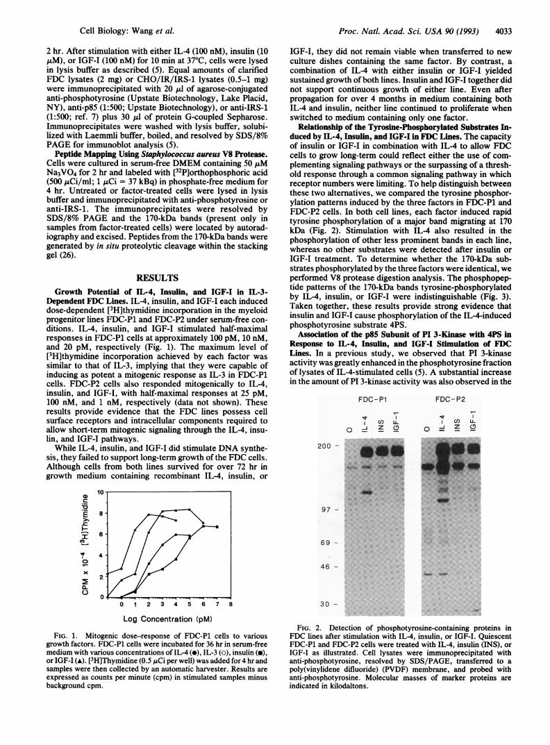
Relationship of the Tyrosine-Phosphorylated Substrates In-duced by IL-4, Insulin, and IGF-I in FDC Lines. The capacityof insulin or IGF-I in combination with IL-4 to allow FDCcells to grow long-term could reflect either the use of com-plementing signaling pathways or the surpassing of a thresh-old response through a common signaling pathway in whichreceptor numbers were limiting. To help distinguish betweenthese two alternatives, we compared the tyrosine phosphor-ylation patterns induced by the three factors in FDC-P1 andFDC-P2 cells. In both cell lines, each factor induced rapidtyrosine phosphorylation of a major band migrating at 170kDa (Fig. 2). Stimulation with IL-4 also resulted in thephosphorylation of other less prominent bands in each line,whereas no other substrates were detected after insulin orIGF-I treatment. To determine whether the 170-kDa sub-strates phosphorylated by the three factors were identical, weperformed V8 protease digestion analysis. The phosphopep-tide patterns of the 170-kDa bands tyrosine-phosphorylatedby IL-4, insulin, or IGF-I were indistinguishable (Fig. 3).Taken together, these results provide strong evidence thatinsulin and IGF-I cause phosphorylation of the IL-4-inducedphosphotyrosine substrate 4PS.
Association of the p85 Subunit of PI 3-Kinase with 4PS inResponse to IL-4, Insulin, and IGF-I Stimulation of FDCLines. In a previous study, we observed that PI 3-kinaseactivity was greatly enhanced in the phosphotyrosine fractionof lysates of IL-4-stimulated cells (5). A substantial increasein the amount of PI 3-kinase activity was also observed in the
FDC-P1
i cn L-O 'i Z CD
200 - D
. ..-..:
.m it
Am
97 -
FDC-P2
CD LUJ Z 0
4-11 -,::`-
69 -
46 -
30 -
FIG. 2. Detection of phosphotyrosine-containing proteins inFDC lines after stimulation with IL-4, insulin, or IGF-I. QuiescentFDC-P1 and FDC-P2 cells were treated with IL-4, insulin (INS), orIGF-I as illustrated. Cell lysates were immunoprecipitated withanti-phosphotyrosine, resolved by SDS/PAGE, transferred to apoly(vinylidene difluoride) (PVDF) membrane, and probed withanti-phosphotyrosine. Molecular masses of marker proteins areindicated in kilodaltons.
Cell Biology: Wang et al.

Proc. Natl. Acad. Sci. USA 90 (1993)
FDC-P2 CHO/IR/IRS-1
IL-4 INS IGF-1 INS INS
200- *94- _69
46 -
..
30
14
s. I*::
s: :v:.w w.
* ^.. :. .:_
J ::: wJ- : _*s. ::.:.. .:...:w.*i: ::: ::.s
w :w
w.
Ptyr a Ptyr axPtyr acPtyr (xLIRS-1
FIG. 3. V8 digestion pattern oftyrosine-phosphorylated 170-kDaproteins' after factor stimulation of FDC-P2 and CHO/IR/IRS-1cells. Partial V8 digestion was performed on IL-4-, insulin (INS)-, orIGF-I-treated [32P]orthophosphate-labeled 170-kDa proteins immu-noprecipitated with anti-phosphotyrosine (aPtyr) or anti-IRS-1(aIRS-1) from FDC-P2 and CHO/IR/IRS-1 cells.
phosphotyrosine fraction of lysates of insulin- or IGF-I-treated FDC-Pi or FDC-P2 cells as determined by theconversion ofPI to PI 3-phosphate in an in vitro reaction (datanot shown). To more specifically define the interaction of PI3-kinase with the tyrosine-phosphorylated substrates in-duced in FDC lines, lysates were first immunoprecipitatedwith anti-p85 and then immunoblotted with anti-phosphoty-rosine. Chinese hamster ovary cells transfected with both thehuman insulin receptor and rat IRS-1 (CHO/IR/IRS-1) werealso analyzed for comparison (7, 22). A strong band migrating
A FDC-P1 FDC- P2 CHO/IRIIRS-1
qt CED ILcOLi- nCu- CO
*....~~~~~~~~~~~~~~~~~~..... .. = ..
200-ii il
97....-,...sfi * .:' .....
68.__l__ |s
Blot: a- PtyrIP: a-p85
at 170 kDa was observed when either FDC line was treatedwith IL-4, insulin, or IGF-I or when CHO/IR/IRS-1 wastreated with insulin (Fig. 4A). When lysates were immuno-precipitated with anti-phosphotyrosine and immunoblottedwith anti-p85, the amount of p85 was substantially increasedin the IL-4-, insulin-, or IGF-I-treated FDC and insulin-stimulated CHO/IR/IRS-1 samples when compared with thelysates of untreated cells (Fig. 4B). Thus, 4PS stronglyassociates with the p85 subunit of PI 3-kinase in response toinsulin, IGF-I, or IL-4 stimulation of hematopoietic cells justas IRS-1 does in nonhematopoietic cells after insulin stimu-lation.
Relationship of 4PS to IRS-1. To determine the relationshipof 4PS to IRS-1, lysates from factor-stimulated FDC-P2 andCHO/IR/IRS-1 cells were immunoprecipitated with a poly-clonal antibody directed against baculovirus-generated ratIRS-1 or with anti-phosphotyrosine and subsequently immu-noblotted with anti-phosphotyrosine (Fig. 5). Although anti-IRS-1 immunoprecipitated a modest amount of 4PS fromlysates of IL-4- or insulin-treated FDC-P2 cells, it was muchless than that immunoprecipitated by anti-phosphotyrosine.By contrast, anti-IRS-i immunoprecipitated a much greaterfraction of that recognized by anti-phosphotyrosine from theinsulin-stimulated CHO/IR/IRS-1 lysate. Moreover, whenrabbit antibodies directed against either amino- or carboxyl-terminal peptides ofrat/mouse IRS-1 (13, 27) were utilized insimilar experiments or for direct immunoblot analysis, 4PSwas not detected whereas IRS-1 was readily observed (datanot shown).V8 protease digestion analysis was carried out on 4PS
immunoprecipitated with anti-phosphotyrosine from lysatesof IL-4- or insulin-stimulated FDC-P2 cells and on IRS-1immunoprecipitated with anti-IRS-1 or anti-phosphotyrosinefrom insulin-treated CHO/IR/IRS-1 cells. Although the pat-terns of phosphopeptides derived from 4PS (from FDC-P2cells) and IRS-1 (from CHO/IR/IRS-1 cells) showed simi-larities, they were not identical (Fig. 3). While a 34-kDapeptide as well as other less distinct peptides in the 12- to24-kDa range were found in digests from both substrates, theIRS-1 digest contained a prominent 25- to 29-kDa peptide notfound in the 4PS digest (Fig. 3). Although these results further
B FDC-P1 FDC-P2 CHO/IRIIRS-1
Stcn)LL urcL cO
O ZZ oOZ
200 -
97 -
p85 - S
68 -
Blot: (l-p85iP: a-Ptyr
FIG. 4. Association of the p85 subunit of PI 3-kinase with 4PS in FDC lines after stimulation with IL-4, insulin (INS), or IGF-I. (A) Lysatesof IL-4-, insulin-, or IGF-I-treated FDC-P2 and insulin-stimulated CHO/IR/IRS-1 cells were immunoprecipitated (IP) with anti-p85, subjectedto SDS/PAGE, and immunoblotted with anti-phosphotyrosine (a-Ptyr). (B) The same lysates were immunoprecipitated with anti-phosphotyrosine, and transferred proteins were immunoblotted with anti-p85.
4034 Cefl Biology: Wang et al.
Proc. Natl. Acad. Sci. USA 90 (1993) 4035
CHO/FDC-P2 IR/
IRS-1
U)
I0(1
CHO/
FDC-P2 IR/IRS-1
U) CO)O =2 0Z
BFDC-P2
CHO/IR/
IRS-1
0 =4 o
200 -
97 -
IP: a-Ptyr IP: a-IRS-1 IP: a-IRS-1Blot: a-Ptyr Blot: a-Ptyr Blot: a-Ptyr
FIG. 5. Weak recognition of 4PS by an IRS-i-specific antibody.(A) Lysates of IL-4- or insulin (INS)-treated FDC-P2 or insulin-treated CHO/IR/IRS-1 cells were immunoprecipitated (IP) withanti-phosphotyrosine (a-Ptyr) or anti-rat IRS-1, resolved by SDS/PAGE, and immunoblotted with anti-phosphotyrosine. (B) Sameanti-phosphotyrosine immunoblot of anti-IRS-1 immunoprecipitatesas in A, except that the autoradiograph was exposed for 72 hr insteadof 24 hr.
indicate that 4PS and IRS-1 are not identical, the finding ofphosphopeptides ofcommon size and the weak recognition of4PS by polyclonal anti-rat IRS-1 suggest that they may berelated to one another.
IL-4 Induces Tyrosine Phosphorylation of Rat IRS-1 inFDC-Pl Cells Transfected with an IRS-1 Expression Vector. Tomore fully investigate the relationship between insulin- andIL-4-mediated signals, we sought to determine whether IL-4could induce tyrosine phosphorylation of the protein encodedby the cloned IRS-1 gene in the FDC background. An expres-sion vector containing rat IRS-1 was introduced into theFDC-P1 line by electroporation. Bands of 170 kDa werereadily detected in lysates of IL-4- and insulin-treated FDC-Pl/IRS-i cells after immunoprecipitation with anti-IRS-i fol-lowed by immunoblot analysis with anti-phosphotyrosine(Fig. 6B). The relative amount of tyrosine-phosphorylatedIRS-1 compared to total tyrosine-phosphorylated protein rec-
AFDC- P1/IRS-1
CHO/IR/
IRS-1
0 IL-4 INS 0 INS
200 -
97 -
IP: a-PtyrBlot: a-Ptyr
B
FDC-P1/IRS-i IRS-IS IRS-10 IL-4 INS 0 INS
200 -.... ...: .. .....
97-
IP: (x-IRS-1
Blot: (z-Ptyr
FIG. 6. Detection of tyrosine-phosphorylated IRS-1 in responseto IL-4 or insulin (INS) stimulation of the FDC-Pl/IRS-1 transfec-tant. (A) Lysates from factor-stimulated FDC-Pl/IRS-1 or CHO/IR/IRS-1 cells were immunoprecipitated (IP) and subsequentlyimmunoblotted with anti-phosphotyrosine (a-Ptyr). (B) Identicallysates were immunoprecipitated with anti-rat IRS-1 and immuno-blotted with anti-phosphotyrosine.
ognized in FDC-P1/IRS-1 lysates was equivalent to thatobserved in the CHO/IR/IRS-1 lysate (Fig. 6). These resultsimply that IRS-1 can be efficiently phosphorylated on tyrosinein response to IL-4 or insulin stimulation in the FDC hema-topoietic cell background.
DISCUSSIONThe results demonstrate that IL-4, insulin, and IGF-I eachinduce pronounced tyrosine phosphorylation of a 170-kDasubstrate in murine IL-3-dependent myeloid progenitor celllines. We have also observed tyrosine phosphorylation of a170-kDa substrate in megakaryocytic, mast cell, and B lym-phoid lines in response to IL-4, insulin, or IGF-I treatmentand in lipopolysaccharide-stimulated B cells and Con A-stim-ulated T cells after treatment with IL-4 (unpublished work).Thus, IL-4 and insulin induce similar tyrosine phosphoryla-tion events in very different hematopoietic cell lineages.Interestingly, the concentrations of both IL-4 and insulinrequired to detect phosphorylation of and PI 3-kinase asso-ciation with 4PS correlated precisely with those needed toevoke DNA synthesis (data not shown), suggesting that thissubstrate may play a crucial role in hematopoietic cell signaltransduction.Our results indicate that the 170-kDa protein phosphory-
lated in response to insulin and IGF-I in the FDC lines isidentical to that phosphorylated in response to IL-4. Thesubstrate strongly associated with the 85-kDa subunit of PI3-kinase after stimulation with each factor. Moreover, thephosphopeptide patterns generated by V8 digestion of the170-kDa proteins were identical to each other. Thus, itappears that IL-4, insulin, and IGF-I activate tyrosine ki-nases in FDC cells (and presumably other hematopoietic-lineage cells) which phosphorylate an identical substrate,4PS. The results obtained suggest that 4PS is probably relatedto but not identical to IRS-1. While antibodies directedagainst amino- and carboxyl-terminal peptides of rat/mouseIRS-1 did not recognize 4PS (data not shown), a polyclonalantibody directed against the entire rat IRS-1 protein wasable to weakly detect 4PS. V8 digestion of 4PS and IRS-1generated some peptides with similar mobilities and otherswhich were distinct. Although 4PS and IRS-1 do not appearto be identical proteins, IL-4 was able to induce readilydetectable tyrosine phosphorylation of rat IRS-1 expressed inthe FDC-P1/IRS-1 transfectant. Thus, the tyrosine kinaseactivated by IL-4 stimulation is capable of phosphorylatingIRS-1, just as insulin or IGF-I receptor activation can medi-ate tyrosine phosphorylation of 4PS. These results suggestthat 4PS and IRS-1 may be encoded by evolutionarily relatedgenes which are preferentially expressed in different celltypes and may play similar functional roles in signal trans-duction.To date, no other factor has been shown to induce phos-
phorylation of 4PS or IRS-1. While IL-3 stimulated pro-nounced tyrosine phosphorylation of several substrates in theFDC lines, it did not detectably phosphorylate 4PS (5). Wehave recently observed weak IL-3-induced phosphorylationof an -170-kDa protein in certain cell lines. Whether thissubstrate is related to 4PS remains to be determined. Othercytokines whose receptors are members of the hematopoietinsuperfamily, including granulocyte/macrophage-colony-stimulating factor, IL-2, IL-7, and erythropoietin, have alsobeen reported to induce tyrosine phosphorylation in variouscell lines (28-33). However, no substrate in the 170-kDa sizerange was described in any of these studies. These resultssuggest that the tyrosine kinase activated by IL-4 stimulationdiffers from those most commonly induced by other cyto-kines.An important question which arises from this study is how
IL-4-mediated signaling leads to the tyrosine phosphoryla-
Cell Biology: Wang et al.

Proc. Natl. Acad. Sci. USA 90 (1993)
tion of 4PS. In contrast to insulin and IGF-I receptors, theIL-4 receptor possesses no known tyrosine kinase domain(2-4, 34, 35). A previous study demonstrated that tyrosineresidues found in the YXXM motifs of IRS-1 were excellentsubstrates for an activated insulin receptor kinase (16). Thus,it is likely that activated insulin or IGF-I receptors directlyphosphorylate 4PS in the FDC lines. It is tempting to spec-ulate that IL-4 might channel its signal by transactivatingeither insulin or IGF-I receptor tyrosine kinases. While wewere able to observe insulin-dependent insulin receptor au-tophosphorylation in the FDC lines, IL-4 treatment did notdetectably affect this receptor (data not shown). Thus, it ismore likely that IL-4 induces phosphorylation of 4PS byactivating an independent tyrosine kinase.Although our results provide evidence that IL-4, insulin,
and IGF-I share similarities in some of the biological andbiochemical cascades they induce in the FDC lines, it re-mains to be determined whether these factors activate otheroverlapping signaling events. Insulin has been demonstratedto stimulate tyrosine phosphorylation of mitogen-activatedprotein kinase, phosphorylation ofRaf kinase, and activationof the cellular Ras protein (36-41). By contrast, others havereported that IL-4 does not affect any of these criticalsignaling molecules (42-45). Thus, it seems likely that IL-4and insulin signaling cascades share some coincident andother divergent elements that account for their distinct phys-iological roles.
We thank Charles Knicley and Jennifer Artrip for excellenttechnical assistance.
1. Paul, W. E. (1991) Blood 77, 1859-1870.2. Mosley, B., Beckmann, M. P., March, C. J., Idzerda, R. L.,
Gimpel, S. D., VandenBos, T., Friend, D., Alpert, A., Ander-son, D., Jackson, J., Wignall, J., Smith, C., Gallis, B., Sims,J. E., Urdal, D. L., Widmer, M. B., Cosman, D. & Park, L. S.(1989) Cell 59, 335-344.
3. Galizzi, J.-P., Zuber, C. E., Harada, N., Gorman, D. M.,Djossou, O., Kastelein, R., Banchereau, J., Howard, M. &Miyajima, A. (1990) Int. Immunol. 2, 669-679.
4. Idzerda, R. L., March, C. J., Mosely, B., Lyman, S. D.,VandenBos, T., Gimpel, S. D., Din, W. S., Grabstein, K. H.,Widmer, M. B., Park, L. S., Cosman, D. & Beckmann, M. P.(1990) J. Exp. Med. 171, 861-876.
5. Wang, L.-M., Keegan, A. D., Paul, W. E., Heidaran, M. A.,Gutkind, J. S. & Pierce, J. H. (1992) EMBO J. 11, 4899-4908.
6. Ruderman, N. B., Kapeller, R., White, M. F. & Cantley, L. C.(1990) Proc. Natl. Acad. Sci. USA 87, 1411-1415.
7. Backer, J. M.,Myers,M. G., Jr., Shoelson, S. E., Chin,D. J.,Sun, X.-J.,Miralpeix, M., Hu,P., Margolis,B., Skolnik,E. Y.,Schlessinger, J. & White, M. F. (1992) EMBOJ. 11,3469-3479.
8. Lavan, B. E., Kuhne, M. R., Gamer, C. W., Anderson, D.,Reedijk, M., Pawson, T. & Lienhard, G. E. (1992) J. Biol.Chem. 267, 11631-11636.
9. Cantley, L. C., Auger, K. R., Carpenter, C., Duckworth, B.,Graziani, A., Kapeller, R. & Soitoff, S. (1991) Cell 64,281-303.
10. White, M. F., Maron, R. & Kahn, C. R. (1985) Nature (Lon-don) 318, 183-186.
11. White, M. F., Stegmann, E. W., Dull, T. J., Ullrich, A. &Kahn, C. R. (1987) J. Biol. Chem. 262, 9769-9777.
12. Izumi, T., White, M. F., Kadowaki, T., Takaku, F., Akanuma,Y. & Kasuga, M. (1987) J. Biol. Chem. 262, 1282-1287.
13. Keller, S. R., Kitagawa, K., Aebersold, R., Lienhard, G. E. &Gamer, C. W. (1991) J. Biol. Chem. 266, 12817-12820.
14. Sun, X. J., Rothenberg, P., Kahn, C. R., Backer, J. M., Araki,E., Wilden, P. A., Cahill, D. A., Goldstein, B. J. & White,M. F. (1991) Nature (London) 352, 73-77.
15. Keller, S. R., Aebersold, R., Garner, C. W. & Lienhard,G. E., Biochim. Biophys. Acta, in press.
16. Shoelson, S. E., Chattedjee, S., Chaudhuri, M. & White, M. F.(1992) Proc. Natl. Acad. Sci. USA 89, 2027-2031.
17. Heldin, C. H. (1991) Trends Biochem. Sci. 16, 450-452.18. Koch, C. A., Anderson, D., Moran, M. F., Ellis, C. & Pawson,
T. (1991) Science 252, 668-674.19. White, M. F., Livingston, J. N., Backer, J. M., Lauris, V.,
Dull, T. J., Ullrich, A. & Kahn, C. R. (1988) Cell 54,641-649.20. Backer, J. M., Kahn, C. R., Cahill, D. A., Ullrich, A. & White,
M. F. (1990) J. Biol. Chem. 265, 16450-16454.21. Backer, J. M., Schroeder, G. G., Cahill, D. A., Ullrich, A.,
Siddle, K. & White, M. F. (1991) Biochemistry 30, 6366-6372.22. Backer, J. M., Schroeder, G. G., Kahn, C. R., Myers, M. G.,
Jr., Wilden, P. A., Cahill, D. A. & White, M. F. (1992) J. Biol.Chem. 267, 1367-1374.
23. Dexter, T. M., Garland, J., Scott, D., Scolnick, E. & Metcalf,D. (1980) J. Exp. Med. 152, 1036-1047.
24. Le Gros, G. S., Gillis, S. & Watson, J. D. (1985) J. Immunol.135, 4009-4014.
25. Pierce, J. H., Ruggiero, M., Fleming, T. P., Di Fiore, P. P.,Greenberger, J. S., Varticovski, L., Schlessinger, J., Rovera,G. & Aaronson, S. A. (1988) Science 239, 628-631.
26. Cleveland, D. W. (1983) Methods Enzymol. 96, 222-229.27. Lampere, L. & Lienhard, G. (1992) Endocrinology 131, 2196-
2202.28. Morla, A. O., Schreurs, J., Miyajima, A. & Wang, J. Y. J.
(1988) Mol. Cell. Biol. 88, 2214-2221.29. Ferris, D. K., Willette-Brown, J., Linnekin, D. & Farrer,
W. L. (1989) Lymphokine Res. 8, 215-224.30. Augustine, J. A., Schlager, J. W. & Abraham, R. T. (1990)
Biochim. Biophys. Acta 1052, 313-319.31. Kanakura, Y., Druker, B., Cannistra, S. A., Furukawa, Y.,
Torimoto, Y. & Griffin, J. D. (1990) Blood 76, 706-711.32. Quelle, F. V. & Wojchowski, D. M. (1991) J. Biol. Chem. 266,
609-614.33. Uckun, F. M., Dibirdik, I., Smith, R., Tuel-Ahlgren, L.,
Chanden-Langlie, M., Schieven, G. L., Weddick, K. G., Han-son, M. & Ledbetter, J. A. (1991) Proc. Natl. Acad. Sci. USA88, 3589-3593.
34. Kasuga, M., Karlsson, F. A. & Kahn, C. R. (1982) Science 215,185-187.
35. Ullrich, A., Gray, A., Tam, A. W., Yang-Feng, T., Tsu-bokawa, M., Collins, C., Henzel, W., Le Bon, T., Kathuria, S.& Chen, E. (1986) EMBO J. 5, 2503-2512.
36. Ray, L. B. & Sturgill, T. W. (1987) Proc. Natl. Acad. Sci. USA84, 1502-1506.
37. Blackshear,P. J.,Haupt,D. M.,App,H.& Rapp, U. R. (1990)J. Biol. Chem. 265, 12131-12134.
38. Boulton, T. G., Nye, S. H., Robbins, D. J., Ip, N. Y., Road-ziejewska, E., Morgenbesser, S. D., DePinho, R. A., Panay-otatos, N., Cobb, M. H. & Yancopoulos, G. D. (1991) Cell 65,663-675.
39. Kovacina, K. S., Yonezawa, K., Brautigan, D., Tonks, N. K.,Rapp, U. R. & Roth, R. A. (1990) J. Biol. Chem. 265, 12115-12118.
40. Burgering, B. M., Snijders, A. J., Maassen, J. A., van der Eb,A. J. & Bos, J. L. (1989) Mol. Cell. Biol. 9, 4312-4322.
41. Izumi, T., Tamemoto, H., Nagao, M., Kadowaki, T., Takaku,F. & Kasuga, M. (1991) J. Biol. Chem. 266, 7933-7939.
42. Turner, B., Rapp, U., App, H., Greene, M., Dobashi, K. &Reed, J. (1991) Proc. Natl. Acad. Sci. USA 88, 1227-1231.
43. Satoh, T., Nakafuka, M., Miyajima, A. & Kajiro, Y. (1991)Proc. Natl. Acad. Sci. USA 88, 3314-3318.
44. Duronio, V., Welhem, M. J., Abraham, S., Dryden, R. &Schrader, J. W. (1992) Proc. Natl. Acad. Sci. USA 89, 1587-1591.
45. Welham, M. J., Duronio, V., Sanghera, J. S., Pelech, S. L. &Schrader, J. W. (1992) J. Immunol. 149, 1683-1693.
4036 Cell Biology: Wang et al.