common nonmutational notch1 activation in chronic ... nonmutational notch1 activation in chronic...
TRANSCRIPT

Common nonmutational NOTCH1 activation in chroniclymphocytic leukemiaGiulia Fabbria, Antony B. Holmesa, Mara Viganottia, Claudio Scuoppoa, Laura Belvera, Daniel Herranza, Xiao-Jie Yanb,Yasmine Kiesob, Davide Rossic,d, Gianluca Gaidanoe, Nicholas Chiorazzib, Adolfo A. Ferrandoa,f,g,and Riccardo Dalla-Faveraa,f,h,i,1
aInstitute for Cancer Genetics and the Herbert Irving Comprehensive Cancer Center, Columbia University, New York, NY 10032; bThe Feinstein Institute forMedical Research, Northwell Health, Manhasset, New York, NY 11030; cDivision of Hematology, Oncology Institute of Southern Switzerland, 6500Bellinzona, Switzerland; dLymphoma and Genomics Research Program, Institute of Oncology Research, 6500 Bellinzona, Switzerland; eDivision ofHaematology, Department of Translational Medicine, Università del Piemonte Orientale, 28100 Novara, Italy; fDepartment of Pathology and Cell Biology,Columbia University, New York, NY 10032; gDepartment of Pediatrics, Columbia University, New York, NY 10032; hDepartment of Genetics & Development,Columbia University, New York, NY 10032; and iDepartment of Microbiology & Immunology, Columbia University, New York, NY 10032
Contributed by Riccardo Dalla-Favera, February 15, 2017 (sent for review December 20, 2016; reviewed by Carlo M. Croce and Louis M. Staudt)
Activating mutations of NOTCH1 (a well-known oncogene in T-cellacute lymphoblastic leukemia) are present in ∼4–13% of chroniclymphocytic leukemia (CLL) cases, where they are associated withdisease progression and chemorefractoriness. However, the specificrole of NOTCH1 in leukemogenesis remains to be established. Here,we report that the active intracellular portion of NOTCH1 (ICN1)is detectable in ∼50% of peripheral blood CLL cases lacking genemutations. We identify a “NOTCH1 gene-expression signature” inCLL cells, and show that this signature is significantly enriched inprimary CLL cases expressing ICN1, independent of NOTCH1 muta-tion. NOTCH1 target genes include key regulators of B-cell prolifera-tion, survival, and signal transduction. In particular, we show thatNOTCH1 transactivates MYC via binding to B-cell–specific regulatoryelements, thus implicating this oncogene in CLL development. Theseresults significantly extend the role of NOTCH1 in CLL pathogenesis,and have direct implications for specific therapeutic targeting.
chronic lymphocytic leukemia | NOTCH1 | transcriptional network
Chronic lymphocytic leukemia (CLL) is a common hematologictumor characterized by the clonal expansion of CD5+ B cells
(1, 2). Recent investigations have provided a comprehensive pic-ture of the CLL genome, revealing its relatively low burden ofgenetic lesions, with a small number of frequently mutated “driver”genes. CLL mutated genes include the NOTCH1 oncogene, thesplicing regulator SF3B1, the tumor-suppressors TP53 and ATM,and several B-cell receptor (BCR)/NF-κB regulators, such asMYD88, BIRC3, and NFKBIE, among others (3–7).NOTCH1, a well-known oncogene in T-cell acute lymphoblastic
leukemia (T-ALL) (8, 9), has emerged as the most commonlymutated gene in CLL at diagnosis, accounting for ∼4–13% ofpatients (3–7). NOTCH1 encodes a transmembrane receptor that,upon binding to a ligand expressed on the surface of a “signal-sending” cell, undergoes a series of conformational changes andproteolytic cleavages, ultimately allowing the translocation of itsintracellular, cleaved, and active portion (hereinafter referred to as“ICN1”) to the nucleus (10, 11). Once in the nucleus, ICN1 bindsto the DNA-binding protein RBPJ, the main effector of NOTCH-signaling, and recruits a series of coactivator proteins to inducetranscriptional activation of target genes (10, 11). NOTCH1 targetgenes mediate regulation of fundamental biological processes, suchas development, cell differentiation, cell-fate decision, prolifera-tion, and apoptosis (10, 11).In contrast to T-ALL, where the majority of mutations are
represented by constitutively activated ligand-independent al-leles affecting the heterodimerization domain of the protein,most NOTCH1 mutational events in CLL are representedby PEST [proline (P), glutamic acid (E), serine (S), threonine(T)-rich protein sequence] -truncations removing the phosphode-gron sequence required for FBXW7-mediated ICN1 proteasomaldegradation; in a minority of cases, point mutations in the 3′UTR
of the NOTCH1 mRNA lead to aberrant splicing events that alsoremove the PEST domain of the NOTCH1 protein (3–7). NOTCH1mutations in CLL were shown to associate with poor prognosis,including a specific subset of patients carrying trisomy 12, diseaseprogression, transformation to highly aggressive diffuse large B-celllymphomas, termed Richter syndrome, and immunochemotherapyresistance (3, 4, 12–16).Despite this potentially relevant role in the CLL clinical
course, the oncogenic role of NOTCH1 in this disease remainspoorly understood. Although few NOTCH1 targets have beenshown to be overexpressed in NOTCH1-mutated cases comparedwith wild-type CLL (4, 17, 18), the full spectrum of genes con-trolled by NOTCH1 and their contribution to the disease path-ogenesis have not been identified. Moreover, recent reportsdocumented that CLL cells in the lymph node frequently expressICN1, independent of NOTCH1 PEST-truncation (19, 20), es-pecially within the proliferation centers, which represent the keymicroanatomical sites of interaction of CLL cells with accessorycells and proliferation (21). Accordingly, these findings havebeen interpreted as the result of microenvironmental signals ac-tivating the NOTCH1 cascade. Conversely, the status of NOTCH1
Significance
A pathogenetic role of NOTCH1 in chronic lymphocytic leukemia(CLL) has been implied by the presence of deregulating mutationsin a relatively small fraction of cases. Our results now indicatethat ∼50% of CLL cases devoid of mutations express the activeform of NOTCH1 ICN1 (intracellular portion of NOTCH1), thusimplicating a much broader role of this transcription factor in thedisease. ICN1+ CLL cases display equivalent NOTCH1-dependenttranscriptional responses regardless of the gene mutation status,indicating that the detection of ICN1 represents a reliable bio-marker of NOTCH1 activation for diagnostic and therapeutictargeting. Finally, our results identify the NOTCH1-dependenttranscriptional program in CLL cells, thus providing direct in-sights into the pathogenesis of a large fraction of CLL cases.
Author contributions: G.F., A.A.F., and R.D.-F. designed research; G.F., M.V., C.S., L.B., andD.H. performed research; A.B.H., X.-J.Y., Y.K., D.R., G.G., and N.C. contributed new re-agents/analytic tools; X.-J.Y., Y.K., D.R., G.G., and N.C. provided subject samples; G.F., A.A.F.,and R.D.-F. analyzed data; and G.F. and R.D.-F. wrote the paper.
Reviewers: C.M.C., The Ohio State University; and L.M.S., National Cancer Institute, Na-tional Institutes of Health.
The authors declare no conflict of interest.
Data deposition: The data reported in this paper have been deposited in the Gene Ex-pression Omnibus (GEO) database, www.ncbi.nlm.nih.gov/geo [accession nos. GSE12195(GeneChip Human Genome U133 Plus 2.0, Affymetrix; expression data from normal ma-ture B-cell subsets), GSE92626 (RNA-Seq data), and GSE92701 (ChIP-Seq data)].1To whom correspondence should be addressed. Email: [email protected].
This article contains supporting information online at www.pnas.org/lookup/suppl/doi:10.1073/pnas.1702564114/-/DCSupplemental.
www.pnas.org/cgi/doi/10.1073/pnas.1702564114 PNAS | Published online March 17, 2017 | E2911–E2919
MED
ICALSC
IENCE
SPN
ASPL
US

activation in the peripheral blood (PB) compartment of CLLpatients is less clear (4, 7, 18, 22).To address these questions, we have analyzed the functional
status of ICN1 in normal mature B cells and in a panel of PBCLL cells including both NOTCH1-mutated and wild-type cases.We report broader NOTCH1 activation, significantly extendingbeyond the mutated cases and the transcriptional consequencesof this activation in leukemic B cells.
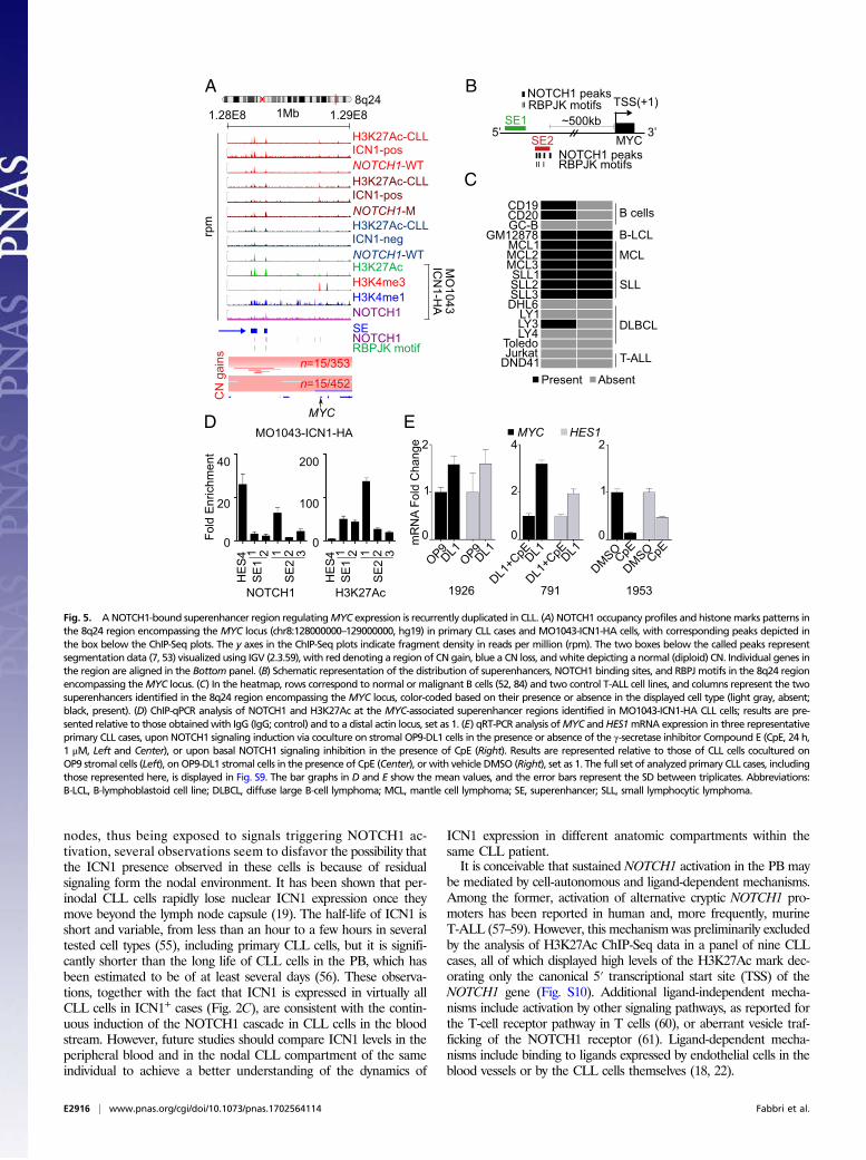
ResultsNOTCH1 Is Activated in Naïve and Memory B Cells, the Putative NormalCounterparts of CLL. To obtain a comparative baseline before in-vestigating the activity of NOTCH1 in CLL, we first defined theexpression and activation pattern of NOTCH1 in normal matureB-cell subsets. We performed gene-expression profiling as well asICN1 immunoblot analysis in naïve, germinal center (GC) andmemory B cells isolated from human tonsils (23). Although thephenotype of the B cell expanding to generate overt CLL remains amatter of debate, naïve and memory B cells are considered the mostlikely putative normal counterparts of this disease (24–26). Thelevels of NOTCH1 mRNA and of the cleaved and active intracel-lular portion of NOTCH1 ICN1 were abundant in naïve andmemory B cells, whereas they were almost undetectable in GC Bcells (Fig. 1 A and B). Immunofluorescence staining of humantonsillar biopsies confirmed these findings, revealing ICN1 nuclearstaining in the B-cell fraction populating the mantle zone of theGCs, which is highly enriched in naïve B cells (Fig. 1C and Fig. S1).Gene set enrichment analysis (GSEA) using the “Hallmark_
Notch_Signaling” signature from the Molecular Signature Data-base (27) confirmed that ICN1 expression in naïve and memoryB cells is associated with NOTCH1 transcriptional activity (Fig.1D). Taken together, these data indicate that NOTCH1 is physi-ologically expressed and activated in the cells of origin of CLL.
PB CLL Cells Express ICN1 in both NOTCH1-Mutated and Wild-TypeCases. We next investigated the incidence of NOTCH1 pathwayactivation in CLL by analyzing peripheral blood CLL cells(>70% purity in 90 of 93 cases analyzed by cytofluorimetryanalysis for CD5+/CD19+) (Materials and Methods) from a co-hort of primary CLL cases (n = 124). Twenty-two percent ofthese cases carried NOTCH1 PEST-truncating events (n = 27 of124) (Dataset S1), representative of the prototypical p.P2515fsmutation (n = 17 of 29, 58.6%), frameshift deletions (n = 3 of 29,10.3%), and nonsense mutations (n = 5 of 29, 17.2%). In addi-tion, four cases carried 3′UTR NOTCH1 mutations known tolead to aberrant splicing events disrupting the PEST domain ofthe ICN1 protein (7). The majority of NOTCH1-mutated CLLcases carried unmutated IGHV genes (n = 23 of 24 with knownIGHV status, 95.8%), as previously reported (3, 4, 12).Notably, ICN1 was detectable by immunoblot analysis in
50.5% (n = 49 of 97) of NOTCH1–wild-type cases (Fig. 2 A andB and Fig. S2A). Among these, ICN1 expression occurred in53.3% (n = 24 of 45) of IGHV mutated and 41.9% (n = 13 of 31)of IGHV-unmutated cases, respectively (Dataset S1).As expected, all NOTCH1 PEST-disrupted CLL cases expressed
a truncated active form of the protein (Fig. 2 A and B and Fig. S2A)(4, 7). The levels of ICN1 expression were variable across the panel,with NOTCH1-mutated cases often displaying higher levels ofICN1 compared with wild-type samples (Fig. S2A). ICN1 levels inNOTCH1–wild-type cases were responsive to NOTCH inhibition bythe γ-secretase inhibitor Compound-E (28) (Fig. S2B).Immunofluorescence analysis showed that strong nuclear
ICN1 expression was detectable in virtually 100% of CLL cells inboth NOTCH1-mutated and wild-type ICN1+ CLL cases (Fig. 2C).Moreover, ICN1 was not expressed in PB mononuclear cells(PBMCs) from healthy, age-matched elderly individuals (Fig. S2C).These observations exclude the possibility that ICN1 expression inNOTCH1–wild-type cases was because of residual contamination by
normal cells. Altogether, these results suggest that the activation ofthe NOTCH1 oncogene in CLL is more common than what iscurrently known based on the frequency of NOTCH1 activatingmutations (Discussion).
Identification of the CLL NOTCH1-Direct Transcriptional Program. Asa tool to further interrogate the functional activity of NOTCH1 inICN1+ CLL cells and to shed light on the NOTCH1-controlledbiological functions in CLL, we next investigated the NOTCH1-dependent CLL transcriptional program. We used a CLL cell line(MO1043) (29), which carries a hemizygous NOTCH1 PEST-truncation and expresses a truncated ICN1 that is responsive toNOTCH inhibition by the γ-secretase inhibitor Compound-E (28).Given the low intrinsic signaling activity of PEST-truncated allelesalone (8) (Fig. S3A), we sought to investigate NOTCH1-dependentprograms using an inducible lentiviral system expressing either anHA-tagged constitutively active form of NOTCH1 (ICN1-HA) orcontrol eGFP upon doxycycline addition (Fig. S3B) (30). Inductionof NOTCH1 signaling was demonstrated by HA immunoblot and
NOTCH1 MYC HES1 BCL6
A C
D
IB:BCL6
IB:MYC
IB:β-actin
B
20x
GC
M
Nai
ve
GC
-cel
ls
Mem
ory
Naïve Memory GC-cells
Run
ning
Enr
ichm
ent S
core
(RE
S)
IB:ICN1
NaïveMemoryn=10
GCn=10
NES=1.45P=0.047
DAPI ICN1
20x
M
GC
LZ
DZ
AID ICN1Hallmark NOTCH Signaling
(n=32)
CD20 ICN1
M
GC40x
-1.5 1.5
0 15000
00.
5
5000 10000
Fig. 1. NOTCH1 is expressed and activated in naïve andmemory B cells, putativenormal counterparts of CLL. (A) Gene-expression profile analysis (HG-U133 Plus 2.0Array) of NOTCH1, MYC, HES1, and BCL6 in normal mature naive, GC, andmemory B-cell subpopulations isolated from human tonsils (23). Each columncorresponds to an independent sample. ThemRNA expression pattern ofNOTCH1in naïve and memory B cells is similar to that ofMYC, typically expressed only in asmall fraction of GC–B cells (69), and opposite to that of BCL6, a known GCmasterregulator (81). Moreover, NOTCH1 expression levels are concordant with those ofHES1, a NOTCH1 target in multiple tissue types (11). (B) Immunoblot (IB) analysisof ICN1, BCL6, MYC, and control β-actin in mature B-cell subpopulations isolatedfrom human tonsils. (C) Immunofluorescence (IF) staining of ICN1, the dark-zoneGC-marker AID (82), and the B-cell–specific surface antigen CD20 in a human tonsilsection. (D) Tracking of the HALLMARK_NOTCH_SIGNALING geneset from theMolecular Signatures Database v5.1 (software.broadinstitute.org/gsea/msigdb/index.jsp) in normal mature B-cell subpopulations by GSEA. Abbreviations: DZ,dark zone; LZ, light zone; M, mantle zone.
E2912 | www.pnas.org/cgi/doi/10.1073/pnas.1702564114 Fabbri et al.

quantitative RT-PCR (qRT-PCR) for DTX1, a well-establishedNOTCH1-direct target gene (11) (Fig. S3 C–E). This experimentalsystem was used to identify genetic elements bound and directlyregulated by NOTCH1 by integrating RNA-Seq and NOTCH1ChIP-Seq data (Datasets S1–S3).Unsupervised clustering of RNA-Seq data showed that the ex-
pression profiles of MO1043-ICN1-HA and -eGFP cells clusterseparately (Fig. 3A). Supervised analysis revealed that ICN1-HAinduction leads to up-regulation of over 700 transcripts [false-discovery rate (FDR) < 0.001, median fold-change 1.7, range =1.1–170.4], including known NOTCH1 targets, such as HES/HEYfamily members, NRARP, DTX1, and NOTCH1 itself, as expected(Fig. 3B), as well as genes involved in immune and signalingpathways relevant for the development and activation of B cells(Datasets S4 and S5).ChIP-Seq analysis identified a total of 4,737 NOTCH1 binding
sites, mapping to promoters in ∼40% of the cases, and to in-tragenic or distal regulatory regions of the genome in ∼60% ofthe cases (Fig. 3 C and D). The integration of NOTCH1 ChIP-Seq profiles with those of the H3K4me3, H3K4me, H3K27Ac,and H3K27me3 histone modifications (Fig. S4) revealed that∼94% of NOTCH1 proximal binding sites displayed chromatinmarks characteristic of active promoters, whereas ∼37% of thedistal ones were associated with putative active enhancers (Fig.3E) (31–33). Analysis of H3K27Ac patterns across the genomeidentified 917 superenhancers (34), many of which involved
genes defining key functions of B cells and displayed sequencemotif enrichment of transcription factors known to be masterregulators of B-cell identity (Fig. 3F, Fig. S5 A and B, andDatasets S6 and S7) (35–38). NOTCH1-binding at super-enhancer regions (n = 698 of 4,737 binding sites, 14.7%) wasprominently involved in the activation of genes implicated inB-cell differentiation and activation and antiapoptotic functions(Fig. 3 G and H and see below).- - -
A B
C
Primary CLL
IB:ICN1
IB:β-actin
+ + + + +-
MO
1043
+DL1
(CLL
)C
UTL
L1 (T
-ALL
)
Primary CLL(n=124)
38.7
39.5
21.8
NOTCH1-M ICN1-posNOTCH1-WT ICN1-posNOTCH1-WT ICN1-neg
CUTLL1(+)856 2008 1961 1988
CUTLL1CpE(-)
DA
PI
ICN
1m
erge
ΔPEST - - -
ICN1-pos ICN1-neg 20X
NOTCH1WT WT M TW
Fig. 2. Primary CLL cases express ICN1 because of NOTCH1 PEST-truncations oralternative mechanisms. (A) IB analysis of ICN1 and control β-actin in 10 represen-tative PB CLL cases, 4 carrying NOTCH1 PEST-truncations (ΔPEST) and 6 NOTCH1–wild-type (WT), in the control T-ALL cell line CUTLL1 (83) and in MO1043 CLL cellscocultured with OP9 stromal cells expressing the NOTCH1 ligand DL1 (54). The fullset of analyzed primary CLL cases, including those reported here, is displayed in Fig.S2. (B) Frequency of ICN1 positivity in 124 primary CLL cases. (C) IF staining of ICN1 inprimary ICN1+ (pos) and ICN1− (neg) CLL cells and in the control CUTLL1 T-ALL cellline in basal conditions (+) and upon Compound E (CpE, 24 h, 1 μM) treatment (−).
A B C
D E
F G H
I J
< 0.001
Fig. 3. Identification of NOTCH1 direct targets in CLL. (A) Hierarchical clusteringof RNA-Seq profiles of MO1043-ICN1-HA and -eGFP cells (Pearson correlationwith average linkage, minimum log2 expression 5 andminimum SD 1). (B) Scatterplot of log2-transformed RNA-Seq FPKM values of differentially expressed genesbetween MO1043-ICN1-HA and -eGFP control CLL cells (FDR < 0.001). (C and D)Distribution of NOTCH1 binding sites (BS) in the genome of MO1043-ICN1-HACLL cells. (E) Functional classification of NOTCH1-BS mapping to proximal pro-moters and distal regions of the genome based on their overlap with theH3K4me3, H3K4me, H3K27Ac and H3K27me3 histone marks. (F) Rank order ofincreasing H3K27Ac fold-enrichment at enhancer loci in in MO1043-ICN1-HA CLLcells. (G) Overlap between NOTCH1-BS and superenhancers identified with theROSE algorithm (35, 36). (H) Representative examples of genes regulated byNOTCH1 via binding to superenhancer regions. (I) Intersection between RNA-Seqand ChIP-Seq data obtained in MO1043-ICN1-HA CLL cells. (J) Top three signifi-cantly (P = 1.00E-15) enriched transcription factor motifs lying ±200 bp ofNOTCH1-BS. Abbreviations: NoExp, transcripts not expressed in MO1043-ICN1-HA cells; NoMov, transcripts not moving upon ICN1-HA expression; SEs,superenhancers; TF, transcription factor.
Fabbri et al. PNAS | Published online March 17, 2017 | E2913
MED
ICALSC
IENCE
SPN
ASPL
US

The intersection between the genes differentially expressedupon ICN1-HA induction as identified by RNA-Seq and theNOTCH1 binding profiles obtained through ChIP-Seq revealeda significant overlap between the two sets (P < 0.001), with ∼39%of genes induced by ICN1-HA (FDR < 0.001) being bound byNOTCH1 (Fig. 3I and Fig. S6). Notably, genes associated withNOTCH1 binding sites in superenhancer regions were more oftenup-regulated upon ICN1-HA induction compared with those asso-ciated with NOTCH1 binding sites in other genomic regions (52%vs. 29%, P < 0.001) (Fig. S5C). Motif enrichment analysis of se-quences surrounding the NOTCH1 binding sites (±200 bp) andassociated with significant (FDR < 0.001) up-regulation of the
corresponding genes (n = 503) confirmed significant (P = 1.00E-15)enrichment of the DNA motifs of RBPJ, the main effector ofNOTCH signaling (11), as well as of binding sequences of otherpotential cooperating cofactors, including NF-κB, PU.1, ETS, andSTAT family members (Fig. 3J and Dataset S8).
The NOTCH1-Dependent CLL Signature Is Detectable in NOTCH1–Wild-Type CLL Cases Expressing ICN1. To identify bona fide directNOTCH1 targets to be used as a tool to interrogate primary CLLcells (NOTCH1 CLL signature), we selected genes bound byNOTCH1 and the transcripts of which were up-regulated byICN1-HA with a FDR < 0.001. Two-hundred and ninety-one
A BZNF318STRADBPLAC8MALT1FAM65BNT5ECPNE5CDK19PTPN6SMPD2CD27CHI3L2IRF2RAB5BGLIPR1ENTPD1FGRUBL7DENND2DCALCOCO1WDTC1PACSIN2DNASE2SCYL3SLC37A2DNASE1L3LOC283710OSTF1SLC46A3BADCD53LOC648987IL7IGFBP4EIF2AK4TBC1D9SSH2SQRDLMUM1ESYT2METTL8PDLIM1CR2GALNT10PRR18PTK2BBCAT1H6PDM6PRZHX2XYLT1CPPED1BLNKPTP4A2RNF130CPOXPPP1R9BPPP1CARALGPS1ZNF398HERPUD2PLEKHA2ZFP36L1FAM134CRABGAP1LST6GAL1HIVEP2USTFBXO18NHLRC3MX2SH3BP5RAD23BERAP2PINK1TMEM140FBXL17OXR1GPR155IGF1RRIN3PANK1RFTN1IL4I1SLC38A1LRRC1LOC100506639DLGAP4NCF1CSTARD10
Run
ning
Enr
ichm
ent S
core
(RE
S)
NOTCH1 CLL signature (n=291)
-2 2
NES=1.59 P=0.002
NES=1.37 P=0.04
NES=1.44 P=0.03
NOTCH1-MICN1-pos(n=10)
NOTCH1-WTICN1-neg
(n=26)
NOTCH1-WTICN1-neg
(n=26)
NOTCH1-WTICN1-pos(n=13)
ICN1-pos ICN1-neg(n=26)
0 5000 10000 15000
Lead
ing
Edg
e (n
=90)
ICN1-negWT
ICN1-posNOTCH1M
(n=23)
Fig. 4. The NOTCH1 CLL signature is enriched in primary CLL cases expressing ICN1. (A) GSEA enrichment plots depicting significant enrichment of theNOTCH1 CLL signature in NOTCH1-mutated (M) and wild-type (WT) primary CLL cases expressing ICN1+ (ICN1-pos) compared with ICN1− (ICN1-neg) cases, andheatmap of RNA-Seq profiles of corresponding leading edge genes (n = 90) (B).
E2914 | www.pnas.org/cgi/doi/10.1073/pnas.1702564114 Fabbri et al.
genes met these criteria, including NOTCH1 target genes, suchas HES1, DTX1, JAG1, and NOTCH1 itself, among others, aswell as NF-κB, antiapoptotic, and cytokine-chemokine genes(Dataset S9).To determine whether NOTCH1–wild-type ICN1+ CLL cases
display evidence of NOTCH1 signaling activation analogous toNOTCH1-mutated cases, we explored the presence of theNOTCH1 CLL signature identified above in RNA-Seq data from49 PB CLL samples fully characterized in terms of NOTCH1mutation and ICN1 protein expression. This panel included 10NOTCH1-mutated cases expressing ICN1 (including three casescarrying NOTCH1 3′UTR events), 13 NOTCH1–wild-type casesexpressing ICN1, and 26 cases devoid of both NOTCH1mutationsand ICN1 expression (Fig. S2A). GSEA analysis (27) revealedsignificant enrichment of the NOTCH1 CLL signature both inNOTCH1-mutated (P = 0.002) andNOTCH1–wild-type (P = 0.04)cases expressing ICN1 compared with ICN1− cases (Fig. 4A).Leading edge genes (n = 90) determining a significant (P = 0.03)enrichment of the NOTCH1 CLL signature in ICN1+ cases wereoverall expressed at similar levels in NOTCH1-mutated and wild-type ICN1+ cases [average fragments per kilobase of transcript permillion mapped reads (FPKM) 54.3 and 51.3, respectively], withfew genes expressed at higher levels in the NOTCH1-mutatedones (n = 9 of 90, P < 0.05) (Fig. 4B and Dataset S10).Thus, ICN1 is also functionally active in ICN1+ CLL cases
devoid of NOTCH1 mutations, indicating that they are func-tionally equivalent in terms of NOTCH1-dependent transcrip-tional responses to NOTCH1-mutated ones (Discussion).
NOTCH1 Regulates Genes with Key Functions in B-Cell Physiology.Functional annotation of the full set of genes bound (ChIP-Seq)and dynamically connected (RNA-Seq) to NOTCH1 revealed thatNOTCH1 directly regulates general functions involved in cell pro-liferation and survival (Datasets S11 and S12). The former includedCCND3, which encodes a cyclin necessary for G1/G2 transition (39)via direct binding to the gene promoter, consistent with a previousreport in T-ALL (40). Among the latter, BCL2 and MCL1, twoantiapoptotic genes with a well-established role in the pathogenesisof CLL, emerged as novel targets of NOTCH1, likely regulatedthrough long-range dynamic interactions (24, 41–43).The NOTCH1 transcriptional program included also a cadre of
genes with specific functions in B-cell physiology (Fig. S7). Amongthese are BCR signaling pathway genes, including upstream pathwaymembers (e.g., LYN, SYK, BLK, BLNK, CR2, and PIK3CD), as wellas downstream effectors, such as MAPK (e.g., MAP3K1, KRAS, andRRAS) and NF-κB cascade members (e.g., IKBKB, NFKB1, and theCBM signalosome complex member MALT1) (44). NOTCH1 alsoappears to activate the NF-κB target NFKBIA, which encodesthe NF-κB repressor IκBα (44), and PTPN6, encoding SHP-1, animportant negative modulator of antigen-receptor signaling in lym-phocytes (45), suggesting a role of NOTCH1 in a delayed negative-feedback of activation of this cascade (46). CXCR4, which encodes achemokine receptor relevant for the chemotaxis of CLL cells towardmicroenvironmental cells expressing the CXCL12 ligand (47),emerged as a novel NOTCH1 target in CLL. This axis is funda-mental for the exit of CLL cells from lymph nodes and, accordingly,the expression of the CXCR4 receptor has been shown to associatewith a higher risk of lymphoid organ infiltration and poor diseaseoutcome (48). Finally, among several NOTCH-pathway relatedgenes, NOTCH1 induced the expression of JAG1, which encodesfor a ligand of NOTCH1 reported to be expressed on the surfaceof CLL cells (22), suggesting a positive feed-forward loop insignaling activation.
NOTCH1 Transactivates MYC in CLL. MYC is a central oncogene inhuman malignancy, an established NOTCH1 target in T-ALLand is involved in CLL progression (3, 13, 49–51). Thus, we in-vestigated the relationship between NOTCH1 activation and
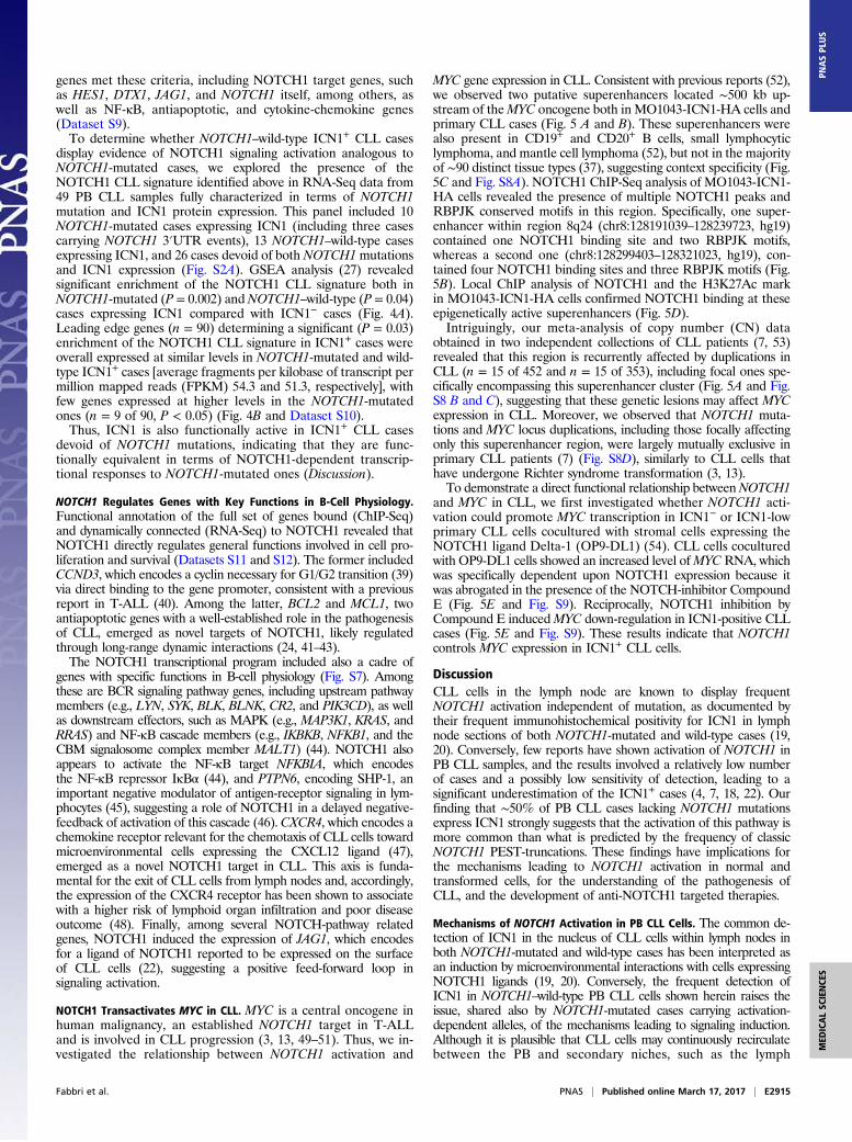
MYC gene expression in CLL. Consistent with previous reports (52),we observed two putative superenhancers located ∼500 kb up-stream of theMYC oncogene both in MO1043-ICN1-HA cells andprimary CLL cases (Fig. 5 A and B). These superenhancers werealso present in CD19+ and CD20+ B cells, small lymphocyticlymphoma, and mantle cell lymphoma (52), but not in the majorityof ∼90 distinct tissue types (37), suggesting context specificity (Fig.5C and Fig. S8A). NOTCH1 ChIP-Seq analysis of MO1043-ICN1-HA cells revealed the presence of multiple NOTCH1 peaks andRBPJK conserved motifs in this region. Specifically, one super-enhancer within region 8q24 (chr8:128191039–128239723, hg19)contained one NOTCH1 binding site and two RBPJK motifs,whereas a second one (chr8:128299403–128321023, hg19), con-tained four NOTCH1 binding sites and three RBPJK motifs (Fig.5B). Local ChIP analysis of NOTCH1 and the H3K27Ac markin MO1043-ICN1-HA cells confirmed NOTCH1 binding at theseepigenetically active superenhancers (Fig. 5D).Intriguingly, our meta-analysis of copy number (CN) data
obtained in two independent collections of CLL patients (7, 53)revealed that this region is recurrently affected by duplications inCLL (n = 15 of 452 and n = 15 of 353), including focal ones spe-cifically encompassing this superenhancer cluster (Fig. 5A and Fig.S8 B and C), suggesting that these genetic lesions may affect MYCexpression in CLL. Moreover, we observed that NOTCH1 muta-tions and MYC locus duplications, including those focally affectingonly this superenhancer region, were largely mutually exclusive inprimary CLL patients (7) (Fig. S8D), similarly to CLL cells thathave undergone Richter syndrome transformation (3, 13).To demonstrate a direct functional relationship betweenNOTCH1
and MYC in CLL, we first investigated whether NOTCH1 acti-vation could promote MYC transcription in ICN1− or ICN1-lowprimary CLL cells cocultured with stromal cells expressing theNOTCH1 ligand Delta-1 (OP9-DL1) (54). CLL cells coculturedwith OP9-DL1 cells showed an increased level ofMYCRNA, whichwas specifically dependent upon NOTCH1 expression because itwas abrogated in the presence of the NOTCH-inhibitor CompoundE (Fig. 5E and Fig. S9). Reciprocally, NOTCH1 inhibition byCompound E inducedMYC down-regulation in ICN1-positive CLLcases (Fig. 5E and Fig. S9). These results indicate that NOTCH1controls MYC expression in ICN1+ CLL cells.
DiscussionCLL cells in the lymph node are known to display frequentNOTCH1 activation independent of mutation, as documented bytheir frequent immunohistochemical positivity for ICN1 in lymphnode sections of both NOTCH1-mutated and wild-type cases (19,20). Conversely, few reports have shown activation of NOTCH1 inPB CLL samples, and the results involved a relatively low numberof cases and a possibly low sensitivity of detection, leading to asignificant underestimation of the ICN1+ cases (4, 7, 18, 22). Ourfinding that ∼50% of PB CLL cases lacking NOTCH1 mutationsexpress ICN1 strongly suggests that the activation of this pathway ismore common than what is predicted by the frequency of classicNOTCH1 PEST-truncations. These findings have implications forthe mechanisms leading to NOTCH1 activation in normal andtransformed cells, for the understanding of the pathogenesis ofCLL, and the development of anti-NOTCH1 targeted therapies.
Mechanisms of NOTCH1 Activation in PB CLL Cells. The common de-tection of ICN1 in the nucleus of CLL cells within lymph nodes inboth NOTCH1-mutated and wild-type cases has been interpreted asan induction by microenvironmental interactions with cells expressingNOTCH1 ligands (19, 20). Conversely, the frequent detection ofICN1 in NOTCH1–wild-type PB CLL cells shown herein raises theissue, shared also by NOTCH1-mutated cases carrying activation-dependent alleles, of the mechanisms leading to signaling induction.Although it is plausible that CLL cells may continuously recirculatebetween the PB and secondary niches, such as the lymph
Fabbri et al. PNAS | Published online March 17, 2017 | E2915
MED
ICALSC
IENCE
SPN
ASPL
US
nodes, thus being exposed to signals triggering NOTCH1 ac-tivation, several observations seem to disfavor the possibility thatthe ICN1 presence observed in these cells is because of residualsignaling form the nodal environment. It has been shown that per-inodal CLL cells rapidly lose nuclear ICN1 expression once theymove beyond the lymph node capsule (19). The half-life of ICN1 isshort and variable, from less than an hour to a few hours in severaltested cell types (55), including primary CLL cells, but it is signifi-cantly shorter than the long life of CLL cells in the PB, which hasbeen estimated to be of at least several days (56). These observa-tions, together with the fact that ICN1 is expressed in virtually allCLL cells in ICN1+ cases (Fig. 2C), are consistent with the contin-uous induction of the NOTCH1 cascade in CLL cells in the bloodstream. However, future studies should compare ICN1 levels in theperipheral blood and in the nodal CLL compartment of the sameindividual to achieve a better understanding of the dynamics of
ICN1 expression in different anatomic compartments within thesame CLL patient.It is conceivable that sustained NOTCH1 activation in the PB may
be mediated by cell-autonomous and ligand-dependent mechanisms.Among the former, activation of alternative cryptic NOTCH1 pro-moters has been reported in human and, more frequently, murineT-ALL (57–59). However, this mechanism was preliminarily excludedby the analysis of H3K27Ac ChIP-Seq data in a panel of nine CLLcases, all of which displayed high levels of the H3K27Ac mark dec-orating only the canonical 5′ transcriptional start site (TSS) of theNOTCH1 gene (Fig. S10). Additional ligand-independent mecha-nisms include activation by other signaling pathways, as reported forthe T-cell receptor pathway in T cells (60), or aberrant vesicle traf-ficking of the NOTCH1 receptor (61). Ligand-dependent mecha-nisms include binding to ligands expressed by endothelial cells in theblood vessels or by the CLL cells themselves (18, 22).
A B
H3K27Ac-CLLICN1-posNOTCH1-WTH3K27Ac-CLLICN1-posNOTCH1-MH3K27Ac-CLLICN1-negNOTCH1-WTH3K27AcH3K4me3H3K4me1NOTCH1SENOTCH1RBPJK motif
MO
1043IC
N1-H
AMYC
n=15/353
n=15/452
CN
gai
nsrp
m
1.28E8 1.29E81Mb
SE2
SE1
MYC5’ 3’
TSS(+1)RBPJK motifs~500kb
RBPJK motifsNOTCH1 peaks
NOTCH1 peaks8q24
CD19CD20
GM12878MCL1MCL2MCL3
DHL6LY1LY3LY4
ToledoJurkat
B cells
B-LCL
MCL
DLBCL
T-ALL
C
SLLSLL1SLL2SLL3
GC-B
DND41Present Absent
D
Fold
Enr
ichm
ent
NOTCH1 H3K27Ac
40
20
0 0
100
200
HE
S4 1 2 1 2 3
SE
1
SE
2
HE
S4 1 2 1 2 3
SE
1
SE
2
MO1043-ICN1-HAE
mR
NA
Fold
Cha
nge
OP9DL1OP9DL1
DMSOCpEDMSOCpE
DL1+C
pEDL1DL1
+CpEDL1
1926 791 1953
MYC HES1
0 0 0
242
1 2 1
Fig. 5. ANOTCH1-bound superenhancer region regulatingMYC expression is recurrently duplicated in CLL. (A) NOTCH1 occupancy profiles and histonemarks patterns inthe 8q24 region encompassing the MYC locus (chr8:128000000–129000000, hg19) in primary CLL cases and MO1043-ICN1-HA cells, with corresponding peaks depicted inthe box below the ChIP-Seq plots. The y axes in the ChIP-Seq plots indicate fragment density in reads per million (rpm). The two boxes below the called peaks representsegmentation data (7, 53) visualized using IGV (2.3.59), with red denoting a region of CN gain, blue a CN loss, andwhite depicting a normal (diploid) CN. Individual genes inthe region are aligned in the Bottom panel. (B) Schematic representation of the distribution of superenhancers, NOTCH1 binding sites, and RBPJ motifs in the 8q24 regionencompassing theMYC locus. (C) In the heatmap, rows correspond to normal or malignant B cells (52, 84) and two control T-ALL cell lines, and columns represent the twosuperenhancers identified in the 8q24 region encompassing the MYC locus, color-coded based on their presence or absence in the displayed cell type (light gray, absent;black, present). (D) ChIP-qPCR analysis of NOTCH1 and H3K27Ac at the MYC-associated superenhancer regions identified in MO1043-ICN1-HA CLL cells; results are pre-sented relative to those obtainedwith IgG (IgG; control) and to a distal actin locus, set as 1. (E) qRT-PCR analysis ofMYC andHES1mRNA expression in three representativeprimary CLL cases, upon NOTCH1 signaling induction via coculture on stromal OP9-DL1 cells in the presence or absence of the γ-secretase inhibitor Compound E (CpE, 24 h,1 μM, Left and Center), or upon basal NOTCH1 signaling inhibition in the presence of CpE (Right). Results are represented relative to those of CLL cells cocultured onOP9 stromal cells (Left), onOP9-DL1 stromal cells in the presence of CpE (Center), or with vehicle DMSO (Right), set as 1. The full set of analyzed primary CLL cases, includingthose represented here, is displayed in Fig. S9. The bar graphs in D and E show the mean values, and the error bars represent the SD between triplicates. Abbreviations:B-LCL, B-lymphoblastoid cell line; DLBCL, diffuse large B-cell lymphoma; MCL, mantle cell lymphoma; SE, superenhancer; SLL, small lymphocytic lymphoma.
E2916 | www.pnas.org/cgi/doi/10.1073/pnas.1702564114 Fabbri et al.
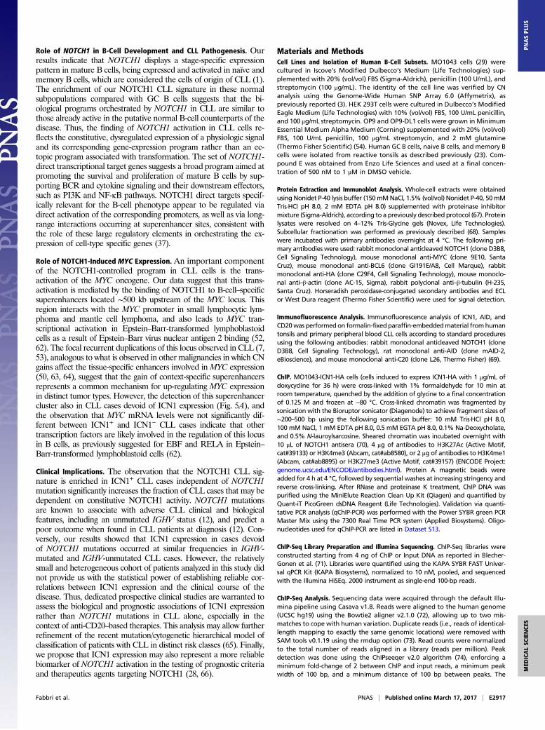
Role of NOTCH1 in B-Cell Development and CLL Pathogenesis. Ourresults indicate that NOTCH1 displays a stage-specific expressionpattern in mature B cells, being expressed and activated in naïve andmemory B cells, which are considered the cells of origin of CLL (1).The enrichment of our NOTCH1 CLL signature in these normalsubpopulations compared with GC B cells suggests that the bi-ological programs orchestrated by NOTCH1 in CLL are similar tothose already active in the putative normal B-cell counterparts of thedisease. Thus, the finding of NOTCH1 activation in CLL cells re-flects the constitutive, dysregulated expression of a physiologic signaland its corresponding gene-expression program rather than an ec-topic program associated with transformation. The set of NOTCH1-direct transcriptional target genes suggests a broad program aimed atpromoting the survival and proliferation of mature B cells by sup-porting BCR and cytokine signaling and their downstream effectors,such as PI3K and NF-κB pathways. NOTCH1 direct targets specif-ically relevant for the B-cell phenotype appear to be regulated viadirect activation of the corresponding promoters, as well as via long-range interactions occurring at superenhancer sites, consistent withthe role of these large regulatory elements in orchestrating the ex-pression of cell-type specific genes (37).
Role of NOTCH1-Induced MYC Expression. An important componentof the NOTCH1-controlled program in CLL cells is the trans-activation of the MYC oncogene. Our data suggest that this trans-activation is mediated by the binding of NOTCH1 to B-cell–specificsuperenhancers located ∼500 kb upstream of the MYC locus. Thisregion interacts with the MYC promoter in small lymphocytic lym-phoma and mantle cell lymphoma, and also leads to MYC tran-scriptional activation in Epstein–Barr-transformed lymphoblastoidcells as a result of Epstein–Barr virus nuclear antigen 2 binding (52,62). The focal recurrent duplications of this locus observed in CLL (7,53), analogous to what is observed in other malignancies in which CNgains affect the tissue-specific enhancers involved in MYC expression(50, 63, 64), suggest that the gain of context-specific superenhancersrepresents a common mechanism for up-regulating MYC expressionin distinct tumor types. However, the detection of this superenhancercluster also in CLL cases devoid of ICN1 expression (Fig. 5A), andthe observation that MYC mRNA levels were not significantly dif-ferent between ICN1+ and ICN1− CLL cases indicate that othertranscription factors are likely involved in the regulation of this locusin B cells, as previously suggested for EBF and RELA in Epstein–Barr-transformed lymphoblastoid cells (62).
Clinical Implications. The observation that the NOTCH1 CLL sig-nature is enriched in ICN1+ CLL cases independent of NOTCH1mutation significantly increases the fraction of CLL cases that may bedependent on constitutive NOTCH1 activity. NOTCH1 mutationsare known to associate with adverse CLL clinical and biologicalfeatures, including an unmutated IGHV status (12), and predict apoor outcome when found in CLL patients at diagnosis (12). Con-versely, our results showed that ICN1 expression in cases devoidof NOTCH1 mutations occurred at similar frequencies in IGHV-mutated and IGHV-unmutated CLL cases. However, the relativelysmall and heterogeneous cohort of patients analyzed in this study didnot provide us with the statistical power of establishing reliable cor-relations between ICN1 expression and the clinical course of thedisease. Thus, dedicated prospective clinical studies are warranted toassess the biological and prognostic associations of ICN1 expressionrather than NOTCH1 mutations in CLL alone, especially in thecontext of anti-CD20–based therapies. This analysis may allow furtherrefinement of the recent mutation/cytogenetic hierarchical model ofclassification of patients with CLL in distinct risk classes (65). Finally,we propose that ICN1 expression may also represent a more reliablebiomarker of NOTCH1 activation in the testing of prognostic criteriaand therapeutics agents targeting NOTCH1 (28, 66).
Materials and MethodsCell Lines and Isolation of Human B-Cell Subsets. MO1043 cells (29) werecultured in Iscove’s Modified Dulbecco’s Medium (Life Technologies) sup-plemented with 20% (vol/vol) FBS (Sigma-Aldrich), penicillin (100 U/mL), andstreptomycin (100 μg/mL). The identity of the cell line was verified by CNanalysis using the Genome-Wide Human SNP Array 6.0 (Affymetrix), aspreviously reported (3). HEK 293T cells were cultured in Dulbecco’s ModifiedEagle Medium (Life Technologies) with 10% (vol/vol) FBS, 100 U/mL penicillin,and 100 μg/mL streptomycin. OP9 and OP9-DL1 cells were grown in MinimumEssential Medium Alpha Medium (Corning) supplemented with 20% (vol/vol)FBS, 100 U/mL penicillin, 100 μg/mL streptomycin, and 2 mM glutamine(Thermo Fisher Scientific) (54). Human GC B cells, naive B cells, and memory Bcells were isolated from reactive tonsils as described previously (23). Com-pound E was obtained from Enzo Life Sciences and used at a final concen-tration of 500 nM to 1 μM in DMSO vehicle.
Protein Extraction and Immunoblot Analysis. Whole-cell extracts were obtainedusing Nonidet P-40 lysis buffer (150mMNaCl, 1.5% (vol/vol) Nonidet P-40, 50mMTris·HCl pH 8.0, 2 mM EDTA pH 8.0) supplemented with proteinase inhibitormixture (Sigma-Aldrich), according to a previously described protocol (67). Proteinlysates were resolved on 4–12% Tris-Glycine gels (Novex, Life Technologies).Subcellular fractionation was performed as previously described (68). Sampleswere incubated with primary antibodies overnight at 4 °C. The following pri-mary antibodies were used: rabbit monoclonal anticleaved NOTCH1 (clone D3B8,Cell Signaling Technology), mouse monoclonal anti-MYC (clone 9E10, SantaCruz), mouse monoclonal anti-BCL6 (clone GI191E/A8, Cell Marque), rabbitmonoclonal anti-HA (clone C29F4, Cell Signaling Technology), mouse monoclo-nal anti–β-actin (clone AC-15, Sigma), rabbit polyclonal anti–β-tubulin (H-235,Santa Cruz). Horseradish peroxidase-conjugated secondary antibodies and ECLor West Dura reagent (Thermo Fisher Scientific) were used for signal detection.
Immunofluorescence Analysis. Immunofluorescence analysis of ICN1, AID, andCD20was performedon formalin-fixedparaffin-embeddedmaterial fromhumantonsils and primary peripheral blood CLL cells according to standard proceduresusing the following antibodies: rabbit monoclonal anticleaved NOTCH1 (cloneD3B8, Cell Signaling Technology), rat monoclonal anti-AID (clone mAID-2,eBioscience), and mouse monoclonal anti-C20 (clone L26, Thermo Fisher) (69).
ChIP. MO1043-ICN1-HA cells (cells induced to express ICN1-HA with 1 μg/mL ofdoxycycline for 36 h) were cross-linked with 1% formaldehyde for 10 min atroom temperature, quenched by the addition of glycine to a final concentrationof 0.125 M and frozen at −80 °C. Cross-linked chromatin was fragmented bysonication with the Bioruptor sonicator (Diagenode) to achieve fragment sizes of∼200–500 bp using the following sonication buffer: 10 mM Tris·HCl pH 8.0,100 mM NaCl, 1 mM EDTA pH 8.0, 0.5 mM EGTA pH 8.0, 0.1% Na-Deoxycholate,and 0.5% N-lauroylsarcosine. Sheared chromatin was incubated overnight with10 μL of NOTCH1 antisera (70), 4 μg of antibodies to H3K27Ac (Active Motif,cat#39133) or H3K4me3 (Abcam, cat#ab8580), or 2 μg of antibodies to H3K4me1(Abcam, cat#ab8895) or H3K27me3 (Active Motif, cat#39157) (ENCODE Project:genome.ucsc.edu/ENCODE/antibodies.html). Protein A magnetic beads wereadded for 4 h at 4 °C, followed by sequential washes at increasing stringency andreverse cross-linking. After RNase and proteinase K treatment, ChIP DNA waspurified using the MiniElute Reaction Clean Up Kit (Qiagen) and quantified byQuant-iT PicoGreen dsDNA Reagent (Life Technologies). Validation via quanti-tative PCR analysis (qChIP-PCR) was performed with the Power SYBR green PCRMaster Mix using the 7300 Real Time PCR system (Applied Biosystems). Oligo-nucleotides used for qChIP-PCR are listed in Dataset S13.
ChIP-Seq Library Preparation and Illumina Sequencing. ChIP-Seq libraries wereconstructed starting from 4 ng of ChIP or Input DNA as reported in Blecher-Gonen et al. (71). Libraries were quantified using the KAPA SYBR FAST Univer-sal qPCR Kit (KAPA Biosystems), normalized to 10 nM, pooled, and sequencedwith the Illumina HiSEq. 2000 instrument as single-end 100-bp reads.
ChIP-Seq Analysis. Sequencing data were acquired through the default Illu-mina pipeline using Casava v1.8. Reads were aligned to the human genome(UCSC hg19) using the Bowtie2 aligner v2.1.0 (72), allowing up to two mis-matches to cope with human variation. Duplicate reads (i.e., reads of identical-length mapping to exactly the same genomic locations) were removed withSAM tools v0.1.19 using the rmdup option (73). Read counts were normalizedto the total number of reads aligned in a library (reads per million). Peakdetection was done using the ChIPseeqer v2.0 algorithm (74), enforcing aminimum fold-change of 2 between ChIP and input reads, a minimum peakwidth of 100 bp, and a minimum distance of 100 bp between peaks. The
Fabbri et al. PNAS | Published online March 17, 2017 | E2917
MED
ICALSC
IENCE
SPN
ASPL
US
threshold for statistical significance of peaks was set at 10−5 for NOTCH1, and10−15 for H3K4me1, H3K4me3, and H3K27Ac (Dataset S2). Peaks within 1 kb ofcentromeric or telomeric regions were removed. H3K4me1 and H3K27Acpeaks were stitched together into regions if located within ±2 kb and ±12.5 kbof each other, respectively, unless they started within a 2-kb window aroundthe TSS. H3K27me3 peaks were called using the RSEG algorithm (75) with 100-bpbin size; only peaks above 5 kb in size were considered.
Motif Enrichment Analysis. Regions within 200 bp of the center of each bindingsite were searched for motifs from the TRANSFAC 2010 Database. Motifs wererepresented as position weighted matrices. Using a moving window, motifswere scored against a reference DNA sequence using a log odds ratio com-paring the motif’s score to a hypothetical score where every base is equallyprobable. Motifs scoring higher than a given threshold were considered aspotentially bound in a location. To determine enrichment in a set of peaks/locations a hypergeometric model was used comparing motifs bound in thepeaks to a GC and length controlled set of random genomic sequences.
Definition of Functional Chromatin States of NOTCH1-Bound Genomic Loci.Significant NOTCH1-bound regions occurring at proximal promoters (i.e.,within −2/+1 kb from the TSS of an annotated gene) were classified as activeif overlapping with H3K4me3, but not H3K27me3, poised if occupied by bothH3K4me3 and H3K27me3, and silenced if decorated only by H3K27me3. Dis-tally NOTCH1-bound genomic regions (intergenic or intragenic) were classifiedas active enhancers if occupied by H3K4me and H3K27Ac, but not H3K4me3,poised if occupied by H3K4me and H3K27me3, and silenced or primed if oc-cupied only by H3K27me3 or H3K4me, respectively. For the identificationof superenhancers, we applied the ROSE algorithm (https://bitbucket.org/young_computation/rose) to our H3K27Ac ChIP-Seq datasets (MO1043-ICN1-HAand primary CLL cells). Occupancy of NOTCH1 at superenhancers was then de-termined based on the overlap between NOTCH1 peaks and genomic regionsidentified by ROSE. NOTCH1-bound superenhancers were assigned to the near-est expressed and transcriptionally active gene (i.e., distance from superenhancercenter to TSS marked by H3K4me3) as the most likely candidate target gene (38).
Primary CLL Cases. Primary CLL cells from the PB of CLL patients (n = 124) wereobtained from the Feinstein Institute for Medical Research and the Divisionof Hematology and the Department of Translational Medicine and theAmedeo Avogadro University of Eastern Piedmont. Diagnosis of CLL wasbased on International Workshop on Chronic Lymphocytic Leukemia-National Cancer Institute Working Group criteria (76) and confirmed by aflow cytometry score >3. The percentage of tumor cells of CLL cases wasestimated by cytofluorimetry analysis for CD5+/CD19+ PB cells of 93 of 124CLL cases, and it was ≥70% in 90 cases and between 57% and 60% in 3 cases.CLL cases included in the RNA-Seq panel are highlighted in Fig. S2A. ICN1+
NOTCH1–wild-type cases were selected for RNA-Seq analysis based on a ratioof ICN1 expression > 0.1 compared with the levels observed in theCUTLL1 cell line in the low-exposure immunoblot image. Quantitation ofsignal intensity was obtained with the ImageJ software (https://imagej.nih.gov/ij/) by subtracting the background signal measured above each bandfrom the signal measured in each band; areas of the same size (set on theimage of ICN1 in the CUTLL1 cell line) were used for all measurements.Values were expressed as ratio relative to the CUTLL1 protein sample, set at1, after normalization for the β-actin loading control. The study was ap-proved by the Institutional Review Board of Columbia University, by theEthical Committee of the Azienda Ospedaliera Maggiore della Carità diNovara, Amedeo Avogadro University of Eastern Piedmont, and by theNorthwell Health’s Institutional Review Board and was conducted accordingto the principles of the World Medical Association Declaration of Helsinki.
DNA Extraction, IGHV Mutational Status, and Sanger Sequencing of NOTCH1.Genomic DNA was extracted with the QIAamp DNA Mini Kit (Qiagen) andverified for integrity by gel electrophoresis. IGHV mutational status wasperformed as previously described (3, 13). The NOTCH1 gene portionencoding the PEST domain and the 3′UTR of the NOTCH1 gene werescreened by Sanger targeted sequencing, as previously reported (3, 7).
Gene-Expression Profiling of Human Mature B-Cell Subsets. Raw expression valuesof GeneChip Human Genome U133 Plus 2.0 (Affymetrix) data from normal
mature B-cell subsets were normalized using the Robust Multiarray Averagingalgorithm in GenePattern (https://www.broadinstitute.org/cancer/software/genepattern/), and multiple probes corresponding to the same gene werecollapsed to a single probe based on the maximum t-statistic/maximum SD.
RNA Extraction, cDNA Synthesis, and Quantitative Real-Time PCR. Total RNAwas extracted from primary CLL cases with the RNeasy Mini Kit (Qiagen) withon-column DNase treatment. cDNA synthesis was performed using the Su-perScript First-Strand Synthesis System (Life Technologies), according to themanufacturer’s instructions. The ABsolute QPCR SYBR green mix (ThermoScientific) was used to amplify specific cDNA fragments with the oligonu-cleotides listed in Dataset S13, in the 7300 Real-Time PCR system (AppliedBiosystems). Data were analyzed by the change-in-threshold (2−ΔΔCT)method (77), using GAPDH as a housekeeping reference gene.
RNA-Sequencing of ICN1-HA and Control eGFP MO1043 Cells and Primary CLLCases. Four MO1043-ICN1-HA and 4 MO1043-eGFP replicates and 49 primaryCLL cases were subjected to RNA-Sequencing. Briefly, poly-A pull-down wasperformed to enrichmRNAs from total RNA samples and librarieswere preparedusing the Illumina TruSeq RNA prep kit and sequenced using the IlluminaHiSeq.2000 instrument at the Columbia Genome Center. MO1043-ICN1-HA andMO1043-eGFP samplesweremultiplexed toobtainanaverageof33,022,292 single-end 100-bp reads per sample; primary CLL samples were multiplexed to obtain anaverage of 61,266,691 paired-end 100-bp reads per sample (Dataset S3). Real-timeanalysis (Illumina) was used for base calling and bcl2fastq (v1.8.4) for convertingBCL to fastq format, coupled with adaptor trimming. Reads were mapped to thereference genome (Human: NCBI/build37.2) using Tophat (v2.0.4) with 4 mis-matches (–read-mismatches = 4) and 10 maximum multiple hits (–max-multihits =10) (78). The relative abundance of genes was assessed using cufflinks (v2.0.2) withdefault settings (79). Hierarchical clustering of MO1043-ICN1-HA and -eGFP pro-files was performed using the Pearson correlation average linkage, filtering forgenes with a minimum log2-transformed expression value of 5 and a minimum SDof 1. Genes differentially expressed between MO1043-ICN1-HA and -eGFP profileswere determined by an unpaired unequal variance two-tailed Student’s t testusing a FDR ≤ 0.001 (after Benjamini–Hochberg correction) (80). For visualizationof gene-expression intensity, expression data were converted to z-scores.
GSEA. Gene-expression profile data from mature B cells, MO1043-ICN1-HAand MO1043-eGFP cells, and from primary CLL cases were analyzed for en-richment in NOTCH1-related gene sets with GSEA-2.0 and 1,000 phenotypepermutations (27). Enrichments were considered significant with a P <0.05 after correction for multiple hypothesis.
Functional Categories and Pathways Analyses of the NOTCH1-Regulated Genes.Genes directly regulated by NOTCH1 in CLL were assigned to functional cat-egories or annotated pathways using the publicly available bioinformatic toolDAVID 2008 6.7 (Database for Annotation, Visualization and Integrated Dis-covery, https://david-d.ncifcrf.gov) and the Molecular Signatures Databasefrom the Broad Institute (MSigDBv5.1, CP Geneset, https://www.broadinstitute.org/gsea/msigdb/index.jsp). Only pathways relevant for B-cell biology, based oncurrent knowledge, were selected for further discussion.
Statistical Analyses. Statistical analysis was performed using the GraphPadPrism 5 software (GraphPad Software). The specific test adopted for eachanalysis is described in each figure legend.
ACKNOWLEDGMENTS. We thank Jon Aster for providing the NOTCH1 antiseraused for ChIP-Seq of NOTCH1 in chronic lymphocytic leukemia cells; Ben K. Seon forthe MO1043 cell line; Elias Campo and Jennifer Brown for sharing usefulinformation; and the Genomic Technologies Shared Resource for sequencing theChIP-Seq and the RNA-Seq libraries. This work was supported by the US NationalInstitutes of Health Grant R01-CA177319 (to R.D.-F. and A.A.F.); Special ProgramMolecular Clinical Oncology 5 × 1000 No. 10007, Associazione Italiana per la Ricercasul Cancro Foundation Milan, Italy and Progetto Ricerca Finalizzata RF-2011-02349712, Ministero della Salute, Rome, Italy (to G.G.); National Institutes of HealthGrant K99/R00 CA197869 and an Alex’s Lemonade Stand Foundation Young Inves-tigator grant (to D.H.); and a Leukemia & Lymphoma Society Fellowship (to C.S.).
1. Fabbri G, Dalla-Favera R (2016) The molecular pathogenesis of chronic lymphocytic
leukaemia. Nat Rev Cancer 16(3):145–162.2. Pekarsky Y, Zanesi N, Croce CM (2010) Molecular basis of CLL. Semin Cancer Biol 20(6):
370–376.
3. Fabbri G, et al. (2011) Analysis of the chronic lymphocytic leukemia coding genome:
Role of NOTCH1 mutational activation. J Exp Med 208(7):1389–1401.4. Puente XS, et al. (2011) Whole-genome sequencing identifies recurrent mutations in
chronic lymphocytic leukaemia. Nature 475(7354):101–105.
E2918 | www.pnas.org/cgi/doi/10.1073/pnas.1702564114 Fabbri et al.
5. Wang L, et al. (2011) SF3B1 and other novel cancer genes in chronic lymphocyticleukemia. N Engl J Med 365(26):2497–2506.
6. Quesada V, et al. (2011) Exome sequencing identifies recurrent mutations of thesplicing factor SF3B1 gene in chronic lymphocytic leukemia. Nat Genet 44(1):47–52.
7. Puente XS, et al. (2015) Non-coding recurrent mutations in chronic lymphocytic leu-kaemia. Nature 526(7574):519–524.
8. Weng AP, et al. (2004) Activating mutations of NOTCH1 in human T cell acute lym-phoblastic leukemia. Science 306(5694):269–271.
9. Belver L, Ferrando A (2016) The genetics and mechanisms of T cell acute lymphoblasticleukaemia. Nat Rev Cancer 16(8):494–507.
10. Kopan R, Ilagan MX (2009) The canonical Notch signaling pathway: Unfolding theactivation mechanism. Cell 137(2):216–233.
11. Guruharsha KG, Kankel MW, Artavanis-Tsakonas S (2012) The Notch signalling system:Recent insights into the complexity of a conserved pathway. Nat Rev Genet 13(9):654–666.
12. Rossi D, et al. (2012) Mutations of NOTCH1 are an independent predictor of survival inchronic lymphocytic leukemia. Blood 119(2):521–529.
13. Fabbri G, et al. (2013) Genetic lesions associated with chronic lymphocytic leukemiatransformation to Richter syndrome. J Exp Med 210(11):2273–2288.
14. Balatti V, et al. (2012)NOTCH1mutations in CLL associatedwith trisomy12.Blood 119(2):329–331.15. Stilgenbauer S, et al. (2014) Gene mutations and treatment outcome in chronic
lymphocytic leukemia: Results from the CLL8 trial. Blood 123(21):3247–3254.16. Pozzo F, et al. (2016) NOTCH1 mutations associate with low CD20 level in chronic lym-
phocytic leukemia: Evidence for a NOTCH1 mutation-driven epigenetic dysregulation.Leukemia 30(1):182–189.
17. Maura F, et al. (2015) Insulin growth factor 1 receptor expression is associated withNOTCH1 mutation, trisomy 12 and aggressive clinical course in chronic lymphocyticleukaemia. PLoS One 10(3):e0118801.
18. Arruga F, et al. (2014) Functional impact of NOTCH1 mutations in chronic lymphocyticleukemia. Leukemia 28(5):1060–1070.
19. Kluk MJ, et al. (2013) Gauging NOTCH1 activation in cancer using immunohisto-chemistry. PLoS One 8(6):e67306.
20. Onaindia A, et al. (2015) Chronic lymphocytic leukemia cells in lymph nodes showfrequent NOTCH1 activation. Haematologica 100(5):e200–e203.
21. Stein H, et al. (1980) Immunohistologic analysis of the organization of normal lym-phoid tissue and non-Hodgkin’s lymphomas. J Histochem Cytochem 28(8):746–760.
22. Rosati E, et al. (2009) Constitutively activated Notch signaling is involved in survivaland apoptosis resistance of B-CLL cells. Blood 113(4):856–865.
23. Klein U, et al. (2003) Transcriptional analysis of the B cell germinal center reaction.Proc Natl Acad Sci USA 100(5):2639–2644.
24. Klein U, et al. (2001) Gene expression profiling of B cell chronic lymphocytic leukemia re-veals a homogeneous phenotype related to memory B cells. J Exp Med 194(11):1625–1638.
25. Seifert M, et al. (2012) Cellular origin and pathophysiology of chronic lymphocyticleukemia. J Exp Med 209(12):2183–2198.
26. Kulis M, et al. (2012) Epigenomic analysis detects widespread gene-body DNA hy-pomethylation in chronic lymphocytic leukemia. Nat Genet 44(11):1236–1242.
27. Subramanian A, et al. (2005) Gene set enrichment analysis: A knowledge-based approach forinterpreting genome-wide expression profiles. Proc Natl Acad Sci USA 102(43):15545–15550.
28. Andersson ER, Lendahl U (2014) Therapeutic modulation of Notch signalling—Are wethere yet? Nat Rev Drug Discov 13(5):357–378.
29. Kawata A, et al. (1993) Establishment and characterization of the tumors of chroniclymphocytic leukemia cell line in nude and SCID mice. Leuk Res 17(10):883–894.
30. Meerbrey KL, et al. (2011) The pINDUCER lentiviral toolkit for inducible RNA in-terference in vitro and in vivo. Proc Natl Acad Sci USA 108(9):3665–3670.
31. Shlyueva D, Stampfel G, Stark A (2014) Transcriptional enhancers: From properties togenome-wide predictions. Nat Rev Genet 15(4):272–286.
32. Bernstein BE, et al. (2006) A bivalent chromatin structure marks key developmentalgenes in embryonic stem cells. Cell 125(2):315–326.
33. Béguelin W, et al. (2013) EZH2 is required for germinal center formation and somaticEZH2 mutations promote lymphoid transformation. Cancer Cell 23(5):677–692.
34. Pott S, Lieb JD (2015) What are super-enhancers? Nat Genet 47(1):8–12.35. Whyte WA, et al. (2013) Master transcription factors and mediator establish super-
enhancers at key cell identity genes. Cell 153(2):307–319.36. Lovén J, et al. (2013) Selective inhibition of tumor oncogenes by disruption of super-
enhancers. Cell 153(2):320–334.37. Hnisz D, et al. (2013) Super-enhancers in the control of cell identity and disease. Cell
155(4):934–947.38. Chapuy B, et al. (2013) Discovery and characterization of super-enhancer-associated
dependencies in diffuse large B cell lymphoma. Cancer Cell 24(6):777–790.39. Ewen ME, et al. (1993) Functional interactions of the retinoblastoma protein with
mammalian D-type cyclins. Cell 73(3):487–497.40. Joshi I, et al. (2009) Notch signaling mediates G1/S cell-cycle progression in T cells via
cyclin D3 and its dependent kinases. Blood 113(8):1689–1698.41. Cimmino A, et al. (2005) miR-15 and miR-16 induce apoptosis by targeting BCL2. Proc
Natl Acad Sci USA 102(39):13944–13949.42. De Falco F, et al. (2015) Notch signaling sustains the expression of Mcl-1 and the
activity of eIF4E to promote cell survival in CLL. Oncotarget 6(18):16559–16572.43. Roberts AW, et al. (2016) Targeting BCL2 with venetoclax in relapsed chronic lym-
phocytic leukemia. N Engl J Med 374(4):311–322.44. Hayden MS, Ghosh S (2008) Shared principles in NF-kappaB signaling. Cell 132(3):344–362.45. Cornall RJ, et al. (1998) Polygenic autoimmune traits: Lyn, CD22, and SHP-1 are limiting elements
of a biochemical pathway regulating BCR signaling and selection. Immunity 8(4):497–508.46. Reth M, Brummer T (2004) Feedback regulation of lymphocyte signalling. Nat Rev
Immunol 4(4):269–277.47. Burger JA (2011) Nurture versus nature: The microenvironment in chronic lymphocytic
leukemia. Hematology: ASH Education Program 2011(1):96–103.
48. Calissano C, et al. (2009) In vivo intraclonal and interclonal kinetic heterogeneity inB-cell chronic lymphocytic leukemia. Blood 114(23):4832–4842.
49. Rossi D, et al. (2011) The genetics of Richter syndrome reveals disease heterogeneityand predicts survival after transformation. Blood 117(12):3391–3401.
50. Herranz D, et al. (2014) A NOTCH1-driven MYC enhancer promotes T cell develop-ment, transformation and acute lymphoblastic leukemia. Nat Med 20(10):1130–1137.
51. Palomero T, et al. (2006) NOTCH1 directly regulates c-MYC and activates a feed-forward-loop transcriptional network promoting leukemic cell growth. Proc NatlAcad Sci USA 103(48):18261–18266.
52. Ryan RJ, et al. (2015) Detection of enhancer-associated rearrangements reveals mechanismsof oncogene dysregulation in B-cell lymphoma. Cancer Discov 5(10):1058–1071.
53. Edelmann J, et al. (2012) High-resolution genomic profiling of chronic lymphocyticleukemia reveals new recurrent genomic alterations. Blood 120(24):4783–4794.
54. Holmes R, Zuniga-Pflucker JC (2009) The OP9-DL1 system: Generation of T-lymphocytes fromembryonic or hematopoietic stem cells in vitro. Cold Spring Harb Protoc 2009(2):pdb.prot5156.
55. O’Neil J, et al. (2007) FBW7 mutations in leukemic cells mediate NOTCH pathwayactivation and resistance to gamma-secretase inhibitors. J Exp Med 204(8):1813–1824.
56. Messmer BT, et al. (2005) In vivo measurements document the dynamic cellular ki-netics of chronic lymphocytic leukemia B cells. J Clin Invest 115(3):755–764.
57. Gómez-del Arco P, et al. (2010) Alternative promoter usage at the Notch1 locus supports ligand-independent signaling in T cell development and leukemogenesis. Immunity 33(5):685–698.
58. Ashworth TD, et al. (2010) Deletion-basedmechanisms of Notch1 activation in T-ALL: Key rolesfor RAG recombinase and a conserved internal translational start site in Notch1. Blood 116(25):5455–5464.
59. Haydu JE, et al. (2012) An activating intragenic deletion in NOTCH1 in human T-ALL.Blood 119(22):5211–5214.
60. Guy CS, et al. (2013) Distinct TCR signaling pathways drive proliferation and cytokineproduction in T cells. Nat Immunol 14(3):262–270.
61. Fortini ME, Bilder D (2009) Endocytic regulation of Notch signaling. Curr Opin Genet Dev 19(4):323–328.
62. Zhao B, et al. (2011) Epstein-Barr virus exploits intrinsic B-lymphocyte transcription pro-grams to achieve immortal cell growth. Proc Natl Acad Sci USA 108(36):14902–14907.
63. Zhang X, et al. (2016) Identification of focally amplified lineage-specific super-enhancers in human epithelial cancers. Nat Genet 48(2):176–182.
64. Shi J, et al. (2013) Role of SWI/SNF in acute leukemia maintenance and enhancer-mediated Myc regulation. Genes Dev 27(24):2648–2662.
65. Rossi D, et al. (2013) Integrated mutational and cytogenetic analysis identifies newprognostic subgroups in chronic lymphocytic leukemia. Blood 121(8):1403–1412.
66. Wu Y, et al. (2010) Therapeutic antibody targeting of individual Notch receptors.Nature 464(7291):1052–1057.
67. Bereshchenko OR, Gu W, Dalla-Favera R (2002) Acetylation inactivates the tran-scriptional repressor BCL6. Nat Genet 32(4):606–613.
68. Pasqualucci L, Kitaura Y, Gu H, Dalla-Favera R (2006) PKA-mediated phosphorylationregulates the function of activation-induced deaminase (AID) in B cells. Proc NatlAcad Sci USA 103(2):395–400.
69. Dominguez-Sola D, et al. (2012) The proto-oncogene MYC is required for selection inthe germinal center and cyclic reentry. Nat Immunol 13(11):1083–1091.
70. Wang H, et al. (2011) Genome-wide analysis reveals conserved and divergent featuresof Notch1/RBPJ binding in human and murine T-lymphoblastic leukemia cells. ProcNatl Acad Sci USA 108(36):14908–14913.
71. Blecher-Gonen R, et al. (2013) High-throughput chromatin immunoprecipitation forgenome-wide mapping of in vivo protein-DNA interactions and epigenomic states.Nat Protoc 8(3):539–554.
72. Langmead B, Salzberg SL (2012) Fast gapped-read alignment with Bowtie 2. NatMethods 9(4):357–359.
73. Li H, et al.; 1000 Genome Project Data Processing Subgroup (2009) The SequenceAlignment/Map format and SAMtools. Bioinformatics 25(16):2078–2079.
74. Giannopoulou EG, Elemento O (2011) An integrated ChIP-seq analysis platform withcustomizable workflows. BMC Bioinformatics 12:277.
75. Song Q, Smith AD (2011) Identifying dispersed epigenomic domains from ChIP-Seqdata. Bioinformatics 27(6):870–871.
76. Hallek M, et al.; International Workshop on Chronic Lymphocytic Leukemia (2008)Guidelines for the diagnosis and treatment of chronic lymphocytic leukemia: A reportfrom the International Workshop on Chronic Lymphocytic Leukemia updating theNational Cancer Institute-Working Group 1996 guidelines. Blood 111(12):5446–5456.
77. Livak KJ, Schmittgen TD (2001) Analysis of relative gene expression data using real-time quantitative PCR and the 2(-Delta Delta C(T)) method. Methods 25(4):402–408.
78. Trapnell C, Pachter L, Salzberg SL (2009) TopHat: Discovering splice junctions withRNA-Seq. Bioinformatics 25(9):1105–1111.
79. Trapnell C, et al. (2010) Transcript assembly and quantification by RNA-Seq revealsunannotated transcripts and isoform switching during cell differentiation. NatBiotechnol 28(5):511–515.
80. Benjamini YH (1995) Controlling the false discovery rate—A practical and powerfulapproach to multiple testing. J R Stat Soc Series B Stat Methodol 57:289–300.
81. Basso K, Dalla-Favera R (2015) Germinal centres and B cell lymphomagenesis. Nat RevImmunol 15(3):172–184.
82. Victora GD, et al. (2012) Identification of human germinal center light and dark zonecells and their relationship to human B-cell lymphomas. Blood 120(11):2240–2248.
83. Palomero T, et al. (2006) CUTLL1, a novel human T-cell lymphoma cell line with t(7;9)rearrangement, aberrant NOTCH1 activation and high sensitivity to gamma-secretaseinhibitors. Leukemia 20(7):1279–1287.
84. Khan A, Zhang X (2016) dbSUPER: A database of super-enhancers in mouse and hu-man genome. Nucleic Acids Res 44(D1):D164–D171.
Fabbri et al. PNAS | Published online March 17, 2017 | E2919
MED
ICALSC
IENCE
SPN
ASPL
US