common problems of the shoulder, examination … · objectives •at the conclusion of this...
TRANSCRIPT
COMMON PROBLEMS OF THE
SHOULDER, EXAMINATION AND OMT
Richard Margaitis DO
Assistant Professor Family Medicine/NMM/SM Florida Hospital
October 19th 2015
Objectives• At the conclusion of this lecture, the attendee should be able to:
Identify basic anatomic landmarks of the shoulder
Identify typical patient symptoms/complaints
Differentiate various medical diagnoses of the shoulder
Perform & understand the indications of specific
shoulder tests
Identify various diagnostic and treatment modalities
Perform various OMT techniques for shoulder
dysfunctions

Pre-Test Question #1
1) Which nerve is most commonly injured with a gleno-
humeral shoulder dislocation?
a) Axillary Nerve
b) Suprascapular Nerve
c) Musculo-cutaneous Nerve
d) Radial Nerve
c) Ulnar Nerve
Answer: A) Axillary Nerve
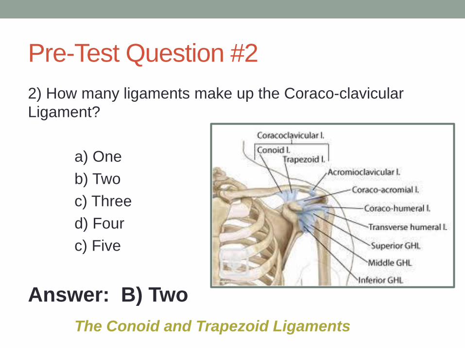
Pre-Test Question #2
2) How many ligaments make up the Coraco-clavicular
Ligament?
a) One
b) Two
c) Three
d) Four
c) Five
Answer: B) Two
The Conoid and Trapezoid Ligaments

Pre-Test Question #3
3) Which of the following tests is used to evaluate for
Bicipital Tendonitis?
a) Jobe
b) Apprehension
c) Hawkins’
d) Apleys
e) Speeds
Answer: E) Speeds

Pre-Test Question #4
4) How many muscles either attach or originate on the
Scapula?
a) 7
b) 10
c) 15
d) 17
e) 21
Answer: D) 17
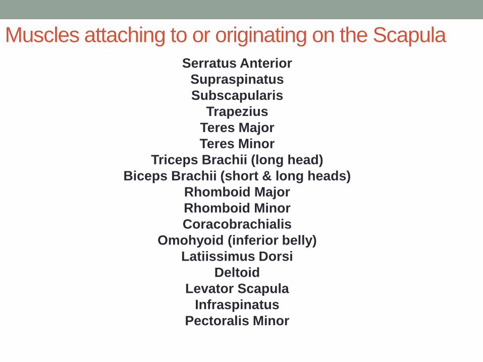
Muscles attaching to or originating on the Scapula
Serratus Anterior
Supraspinatus
Subscapularis
Trapezius
Teres Major
Teres Minor
Triceps Brachii (long head)
Biceps Brachii (short & long heads)
Rhomboid Major
Rhomboid Minor
Coracobrachialis
Omohyoid (inferior belly)
Latiissimus Dorsi
Deltoid
Levator Scapula
Infraspinatus
Pectoralis Minor

Pre-Test Question #55) What is the name of the (true AP) radiographic view that
is taken in the plane of the scapula (30-45o medial to lateral)
a) Scapular Y
b) Swimmers
c) Zanca
d) Serendipity
e) Grashey
Answer: E) Grasheya) Scapular Y – Lateral view for dislocations
b) Swimmers – Lateral view for better visualization of C7-T3
c) Zanca – AP view with Cephalic tilt to view AC joint
d) Serendipity – 40o Cephalic tilt to view SC joint

Pre-Test Question #6
6) Which of the following Osteopathic Manipulative
Medicine Techniques will work best at improving ROM of
the Glenohumeral Joint?
a) Miller Pump
b) Dalrymple Pump
c) Spencers Technique
d) AC Joint Counterstrain
e) Myofascial Release of the Scapulothoracic Joint
Answer: C) Spencers Technique

History and Physical Exam are Key• Is this problem Acute or Chronic (> 3 mos)?
• Was there a History of Trauma/MOI or did it come on slowly?
• Has this problem occurred before?
• What activities exacerbate the symptoms?
• Does the pain radiate from any other area?
• Does this prevent you from doing certain things?
• Are the symptoms getting worse?
• What alleviates the symptoms?
• On a pain scale 1-10, how severe is your pain now and what is it
at it’s worst?
• Have you seen anyone else for this problem before?
• Have you tried anything to self treat or done anything at the
advice of another physician?

Anterior Shoulder

Posterior Shoulder

Lateral Shoulder
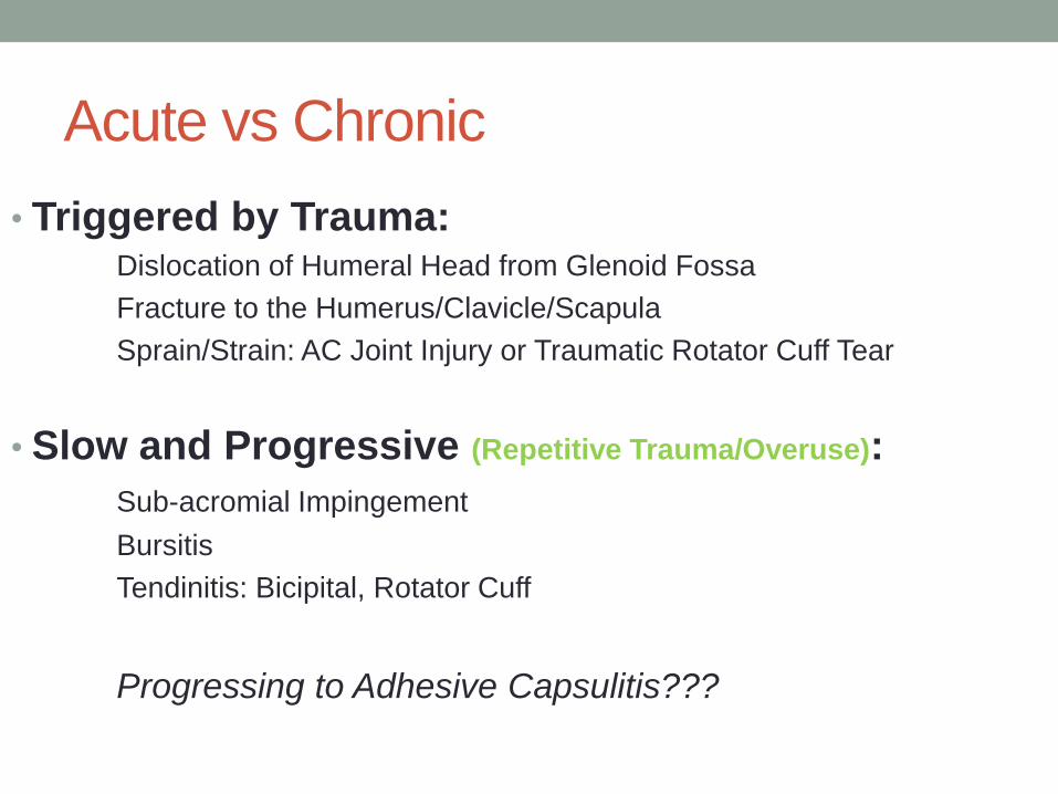
Acute vs Chronic
• Triggered by Trauma:Dislocation of Humeral Head from Glenoid Fossa
Fracture to the Humerus/Clavicle/Scapula
Sprain/Strain: AC Joint Injury or Traumatic Rotator Cuff Tear
• Slow and Progressive (Repetitive Trauma/Overuse):
Sub-acromial Impingement
Bursitis
Tendinitis: Bicipital, Rotator Cuff
Progressing to Adhesive Capsulitis???

Acromion Types• Type I – {FLAT} least likely to cause impingement
• Type II – {CURVED} more likely to cause impingement
• Type III – {BEAKED} most likely to cause impingement, usually requires surgical
debridement prior to rehabilitation
• Subacromial impingement of the Supraspinatus with overhead activities

Gross Shoulder Anatomy Landmarks

Functional Shoulder Articulations
• Structural• Thoracic cage
• Scapula
• Clavicle
• Humerus
• Functional• Scapulothoracic
• Acromioclavicular
• Sternoclavicular
• Glenohumeral
Hoppenfeld

Common Ailments of the Shoulder
• Tendonitis (Supraspinatus, Biceps)
• Bursitis (Sub-acromial)
• Dislocation/Subluxation (Gleno-humeral)
• Sprain (AC Joint)
• Strain/Tear (Rotator Cuff – SITS muscles, Biceps tendon)
• Osteoarthritis (AC Joint)
• Fracture (Clavicular, Scapular, Humeral)
• * Adhesive Capsulitis
• Rule out Cervical Radiculopathy with Spurling’s maneuver and
Neurologic Examination: DTRs, Sensory Testing, and Motor Strength

Case #1
• 54 yo female environmental engineer presents for re-evaluation
of right shoulder pain x 1 week after falling on the same
shoulder. She was initially seen in the ER and x-rays were
negative, she was sent home with a sling. Pain has become
progressively worse and she now has limited ROM in all planes
(sling use x 1 week). No prior injury or shoulder issues
reported. She has been taking Cataflam with limited relief. Pain
7/10 on pain scale.
• Exam is very limited due to pain
• Suspected Frozen Shoulder 2/2 trauma
• CS injection done of the sub-acromial space
• Slight improvement of pain, 5/10, and handout given w/
instructions for ROM exercises and advised discontinued use of
sling

Case #1 Continued
• Patient returns in one week with little to no improved pain
and continued very limited ROM and limited exam 2/2
guarding.
• MRI performed demonstrating
• Massive full thickness tear of the supraspinatus, infraspinatus and
subscapularis muscles, also the biceps tendon is not seen and
appears completely torn
• Surgery referral place
• * Review of history showing Poorly controlled asthma and
taking Prednisone 20 mg daily
• **No OMT done in this case

Frozen Shoulder• Adhesive Capsulitis - refers to a stiffened GH joint that has lost significant ROM
- shoulder motion is more scapulothoracic than GH
- persistent dull ache, unable to lift arm above head or internally rotate GH joint
- Due to lack of use from shoulder pain, mcc rotator cuff tendinopathy
• Tx: OMT, Physical Therapy and Pain Relief (NSAIDs), CS injections
* Consider MUA if not better in 6-18 mos
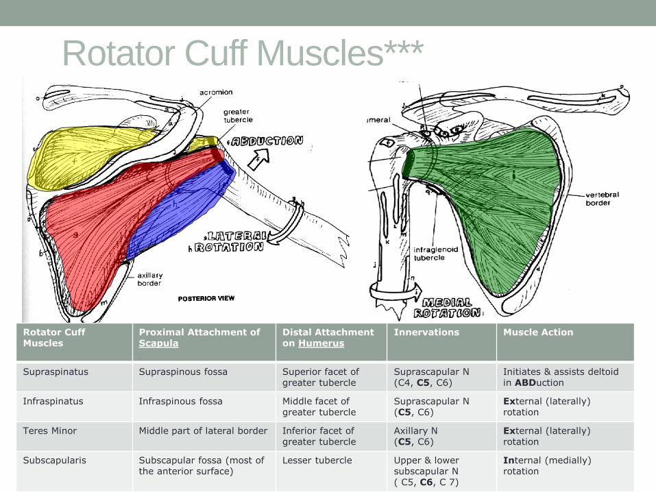
Rotator Cuff Muscles***
Rotator Cuff Muscles
Proximal Attachment of Scapula
Distal Attachment on Humerus
Innervations Muscle Action
Supraspinatus Supraspinous fossa Superior facet of greater tubercle
Suprascapular N(C4, C5, C6)
Initiates & assists deltoid in ABDuction
Infraspinatus Infraspinous fossa Middle facet of greater tubercle
Suprascapular N(C5, C6)
External (laterally) rotation
Teres Minor Middle part of lateral border Inferior facet of greater tubercle
Axillary N (C5, C6)
External (laterally) rotation
Subscapularis Subscapular fossa (most of the anterior surface)
Lesser tubercle Upper & lower subscapular N( C5, C6, C 7)
Internal (medially) rotation

Rotator Cuff Muscles Remember {SITS} Stabilizes humeral head in glenoid fossa along with
many ligaments
Supraspinatus – mc tear, attaches to the greater tubercle of the superior-lateral humeral head
* Mainly involved w/ ABduction Infraspinatus – attaches to the greater tubercle and
Externally Rotates Arm Teres Minor - attaches to the greater tubercle and Externally
Rotates Arm Subscapularis – attaches to the lesser tubercle and
Internally Rotates Arm
* Pain commonly radiates to the Deltoid insertion with rotator cuff pathology
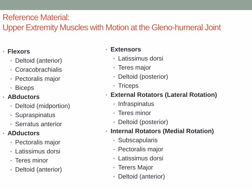
Reference Material:
Upper Extremity Muscles with Motion at the Gleno-humeral Joint
• Flexors
• Deltoid (anterior)
• Coracobrachialis
• Pectoralis major
• Biceps
• ABductors
• Deltoid (midportion)
• Supraspinatus
• Serratus anterior
• ADductors
• Pectoralis major
• Latissimus dorsi
• Teres minor
• Deltoid (anterior)
• Extensors
• Latissimus dorsi
• Teres major
• Deltoid (posterior)
• Triceps
• External Rotators (Lateral Rotation)
• Infraspinatus
• Teres minor
• Deltoid (posterior)
• Internal Rotators (Medial Rotation)
• Subscapularis
• Pectoralis major
• Latissimus dorsi
• Terers Major
• Deltoid (anterior)

Case #2
• 51 yo male presents for evaluation of right shoulder/arm pain
x 1 day. He helped a driver who had got stuck with his
corvette on the beach. While lifting the back end and
pushing the car, with the driver hitting the accelerator, he felt
a snap in his shoulder and arm.
• He immediately feels pain and weakness in the right arm.
• Examination reveals weakness in his right arm, 4/5 with arm
flexion (biceps)
• Tenderness to palpation of the bicipital tuberosity on the
proximal radius
• Pain and weakness with Speeds test and Yergasons test
• Also noted, slight muscular deformity proximally

Case #2 Continued
• MRI performed
confirming distal
bicipital tendon rupture
• Surgery consulted
• No OMT performed

Bicipital Tendinitis• Usually involves Long Head biceps tendon
• L.H. of Biceps tendon passes through the Bicipital Groove of the anterior humerus {Anterior Shoulder Pain}
L.H. attaches to supraglenoid tubercle on humeral head
S.H. attaches to coracoid process of scapula
• Frequently seen w/ Rotator Cuff pathology• Yergason’s Test – elbow flexed to 90o and wrist pronated, pt will attempt to
externally rotate arm and supinate against resistance
• Speed’s Test – Arm Flexed up to 90o and continued Flexion against physician resistance
* (+) if pain in bicipital tendon or tendon slips out of bicipital groove (held in place by transverse humeral ligament)

Speed’s Test• Testing for Bicipital tendonitis,
(long head)
• Patient with Arm Flexed to 80-
90o and Forearm Supinated
• Examiner Resists Forward
Flexion from patient w/
downward force on patient’s
wrist
• Pain in the anterior shoulder,
site of biceps tendon, = + TEST
Yergasons TestTesting for Bicipital tendonitis, bicipital tendon
subluxation
Patient with Arm Pronated and Elbow at 90o
Examiner resists and externally rotates arm
while the patient supinates and externally
rotate the arm against resistance, can add
elbow flexion
Pain in the anterior shoulder, and/or subluxation
of biceps tendon = + Test

Steps to perform Counterstrain (of the shoulder)
• Diagnose the somatic dysfunction by identifying significant tender points.• Test the regions and determine the worst.
• Position the patient to reduce the tenderness.• First stage: place the patient in the classic position.
• Second stage: Fine tune three times to attempt to reduce tenderness to O%.
• Quantify your results each time you position the patient:• assign the initial value of tenderness to 10 on a scale of 0-10
• ask the patient how much tenderness is left on a scale of 0-10 each time you reposition
• “If your original pain was a 10 on a scale of 0-10, what is it now on a scale of 0-10 when I press on it?”
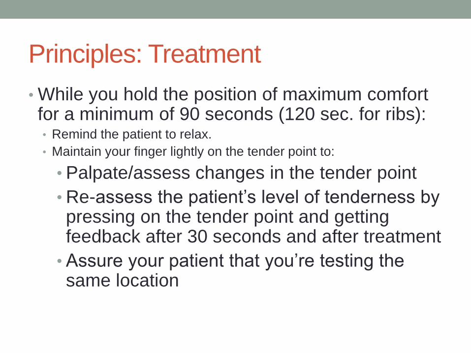
Principles: Treatment
• While you hold the position of maximum comfort for a minimum of 90 seconds (120 sec. for ribs):• Remind the patient to relax.
• Maintain your finger lightly on the tender point to:
• Palpate/assess changes in the tender point
• Re-assess the patient’s level of tenderness by pressing on the tender point and getting feedback after 30 seconds and after treatment
• Assure your patient that you’re testing the same location

Principles: Treatment
• Return the patient to the neutral position. This must be done very slowly and totally passively (talk them out of the position).
• “The only difference between the cause and the cure is the speed of return to the neutral position.”
• Maintain your finger on the tender point while you return the patient to neutral.

Principles: Treatment
• In neutral position, retest the tender point for desired effect, i.e., resolution or significant reduction in tenderness.• reduction of pain to about 30% of the original tenderness level
• Remaining tenderness of:
• 3/10 on tenderness scale or 30% of original tenderness still present
• resolution of “jump sign”
Although some tenderness may be present, this is a significant reduction.

LH Biceps
• Tendon of the biceps
muscle’s long head in
the bicipital groove

LH Biceps
• Flexion of elbow, flexion of shoulder
• Slight abduction
• Internal rotation of shoulder
• Patient should be lying comfortably in the supine
position as seen below with their dorsal wrist/forearm
placed on their forehead
Case #3
• 49 yo female county office worker presents with h/o 3
months worsening right shoulder pain. Denies any
specific trauma, but states pain came on after having a
two week duration of shifting boxes/files in a storage area.
She notes having difficulty with putting her bra strap on
now, unable to talk on phone while holding phone on the
right side. Pain is 9/10 on pain scale and responds
somewhat to Naproxen. Pain resolves when the shoulder
is being held still and not moving.

Special TestsApley Scratch Test
The positions at the lower left test for internal rotation and adduction of the shoulder.
The patient reaches behind his back to touch the inferior angle of the opposite scapula.
The position at the upper left arm tests for external rotation and abduction of the shoulder.The patient reaches behind his or her head to attempt to touch the superior medial angle of the opposite scapula.

Apley’s Scratch TestNotice the limited internal
rotation on the right side *

Normal ROM
• Flexion – 160-180 degrees
• Extension – 45 degrees
• Abduction – 160-180 degrees
• External Rotation – 45-90 degrees
• The Apley Scratch Test used to assess rotation of the shoulder joint. Patients with normal glenohumeral motion should be able to scratch the midback ( T8 to T10 level)
*patients with glenohumeral osteoarthritis (or frozen shoulder and possibly acute rotator cuff tendinitis) have limited ROM when compared with the healthy arm
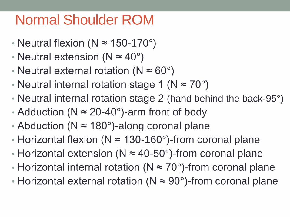
Normal Shoulder ROM
• Neutral flexion (N ≈ 150-170°)
• Neutral extension (N ≈ 40°)
• Neutral external rotation (N ≈ 60°)
• Neutral internal rotation stage 1 (N ≈ 70°)
• Neutral internal rotation stage 2 (hand behind the back-95°)
• Adduction (N ≈ 20-40°)-arm front of body
• Abduction (N ≈ 180°)-along coronal plane
• Horizontal flexion (N ≈ 130-160°)-from coronal plane
• Horizontal extension (N ≈ 40-50°)-from coronal plane
• Horizontal internal rotation (N ≈ 70°)-from coronal plane
• Horizontal external rotation (N ≈ 90°)-from coronal plane

Case #3 Continued
• Pain with Hawkins and Neers tests
• Pain with Flexion and Abduction > 90o of motion

Dx
•Sub-acromial Bursitis/Tendonitis
• leading to Adhesive capsulitis

CS injection (sub-acromial)

1
2
3
4
5a5b
6
7
Spencer Technique
4

Case #4
• 21 yo softball pitcher with c/o right shoulder pain and a
heaviness with right upper extremity tingling which occurs
after throwing 40-50 pitches. Pain and numbness/tingling
has been getting worse over the past 4 months. Pain and
tingling resolve shortly after a pitching sensation has
ended. She denies any specific injuries to the shoulder.
No h/o prior shoulder trauma, including
subluxation/dislocation. Meds don’t help with the pain.
• Examination (+) Roos, (+) Adsons, (+) Wrights tests
• Dx: ?
• Thoracic Outlet Syndrome (TOS)
• To confirm Dx of TOS; EMG/NCS is commonly performed
to determine

Thoracic Outlet Syndrome (TOS) Tests• Roos Test – Shoulder Abduction/External Rotation; Elbows flexed to 90
degrees, hands perform repetitive opening/closing of hands x 3 mins
(Paresthesias) Numbness/Tingling + test
• Adsons Test – Shoulder/Arm held in Slight Extension and Abduction, slight
extension of head turned toward affected side, physician palpates wrist for
Weakened radial pulse or paresthesias + test
(Scalene Triangle Impingement)
• Wrights Test – Hyperabduct arm above the head in the coronal plane,
palpate radial pulse, notice change in pulse with patient turning head away
Weakened radial pulse or Paresthesias + test
(Pectoralis Minor Impingement)
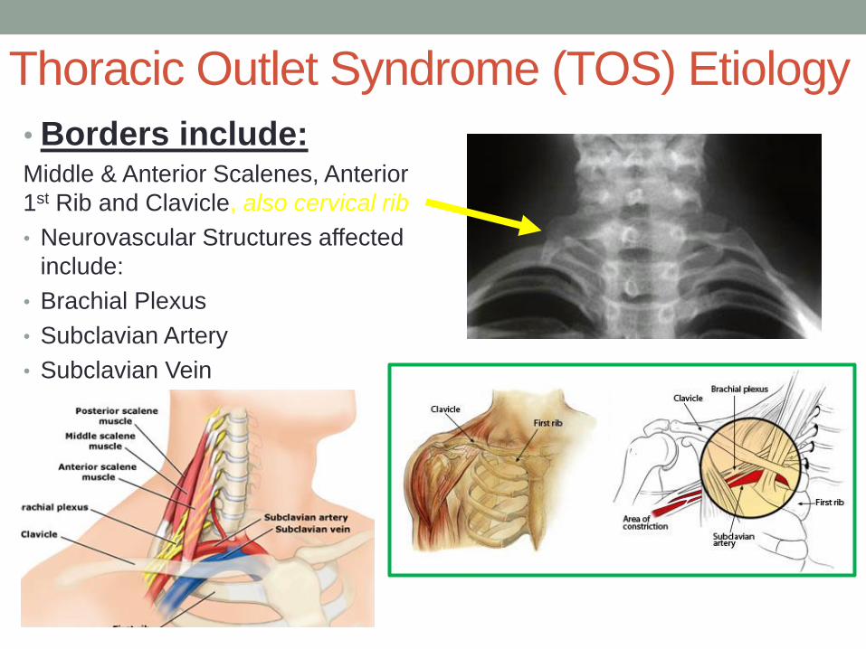
Thoracic Outlet Syndrome (TOS) Etiology
• Borders include:Middle & Anterior Scalenes, Anterior
1st Rib and Clavicle, also cervical rib
• Neurovascular Structures affected
include:
• Brachial Plexus
• Subclavian Artery
• Subclavian Vein
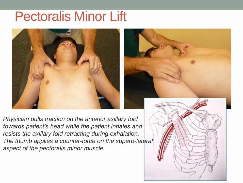
Pectoralis Minor Lift
Physician pulls traction on the anterior axillary fold
towards patient’s head while the patient inhales and
resists the axillary fold retracting during exhalation.
The thumb applies a counter-force on the supero-lateral
aspect of the pectoralis minor muscle
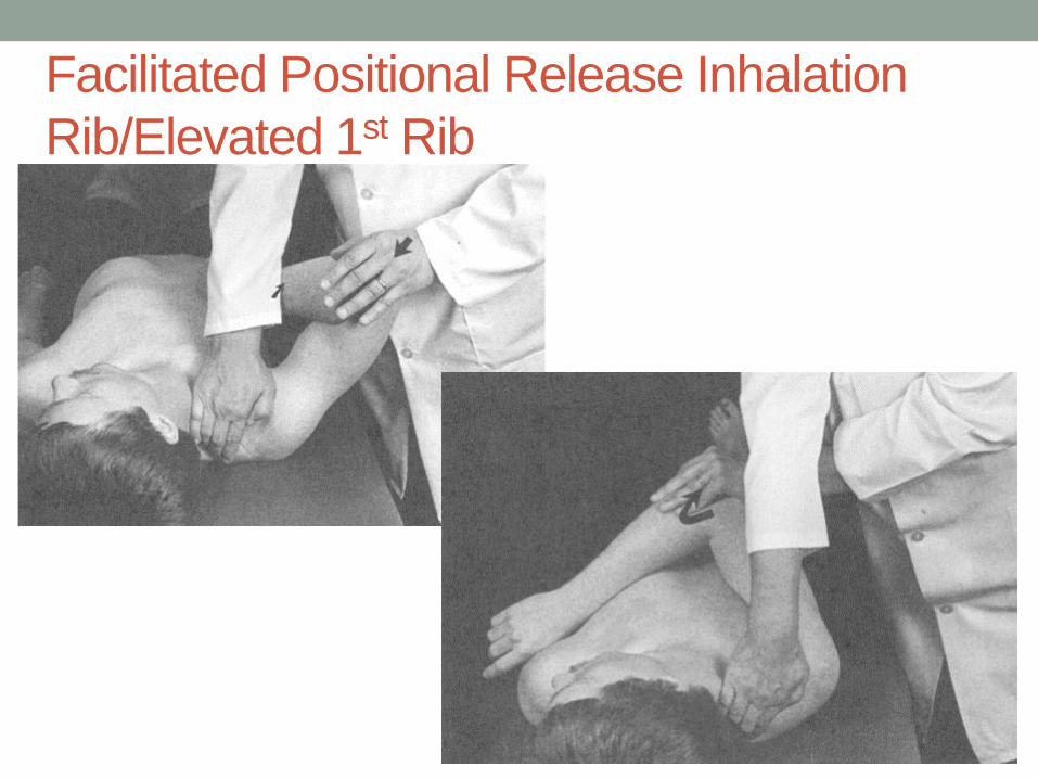
Facilitated Positional Release Inhalation
Rib/Elevated 1st Rib


Still Technique (seated) Superior 1st Rib, LeftA B
C D

Case #5
• A 17 yo male swimmer presents for evaluation of left
shoulder pain x 4 weeks. Pain is worse with overhead
activity such as when he is doing the breaststroke. He is
now starting to have pain when he is lying on his left
shoulder in bed. He denies weakness, but has continued
pain with overhead activities. He has a h/o left shoulder
subluxation previously while water skiing over the
summertime. Pain with activities is 8/10 on pain scale.
Ibuprofen helps minimally with pain.
• (+) O’Briens and Biceps load tests, (-) Drop arm test and
JOBE tests, (+)mild tenderness to palpation of the AC joint,
(+) Apprehension/Relocation test

Drop Arm Test• To screen for possible rotator cuff
tear• Have the patient fully ABduct the
arm (160-180o)• Instruct the patient to slowly lower
the arm to the side.• Tears in the rotator cuff muscles
(specifically the supraspinatus) cause the arm to drop to the side once the arm has been lowered to about 90º of abduction.
Deltoid controls ABduction > 90o
• The patient will not be able to lower the arm slowly and smoothly no matter how many times he tries.
*differentiate from an Axillary Nerve Palsy due to weak, atrophied Deltoid
Watch for shoulder shrugging (cheating) by the patient

Special Tests
Empty Can/JOBE TestTo perform the Empty Can Test aka JOBE Test, have the patient assume a shoulder abduction angle as shown below (90° Abduction with 30° horizontal flexion anterior of coronal plane) and internal rotation (Thumbs DOWN), “like to empty a can”
Doctor applies slight pressure downward on the distal forearm bilaterally to test for Supraspinatus weakness/pain

Apprehension/Relocation Tests• Patient should be placed in the Supine Position w/ arm off table as shown in pictures
• Apprehension Test is used to Evaluate an Unstable Shoulder – one that demonstrates ligamentous laxity/injury and has dislocated/subluxed
• Dislocated – Remains Dislocated and Needs to be Reduced Back into the Glenoid Fossa
• Subluxed – Relocates Spontaneously After Coming Out of the Glenoid Fossa
• Shoulder is Vulnerable in ABduction and/or External Rotation (Positon of Apprehension/Relocation Test)
• Relocation Test – Posterior pressure is placed on the GH joint being tested, which gives relief of pain and sense of possible dislocation

Apprehension/Relocation Tests
Apprehension Test Relocation Test

SLAP Lesion Superior Labrum Anterior to Posterior tear
Pain with lying on affected arm Feeling of Instability, Grinding with GH Joint
O’Brien Test – forward flex shoulder w/ elbow extended, bring arm to midline 15 degrees, internally rotate (THUMB DOWN), resist force downward, then test with THUMB UP
(+) Test if pain is present/worse with THUMB DOWN Bicipital Load Test – Arm is Abducted 100o and Forearm
Supinated, Flexed 90o and Patient attempts to Flex (fist towards head) against resistance
Pain on top of shoulder - think AC joint injury Pain in shoulder joint - think injury to labrum Pain @ Deltoid Insertion - think Rotator Cuff
If this is being suspected and an MRI is being ordered, get it with an Arthrogram to better visualize a possible labral tear!

SLAP Lesion
• Superior Labral Anterior-Posterior Tear
• O’Brien’s Test Shoulder: Horizontal Flexion (90o)
Adduction (15o)
Internal rotation
Patient resists further motion in these directions
Thumb down (+) pain, possible tear
Thumb up (-) pain, alleviates

Glenohumeral Joint• Labrum
- fibro-cartilagenous structure
- deepens the glenoid fossa
- provides increased stability

Anatomy• The Glenohumeral (GH) joint is loosely constrained within a thin
capsule bounded by surrounding muscles and ligaments.
• The shoulder's great mobility is due to the shallow depth of the glenoid & the limited contact between the glenoid and the humeral head.
• Only 25% of the humeral head surface makes contact with the glenoid
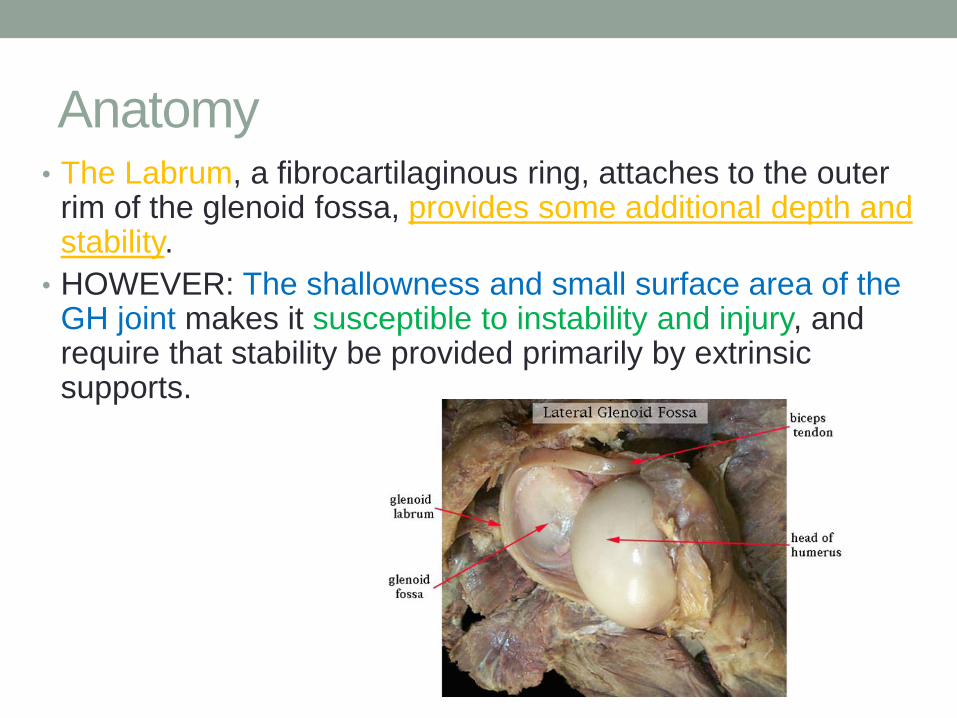
Anatomy• The Labrum, a fibrocartilaginous ring, attaches to the outer
rim of the glenoid fossa, provides some additional depth and stability.
• HOWEVER: The shallowness and small surface area of the GH joint makes it susceptible to instability and injury, and require that stability be provided primarily by extrinsic supports.

Infraspinatus/Teres Minor• Both tendons attach to the lateral humeral head (Greater Tubercle)
• Primarily involved w/ External Rotation of Shoulder
• Typically torn together, seen w/ massive rotator cuff tears
Watch for Shoulder Shrug while attempting to lift arm
• Tested with Patient Attempting to Externally Rotate Against the examiners counterforce
• DROPPING test – aka External Rotation Lag Test, flex elbow to 90, arm tight at side, ROTATE Externally to max point, then release, (+) if arm involuntarily drifts back to Neutral position
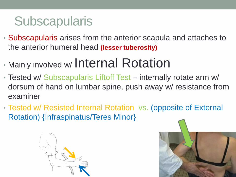
Subscapularis• Subscapularis arises from the anterior scapula and attaches to
the anterior humeral head (lesser tuberosity)
• Mainly involved w/ Internal Rotation• Tested w/ Subscapularis Liftoff Test – internally rotate arm w/
dorsum of hand on lumbar spine, push away w/ resistance from
examiner
• Tested w/ Resisted Internal Rotation vs. (opposite of External
Rotation) {Infraspinatus/Teres Minor}

Case #6
• A 27 yo male presents with h/o recurrent right shoulder
pain. He initially injured the shoulder playing football in
high school. He states that the pain is on the top of his
shoulder and also in the upper back radiating up his neck.
He states getting a steroid injection in his shoulder about
2 years ago which helped with the shoulder pain. He is
now getting the pain whenever he is lifting weights such
as with bench press, doing dips and also push ups. He
takes Tylenol occasionally for pain, which doesn’t really
work. Pain is rated at 8/10 on pain scale with
exacerbating activities and 2/10 with rest following a work
out.

Case #6 continued
• Examination reveals pain to palpation at the right AC joint
with pain exacerbated with cross arm Adduction test
• He also has pain on the right upper back at the superior-
medial scapular border radiating up to his ipsilateral
cervical spine

AC Joint Injuries
• The AC joint has a cartilaginous disk and synovial membrane
• Injuries to the AC joint are classified according to the position of the clavicle with respect to the acromion and coracoid
• Injuries usually due to direct trauma superior or laterally, or forced adduction of the arm
• the distal clavicle is held in alignment with the acromion by the strong coracoclavicular (CC) ligament comprised of 1) conoid, and 2) trapezoid ligaments

Acromioclavicular (AC) Joint

AC Joint Sprains/Injuries• Seen w/ pain to direct palpation of the AC joint
• Crossed Arm Adduction Test also indicative
• 6 Types of AC joint injuries
1) Partial tear of the AC ligaments, and manifest as a tender AC joint that often
has mild swelling but no deformity
2) Represent a complete tear of the AC ligaments and partial tear of the
CC ligaments
3) Involves a complete disruption of both the AC and CC ligaments
4) Occurs with forceful shoulder trauma that causes disruption of the AC and
CC ligaments, and displaces the distal clavicle into or through the
trapezius
5) Represents significant disruption of the AC and CC ligaments, along with
disruption of the muscular and fascial attachments of the distal clavicle
6) rare and involve severe dislocations of the AC joint in which the distal
clavicle is forced into the subacromial or subcoracoid position

Ligament Sprain/Tear
• Treatment Options:
• OMT
• +/- Short Period of Immobilization for Pain Relief
• Physical Therapy/Daily Stretching
• NSAIDs
• Corticosteroid Injection or Prolotherapy
• Surgery if unstable/continued pain/failed conservative management
• Corticosteroid injection: 0.5 mL/0.5mL/0.5-1 mL {lidocaine/marcaine/betamethasone or triamcinolone}
• Prolotherapy: Dextrose 50% MIXED with Lidocaine to promote inflammation and healing (no NSAIDs
or CS 1 week prior or for 4 weeks after injections

Imaging
AP view – If looking for a separation of the AC joint: Normal spacing intervals are:
1) separation from acromion and clavicle should be < 8 mm2) separation of coracoid and clavicle should be < 13 mm
x-ray should be taken as the patient holds a weight in his or her affected shoulder

Radiographs of AC Joint Injuries
Type 1 AC Joint Injury: tearing of the
AC ligament
Type 3 AC Joint Injury: tearing of the
AC and CC ligaments

OA of Shoulder Joint
• Osteoarthritis of the GH joint represents wear-and-tear of the articular cartilage of the glenoid, labrum, and humeral
head (more of chronic problem, however RARE)• Seen with prior h/o fractures, dislocations, major rotator
cuff tears
• Also consider Metabolic Disease (DM, Hypothyroidism, RA) as a presdisposing/contributing factors

Anatomy
•Levator Scapulae • originates on TP of C1-4,
• inserts on superior-medial angle of scapula
* innervated by C2-4
• Commonly seen is muscle spasm especially @ supero-
medial scapular border• Tx: Trigger Point injection with Lidocaine/Marcaine {50/50 ratio} +/- Homeopathic agent

Levator Scapulae
• Place the arm behind the back to
rotate the scapula medially.
• Push the scapula superiorly and
medially from the inferior angle to
shorten the muscle.
• Patient’s head placed on a pillow to
achieve side bending of the neck
toward the dysfunctional side.
• Follow the other steps for
performing Counterstrain treatment
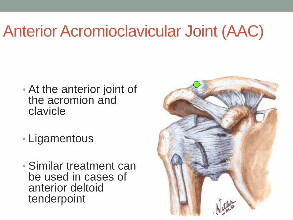
Anterior Acromioclavicular Joint (AAC)
• At the anterior joint of the acromion and clavicle
• Ligamentous
• Similar treatment can be used in cases of anterior deltoid tenderpoint

AAC
• Flexion, slight internal rotation, adduction with traction across the body

Posterior Acromioclavicular Joint (PAC)
• At the posterior joint of the
acromion and clavicle
• Ligamentous
• Posterior deltoid dysfunction
can be treated approximately
the same way, with fine tuning

PAC• Patient prone
• Pull arm back into
extension and slight
horizontal adduction
• hand held at about
midline near/behind
waist

Shoulder PainViscero-somatic vs. Somatic
• Where is the pain coming from?
• NOT ALL SHOULDER PAIN IS
ACTUALLY SHOULDER PAIN
• - Check elbow/cervical/thoracic spines
• Visceral dysfunction needs to also be
considered in the diagnosis.

Referred Shoulder Pain
Boa’s Sign
• Pain in right shoulder/upper
back
• Referred pain from abdominal
organs on the right
• Indicates possible
cholecystitis, pyloric stenosis
or duodenal ulcer
Kehr’s Sign
• Pain in left shoulder/upper
back
• Referred pain from abdominal
organs on the left
• Indicates possible splenic
injury (splenic rupture in
supine patient), gastritis,
gastric ulcer or even renal
stone
• Also possible Cardiac
etiology
Post-Test Question #1
1) Which nerve is most commonly injured with a gleno-
humeral shoulder dislocation?
a) Axillary Nerve
b) Suprascapular Nerve
c) Musculo-cutaneous Nerve
d) Radial Nerve
c) Ulnar Nerve
Answer: A) Axillary Nerve
Post-Test Question #2
2) How many ligaments make up the Coraco-clavicular
Ligament?
a) One
b) Two
c) Three
d) Four
c) Five
Answer: B) Two
The Conoid and Trapezoid Ligaments

Post-Test Question #3
3) Which of the following tests is used to evaluate for
Bicipital Tendonitis?
a) Jobe
b) Apprehension
c) Hawkins’
d) Apleys
e) Speeds
Answer: E) Speeds
Post-Test Question #4
4) How many muscles either attach or originate on the
Scapula?
a) 7
b) 10
c) 15
d) 17
e) 21
Answer: D) 17

Post-Test Question #55) What is the name of the (true AP) radiographic view that
is taken in the plane of the scapula (30-45o medial to lateral)
a) Scapular Y
b) Swimmers
c) Zanca
d) Serendipity
e) Grashey
Answer: E) Grashey

Post-Test Question #6
6) Which of the following Osteopathic Manipulative
Medicine Techniques will work best at improving ROM of
the Glenohumeral Joint?
a) Miller Pump
b) Dalrymple Pump
c) Spencers Technique
d) AC Joint Counterstrain
e) Myofascial Release of the Scapulothoracic Joint
Answer: C) Spencers Technique