comparative analysis of gingival tissue antigen
TRANSCRIPT

University of Kentucky University of Kentucky
UKnowledge UKnowledge
Center for Oral Health Research Faculty Publications Oral Health Research
4-2014
Comparative Analysis of Gingival Tissue Antigen Presentation Comparative Analysis of Gingival Tissue Antigen Presentation
Pathways in Ageing and Periodontitis Pathways in Ageing and Periodontitis
Octavio A. Gonzalez University of Kentucky, [email protected]
Michael John Novak University of Kentucky, [email protected]
Sreenatha S. Kirakodu University of Kentucky, [email protected]
Luis Orraca University of Puerto Rico, Puerto Rico
Kuey-Chu Chen University of Kentucky, [email protected]
See next page for additional authors Follow this and additional works at: https://uknowledge.uky.edu/cohr_facpub
Part of the Periodontics and Periodontology Commons
Right click to open a feedback form in a new tab to let us know how this document benefits you. Right click to open a feedback form in a new tab to let us know how this document benefits you.
Repository Citation Repository Citation Gonzalez, Octavio A.; Novak, Michael John; Kirakodu, Sreenatha S.; Orraca, Luis; Chen, Kuey-Chu; Stromberg, Arnold J.; Gonzalez-Martinez, Janis; and Ebersole, Jeffrey L., "Comparative Analysis of Gingival Tissue Antigen Presentation Pathways in Ageing and Periodontitis" (2014). Center for Oral Health Research Faculty Publications. 7. https://uknowledge.uky.edu/cohr_facpub/7
This Article is brought to you for free and open access by the Oral Health Research at UKnowledge. It has been accepted for inclusion in Center for Oral Health Research Faculty Publications by an authorized administrator of UKnowledge. For more information, please contact [email protected].

Comparative Analysis of Gingival Tissue Antigen Presentation Pathways in Comparative Analysis of Gingival Tissue Antigen Presentation Pathways in Ageing and Periodontitis Ageing and Periodontitis
Digital Object Identifier (DOI) http://dx.doi.org/10.1111/jcpe.12212
Notes/Citation Information Notes/Citation Information Published in Journal of Clinical Periodontology, v. 41, issue 4, p. 327-339.
© 2014 John Wiley & Sons A/S.
This is the peer reviewed version of the following article: Gonzalez OA, Novak MJ, Kirakodu S, Orraca L, Chen K-C, Stromberg A, Gonzalez-Martinez J, Ebersole JL Comparative analysis of gingival tissue antigen presentation pathways in ageing and periodontitis. J Clin Periodontol 2014; 41: 327–339, which has been published in final form at http://dx.doi.org/10.1111/jcpe.12212. This article may be used for non-commercial purposes in accordance with Wiley Terms and Conditions for Self-Archiving.
Authors Authors Octavio A. Gonzalez, Michael John Novak, Sreenatha S. Kirakodu, Luis Orraca, Kuey-Chu Chen, Arnold J. Stromberg, Janis Gonzalez-Martinez, and Jeffrey L. Ebersole
This article is available at UKnowledge: https://uknowledge.uky.edu/cohr_facpub/7

This is the peer reviewed version of the following
article: Gonzalez OA, Novak MJ, Kirakodu S, Orraca
L, Chen K-C, Stromberg A, Gonzalez-Martinez J,
Ebersole JL Comparative analysis of gingival tissue
antigen presentation pathways in ageing and
periodontitis. J Clin Periodontol 2014; 41: 327–339,
which has been published in final form at
http://dx.doi.org/10.1111/jcpe.12212. This article
may be used for non-commercial purposes in
accordance with Wiley Terms and Conditions for
Self-Archiving.

Comparative Analysis of Gingival Tissue Antigen PresentationPathways in Aging and Periodontitis
O.A. Gonzalez1,*, M.J. Novak1, S. Kirakodu1, L. Orraca4, K.C. Chen2, A. Strom-berg3, J.Gonzalez-Martinez5, and J. L. Ebersole1
1Center for Oral Health Research, College of Dentistry, University of Kentucky, Lexington, KY2Microarray Core Facility, College of Medicine, University of Kentucky, Lexington, KY3Department of Statistics, College of Arts & Sciences, University of Kentucky, Lexington, KY4School of Dental Medicine, University of Puerto Rico, San Juan, PR5Caribbean Primate Research Center, University of Puerto Rico, Sebana Seca, PR
AbstractAim—Gingival tissues of periodontitis lesions contribute to local elevations in mediators,including both specific T cell and antibody immune responses to oral bacterial antigens. Thus,antigen processing and presentation activities must exist in these tissues to link antigen-presentingcells with adaptive immunity. We hypothesized that alterations in the transcriptome of antigenprocessing and presentation genes occur in aging gingival tissues and that periodontitis enhancesthese differences reflecting tissues less capable of immune resistance to oral pathogens.
Materials and Methods—Rhesus monkeys (n=34) from 3–23 years of age were examined. Abuccal gingival sample from healthy or periodontitis sites were obtained, total RNA isolated, andmicroarray analysis was used to describe the transcriptome.
Results—The results demonstrated increased transcription of genes related to the MHC class IIand negative regulation of NK cells with aging in healthy gingival tissues. In contrast, both adultand aging periodontitis tissues showed decreased transcription of genes for MHC class II antigens,coincident with up-regulation of MHC class I-associated genes.
Conclusion—These transcriptional changes suggest a response of healthy aging tissues throughthe class II pathway (i.e., endocytosed antigens) and altered responses in periodontitis that couldreflect host-associated self-antigens or targeting cytosolic intra-cellular microbial pathogens.
KeywordsPeriodontitis; aging; antigen presentation; adaptive immunity
INTRODUCTIONPeriodontitis is a chronic infection of the oral cavity with pathogenic biofilms triggering apersistent inflammatory response in gingival tissues that lead to loss of function of theperiodontium and consequently loss of teeth (Jakubovics and Kolenbrander, 2010, Armitage
*Corresponding author: Octavio A. Gonzalez, Complete Address: Center for Oral Health Research, College of Dentistry, University ofKentucky, 1095 VA Drive. HSRB 414 Lexington, KY 40536-0305. Phone: (859)-323-0125. Fax number: (859)[email protected].
Conflict of interestThe authors declare that there are no conflicts of interest related to this study.
NIH Public AccessAuthor ManuscriptJ Clin Periodontol. Author manuscript; available in PMC 2015 April 01.
Published in final edited form as:J Clin Periodontol. 2014 April ; 41(4): 327–339. doi:10.1111/jcpe.12212.
NIH
-PA Author Manuscript
NIH
-PA Author Manuscript
NIH
-PA Author Manuscript

and Cullinan, 2010). Historically, a range of immune and non-immune cell responses to oralbacteria in vitro and in gingival crevicular fluid of periodontitis have been described(Belibasakis and Guggenheim, 2011, Yin et al., 2010, Peyyala et al., 2012, Bodet et al.,2006, Kinane and Bartold, 2007). More recently, numerous investigations have emphasizedthe importance of the innate immune system in oral mucosal tissues, producing an array ofbiomolecules to maintain homeostasis (DeSantis et al., 2006). Nevertheless, the apparentinability of innate immunity and the inflammatory response to control oral infections resultsin the generation of more specific adaptive immune responses (Hayman et al., 2011,Ebersole, 2003a).
Both local and systemic immune responses result from periodontal infections, and arecomposed of antigen specific T cells and antibody of diverse isotypes and subclasses(Ebersole, 2003b). Various studies have documented that the phenotype and function of Tcells in the periodontium reflect the types of antigens inducing the local responses andcontribute to communicating with osteogenic processes leading to a potential control of thebone resorptive processes (Vernal et al., 2006, Kawai et al., 2006). Additionally, elevatedlevels of antibodies are detected to bacteria considered to be pathogens in oral biofilms(Hayman et al., 2011, Ramseier et al., 2009, Kinane and Bartold, 2007, Takeuchi et al.,2006). The breadth of adaptive immune responses, coupled with the detection and proposedrole of professional antigen presenting cells (APCs), macrophages (Ku et al., 2011, Artese etal., 2011, Ren et al., 2009) and den-dritic cells (Jotwani et al., 2001, Cutler and Jotwani,2006) supports that local antigen uptake, processing, and presentation must occur and play arole in control of periodontal infections.
Existing epidemiological data demonstrate increases in the prevalence and severity ofperiodontitis with aging in the presence of altered immune responses that may contribute toboth protection and tissue destructive processes (Huttner et al., 2009). The acceptedparadigm from these observations is that the disease in aging represents an accumulation ofnoxious challenge over time linked with more general disruptions in the integrity of theperiodontal tissues (Hajishengallis, 2010, Gonzalez et al., 2011, Ebersole et al., 2008b,Ebersole et al., 2008a). However, substantial literature from other models of infection hasdemonstrated significant age-associated increases in susceptibility to infections. Theseobservations have identified decreases in the capacity of older individuals to producespecific antibody (Frasca et al., 2011), and alterations in T cell activation profiles that couldaffect antibody levels/functions (Ebersole et al., 2008b, McArthur et al., 1995, Haynes andSwain, 2012).
Various aspects of human periodontal disease may be assessed in animal models thatpossess similar oral structures to the human periodontium (Graves et al., 2012, Oz andPuleo, 2011, Struillou et al., 2010, Yoshinari et al., 2006, Persson, 2005, Hardham et al.,2005, Ebersole et al., 2002, Assuma et al., 1998, Persson et al., 1994, Schou et al., 1993,Persson et al., 1993, Dreyer et al., 1986),. These animal models of periodontal bone loss alsoinclude extensive studies in nonhuman primates (Roberts et al., 2004, Ebersole et al., 2002,Ebersole et al., 2000a, Schou et al., 1993, Holt et al., 1988, Ebersole et al., 1999, Moritz etal., 1998, Beem et al., 1991), in which significant bone loss results from ligature-induceddisease, enable the examination of microbiological, immunological, and clinical features ofperiodontal disease and its prevention and treatment, and provide data supporting diseaserelated to infection by P. gingivalis (Holt et al., 1988) similar to humans. It is clear that theprimate model has provided the essential bridge for understanding the interaction of selectedmembers of the subgingival microbiota with the host, particularly as reflected by thelongitudinal alterations, which occur in the clinical and microbiological progression ofligature-induced periodontitis similar to the human periodontal experience (Madden andCaton, 1994, Persson et al., 1993). We and others have shown that characteristics of the
Gonzalez et al. Page 2
J Clin Periodontol. Author manuscript; available in PMC 2015 April 01.
NIH
-PA Author Manuscript
NIH
-PA Author Manuscript
NIH
-PA Author Manuscript

inflammatory response and systemic humoral immune responses that accompany ligature-induced periodontitis in nonhuman primates parallel those observed in human periodontitis(Ebersole et al., 2010, Ebersole et al., 2009, Ebersole et al., 2008b, Ebersole et al., 2002,Persson et al., 1994). Soluble receptors to IL-1 and TNF significantly inhibited recruitmentof inflammatory cells in close proximity to bone, reduced osteoclast formation, and reducedbone loss in ligature-induced periodontitis in a nonhuman primate animal model (Assuma etal., 1998, Delima et al., 2001). Thus, we hypothesized that nonhuman primates could beemployed to delineate alterations in the transcriptome of antigen processing and presentationgenes that occur in aging gingival tissues and that periodontitis would enhance thesedifferences reflecting tissues less capable of immune resistance to oral pathogens.
MATERIALS AND METHODSOral clinical evaluation
Rhesus monkeys (Macaca mulatta) (n=34; 14 females and 20 males) housed at theCaribbean Primate Research Center (CPRC) at Sabana Seca, Puerto Rico, were used in thesestudies. Animals were selected by age based on the following criteria: <3 years (young;n=5), 3–7 years (adolescent; n=5), 12–16 years (adult; n=12) and 18–23 years (aged; n=12).Nonhuman primates were fed a 20% protein, 5% fat, and 10% fiber commercial monkeydiet (diet 8773, Teklad NIB primate diet modified: Harlan Teklad). The diet wassupplemented with fruits and vegetables, and water was provided ad libi-tum in an enclosedcorral setting.
A protocol approved by the Institutional Animal Care and Use Committee (IACUC) of theUniversity of Puerto Rico, enabled anesthetized animals to be examined for clinicalmeasures of periodontal including probing pocket depth (PD), and bleeding on probing(BOP) (Ebersole et al., 2008a), including measures on 2 interproximal sites per tooth(mesio- and disto-buccal), excluding the canines and 3rd molars.
Tissue sampling and gene expression microarray analysisA buccal gingival sample from either a healthy or periodontitis-affected site from thepremolar/molar maxillary region of each animal was taken using a standard gin-givectomytechnique, and maintained frozen in RNAlater solution. Total RNA from each gingivaltissue was isolated using a standard procedure as we have described, and submitted to themicroarray core to assess RNA quality and analyze the transcriptome using the GeneChip®Rhesus Macaque Genome Array (Affymetrix) (Meka et al., Gonzalez et al., 2011).
qPCR AnalysisBased upon the microarray outcomes we selected 5 genes and performed a qPCR analysisusing a standard technique in our laboratory employing a Roche 480 LightCycler. The oraltissue from M. mulatta was stored in RNAlater (Invitrogen, IN) in −80°C. Oral tissue wasremoved from RNAlater solution and homogenized in TRIZOL reagent (Invitro-gen, IN)and RNA was isolated following venders instruction. RNA was cleaned up using RNeasyMini kit (Qiagen), and subsequently quantified using Nanodrop 1000 (Thermo Scientific).RNA from 4 different animals was pooled from each group and a total of 1ug was used foreach cDNA synthesis reaction. The first strand cDNA synthesis was carried out using 2.5μM anchored-oligo (dT)18 primer, 1X transcriptor RT reaction buffer, 20 units of 40 U/μlRNase inhibitor, 1 mM of deoxynucleotide mix and 10 units of reverse transcriptase (Roche,IN) to a final reaction volume of 20 μl. The reaction mixture was incubated 30 min at 55°Cand the reverse transcriptase was inactivated by incubating 5 min at 85°C.
Gonzalez et al. Page 3
J Clin Periodontol. Author manuscript; available in PMC 2015 April 01.
NIH
-PA Author Manuscript
NIH
-PA Author Manuscript
NIH
-PA Author Manuscript
qPCR primers were designed using software PrimerQuest at Integrated DNA Technologieswebsite (www.idtdna.com) and were synthesized by Integrated DNA Technologies, Inc(Coralville, IA). Primers were prepared for PSME2 (forward -CCACCCAAGGATGATGAGATG; reverse - CAGGGACAGGACTTTCTCATTC), IF130(forward - GAGAAGAGGAGTGCAAACTCAA; reverse -TCTCCATGTCGTCAAACTCTTC), HLA-DPB1 (forward -TTTCTACCCAGGCAGCATTC; reverse - TGGAAGGTCCAGTCTCCATTA), HLA-DRA(forward - TCTCCCAGAGACTACAGAGAAC; reverse -CGCACACCCTTGATGATGA), CTSS (forward - GATGCGAGTCATCCTTCTTTCT;reverse - CACCATAGCCAACCACAAGTA) and GAPDH (forward -GGTGTGAACCATGAGAAGTATGA; reverse - GAGTCCTTCCACGATACCAAAG)genes. The cDNA was diluted 1:4 and used as a template for the real-time PCR analysisusing a LightCycler 480 (Roche, IN). qPCR was performed in a total volume of 20 μlcontaining 2 μl of 10X LightCycler 480 SYBR Green I Master (Roche, IN), 0.5 μM each offorward and reverse primers, and 2 or 4 μl of the diluted cDNA. Real-time qPCR wascarried out with an initial incubation at 95°C for 5 min followed by 45 cycles consisting ofdenaturing at 95°C for 10 s, annealing at 60°C for 5 s followed by amplification at 72°C for5 s.
After amplification, a melting curve analysis was done to determine the specificity of thePCR products by incubating the products for 15 s at 55°C, and then increasing thetemperature to 95°C at a ramp rate of 0.1°C/s. Melting curve profiles was used to identifyand genotype PCR products. Relative quantification analysis was performed withLightCycler 480 software (Roche, IN). The concentration ratios for the target genes werecalculated by normalizing to the housekeeping gene GAPDH. The level of message wasdetermined and those levels compared across the RNA samples prepared from each of thehealthy groups and the 2 periodontitis groups.
Data AnalysisNormalization of values across the chips was accomplished through signal intensitystandardization across each chip using Affymetrix PLIER algorithm. The Ge-neChip®Rhesus Macaque Genome Array contained matched and mismatched pairs allowing theMAS 5 algorithm to be used. For each gene we first determined differences in expressionacross the groups using ANOVA (version 9.3, SAS Inc., Cary, NC). The healthy agedtissues were then compared to the other age groups, or compared with health versusperiodontitis tissues in adults or aged animals using a t-test and accepting a p-value ≤0.05for significance. Because of the cost of these types of nonhuman primate experiments andavailability of primates of the various ages, we did not have sufficient samples to identify ifthe relationship between age and gene expression could be treated using a linear model, thusthe subjects were classified and ANOVA was used for analysis. The choice of LeastSignificant Difference for multiple comparisons (ANOVA followed by t-tests) providedmaximum power given our necessarily small sample sizes. We did determine a correlationwith aging in healthy tissues or periodontitis tissues using a Spearman Rank correlationanalysis that was fit to the gene expression by age. A p-value ≤ 0.05 was used to evaluatethe significance of the correlation. Genes whose expression showed significant correlationwith age were mapped into the Kyoto Encyclopedia of Genes and Genomes (KEGG) antigenprocessing and presentation pathway (www.genome.jp) to develop an ontology analysis.JMP (version 10.0, SAS Inc., Cary, NC) was used to create metagenes independently ofgroup classification using principal components based on the correlation matrix. The plotsare of the first two PCA scores across the healthy tissues and comparing healthy toperiodontitis tissues within the two age groups. The variability is explained by each of the
Gonzalez et al. Page 4
J Clin Periodontol. Author manuscript; available in PMC 2015 April 01.
NIH
-PA Author Manuscript
NIH
-PA Author Manuscript
NIH
-PA Author Manuscript
scores indicated on the plots. The data has been uploaded to http://www.ncbi.nlm.nih.gov/geo/info/submission.html.
RESULTSGene Expression Profiles in Aging Healthy Gingival Tissues
Table 1 lists the genes examined in the antigen presentation and processing pathways. Table2 identifies those genes that were significantly altered in aging healthy gingival tissuescompared to other groups. The results demonstrated that the majority of alterations wereobserved in genes related to the Major Histocompatibility Complex class II (MHC-II)pathway, although selected NK cell genes were up-regulated in the healthy aging gingivaltissues. Table 2 also provides an overview of the genes significantly correlated with age inhealthy tissues. As was noted with the up-regulation of the MHC-II pathway genes, themajority of the correlated genes were in this pathway. Interestingly, this analytic approachalso demonstrated a negative correlation with a primary MHC class I (MHC-I) antigen,MAMU-A, the heat shock protein, HSP90, and NFYA, a nuclear transcription factor.
Gene Expression Profiles in Periodontitis TissuesTable 3 summarizes the variations in gene expression comparing healthy and periodontitistissues. In adults MHC-I and MHC-II pathway genes were up-regulated, while HSP90 weredown-regulated. Similar patterns were observed in periodontitis tissues from aged animals,with HSP70 down-regulation. Additionally various NK antigens were up-regulated inperiodontitis tissues. Table 3 also summarizes the gene expression profiles that correlatedwith age in periodontitis tissues. While a number of these correlated genes overlapped withthose noted in healthy tissues, i.e. aging effect, there were a number of genes in both MHC-Iand II pathways that were significantly related to age, but only in the diseased tissues.Negative correlations were again noted with selected heat shock proteins and an NFYtranscription factor.
The results of a Principal Components Analysis for the groups depicted in Figure 1demonstrate that the transcriptome of aged healthy tissues was somewhat distinct from theother age groups with over 42% of the variation among the age groups described by thesegene patterns. Additionally, the periodontitis tissues from the aged animals demonstrated adistinctive profile of genes expressed compared to healthy adult or aged tissues with 44.7%of the variation between disease and healthy tissues accounted for by the differential geneexpression. Table 4 identifies those genes that were primary determinants in the PC analysis,showing representation of both the class I and class II pathways in discriminating among thehealthy age groups and periodontitis tissues. We subsequently performed a PC Analysis onthe animals, based upon clinical presentation of the gingival tissue for extent of bleeding onprobing and mouth mean pocket depths, rather than stratified based on age. While theclinical characteristics increased with elevated bleeding and pocket depths with age and inthose animals identified with naturally acquired periodontitis, the results demonstrated amuch less robust grouping of the individual animals when age was omitted fromconsideration in this analysis (data not shown).
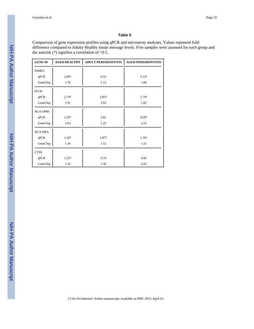
From the list of altered gene expression, we selected 5 genes (PSME2, IF130, HLA-DRB1,HLA-DRA, CTSS) for qPCR validation of the microarray analysis. Table 5 provides theresults of this comparison identifying similar directions for the altered gene expression,although the magnitude varied between these 2 independent analyses. Only 3/15comparisons appeared to be at variance between the methods. Correlation analysesdemonstrated that 11/15 of the comparisons showed a positive correlation of >0.5.
Gonzalez et al. Page 5
J Clin Periodontol. Author manuscript; available in PMC 2015 April 01.
NIH
-PA Author Manuscript
NIH
-PA Author Manuscript
NIH
-PA Author Manuscript
Figure 2 provides a schematic of antigen processing and presentation pathways for both theMHC-I and II antigens as well as T cell interactions with the resulting antigenic moietiesthat have been processed by the APCs. Highlighted within the figure are those genes whoseexpression was altered with aging in healthy gingival tissues. Figures 3A & B provide aschematic of the gene expression profiles in the antigen presentation and processingpathways that were affected by periodontitis in adult or aged animals. In both age groups,periodontitis was reflected by up-regulation of genes within the MHC-I pathway, with fewerincreases in gene expression for NK cell antigens compared to healthy tissues. Additionally,both HSP70 and HSP90 gene expression was down-regulated with disease. The agedperiodontitis tissues continued to demonstrate up-regulation of genes within the MHC-IIprocessing compartment (MIIC)/class II vesicle (CIIV), albeit this was coincident with alower increase in expression of MHC-II antigens for presentation processes.
DISCUSSIONEvidence from humans and nonhuman primate models of periodontitis has demonstrated anarray of immune responses in the local gingival tissues. Innate immune mechanisms arecrucial for maintaining homeostasis, while alterations in the microbial challenge to thesetissues results in an acute inflammatory response (Cobb et al., 2009, Van Dyke and Sheilesh,2005, Kinane and Bartold, 2007, Preshaw et al., 2004). The existing paradigm is that thisacute response is incapable of removing the noxious microbial challenge resulting in achronic or persistent inflammatory response with dys-regulated control of tissue destructivehost mediators, enzymes, and cytokines. However, it has also been demonstrated that anadaptive immune response is taking place in these local tissues reflected by local andsystemic sensitization of T cells and antibody specific for oral bacteria (Ebersole, 2003a,Ebersole et al., 2000b). Antibody development, antigenic specificity, response to therapy,and the impact of vaccination with select periodontopathogens on resistance to periodontitishave all supported the role of the adaptive immune response in the host-bacterial interactionsthat occur within the subgingival sulci (Graves et al., 2012, Schou et al., 1993). However,investigations of adaptive immune responses in other infectious diseases have suggested thatthese responses may wane with aging, albeit, this varies with antigen and pre-existingimmune responses (Frasca and Blomberg, 2011, McElhaney, 2011, Grubeck-Loebenstein etal., 2009).
The principal finding in aging healthy gingival tissues in this study was significant increasesin gene expression in the MHC-II pathway. Beyond a general up-regulation of MAMU-DM,-DO, -DP, -DQ and -DR genes, numerous other genes crucial for the pathway function werealso up-regulated. Presentation of antigen and engagement of TCR on CD4+ T cells in thecontext of MHC-II molecules require processing of native proteins into short peptidefragments. These processes in professional APCs involve antigen denaturation via unfoldingcoupled with proteolysis. IFI30 (GILT; gamma-interferon-inducible lysosomal thiolreductase) can reduce protein disulfide bonds to facilitate unfolding of protein antigens fordegradation and processing (Hastings and Cresswell, 2011). This endosomal processing ofexogenous antigens integrates the resulting peptides with the MHC-II antigens in the contextof the MIIC/CIIV compartment in the cells (Stern et al., 2006).
Besides the MHC-II heterodimers with isoforms that can bind and present different antigensto T cells, CD74 (HLA-DR antigens-associated invariant chain (li)) codes for a chaperoneprotein that associates with MHC-II and regulates antigen presentation in the endoplasmicreticulum (ER) and in endocytic vesicles (Beswick and Reyes, 2009). It stabilizes peptide-free MHC-II heterodimers and directs transport of the complex from the ER to theendosomal/lysosomal system for processing and binding of antigenic peptides to MHC-II(Shachar and Haran, 2011). Further enzymatic processing of the antigen for interaction with
Gonzalez et al. Page 6
J Clin Periodontol. Author manuscript; available in PMC 2015 April 01.
NIH
-PA Author Manuscript
NIH
-PA Author Manuscript
NIH
-PA Author Manuscript

the MHC-II is accomplished by a stepwise activity of pro-teinases including a cysteineproteinase with small leupeptin-induced peptides (SLIP), which also initiate the release of Iichain from MHC-II (Cresswell et al., 1990). The MHC-II proteins are exported from the ERin a vesicle, which is directed by the Ii-chain allowing fusion with a late endosomecontaining the endocytosed, and degraded proteins. This heterodimer is cleaved by cathepsinS (CTSS), a member of the peptidase C1 family of lysosomal cysteine proteases, leavingonly a small fragment of the Ii-chain (CD74) called CLIP that continues to block peptidebinding to the MHC-II protein. MAMU-DM (e.g., MAMU-DMB) is a molecular chaperonethat works in lysosomes and endosomes and binds to MHC-II, releasing CLIP and replacingit with an antigenic peptide from the endosome (Jensen et al., 1999, Zavasnik-Bergant andTurk, 2006). The stable MHC-II-exogenous antigen peptide is then presented on the cellsurface. Of particular interest is that all of these components are up-regulated in aginghealthy gingival tissues.
A corollary to this up-regulation of the exogenous antigen presentation pathway was adown-regulation of MHC-I pathway genes in aging healthy tissues compared to othergroups. Interestingly, β2-microglobulin (B2M) and ERp57 (PDIA3) were increased in theaging healthy tissues. B2M is a serum protein found in association with the MHC-I heavychain on the surface of cells. B2M knockout mice lose surface expression of MHC-I andshow altered stability of the peptide binding groove for antigen presentation (Cooper et al.,2007). This lack of functional surface MHC-I minimizes the development of CD8+ T cellsand as a consequence reduce cytotoxic T cell activities. The PDIA3 gene results in an ERprotein that interacts with lectin chaperones and is a portion of the protein loading complexfor MHC-I proteins in the endogenous pathway (Garbi et al., 2007). Current informationsuggests that complexes of PDIA3 and the lectins mediate protein folding by promotingformation of disulfide bonds. However, while the expression of the genes for proteinsimportant in interfacing with the MHC-I proteins for antigen expression was elevated, thelower level of MHC-I antigens would be expected to negate the overall function of thisantigen processing and presentation pathway in the aging healthy tissues. Additionally, theheat shock protein, HSP90, related to protea-some processing of cytosolic antigens was alsodecreased. This is a member of the heat shock protein family, which is up-regulated inresponse to stress, e.g., heat shock. These are among the most highly expressed cellularproteins across all species. HSP90 assists in protein folding, aids in protein degradation, andtransport of proteins (Tsan and Gao, 2009).
Finally, within the patterns of aging effects in healthy gingival tissues, nuclear factors thathave a role in both MHC-I and MHC-II functions were significantly increased. The CIITA(class II, major histocompatibility complex, transactivator), which is up-regulated in agingtissues, is a transcriptional co-activator that translocates to the nucleus and acts as anessential positive regulator of MHC-II gene transcription (Drozina et al., 2005). Mutations inthis gene severely compromise the immune system and increase susceptibility to rheumatoidarthritis and multiple sclerosis (Friese et al., 2005). Consistent with the alterations inpathway genes identified in the aging healthy tissues, the NFYB (nuclear transcription factorY subunit beta) gene is down-regulated with aging. It is a member of a heterotrimerictranscription factors composed of three components, NFYA, NFYB and NFYC, thatinteracts with the SP1 transcription factor (Jabrane-Ferrat et al., 2002). The NFY complexstimulates the transcription of various genes, such as type 1 collagen, albumin and β-actingenes. Thus, NFY decreased expression parallels aging-related down regulation of MHC-Igenes.
A crucial aspect for a successful antigen processing and presentation requires the capacity ofthe APCs to interact with and stimulate the functions of distinct T cell populations. The datashowed no changes in genes related to interactions with CD4+ helper T cells and a minimal
Gonzalez et al. Page 7
J Clin Periodontol. Author manuscript; available in PMC 2015 April 01.
NIH
-PA Author Manuscript
NIH
-PA Author Manuscript
NIH
-PA Author Manuscript

effect on CD8+ T cells in healthy aging tissues. However, substantive increases inexpression of genes related to NK cell interactions with APCs were observed. Natural killer(NK) cells are lymphocytes that can lyse abnormal/infected cells without previous specificantigen activation. They can also regulate specific humoral and cellular immunity. Theaging tissues showed up-regulation of an array of genes for KIRs (killer cellimmunoglobulin-like receptors). The ligands for these receptors (e.g. KIR3DL3, KIR3DP1,etc), are subsets of MHC-I proteins, which contribute to regulation of immune responses(Parham et al., 2012, Thielens et al., 2012). More specifically, increased expression of theKIR2D4 receptor that binds to HLA-C alleles and inhibits the activity of NK cells was seenin healthy aging gingival tissues (Jamil and Khakoo, 2011). Also altered were levels of thegenes for KLRC1 that is a member of the killer cell lectin-like receptor family preferentiallyexpressed by NK cells (Middleton et al., 2002). These proteins form a complex with KLRD1(CD94) related to binding of MHC class I HLA-E molecules in NK and some cytotoxic T-cells resulting in negative regulation through the inhibitory CD94 receptor (Pegram et al.,2011). KLRC3 acts in a similar fashion through binding of HLA-Bw4 allele and inhibitingthe activity of NK cells. Thus, it appears that the perspective related to increased levels ofgene expression for NK cells in the aging healthy gingival tissues were related to down-regulating the function of these cells in the tissues.
Comparison of the findings with healthy gingival tissues demonstrated quite distinctivedifferences in gene expression profiles for antigen processing and presentation inperiodontitis tissues. We noted that HSP90 and HSP70 gene expression were down-regulated in adult and aged periodontitis tissues. As described above, these are ubiquitouslyexpressed chaperones up-regulated with cell stress and are critical for protein foldingrequirements of the cell (Chen et al., 2007, Udono, 2012). It would be expected that thenoxious microbial challenge to the gingival tissues during periodontitis would be detected asa cellular stress. Thus, the lack of HSP responses under the conditions of periodontitis wouldbe predicted to be a risk for the cells and tissues.
Also observed in the periodontitis tissues from both adults and aged animals was a seemingtransition from up-regulation of the MHC-II pathway in health, to the MHC-I pathway inperiodontitis. In particular, the aging periodontitis tissues presented a minimal effect on allthe molecules in the MHC-II pathway. In adults, the expression of multiple genes in theMIIC/CIIV compartment of this pathway were up-regulated; however, little effect on thevarious members of the HLA class II antigen family was observed. In contrast, agingperiodontitis tissues had up-regulation of the IFNγ gene, a pleiotropic cy-tokine, which playsa central role in promoting innate and adaptive mechanisms of host defense. IFNγ is secretedby Th1 cells, CD8+ T cells, and NK cells and has antiviral, immunoregulatory, and anti-tumor capabilities. Related specifically to these studies, IFNγ stimulates normal cells toincrease expression of MHC-I antigens through induction of antigen processing genesrelated to proteasome activities in APCs. It has also been suggested that this molecule candirectly up-regulate MHC-I heavy chains and β2-microglobulin, as part of the antigen-presentation complex (Zhou, 2009). In addition, in the adult periodontitis tissues, TAPBP(TAP-associated glycoprotein, tapasin) was significantly up-regulated (Lampton et al.,2008). The resulting glycoprotein is present in the lumen of the (ER) and mediatesinteraction between newly-assembled MHC-I molecules and the transporter associated withantigen processing (TAP), which is required for the transport of antigenic peptides acrossthe ER membrane. Thus, taken together these findings support that antigen processing andpresentation pathway activities in naturally occurring periodontitis in adults and agedanimals are generally similar, and appear to primarily engage MHC-I-based processes. Thiscould infer that during periodontitis cytosolic antigens processed through the proteasomeand ER compartments may predominate in the tissues. These could be host-associated self-antigens, enabling some features of localized autoimmune reactions, or more likely
Gonzalez et al. Page 8
J Clin Periodontol. Author manuscript; available in PMC 2015 April 01.
NIH
-PA Author Manuscript
NIH
-PA Author Manuscript
NIH
-PA Author Manuscript

indicating immune responses that are targeting intracellular microbes, e.g. bacteria, viruses.This interpretation is consistent with numerous reports emphasizing the importance of cellinvasion by periodontal pathogens, e.g. P. gingivalis and A. actinomycetemcomitans thatexacerbate local inflammatory responses and tissue destruction.
A study by Papapanou and colleagues (Papapanou et al., 2009) evaluated gene expressionpatterns in human gingival tissues related to colonization by various oral bacteria. Theresults demonstrated that genes represented in the Gene Ontology database that were themost overrepresented GO group, was antigen processing and presentation related to A.actinomycetemcomitans, P. gingivalis, T. forsythia, and T. denti-cola. Stoecklin-Wasmer etal. (Stoecklin-Wasmer et al., 2012) identified difference expression of microRNAs indisease and healthy gingival tissues from humans. As in the previous study, GO analysis ofthe gene families most impacted by altered by specific miRNAs were related to immune celltrafficking of immune system development and function. Most recently Davanian et al.(Davanian et al., 2012) demonstrated a highly significant up-regulation of genes related toimmune system processes in periodontitis gingival tissues of humans. In contrast, a reportevaluating gene expression in tissues from experimental gingivitis, identified numerousimmune response gene that were altered, although there appeared to be a predilection forinflammatory response genes rather than those associated with antigen processing andpresentation (Offenbacher et al., 2009). Consequently, the alterations that we observed innaturally-occurring periodontitis lesions in these nonhuman primates demonstrate changes inthis immune response pathway similar to those reported in humans and supports andimportant role for adaptive immune reactions in the local tissues within the context of thischronic disease.
AcknowledgmentsSource of funding statement
This project was supported by National Institute of Health grants P20GM103538 and UL1TR000117.
We express our gratitude to the Caribbean Primate Research Center (CPRC) supported by grant P40RR03640, andthe Microarray Core of University Kentucky for their invaluable technical assistance
ReferencesArmitage GC, Cullinan MP. Comparison of the clinical features of chronic and aggressive
periodontitis. Periodontol 2000. 2010; 53:12–27. PRD353 [pii]. 10.1111/j.1600-0757.2010.00353.x[PubMed: 20403102]
Artese L, Simon MJ, Piattelli A, Ferrari DS, Cardoso LA, Faveri M, Onuma T, Piccirilli M, Perrotti V,Shibli JA. Immunohistochemical analysis of inflammatory infiltrate in aggressive and chronicperiodontitis: a comparative study. Clinical oral investigations. 2011; 15:233–240.10.1007/s00784-009-0374-1 [PubMed: 20058159]
Assuma R, Oates T, Cochran D, Amar S, Graves DT. IL-1 and TNF antagonists inhibit theinflammatory response and bone loss in experimental periodontitis. J Immunol. 1998; 160:403–409.[PubMed: 9551997]
Beem JE, Hurley CG, Magnusson I, McArthur WP, Clark WB. Subgingival microbiota in squirrelmonkeys with naturally occurring periodontal diseases. Infect Immun. 1991; 59:4034–4041.[PubMed: 1937762]
Belibasakis GN, Guggenheim B. Induction of prostaglandin E(2) and interleukin-6 in gingivalfibroblasts by oral biofilms. FEMS Immunol Med Microbiol. 2011; 63:381–386.10.1111/j.1574-695X.2011.00863.x [PubMed: 22092565]
Beswick EJ, Reyes VE. CD74 in antigen presentation, inflammation, and cancers of thegastrointestinal tract. World journal of gastroenterology: WJG. 2009; 15:2855–2861. [PubMed:19533806]
Gonzalez et al. Page 9
J Clin Periodontol. Author manuscript; available in PMC 2015 April 01.
NIH
-PA Author Manuscript
NIH
-PA Author Manuscript
NIH
-PA Author Manuscript

Bodet C, Chandad F, Grenier D. Inflammatory responses of a macrophage/epithelial cell co-culturemodel to mono and mixed infections with Porphyromonas gingivalis, Treponema denticola, andTannerella forsythia. Microbes Infect. 2006; 8:27–35. [PubMed: 16153871]
Chen Y, Voegeli TS, Liu PP, Noble EG, Currie RW. Heat shock paradox and a new role of heat shockproteins and their receptors as anti-inflammation targets. Inflamm Allergy Drug Targets. 2007;6:91–100. [PubMed: 17692032]
Cobb CM, Costerton JW, Van Dyke TE. Have we become so focused on inflammation and hostresponse that we have neglected the role of bacteria in initiating periodontal disease? CompendContin Educ Dent. 2009; 30:46–48. [PubMed: 19263764]
Cooper JC, Dealtry GB, Ahmed MA, Arck PC, Klapp BF, Blois SM, Fernandez N. An impairedbreeding phenotype in mice with a genetic deletion of beta-2 microglobulin and diminished MHCclass I expression: role in reproductive fitness. Biology of reproduction. 2007; 77:274–279.10.1095/biolreprod.106.057125 [PubMed: 17442853]
Cresswell P, Blum JS, Davis JE, Marks MS. Transport and expression of HLA class-II glycoproteins.Immunol Res. 1990; 9:190–199. [PubMed: 2121862]
Cutler CW, Jotwani R. Dendritic cells at the oral mucosal interface. J Dent Res. 2006; 85:678–689.[PubMed: 16861283]
Davanian H, Stranneheim H, Bage T, Lagervall M, Jansson L, Lundeberg J, Yucel-Lindberg T. Geneexpression profiles in paired gingival biopsies from periodontitis-affected and healthy tissuesrevealed by massively parallel sequencing. PLoS One. 2012; 7:e46440.10.1371/journal.pone.0046440 [PubMed: 23029519]
Delima AJ, Oates T, Assuma R, Schwartz Z, Cochran D, Amar S, Graves DT. Soluble antagonists tointerleukin-1 (IL-1) and tumor necrosis factor (TNF) inhibits loss of tissue attachment inexperimental periodontitis. J Clin Periodontol. 2001; 28:233–240. [PubMed: 11284536]
DeSantis TZ, Hugenholtz P, Larsen N, Rojas M, Brodie EL, Keller K, Huber T, Dalevi D, Hu P,Andersen GL. Greengenes, a chimera-checked 16S rRNA gene database and workbenchcompatible with ARB. Appl Environ Microbiol. 2006; 72:5069–5072.10.1128/AEM.03006-05[PubMed: 16820507]
Dreyer WP, Parker JR, Ebersole JL, Schneider DJ, Viljoen JH, Austin JC, van WKTJ. Thebacteriology of sheep periodontitis with special reference to Actinbacillusactinomycetemcomitans. J Dent Assoc S Afr. 1986; 41:229–233. [PubMed: 3466409]
Drozina G, Kohoutek J, Jabrane-Ferrat N, Peterlin BM. Expression of MHC II genes. Curr TopMicrobiol Immunol. 2005; 290:147–170. [PubMed: 16480042]
Ebersole JL. Humoral immune responses in gingival crevice fluid: local and systemic implications.Periodontol 2000. 2003a; 31:135–166. [PubMed: 12657000]
Ebersole, JL. Immune responses in periodontal diseases. In: Wilson, TG., Jr; Kornman, KS., editors.Fundamentals of Periodontics. 2. Chicago: Quintessence Publishing Co., Inc; 2003b. p. 111-143.
Ebersole JL, Cappelli D, Holt SC, Singer RE, Filloon T. Gingival crevicular fluid inflammatorymediators and bacteriology of gingivitis in nonhuman primates related to susceptibility toperiodontitis. Oral Microbiol Immunol. 2000a; 15:19–26. [PubMed: 11155160]
Ebersole JL, Cappelli D, Mathys EC, Steffen MJ, Singer RE, Montgomery M, Mott GE, Novak MJ.Periodontitis in humans and non-human primates: oral-systemic linkage inducing acute phaseproteins. Ann Periodontol. 2002; 7:102–111. [PubMed: 16013223]
Ebersole JL, Cappelli D, Mott G, Kesavalu L, Holt SC, Singer RE. Systemic manifestations ofperiodontitis in the non-human primate. J Periodontal Res. 1999; 34:358–362. [PubMed:10685361]
Ebersole JL, Cappelli D, Steffen MJ. Antigenic specificity of gingival crevicular fluid antibody toActinobacillus actinomycetemcomitans. J Dent Res. 2000b; 79:1362–1370. [PubMed: 10890714]
Ebersole JL, Novak MJ, Michalowicz BS, Hodges JS, Steffen MJ, Ferguson JE, Diangelis A,Buchanan W, Mitchell DA, Papapanou PN. Systemic immune responses in pregnancy andperiodontitis: relationship to pregnancy outcomes in the Obstetrics and Periodontal Therapy (OPT)study. J Periodontol. 2009; 80:953–960.10.1902/jop.2009.080464 [PubMed: 19485826]
Gonzalez et al. Page 10
J Clin Periodontol. Author manuscript; available in PMC 2015 April 01.
NIH
-PA Author Manuscript
NIH
-PA Author Manuscript
NIH
-PA Author Manuscript

Ebersole JL, Steffen MJ, Gonzalez-Martinez J, Novak MJ. Effects of age and oral disease on systemicinflammatory and immune parameters in nonhuman primates. Clin Vaccine Immunol. 2008a;15:1067–1075. CVI.00258-07 [pii]. 10.1128/CVI.00258-07 [PubMed: 18448617]
Ebersole JL, Steffen MJ, Holt SC, Kesavalu L, Chu L, Cappelli D. Systemic inflammatory responsesin progressing periodontitis during pregnancy in a baboon model. Clinical and experimentalimmunology. 2010; 162:550–559.10.1111/j.1365-2249.2010.04202.x [PubMed: 21070210]
Ebersole JL, Steffen MJ, Reynolds MA, Branch-Mays GL, Dawson DR, Novak KF, Gunsolley JC,Mattison JA, Ingram DK, Novak MJ. Differential gender effects of a reduced-calorie diet onsystemic inflammatory and immune parameters in nonhuman primates. J Periodontal Res. 2008b;43:500–507. JRE1051 [pii]. 10.1111/j.1600-0765.2008.01051.x [PubMed: 18565132]
Frasca D, Blomberg BB. Aging affects human B cell responses. J Clin Immunol. 2011; 31:430–435.10.1007/s10875-010-9501-7 [PubMed: 21318330]
Frasca D, Diaz A, Romero M, Landin AM, Blomberg BB. Age effects on B cells and humoralimmunity in humans. Ageing Res Rev. 2011; 10:330–335.10.1016/j.arr.2010.08.004 [PubMed:20728581]
Friese MA, Jones EY, Fugger L. MHC II molecules in inflammatory diseases: interplay of qualitiesand quantities. Trends Immunol. 2005; 26:559–561.10.1016/j.it.2005.08.011 [PubMed: 16139566]
Garbi N, Hammerling G, Tanaka S. Interaction of ERp57 and tapasin in the generation of MHC classI-peptide complexes. Curr Opin Immunol. 2007; 19:99–105.10.1016/j.coi.2006.11.013 [PubMed:17150345]
Gonzalez OA, Stromberg AJ, Huggins PM, Gonzalez-Martinez J, Novak MJ, Ebersole JL. Apoptoticgenes are differentially expressed in aged gingival tissue. J Dent Res. 2011; 90:880–886.10.1177/0022034511403744 [PubMed: 21471327]
Graves DT, Kang J, Andriankaja O, Wada K, Rossa C Jr. Animal models to study host-bacteriainteractions involved in periodontitis. Front Oral Biol. 2012; 15:117–132.10.1159/000329675[PubMed: 22142960]
Grubeck-Loebenstein B, Della Bella S, Iorio AM, Michel JP, Pawelec G, Solana R.Immunosenescence and vaccine failure in the elderly. Aging Clin Exp Res. 2009; 21:201–209.[PubMed: 19571643]
Hajishengallis G. Too old to fight? Aging and its toll on innate immunity. Mol Oral Microbiol. 2010;25:25–37.10.1111/j.2041-1014.2009.00562.x [PubMed: 20305805]
Hardham J, Dreier K, Wong J, Sfintescu C, Evans RT. Pigmented-anaerobic bacteria associated withcanine periodontitis. Vet Microbiol. 2005; 106:119–128. [PubMed: 15737481]
Hastings KT, Cresswell P. Disulfide reduction in the endocytic pathway: immunological functions ofgamma-interferon-inducible lysosomal thiol reductase. Antioxid Redox Signal. 2011; 15:657–668.10.1089/ars.2010.3684 [PubMed: 21506690]
Hayman L, Steffen MJ, Stevens J, Badger E, Tempro P, Fuller B, McGuire A, Al-Sabbagh M, ThomasMV, Ebersole JL. Smoking and periodontal disease: discrimination of antibody responses topathogenic and commensal oral bacteria. Clin Exp Immunol. 2011; 164:118–126.10.1111/j.1365-2249.2010.04314.x [PubMed: 21303363]
Haynes L, Swain SL. Aged-related shifts in T cell homeostasis lead to intrinsic T cell defects. SeminImmunol. 201210.1016/j.smim.2012.04.001
Holt SC, Ebersole J, Felton J, Brunsvold M, Kornman KS. Implantation of Bacteroides gingivalis innonhuman primates initiates progression of periodontitis. Science. 1988; 239:55–57. [PubMed:3336774]
Huttner EA, Machado DC, de Oliveira RB, Antunes AG, Hebling E. Effects of human aging onperiodontal tissues. Spec Care Dentist. 2009; 29:149–155.10.1111/j.1754-4505.2009.00082.x[PubMed: 19573041]
Jabrane-Ferrat N, Nekrep N, Tosi G, Esserman LJ, Peterlin BM. Major histocompatibility complexclass II transcriptional platform: assembly of nuclear factor Y and regulatory factor X (RFX) onDNA requires RFX5 dimers. Molecular and cellular biology. 2002; 22:5616–5625. [PubMed:12101253]
Jakubovics NS, Kolenbrander PE. The road to ruin: the formation of disease-associated oral biofilms.Oral diseases. 2010; 16:729–739.10.1111/j.1601-0825.2010.01701.x [PubMed: 20646235]
Gonzalez et al. Page 11
J Clin Periodontol. Author manuscript; available in PMC 2015 April 01.
NIH
-PA Author Manuscript
NIH
-PA Author Manuscript
NIH
-PA Author Manuscript

Jamil KM, Khakoo SI. KIR/HLA interactions and pathogen immunity. J Biomed Biotechnol. 2011;2011:298348.10.1155/2011/298348 [PubMed: 21629750]
Jensen PE, Weber DA, Thayer WP, Chen X, Dao CT. HLA-DM and the MHC class II antigenpresentation pathway. Immunol Res. 1999; 20:195–205. [PubMed: 10741860]
Jotwani R, Palucka AK, Al-Quotub M, Nouri-Shirazi M, Kim J, Bell D, Banchereau J, Cutler CW.Mature dendritic cells infiltrate the T cell-rich region of oral mucosa in chronic periodontitis: insitu, in vivo, and in vitro studies. J Immunol. 2001; 167:4693–4700. [PubMed: 11591800]
Kawai T, Matsuyama T, Hosokawa Y, Makihira S, Seki M, Karimbux NY, Goncalves RB, ValverdeP, Dibart S, Li YP, Miranda LA, Ernst CW, Izumi Y, Taubman MA. B and T lymphocytes are theprimary sources of RANKL in the bone resorptive lesion of periodontal disease. Am J Pathol.2006; 169:987–998. [PubMed: 16936272]
Kinane DF, Bartold PM. Clinical relevance of the host responses of periodontitis. Periodontol 2000.2007; 43:278–293. [PubMed: 17214845]
Ku SK, Cho HR, Sung YS, Kang SJ, Lee YJ. Effects of calcium gluconate on experimentalperiodontitis and alveolar bone loss in rats. Basic Clin Pharmacol Toxicol. 2011; 108:241–250.10.1111/j.1742-7843.2010.00646.x [PubMed: 21118355]
Lampton PW, Goldstein CY, Warner CM. The role of tapasin in MHC class I protein trafficking inembryos and T cells. J Reprod Immunol. 2008; 78:28–39.10.1016/j.jri.2007.10.003 [PubMed:18061684]
Madden TE, Caton JG. Animal models for periodontal disease. Methods Enzymol. 1994; 235:106–119. [PubMed: 8057890]
McArthur WP, Bloom C, Taylor M, Smith J, Wheeler T, Magnusson NI. Antibody responses tosuspected periodontal pathogens in elderly subjects with periodontal disease. J Clin Periodontol.1995; 22:842–849. [PubMed: 8550860]
McElhaney JE. Influenza vaccine responses in older adults. Ageing Res Rev. 2011; 10:379–388.10.1016/j.arr.2010.10.008 [PubMed: 21055484]
Meka A, Bakthavatchalu V, Sathishkumar S, Lopez MC, Verma RK, Wallet SM, Bhattacharyya I,Boyce BF, Handfield M, Lamont RJ, Baker HV, Ebersole JL, Kesavalu L. Porphyromonasgingivalis infection-induced tissue and bone transcriptional profiles. Mol Oral Microbiol. 25:61–74. OMI555 [pii]. 10.1111/j.2041-1014.2009.00555.x [PubMed: 20331794]
Middleton D, Curran M, Maxwell L. Natural killer cells and their receptors. Transpl Immunol. 2002;10:147–164. [PubMed: 12216946]
Moritz AJ, Cappelli D, Lantz MS, Holt SC, Ebersole JL. Immunization with Porphyromonas gingivaliscysteine protease: effects on experimental gingivitis and ligature-induced periodontitis in Macacafascicularis. J Periodontol. 1998; 69:686–697. [PubMed: 9660338]
Offenbacher S, Barros SP, Paquette DW, Winston JL, Biesbrock AR, Thomason RG, Gibb RD,Fulmer AW, Tiesman JP, Juhlin KD, Wang SL, Reichling TD, Chen KS, Ho B. Gingivaltranscriptome patterns during induction and resolution of experimental gingivitis in humans. JPeriodontol. 2009; 80:1963–1982.10.1902/jop.2009.080645 [PubMed: 19961380]
Oz HS, Puleo DA. Animal models for periodontal disease. J Biomed Biotechnol. 2011;2011:754857.10.1155/2011/754857 [PubMed: 21331345]
Papapanou PN, Behle JH, Kebschull M, Celenti R, Wolf DL, Handfield M, Pavlidis P, Demmer RT.Subgingival bacterial colonization profiles correlate with gingival tissue gene expression. BMCMicrobiol. 2009; 9:221.10.1186/1471-2180-9-221 [PubMed: 19835625]
Parham P, Norman PJ, Abi-Rached L, Guethlein LA. Human-specific evolution of killer cellimmunoglobulin-like receptor recognition of major histocompatibility complex class I molecules.Philos Trans R Soc Lond B Biol Sci. 2012; 367:800–811.10.1098/rstb.2011.0266 [PubMed:22312047]
Pegram HJ, Andrews DM, Smyth MJ, Darcy PK, Kershaw MH. Activating and inhibitory receptors ofnatural killer cells. Immunol Cell Biol. 2011; 89:216–224.10.1038/icb.2010.78 [PubMed:20567250]
Persson GR. Immune responses and vaccination against periodontal infections. J Clin Periodontol.2005; 32(Suppl 6):39–53. [PubMed: 16128828]
Gonzalez et al. Page 12
J Clin Periodontol. Author manuscript; available in PMC 2015 April 01.
NIH
-PA Author Manuscript
NIH
-PA Author Manuscript
NIH
-PA Author Manuscript

Persson GR, Engel LD, Moncla BJ, Page RC. Macaca nemestrina: a non-human primate model forstudies of periodontal disease. J Periodontal Res. 1993; 28:294–300. [PubMed: 8393106]
Persson GR, Engel LD, Whitney CW, Weinberg A, Moncla BJ, Darveau RP, Houston L, Braham P,Page RC. Macaca fascicularis as a model in which to assess the safety and efficacy of a vaccinefor periodontitis. Oral Microbiol Immunol. 1994; 9:104–111. [PubMed: 8008428]
Peyyala R, Kirakodu SS, Novak KF, Ebersole JL. Oral microbial biofilm stimulation of epithelial cellresponses. Cytokine. 2012; 58:65–72.10.1016/j.cyto.2011.12.016 [PubMed: 22266273]
Preshaw PM, Seymour RA, Heasman PA. Current concepts in periodontal pathogenesis. Dent Update.2004; 31:570–572. 574–578. [PubMed: 15656071]
Ramseier CA, Kinney JS, Herr AE, Braun T, Sugai JV, Shelburne CA, Rayburn LA, Tran HM, SinghAK, Giannobile WV. Identification of pathogen and host-response markers correlated withperiodontal disease. J Periodontol. 2009; 80:436–446.10.1902/jop.2009.080480 [PubMed:19254128]
Ren L, Jiang ZQ, Fu Y, Leung WK, Jin L. The interplay of lipopolysaccharide-binding protein andcytokines in periodontal health and disease. J Clin Periodontol. 2009; 36:619–626.10.1111/j.1600-051X.2009.01436.x [PubMed: 19558463]
Roberts FA, Houston LS, Lukehart SA, Mancl LA, Persson GR, Page RC. Periodontitis vaccinedecreases local prostaglandin E2 levels in a primate model. Infect Immun. 2004; 72:1166–1168.[PubMed: 14742568]
Schou S, Holmstrup P, Kornman KS. Non-human primates used in studies of periodontal diseasepathogenesis: a review of the literature. J Periodontol. 1993; 64:497–508. [PubMed: 8336250]
Shachar I, Haran M. The secret second life of an innocent chaperone: the story of CD74 and B cell/chronic lymphocytic leukemia cell survival. Leuk Lymphoma. 2011; 52:1446–1454.10.3109/10428194.2011.565437 [PubMed: 21417823]
Stern LJ, Potolicchio I, Santambrogio L. MHC class II compartment subtypes: structure and function.Curr Opin Immunol. 2006; 18:64–69.10.1016/j.coi.2005.11.005 [PubMed: 16337363]
Stoecklin-Wasmer C, Guarnieri P, Celenti R, Demmer RT, Kebschull M, Papapanou PN. MicroRNAsand their target genes in gingival tissues. J Dent Res. 2012; 91:934–940.10.1177/0022034512456551 [PubMed: 22879578]
Struillou X, Boutigny H, Soueidan A, Layrolle P. Experimental animal models in periodontology: areview. Open Dent J. 2010; 4:37–47.10.2174/1874210601004010037 [PubMed: 20556202]
Takeuchi Y, Aramaki M, Nagasawa T, Umeda M, Oda S, Ishikawa I. Immunoglobulin G subclassantibody profiles in Porphyromonas gingivalis-associated aggressive and chronic periodontitispatients. Oral Microbiol Immunol. 2006; 21:314–318. OMI296 [pii]. 10.1111/j.1399-302X.2006.00296.x [PubMed: 16922931]
Thielens A, Vivier E, Romagne F. NK cell MHC class I specific receptors (KIR): from biology toclinical intervention. Curr Opin Immunol. 2012; 24:239–245.10.1016/j.coi.2012.01.001 [PubMed:22264929]
Tsan MF, Gao B. Heat shock proteins and immune system. J Leukoc Biol. 2009; 85:905–910.10.1189/jlb.0109005 [PubMed: 19276179]
Udono H. Heat shock protein magic in antigen trafficking within dendritic cells: implications inantigen cross-presentation in immunity. Acta Med Okayama. 2012; 66:1–6. [PubMed: 22358133]
Van Dyke TE, Sheilesh D. Risk factors for periodontitis. J Int Acad Periodontol. 2005; 7:3–7.[PubMed: 15736889]
Vernal R, Dutzan N, Hernandez M, Chandia S, Puente J, Leon R, Garcia L, Del Valle I, Silva A,Gamonal J. High expression levels of receptor activator of nuclear factor-kappa B ligandassociated with human chronic periodontitis are mainly secreted by CD4+ T lymphocytes. JPeriodontol. 2006; 77:1772–1780. [PubMed: 17032122]
Yin L, Swanson B, An J, Hacker BM, Silverman GA, Dale BA, Chung WO. Differential effects ofperiopathogens on host protease inhibitors SLPI, elafin, SCCA1, and SCCA2. J Oral Microbiol.2010; 210.3402/jom.v2i0.5070
Yoshinari N, Kawase H, Noguchi T. The relationship between periodontal disease and osteoporosis inanimals. Clin Calcium. 2006; 16:279–286. [PubMed: 16465030]
Gonzalez et al. Page 13
J Clin Periodontol. Author manuscript; available in PMC 2015 April 01.
NIH
-PA Author Manuscript
NIH
-PA Author Manuscript
NIH
-PA Author Manuscript
Zavasnik-Bergant T, Turk B. Cysteine cathepsins in the immune response. Tissue Antigens. 2006;67:349–355.10.1111/j.1399-0039.2006.00585.x [PubMed: 16671941]
Zhou F. Molecular mechanisms of IFN-gamma to up-regulate MHC class I antigen processing andpresentation. Int Rev Immunol. 2009; 28:239–260.10.1080/08830180902978120 [PubMed:19811323]
Gonzalez et al. Page 14
J Clin Periodontol. Author manuscript; available in PMC 2015 April 01.
NIH
-PA Author Manuscript
NIH
-PA Author Manuscript
NIH
-PA Author Manuscript
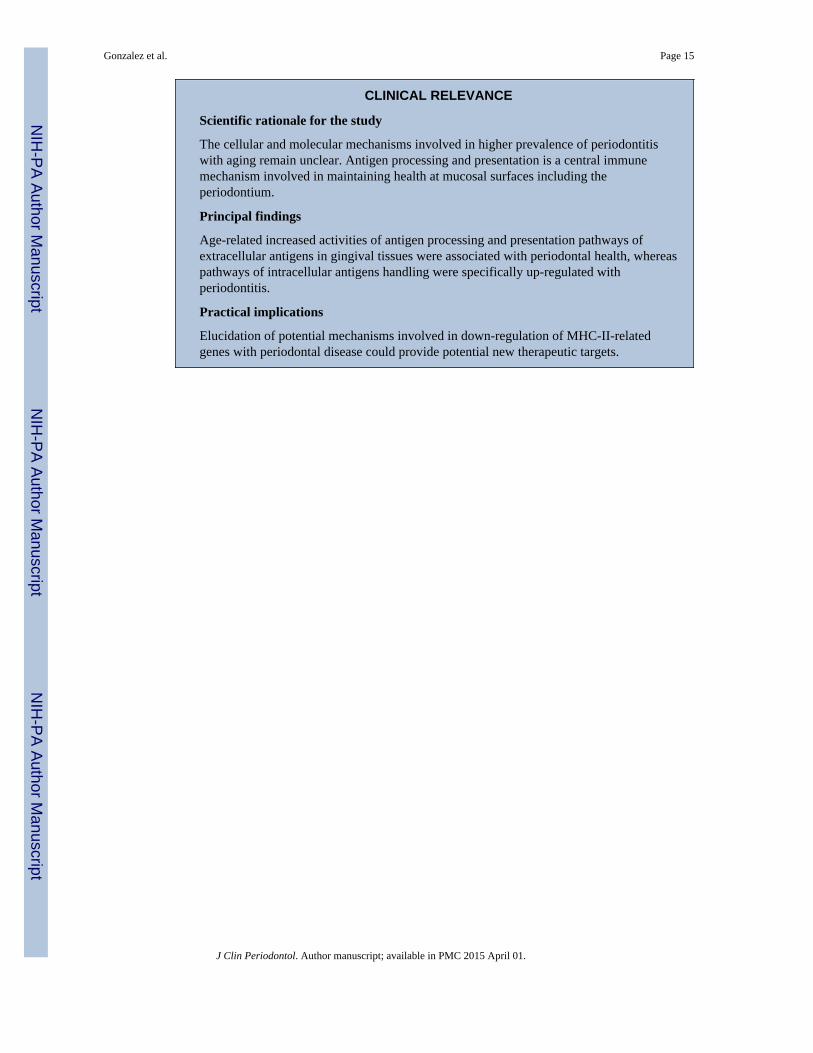
CLINICAL RELEVANCE
Scientific rationale for the study
The cellular and molecular mechanisms involved in higher prevalence of periodontitiswith aging remain unclear. Antigen processing and presentation is a central immunemechanism involved in maintaining health at mucosal surfaces including theperiodontium.
Principal findings
Age-related increased activities of antigen processing and presentation pathways ofextracellular antigens in gingival tissues were associated with periodontal health, whereaspathways of intracellular antigens handling were specifically up-regulated withperiodontitis.
Practical implications
Elucidation of potential mechanisms involved in down-regulation of MHC-II-relatedgenes with periodontal disease could provide potential new therapeutic targets.
Gonzalez et al. Page 15
J Clin Periodontol. Author manuscript; available in PMC 2015 April 01.
NIH
-PA Author Manuscript
NIH
-PA Author Manuscript
NIH
-PA Author Manuscript

Figure 1.Principal Components Analysis of antigen presentation and processing pathway genes (A)across the age groups of healthy gingival tissues and (B) periodontitis tissues of adult andaged animals. Each point denotes the positional values for an individual animal. The redcircle highlights the clustering of the animals (aged) or periodontitis.
Gonzalez et al. Page 16
J Clin Periodontol. Author manuscript; available in PMC 2015 April 01.
NIH
-PA Author Manuscript
NIH
-PA Author Manuscript
NIH
-PA Author Manuscript
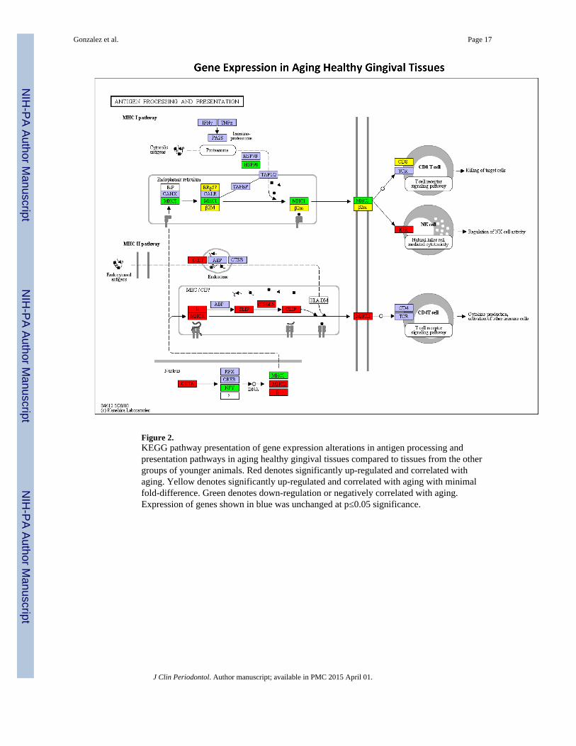
Figure 2.KEGG pathway presentation of gene expression alterations in antigen processing andpresentation pathways in aging healthy gingival tissues compared to tissues from the othergroups of younger animals. Red denotes significantly up-regulated and correlated withaging. Yellow denotes significantly up-regulated and correlated with aging with minimalfold-difference. Green denotes down-regulation or negatively correlated with aging.Expression of genes shown in blue was unchanged at p≤0.05 significance.
Gonzalez et al. Page 17
J Clin Periodontol. Author manuscript; available in PMC 2015 April 01.
NIH
-PA Author Manuscript
NIH
-PA Author Manuscript
NIH
-PA Author Manuscript

Gonzalez et al. Page 18
J Clin Periodontol. Author manuscript; available in PMC 2015 April 01.
NIH
-PA Author Manuscript
NIH
-PA Author Manuscript
NIH
-PA Author Manuscript

Figure 3.KEGG pathway presentation of gene expression alterations in antigen processing andpresentation pathways in (A) adult or (B) aged periodontitis gingival tissues compared totissues from healthy adult and aged animals respectively. Red denotes significantly up-regulated and correlated with aging. Green denotes down-regulation or negatively correlatedwith aging. Orange denotes fewer genes within the family that were significantly changed.Expression of genes shown in blue was unchanged at p≤0.05 significance.
Gonzalez et al. Page 19
J Clin Periodontol. Author manuscript; available in PMC 2015 April 01.
NIH
-PA Author Manuscript
NIH
-PA Author Manuscript
NIH
-PA Author Manuscript

NIH
-PA Author Manuscript
NIH
-PA Author Manuscript
NIH
-PA Author Manuscript
Gonzalez et al. Page 20
Table 1
Genes evaluated in the antigen processing and presentation pathways with their corresponding ProbeIdentification numbers.
GENE ID GENE NAME AFFYMETRIX PROBE
IFNG (KO: K04687) interferon-gamma MmugDNA.41414.1.S1_at
TNF (KO: K03156) tumor necrosis factor Mmu.14298.1.S1_at
PSME1 (KO: K06696) Hypothetical protein LOC700644 MmugDNA.21695.1.S1_at
PSME2 (KO: K06697) proteasome activator subunit 2 (PA28 beta) MmugDNA.22991.1.S1_at
HSPA1A (KO: K03283) similar to Heat shock 70 kDa protein 1 (HSP70.1) (HSP70-1/HSP70-2) MmuSTS.602.1.S1_at
HSPA1B (KO: K03283) heat shock 70kDa protein 1B MmuSTS.603.1.S1_at
HSPA6 (KO: K03283) similar to heat shock 70kDa protein 6 (HSP70B) MmugDNA.25285.1.S1_at
HSPA8 (KO: K03283) Heat shock 70kDa protein 8 MmugDNA.25572.1.S1_at
HSPA4 (KO: K09489) heat shock 70kDa protein 4 MmugDNA.42100.1.S1_at
HSP90AA1 (KO: K04079) Heat shock protein 90kDa alpha (cytosolic), class A member 1 MmuSTS.4086.1.S1_at
HSP90AB1 (KO: K04079) heat shock 90kDa protein 1, beta MmugDNA.32274.1.S1_at
MAMU-A (KO: K06751) major histocompatibility complex, class I, A Mmu.2935.4.S1_x_at
MAMU-B (KO: K06751) major histocompatibility complex, class I, B Mmu.2177.1.S1_x_at
MAMU-E (KO: K06751) major histocompatibility complex, class I, E MmunewRS.790.1.S1_at
MAMU-F (KO: K06751) major histocompatibility complex, class I, F MmugDNA.1042.1.S1_s_at
MAMU-G (KO: K06751) major histocompatibility complex, class I, G Mmu.2935.3.S1_s_at
B2M (KO: K08055) beta-2-microglobulin MmugDNA.5628.1.S1_at
PDIA3 (KO: K08056) protein disulfide isomerase family A, member 3 MmugDNA.2124.1.S1_at
TAPBP (KO: K08058) TAP binding protein (tapasin) MmugDNA.10000.1.S1_at
TAP1 (KO: K05653) transporter 1, ATP-binding cassette, sub-family B (MDR/TAP) MmugDNA.1081.1.S1_at
TAP2 (KO: K05654) antigen peptide transporter 2-like MmugDNA.3066.1.S1_at
CD8A (KO: K06458) similar to T-cell surface glycoprotein CD8 alpha chain precursor (T-lymphocyte differentiation antigen T8/Leu-2) (CD8a antigen)
MmugDNA.5711.1.S1_at
CD8B (KO: K06459) similar to T-cell surface glycoprotein CD8 beta chain precursor (CD8bantigen)
MmugDNA.16367.1.S1_at
KIR3DL (KO: K07980) killer immunoglobulin-like receptor KIR3DL (CD158k antigen) Mmu.7460.14.S1_s_at
KIR2DL4 (KO: K07981) killer cell immunoglobulin-like receptor, two domains, long cytoplasmictail, 4
Mmu.7460.2.S1_x_at
KLRC1 (KO: K06541) killer cell lectin-like receptor subfamily C, member 1 Mmu.1906.10.S1_s_at
KLRC3 (KO: K06541) killer cell lectin-like receptor subfamily C, member 3 Mmu.1906.7.S1_at
KLRD1 (KO: K06516) killer cell lectin-like receptor subfamily D, member 1 MmugDNA.17475.1.S1_s_at
IFI30 (KO: K08059) similar to Gamma-interferon-inducible lysosomal thiol reductase precursor(Gamma- interferon-inducible protein IP-30)
MmunewRS.536.1.S1_at
LGMN (KO: K01369) legumain Mmu.2523.1.S1_at
MAMU-DMB (KO: K06752) similar to HLA class II histocompatibility antigen, DM beta chainprecursor (MHC class II antigen DMB)
MmugDNA.18015.1.S1_at
MAMU-DOA (KO: K06752) similar to HLA class II histocompatibility antigen, DO alpha chainprecursor (MHC class II antigen DOA) (MHC DZ alpha) (MHC DN-alpha)
MmugDNA.36614.1.S1_s_at
J Clin Periodontol. Author manuscript; available in PMC 2015 April 01.

NIH
-PA Author Manuscript
NIH
-PA Author Manuscript
NIH
-PA Author Manuscript
Gonzalez et al. Page 21
GENE ID GENE NAME AFFYMETRIX PROBE
MAMU-DOB (KO: K06752) similar to HLA class II histocompatibility antigen, DO beta chainprecursor (MHC class II antigen DOB)
MmuSTS.84.1.S1_at
MAMU-DPA1 (KO: K06752) major histocompatibility complex, class II, DP alpha MmugDNA.37154.1.S1_at
MAMU-DPB1 (KO: K06752) major histocompatibility complex, class II, DP beta MmugDNA.18601.1.S1_at
MAMU-DQA1 (KO: K06752) major histocompatibility complex, class II, DQ alpha 1 MmugDNA.15798.1.S1_s_at
MAMU-DQB1 (KO: K06752) similar to HLA class II histocompatibility antigen, DQ(1) beta chainprecursor (DC-3 beta chain)
MmuSTS.86.1.S1_at
MAMU-DRA (KO: K06752) major histocompatibility complex, class II, DR alpha MmugDNA.1046.1.S1_s_at
MAMU-DRB1-4 (KO: K06752) similar to HLA class II histocompatibility antigen, DRB1-4 beta chainprecursor (MHC class I antigen DRB1*4) (DR-4) (DR4)
MmunewRS.436.1.S1_s_at
CD74 (KO: K06505) CD74 molecule, major histocompatibility complex, class II invariant chain Mmu.9241.2.S1_at
CTSS (KO: K01368) similar to Cathepsin S MmuSTS.3988.1.S1_at
CD4 (KO: K06454) CD4 molecule Mmu.15035.1.S1_at
CIITA (KO: K08060) class II, major histocompatibility complex, transactivator MmugDNA.20.1.S1_at
RFX5 (KO: K08061) regulatory factor X, 5 MmuSTS.3763.1.S1_at
RFXANK (KO: K08062) similar to regulatory factor X-associated ankyrin-containing proteinisoform a
MmugDNA.24504.1.S1_at
RFXAP (KO: K08063) regulatory factor X-associated protein MmugDNA.10869.1.S1_at
CREB1 (KO: K05870) cAMP responsive element binding protein 1 MmuSTS.3958.1.S1_at
NFYA (KO: K08064) similar to Nuclear transcription factor Y subunit alpha (Nucleartranscription factor Y subunit A) (NF-YA) (CAAT-box DNA-bindingprotein sub- unit A)
MmugDNA.24571.1.S1_at
NFYB (KO: K08065) nuclear transcription factor Y, beta MmugDNA.37609.1.S1_at
J Clin Periodontol. Author manuscript; available in PMC 2015 April 01.
NIH
-PA Author Manuscript
NIH
-PA Author Manuscript
NIH
-PA Author Manuscript
Gonzalez et al. Page 22
Table 2
Differences in gene expression of aging healthy gingival tissues compared to younger animals (negative fold-difference denotes decrease in aging tissues), and correlations in gene expression with aging.
Gene ID Fold Difference P-value Correlation (R) P-value
B2M 1.09 0.023 0.4010 0.05
CD8A 0.4470 0.032
CD74 1.35 0.044 0.5870 0.003
CIITA 1.53 0.102
CREB1 −1.18 0.019
CTSS 1.48 0.007 0.5950 0.003
HSPA1B −1.11 0.050
HSP90AB1 −1.19 0.039 −0.4420 0.035
HLA-A −0.747 <0.001
HLA-DMB 1.44 0.015
HLA-DOB 1.51 0.008 0.5200 0.010
HLA-DPB1 0.5980 0.003
HLA-DQB1 0.5160 0.012
HLA-DRA 1.44 0.013
HLA-DRB1-4 1.48 0.043
IFI30 1.39 0.017
KLRC1 1.52 0.06
KLRC3 1.86 0.022
KLRD1 1.41 0.015 0.64705 <0.001
NFYA −1.19 0.022 −0.4330 0.039
PSME2 1.21 0.008
PDIA3 1.26 0.030
J Clin Periodontol. Author manuscript; available in PMC 2015 April 01.

NIH
-PA Author Manuscript
NIH
-PA Author Manuscript
NIH
-PA Author Manuscript
Gonzalez et al. Page 23
Tabl
e 3
Dif
fere
nces
in g
ene
expr
essi
on b
etw
een
heal
thy
and
peri
odon
titis
gin
giva
l tis
sues
in a
dult
and
aged
ani
mal
s (n
egat
ive
fold
-dif
fere
nce
deno
tes
decr
ease
inpe
riod
ontit
is ti
ssue
s), a
nd c
orre
latio
ns w
ith a
ge in
per
iodo
ntiti
s tis
sues
.
Adu
ltA
ged
Gen
e ID
Fol
d D
iffe
renc
eP
-val
ueF
old
Dif
fere
nce
P-v
alue
Cor
rela
tion
(R
)P
-val
ue
CD
8A1.
430.
045
CD
74
CT
SS1.
500.
017
1.62
0.01
70.
5660
0.06
HSP
A1B
−1.
160.
004
−0.
5750
0.06
HSP
90A
A1
−1.
430.
06−
0.63
500.
032
HL
A-A
1.47
0.03
41.
490.
047
HL
A-F
1.51
0.05
0.68
000.
019
HL
A-D
MB
0.60
300.
047
HL
A-D
OB
1.63
0.03
8
HL
A-D
PB1
0.56
200.
06
HL
A-D
RB
1-4
1.54
0.05
0.60
3000
47
IFI3
0(G
ILT
)1.
330.
020
0.68
000.
019
IFNγ
0.72
100.
011
KL
RC
17.
230.
05
KL
RC
32.
220.
015
KL
RD
12.
310.
061.
420.
023
NFY
A1.
290.
005
NFY
B1.
330.
030
−0.
5800
0.05
PSM
E2
0.56
200.
06
PDIA
30.
7950
0.00
2
RFX
AN
K1.
2000
24
TA
PBP
1.44
0.02
21.
560.
060.
6030
0.04
7
J Clin Periodontol. Author manuscript; available in PMC 2015 April 01.

NIH
-PA Author Manuscript
NIH
-PA Author Manuscript
NIH
-PA Author Manuscript
Gonzalez et al. Page 24
Table 4
Discriminatory gene expression derived from Principal Component Analysis and relationship to antigenprocessing and presentation classes. The + symbols denote the antigen presentation pathway of genes thatcontribute to stratifying the aged or peridontitis tissue profiles.
Healthy Periodontitis
MHC CIass I MHC Class II MHC CIass I MHC Class II
CD74(li) +
CD8A +
CIITA + +
CTSS + +
HLA-A +
HLA-B +
HLA-F +
HLA-DMB +
HLA-DOA + +
HLA-DPA1 + +
HLA-DPB1 + +
HLA-DRA +
HSPA1B +
HSPA8 +
IFI30(GILT) +
KLRC3 +
KLRD1 +
J Clin Periodontol. Author manuscript; available in PMC 2015 April 01.
NIH
-PA Author Manuscript
NIH
-PA Author Manuscript
NIH
-PA Author Manuscript
Gonzalez et al. Page 25
Table 5
Comparison of gene expression profiles using qPCR and microarray analyses. Values represent fold-difference compared to Adulty Healthy tissue message levels. Five samples were assessed for each group andthe asterisk (*) signifies a correlation of >0.5.
GENE ID AGED HEALTHY ADULT PERIODONTITIS AGED PERIODONTITIS
PSME2
qPCR 2.06* 0.55 2.12*
GeneChip 1.76 1.12 1.84
IF130
qPCR 2.74* 2.85* 1.74*
GeneChip 1.91 2.02 1.82
HLA-DPB1
qPCR 1.95* 3.81 8.59*
GeneChip 1.63 1.25 2.53
HLA-DRA
qPCR 1.56* 1.87* 1.18*
GeneChip 1.30 1.52 1.22
CTSS
qPCR 1.53* 0.79 0.82
GeneChip 1.33 1.50 2.02
J Clin Periodontol. Author manuscript; available in PMC 2015 April 01.