comparative anatomical analysis of stem in four...
TRANSCRIPT
COMPARATIVE ANATOMICAL ANALYSIS OF STEM IN FOUR GENERA OF THE TRIBE SALSOLEAE, CHENOPODIACEAE R. Ramazannejad Ghadi, D. Azizian & M. Assadi Ramazannejad Ghadi, R., Azizian, D. & Assadi, M. 2006 12 31: Comparative anatomical analysis of stem in four genera of the tribe Salsoleae, Chenopodiaceae. –Iran. J. Bot. 12 (2):169-182. Tehran. The stem anatomy of 18 species belong to the genera Anabasis L., Haloxylon Bge., Hammada Iljin, Seidlitzia Bge. ex Boiss. of the tribe Salsoleae (Chenopodiaceae) was examined. Anatomical characters with numerical analysis provided some significant data for the delimitation of taxa at the generic and specific levels. Reza Ramazannejad Ghadi, Department of Biology, Golestan University, Gorgan, Iran.- Dina Azizian, Department of Biology, Shahid Beheshti University, Tehran, Iran.- Mostafa Assadi, Research Institute of Forests & Rangelands, P. O. Box 13185-116, Tehran, Iran. Key words. Anatomy, numerical taxonomy, Chenopodiaceae, Salsoleae, Iran.
������ ��� ��� � �� ��� ���� �� ���� ��Salsoleae ��� Chenopodiaceae
��� ���� ����� ����،���� ���!�!" # �$�� %&'(�
از تيره اسفناج مورد Haloxylon , Anabasis , Hammada , Seidlitzia ساختمان تشريحي هجده گونه از چهار جنس
مورفولوژيك و اشكاالت موجود در تفكيك اين تاكسونها بر حسب صفات با وجود تشابهات . بررسي قرار گرفت
قرابت بين ،با استفاده از تاكسونومي عددي . ز كننده اي در سطح جنس و گونه شناسايي شدياصفات آناتومي متم،مذكور
.ري داشته استتاكسونها بررسي شده وتفكيك تاكسونها بر حسب صفات مورفولوژيك با نتايج اين پژوهش تفاوتهاي آشكا
INTRODUCTION Chenopodiaceae is a family of about 100 genera and more than 1500 species which widely distributed in temperate and subtropical area (Heywood 1978). One of the great diversity and richness centers of the family is in southwestern Asia, especially Iran. The family Chenopodiaceae represented in Flora of Iran by 41 genera and about 175 species which grow in various parts of Iran (Assadi 2001). The family comprises annual and perennial herbs mainly growing in saline habitats. Characters of taxonomic value within the family include the vegetative and reproductive organs but mainly on the basis of embryo shape as mentioned in various floras (Assadi 2001, Rechinger1997). Taxonomy of the family based on the morphological characters has been unsatisfactory to determine relationships among the species and genera of the family. Anatomical characters have been used in the taxonomy of this family (Khatib 1959), (A.Butnik 1991), (Pyankov et al. 1997), (Krumbiegel 1998) and other plant families.
In this work four genera, Anabasis L., Haloxylon Bge., Hammada Iljin and Seidlitzia Bge. ex Boiss. Belong to the tribe Salsoleae C. A. Mey., subfamily Salsoloideae C. A. Mey. of the family Chenopodiaceae Vent. has been investigated. Altogether 18 species of four mentioned genera were examined anatomically to determine diagnostic characters to assess interspecific and intergeneric relationships. In comparison with the size of the family and genera few microscopic details on the anatomy of these genera has been published by various authors as Fahn & Arzee (1959), Khatib (1959), Fahn (1963,1982), Fahn & Schori,(1968), Wendelbo & Bokhari(1978), Metcalfe & Chalk (1979), Butnik (1991), and Kadereit & et al. (2003). In order to attain a deeper insight into these genera and to identify further distinctive characters at different taxonomic level, we carried out anatomical studies on stem of those taxa. Because of leaves reduction in some taxa of these genera, anatomical study performed on stem.
IRAN. JOURN. BOT. 12 (2), 2006 Ramazannejad Ghadi & al.
170
MATERIALS AND METHODS Following 19 taxa based on the nomenclature of Assadi (2001) were anatomically examined. Anabasis annua Bge. -A. aphylla L. - A. articulata (Forssk.) Moq., Syn.: A. lachnantha Aellen & Rech. f. - A. calcarea (Charif & Aellen) Bokhari & Wendelbo – A. eriopoda (Shrenk) Volkens -A. eugeniae Iljin –A. haussknechtii Bge. ex Boiss. var. haussknechtii –A. haussknechtii Bge. ex Boiss. var. iranica (Iljin) Assadi, Syn.: A. iranica Iljin– A. jaxartica (Bge.) Benth ex Volkens –A. salsa (C. A. Mey.) Benth ex Volkens -A. setifera Moq. –Haloxylon ammodendron (C. A. Mey.) Bge. -H. persicum Bge. ex Boiss. -Hammada griffithii
(Moq.) Iljin, Syn.: Haloxylon griffithii (Moq.) Boiss. -Hammada salicornica (Moq.) Iljin, Syn.: Haloxylon salicornicum (Moq.) Bge. ex Boiss. - Seidlitzia cinerea (Moq.) Bge. ex Botsch. -S. florida (M.B.) Bge. ex Boiss. - S. rosmarinus (Ehrenb.) Bge. ex Boiss. -S. stocksii (Boiss) Assadi, Syn.: Haloxylon recurvum sensu Boiss.. For light microscopic studies, dried material was obtained from herbarium specimens of Research Institute of Forests & Rangelands (TARI). In addition some fresh specimens were used for some taxa. The list of species is presented in table 1.
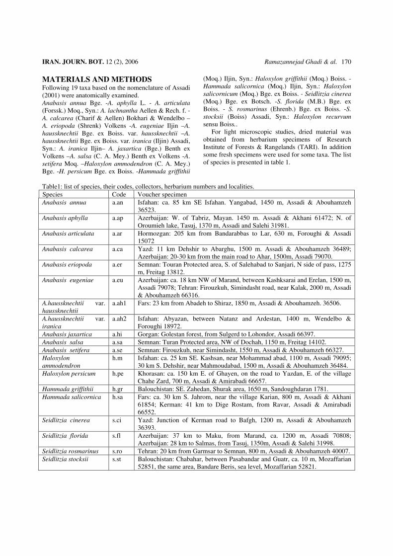
Table1: list of species, their codes, collectors, herbarium numbers and localities.
Voucher specimen Code Species Isfahan: ca. 85 km SE Isfahan. Yangabad, 1450 m, Assadi & Abouhamzeh 36523.
a.an Anabasis annua
Aَzerbaijan: W. of Tabriz, Mayan. 1450 m. Assadi & Akhani 61472; N. of Oroumieh lake, Tasuj, 1370 m, Assadi and Salehi 31981.
a.ap Anabasis aphylla
Hormozgan: 205 km from Bandarabbas to Lar, 630 m, Foroughi & Assadi 15072
a.ar Anabasis articulata
Yazd: 11 km Dehshir to Abarghu, 1500 m. Assadi & Abouhamzeh 36489; Azerbaijan: 20-30 km from the main road to Ahar, 1500m, Assadi 79070.
a.ca Anabasis calcarea
Semnan: Touran Protected area, S. of Salehabad to Sanjari, N side of pass, 1275 m, Freitag 13812.
a.er Anabasis eriopoda
Azerbaijan: ca. 18 km NW of Marand, between Kashksarai and Erelan, 1500 m, Assadi 79078; Tehran: Firouzkuh, Simindasht road, near Kalak, 2000 m, Assadi & Abouhamzeh 66316.
a.eu Anabasis eugeniae
Fars: 23 km from Abadeh to Shiraz, 1850 m, Assadi & Abouhamzeh. 36506. a.ah1 A.haussknechtii var. haussknechtii
Isfahan: Abyazan, between Natanz and Ardestan, 1400 m, Wendelbo & Foroughi 18972.
a.ah2 A.haussknechtii var. iranica
Gorgan: Golestan forest, from Sulgerd to Lohondor, Assadi 66397. a.hi Anabasis jaxartica Semnan: Turan Protected area, NW of Dochah, 1150 m, Freitag 14102. a.sa Anabasis salsa Semnan: Firouzkuh, near Simindasht, 1550 m, Assadi & Abouhamzeh 66327. a.se Anabasis setifera Isfahan: ca. 25 km SE. Kashsan, near Mohammad abad, 1100 m, Assadi 79095; 30 km S. Dehshir, near Mahmoudabad, 1500 m, Assadi & Abouhamzeh 36484.
h.m Haloxylon ammodendron
Khorasan: ca. 150 km E. of Ghayen, on the road to Yazdan, E. of the village Chahe Zard, 700 m, Assadi & Amirabadi 66657.
h.pe Haloxylon persicum
Balouchistan: SE. Zahedan, Shurak area, 1650 m, Sandoughdaran 1781. h.gr Hammada griffithii Fars: ca. 30 km S. Jahrom, near the village Karian, 800 m, Assadi & Akhani 61854; Kerman: 41 km to Dige Rostam, from Ravar, Assadi & Amirabadi 66552.
h.sa Hammada salicornica
Yazd: Junction of Kerman road to Bafgh, 1200 m, Assadi & Abouhamzeh 36393.
s.ci Seidlitzia cinerea
Azerbaijan: 37 km to Maku, from Marand, ca. 1200 m, Assadi 70808; Azerbaijan: 28 km to Salmas, from Tasuj, 1350m, Assadi & Salehi 31998.
s.fl Seidlitzia florida
Tehran: 20 km from Garmsar to Semnan, 800 m, Assadi & Abouhamzeh 40007. s.ro Seidlitzia rosmarinus Balouchistan: Chabahar, between Pasabandar and Guatr, ca. 10 m, Mozaffarian 52851, the same area, Bandare Beris, sea level, Mozaffarian 52821.
s.st Seidlitzia stocksii

Chenopodiaceae anatomy IRAN. JOURN. BOT. 12 (2), 2006 171
In order to study stem characters, materials below the inflorescence were revived by boiling water, cooled and fixed in FAA for 48 hours, and cross sections were prepared by hand using razor blade. Sections were cleared with sodium hypochlorite, dehydrated and stained with methyl green 0.1% and carmine 1% for 30 seconds and 15 minutes respectively, then mounted in gelatin. Observations were carried out with Olympus light microscope. For numerical analysis 40 characters were studied. Qualitative characters were coded as multistate characters and means of quantitative characters were used. The 40 used characters and their coding presented
in table 2. In order to determine the significant differences in quantitative characters between the taxa of Anabasis and or Seidlitzia, analysis of variance (ANOVA) was performed. But because of limitation of samples in two genera, Hammada and Haloxylon, the T- test was performed. For grouping the similar taxa, cluster analysis using WARD and ordination of species on the first two principal component axes (PCA) was performed. In order to determine the most variable anatomical characters among the species, a factor analysis based on principal components analysis (PCA) was performed (Sneath1957) and (Stace1989).
Table 2: The 40 used characters and their coding. 1- Arrangement of epidermal layers: Oriented along the stem = 1 , irregular = 2 2- Shape of epidermal cells: Long (length two time more than width) = 1, short = 2 3- Stomata: Present = 1, absent = 2 4- Stomatal canals: Present = 1, absent = 2 5- Crystal in epidermis: Present = 1, absent = 2 6- Hypodermis: Present = 1, absent = 2 7- Continuity of hypodermis layer: Continued = 1, uncontinued = 2 8- Crystal in hypodermis: Present = 1, absent = 2 9- Palisade like chlorenchyma: Present = 1, absent = 2 10- Crystal in palisade like chlorenchyma: Present = 1, absent = 2 11- Continuity of palisade like layers: Continued = 1, uncontinued = 2 12- Short cell chlorenchyma: Present = 1, absent = 2 13- Continuity of short cell chlorenchyma: Continued = 1, uncontinued = 2 14- Crystal in short cell chlorenchyma: Present = 1, absent = 2 15- Cortical collenchyma: Present = 1, absent = 2 16- Position of collenchyma in transverse section: Symmetric = 1, unsymmetric = 2 17- Type of cortical collenchyma: In 4 equal groups = 1, in 4 unequal groups = 2, in 2 groups = 3 18- Crystal in storage cortical parenchyma: Present = 1, absent = 2 19- Ray elongated parenchyma around vascular cylinder: Present = 1, absent = 2 20- Cortical vascular bundles: Present = 1, absent = 2 21- Arrangement of vascular bundles: Symmetric = 1, unsymmetric = 2 22- Type of symmetry in vascular bundles: In 4 group = 1, in 2 group = 2 23- Size of vascular bundles group: Equal = 1, unequal = 2 24- Crystal in vascular tissues: Present = 1, absent = 2 25- Crystal in pith parenchyma: Present = 1, absent = 2 26- Cortical fiber: Present = 1, absent = 2 27- Arrangement of cortical fiber: Symmetric = 1, unsymmetric = 2 28- Type of symmetry in cortical fiber: Round = 1, in 4 group = 2 29- Shape of stem in transverse section: Rounded = 1, elliptic = 2, tetragonal = 3, irregular = 4 30- Diameter ratio of cortex to vascular cylinder 31- Diameter ratio of vascular cylinder to stem 32- Diameter ratio of pith parenchyma to cortex 33- Diameter ratio of crystal to cortex 34- Diameter ratio of protective tissues to stem 35- Diameter of crystals in cortex: (Micron) 36- Number of epidermal layers: One = 1, two = 2, three = 3, four = 4, more than seven = 5 37- Length of epidermal cell: (Micron) 38- Number of hypodermal layers: One = 1, two = 2 39- Number of cortex layers 40- Diameter ratio of cortex to stem

IRAN. JOURN. BOT. 12 (2), 2006 Ramazannejad Ghadi & al.
172
OBSERVATION The comparison of important anatomical characters of stem for separation of taxa in generic and specific levels are as bellow: Stem TS.: The stem in transverse section was more or less circular in most of the species examined (fig. 2 A-F, fig. 3 A-E, fig.4 E-F). This character was different in genus Haloxylon (fig.3 F,G) and also divided the genus Seiditzia into two groups. Shape of stem in transverse section was more regular in two species; S. cinerea and S. stocksii than other species with irregular shape (fig. 4 A-D). Epidermis: usually consisting of one layer of cells for example in Hammada griffithii and Haloxylon ammodendron (Figs.4 E, 3 F), but frequently more than one layer present in some species, as distinctly two layered in Hammada salicornica and Haloxylon persicum (figs. 4 F, 3 G), and three layered epidermal cells in Seidlitzia rosmarinus (Fig. 4 C) which can be distinguished within genera and species. Therefore number of epidermal layers separated two species of the genera Haloxylon and Hammada (Figs.3 F, 3 G and 4 E, 4 F). This character also distinguishes Seidlitzia rosmarinus with three layered epidermis from the others with one layer, or Anabasis clacarea, with more than seven layers of epidermal cells from the other species of these genera (fig. 2 D). Epidermal cells sometimes have different sizes and shapes as in Anabasis species. Shape of epidermal cells is two types, short and long, that is various and distinguishes the species of Anabasis. Arrangment of epidermal layers had two types; regular or irregular along the stem, and constant in species of two genera, Hammada and Haloxylon with regular arrangment, but distinguished Seidlitzia rosmarinus with irregular type, from the other species of this genus (fig. 4 A-D) and divided the genus Anabasis into two groups. The first group involved Anabasis Jaxartica, A. eugeniae , A. setifera , A. aphylla and A. haussknechtii that had regular layers of epiderm, but other species of Anabasis had irregular epidermis layers (figs. 3 A-E and 2 A-F). Crystals present in epidermal cells of some species, which distinguish S. rosmarinus (fig. 4 C), Anabasis annua, A. articulata and A. calcarea from the other species of their genera. Presence of superficial stomata is only important and various in the genus Anabasis. But presence of stomatal cannals can distinguish Hammada salicornica (fig. E, F) and three species of Anabasis including A. calcarea, A. Jaxartica and A. articulata from the others (figs. 2 D and 3 C, E). Variation in length of epidermal cells is very important character, especially in two genera Hammada and Haloxylon.
Hypodermis: Usually one layer of hypodermis presents in S. stocksii but absent in other species of this genus. Also all species of the genus Anabasis except A. annua had hypodermis. Continiuty of hypodermis layer delimitate A. salsa, A. setifera and A. eugeniae from the other group of this genus with continuted hypodermis. Number of hypodermis layers distinguishes two species of Hammada and also A. eugeniae (with two layers of hypodermis) from the other species of Anabasis (figs. 2-4). Cortex: usually involved these parts; Collenchyma: Cortical collenchyma absent in two genera Hammada and Haloxylon (fig. 3 F, G and 4 E, F). But presence of this layer distinguishes S. stocksii and A. eugeniae , A. salsa and A. annua from the others (figs. 4 B, 2 B, E, 3 D). Type of symmetry in this layers based on transverse section of stem is different in A. eugeniae from the other taxa of Anabasis that have collenchyma (fig. 2 B). Palisade like chlorenchyma: presence of this layer only separates S. stocksii from the other species of Seidlitzia. Continuation of this layer that is caused by absence of collenchyma, divides the genus Anabasis into two groups. The first group involved A. eugeniae, A. salsa, A. annua and A. setifera with no continuous chlorenchyma. Presence of crystals in this layer delimitated A. salsa, A. eriopoda and A. haussknechtii var. haussknechtii from the other taxa of this genus (figs. 2, 3, 4). Short cell Chlorenchyma: This layer located under palisade shape chlorenchyma. Species of two genera Hammada and Haloxylon have this layer, but prese Chenopodiaceae nce of this charcter distinguishes S. stocksii from the other taxa of this genus and assign it to genus Anabasis (fig. 1 up, 4 A-D). Also presence of crystals in this layer distinguishes A. setifera from the other taxa of Anabasis. Continuity of this layer divides the genus Anabasis into two groups similar to last paragraph. Storage cortical parenchyma: Persence of crystals in these layers distinguish two species of Haloxylon (fig. 3 F-G). There are no crystals in cortical cells of H. persicum. This character is not useful in other genera related to presence of crystal in cortical cells of all of them. Existence of cortical bundles was constant in Anabasis but separate S. stocksii with cortical bundles from the other taxa of this genus that lack this character. Presence of ray elongated parenchyma that arranged vertically around the vascular cylinder is distinctive between two species of Haloxylon and also separate S. rosmarinus and A. setifera that had this character from the other taxa of their genera. Presence of cortical fibers was constant in Haloxylon and Hammada but distinguishes S. stocksii and S. rosmarinus from other group of this genus, and also

Chenopodiaceae anatomy IRAN. JOURN. BOT. 12 (2), 2006 173
distinguishes A. annua and A. salsa from the other species of Anabasis. Number of cortical parenchymatous layers is very important in separation of S. florida from the other species of Seidlitzia (fig. 4 A-D). Vascular cylinder: nearly all of genera posses 4 large bundles, but the size of vascular bundles varies among the species and sometimes among 4 bundles of a specimens (figs. 5 A-D, 5 G, 6 A-F, 7 A, B, D) but are different in other taxa (figs. 5 E, F and 7 C, E, F). Each vascular bundle is surrounded by a thick sclerenchymatous sheath. Arrangment of vascular bundles, distinguish S. romarinus from the other species of this genus (fig. 7 C-D). This character distinguishes A. eriopoda and A. jaxartica from the other taxa of Anabasis (fig. 5 E, F). Type of symmetry in vascular bundles based on number, size and position of bundles which distinguish two species of Hammada (fig. 6 B, D) and also some species of Anabasis. Presence of crystals in vascular fibers distinguishes two species of Haloxylon (fig. 7 A, B) and also A. salsa from the other species of Anabasis (fig. 5 and 6). The pith in all species composed of parenchyma cells. Absence of crystals in pith parenchyma only distinguishes A. setifera, from the other taxa of this genus. Quantitative characters mostly changed to ratio of two characters and were important in specific and generic levels too. The main quantitative characters are diameter ratio of; cortex to vascular cylinder, vascular cylinder to stem in transverse section, pith parenchyma to cortex, epidermal layers to cortex, cortex to stem and crystals to cortex. Other characters are number and length of epidermis and hypodermis layers, number of cortex layers and diameter of crystal in cortex region. RESULT OF STATISTICAL ANALYSIS Results of ANOVA and T-test showed the significant difference between quantitative characters. Phenogram of cluster analysis (UPGMA) presented in fig.1. Two major clusters can be recognized as being supported by ordination of the genera based on principal components analysis (fig.1). The first cluster is divided to two groups. The arrangement of species in cluster was written in the left of cluster. The main points in this cluster are location of the genus Seidlitzia, separation of the two species of Haloxylon and also separation of Seidlitzia stocksii from the other species of Seidlitzia. Factor analysis of anatomical characters showed that the first four factors described about 69% of total variance. The first component comprises about 34% of the total variance to which the presence and continuity of hypodermis and chlorenchymatus layers, presence of
Figure1: Above; Cluster analysis of taxa by UPGMA; Two major clusters are recognized. The first one divided to two groups. In a cluster three species of Seidlitzia located. In other cluster one species of Seidlitzia with species of other genera located. Below; Ordination of taxa based on PCA;Three species of Seidlitzia are separated from the others. Then four species of Anabasis with Seidlitzia stocksii have longer distance from the other taxa: a. sa : Anabasis salsa, a. an: A. annua, a.se: A . setifera, a. eu: A. eugeniae, a. er: A. eriopoda, a. ap: A. aphylla, a. ca: A. calcarea, a. ar: A. articulata, a. hi: A. jaxartica, a. h 1: A. h. var. haussknekhtii, a. h 2: A. h .var. iranica, s. st: Seidlitzia stocksii, s. ci: S. cinerea, s. ro: S .rosmarinus, s fl: S. florida, h. sa: Hammada salicornica, h. gr: H. griffithii, h. pe: Haloxylon persicum , h. am: H. ammodendron.

IRAN. JOURN. BOT. 12 (2), 2006 Ramazannejad Ghadi & al.
174
Figure 2: Cortex and epidermal layers: A: Anabasis eriopoda, B: A. eugeniae, C: A. aphylla, D: A. calcarea, E: A. annua, F: A. setifera. All of them have palisade and also spongy shape chlorenchyma. Hypodermis exists below the uni- or multilayered epidermis. This layer has crystals in some species. Some cortical bundles observed in A. eriopoda. This species also has crystals in palisade shape chlorenchyma. Size of crystals in cortex is very bigger than the other layers. A.calcarea has stomatal canal in multilayered epidermis. This species has crystals in epidermis layers too. C. B. = Cortical bundles, COR = Cortex,CR =Crystal, EP= Epidermis, PAL= Palisade chlorenchym, ST. C.=Stomatal canal.(Bar=200 micron).
Chenopodiaceae anatomy IRAN. JOURN. BOT. 12 (2), 2006 175
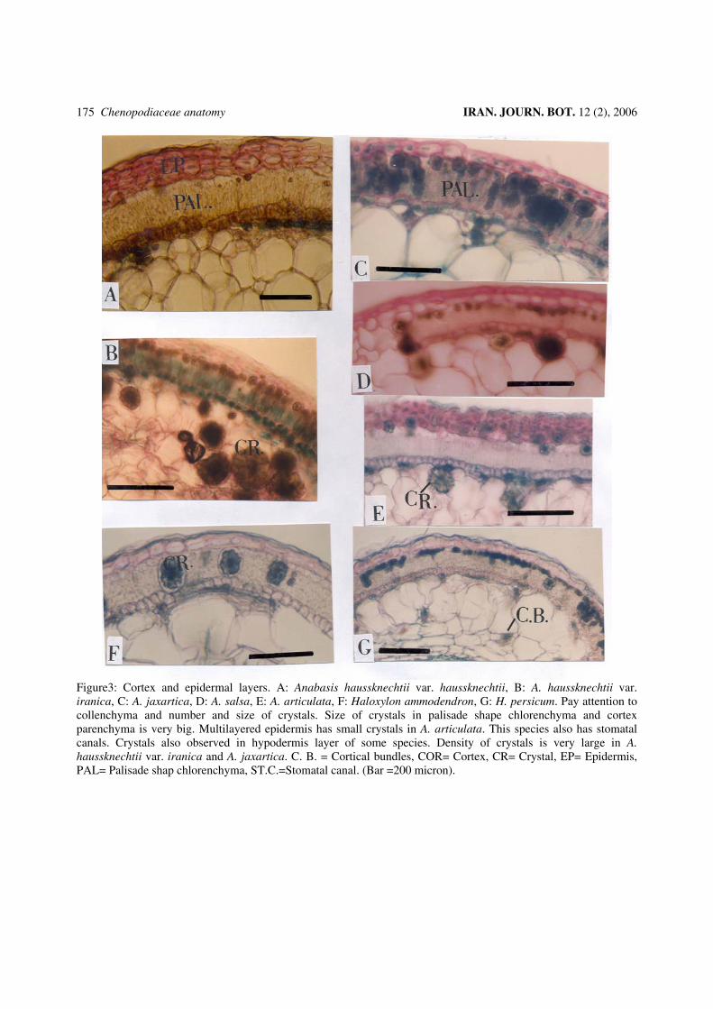
Figure3: Cortex and epidermal layers. A: Anabasis haussknechtii var. haussknechtii, B: A. haussknechtii var. iranica, C: A. jaxartica, D: A. salsa, E: A. articulata, F: Haloxylon ammodendron, G: H. persicum. Pay attention to collenchyma and number and size of crystals. Size of crystals in palisade shape chlorenchyma and cortex parenchyma is very big. Multilayered epidermis has small crystals in A. articulata. This species also has stomatal canals. Crystals also observed in hypodermis layer of some species. Density of crystals is very large in A. haussknechtii var. iranica and A. jaxartica. C. B. = Cortical bundles, COR= Cortex, CR= Crystal, EP= Epidermis, PAL= Palisade shap chlorenchyma, ST.C.=Stomatal canal. (Bar =200 micron).

IRAN. JOURN. BOT. 12 (2), 2006 Ramazannejad Ghadi & al.
176
Figure 4: Cortex and epidermal layers: A: Seidlitzia cinerea, B: S. stocksii, C: S. rosmarinus, D: S. florida, E: Hammada griffithii, F: H. salicornica. Hammada salicornica has two layered hypodermis and stomatal canals. S. cinerea and S. rosmarinus and S. florida don’t have palisade and spongy shape chlorenchyma against the S. stocksii. Density of crystals is limited in these species. C. B. = Cortical bundles , COR = Cortex , CR = Crystal, EP = Epidermis, PAL = Palisade chlorenchym, ST. C.=Stomatal cannal.(Bar =200 micron).

Chenopodiaceae anatomy IRAN. JOURN. BOT. 12 (2), 2006 177
Figure 5: Vascular cylinder or part of it: A: Anabasis articulata, B: A. haussknechtii var. haussknechtii, C: A. annua, D: A. haussknechtii var. iranica, E: A. eriopoda, F: A. jaxartica, G: A. salsa. (Bar = 200 micron).
IRAN. JOURN. BOT. 12 (2), 2006 Ramazannejad Ghadi & al.
178
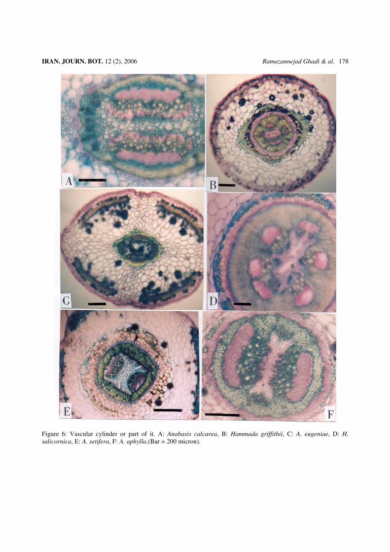
Figure 6: Vascular cylinder or part of it. A: Anabasis calcarea, B: Hammada griffithii, C: A. eugeniae, D: H. salicornica, E: A. setifera, F: A. aphylla.(Bar = 200 micron).
Chenopodiaceae anatomy IRAN. JOURN. BOT. 12 (2), 2006 179
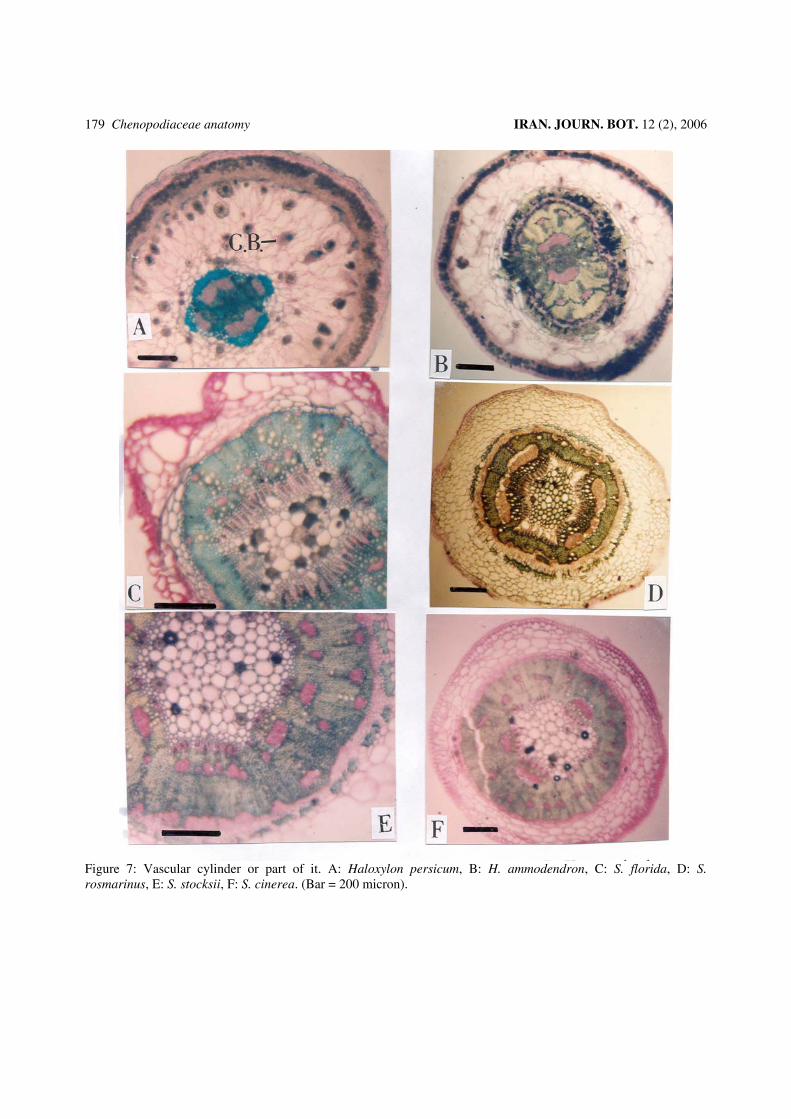
Figure 7: Vascular cylinder or part of it. A: Haloxylon persicum, B: H. ammodendron, C: S. florida, D: S. rosmarinus, E: S. stocksii, F: S. cinerea. (Bar = 200 micron).
IRAN. JOURN. BOT. 12 (2), 2006 Ramazannejad Ghadi & al.
180
crystals in those and presence of cortical bundles are highly correlated. These factors distinguish S. rosmarinus, S. florida and S. cinerea from the other taxa. Diameter of crystals and length of epidermal cells are highly correlated with the second component, these factors separates A. salsa, A. eugeniae, A. setifera , A. annua and S. stocksii from the others. Arrangment and number of epidermal cells, type and symmetry of vascular bundles, and presence of elongated parenchyma are highly correlated with the other factors. These characters are the most variable characters and may be used to differentiation among the genera and species. DISCUSSION The results obtained from this study showed that the anatomical characters can separate the taxa in generic and specific levels, and we made an anatomical key for delimitation of taxa. From morphological point of view we also found that grouping of the genera in this tribe is not correct exactly. In this study many specimens of each species from different areas of Iran were compared, these specimens had same morphological and anatomical characters. The genus Haloxylon is very similar to Anabasis in some Floras such as Flora Iranica (Rechinger 1997), Flora of Iran (Assadi 2001), Flora Palaestina (Zohary 1966). The main morphological difference between these two genera is location of embryo in seed. However in our results general anatomical similarity combined these genera. Two species of Haloxylon are separated by some characters of epidermis and cortex, but split the species of Anabasis. Therefore we need better morphological characters for delimitation of these two genera. The genus Hammada mentioned as a synonym of Haloxylon in Flora Orientalis (Boissier 1879) and Flora Iranica (Rechinger 1997). But Assadi (2001) in Flora of Iran separated these two genera by morphological characters of stamen, inflorescence and phenology of flower. Our anatomical study confirms the distinction of these two genera. Two mentioned species of the genus Hammada are very similar morphologically, as recognized in Flora of Iran (Assadi 2001) and Flora Palaestina (Zohary 1966). These two species located together by anatomical evidence (fig.1 up). Species of Hammada and Anabasis are very similar morphologyically, also based on anatomical analysis, Hammada species located among the species of Anabasis (fig.1 up). So, we think that location of embryo alone is not a good character for delimitation of these two genera. Seidlitzia stocksii presented in the genus Haloxylon in Flora Orientalis (Boissier1879) and Flora Iranica
(Rechinger1997). This species separated from Hammada and Haloxylon by its succulent leaves, phenology of flowers and embryo location, in Flora of Iran (Assadi 2001). In this study this species located among the species of Anabasis because of similar characters (fig.1 up and down). It should be mentioned that oblique embryo in S. stocksii is intermediate between horizontal type in Haloxylon and vertical type in Anabasis. Seidlitzia cinerea and S. florida were regarded as synonyms in Flora Iranica (Rechinger 1997), but separated by some characters of leaves and bracts in Flora of Iran (Assadi 2001). Statistical phenogram in this study separated them but they are very similar and closely related based on anatomical analysis (fig. 1). Anabasis iranica mentioned as a synonym of A. haussknechtii or A. aphylla in Flora Iranica (Rechinger 1997) and Flora Orientalis (Boissier 1879) respectively. But they are separated by two quantitative characters of stem in Flora of Iran (Assadi 2001). In anatomical study they located in variety level of A. haussknechtii . Also A. aphylla is a species that closely related to A. hausskenchtii (fig.1 up and down). Anabasis setifera mentioned as a synonym of A. annua in Flora Iranica (Rechinger 1997), but they were distinguished as perennial or annual respectively in Flora of Iran (Assadi 2001). Our study confirmed to distinguish them by some other characters such as presence or absence of elongated ray parenchyma next to vascular cylinder too. Location of some species in clusters of statistical analysis is similar to morphological delimitation (fig.1 up and down). For example A. salsa to A. eugeniae , location of A. jaxartica between A. aphylla and A. calcarea, A. articulata, A. aphylla to A. haussknechtii, and also A. calcarea to A. articulata, are similar to their position in some Floras such as Flora Iranica (Rechinger1997) and especially Flora of Iran (Assadi 2001).
The key to the genera based on stem anatomical characters 1- Epidermal layers with irregular arrangment 2 - Epidermal layers with regular arrangement 3 2- Stomata and cortical vascular bundlles absent Seidlitzia (S. rosmarinus) - Stomata and cortical bundles present Anabasis (A. calcarea, A. articulata, A. eriopoda) 3- Cortical bundles absent Seidlitzia (S. florida, S. cinerea) - Cortical bundles present 4 4- Crystals in pith parenchyma absent 5 - Crystals in pith parenchyma present 6
Chenopodiaceae anatomy IRAN. JOURN. BOT. 12 (2), 2006 181
5- Cortical collenchyma present Anabasis (A. setifera) - Cortical collenchyma absent Haloxylon spp. 6- Elongated ray parenchyma near the vascular cylinder present 7 - Elongated ray parenchyma near the vascular cylinder absent Anabasis (A. salsa, A. haussknechtii, A. eugeniae, A. annua, A. jaxartica ) 7- Cortical collenchyma absent 8 - Cortical collenchyma present Seidlitzia (S. stocksii) 8- Stomata present Anabasis (A. aphylla) - Stomata absent Hammada spp. Key to the species of the genus Anabasis 1- Cortical collenchyma present 2 - Cortical collenchyma absent 3 2- Epidermis has two layers A. salsa - Epidermis has one layer 4 3- Stomatal canal present 6 - Stomatal canal absent 8 4- Hypodermis has one layer 5 - Hypodermis has two layers A. eugeniae 5- Ray elongated parenchyma present A. Setifera - Ray elongated parenchyma absent A. annua 6- Epidermis has 4 layers 7 - Epidermis has more than 6 layers A. calcarea 7- Epidermis with regular arrangement. Stem tetragonal A. jaxartica - Epidermis with irregular arrangement. Stem round A. articulata 8- Epidermis and vascular bundles arrangement irregular A. eriopoda - Epidermis and vascular bundles arrangement regular 9 9- Epidermis has two layers A. aphylla - Epidermis has three layers A. haussknechtii The key for the delimitation of varieties of A. haussknechtii is as follows 1-Crystals in palisade shape chlorenchyma absent. Size of vascular bundles unequal A. haussknechtii var. iranica - Crystals in palisade shape chlorenchyma present, Size of vascalar bundles equal A. haussknechtii var. haussknechtii Key to the species of the genus Seidlitzia 1-View of stem in transverse section is regular 2 - View of stem in transverse section is irregular 3 2- Hypodermis and cortical bundles present S. stocksii - Hypodermis and cortical bundles absent S. cinerea 3- Epidermis has three layers, S. rosmarinus - Epidermis has one layer S. florida
Key to the species of the genus Haloxylon 1- Epidermis has one layer, H. ammodendron - Epidermis has two layers H . persicum Key to the species of the genus Hammada 1- Epidermis has one layer, Stomatal canal absent H. griffithii - Epidermis has two layers, Stomatal canal present H. salicornica REFERENCES Assadi, M. 2001: Chenopodiaceae Flora of Iran, no. 38:
-Tehran. Boissier, E. 1879: Chenopodiaceae, Salsolaceae Flora
Orientalis, vol. 4: 948-971 -Genevae & Basiliae. Bokhari, M. & Wendelbo, P. 1978: On anatomy,
adapatation to xerophytism and taxonomy of Anabasis inclusive Esfandiaria. -Bot. Notiser 137: 279-292.
Butnik, AA, Nigmanova, RN, Paisieva, SA, Saidov, DK, 1991: Ecological anatomy of desert plants of Middle Asia.V.1.Trees, Shrubs, Semishrubs. -Tashkent: Fan (in Ruassian).
Cutler, D. F.1978: Applied Plant Anatomy - Clarendon press, London .
Fahn, A, 1963: The fleshy cortex of articulated Chenopodiaceae. -Indian Bot. Soc. 42A: 39-45.
Fahn, A.1982: Plant Anatomy (3d.ed). -Pergamon Press, Oxford, New York.
Fahn, A & Arzee, T. 1959: Vascularization of articulated Chenopodiaceae. -Amer. J. Bot. 46: 330 – 338.
Fahn, A & Schori, Y. 1968: The Organization of the secondary conducting tissues in some species of the Chenopodiaceae. Phytomorph. 17: 144 – 154.
Heywood, V. H. 1978: Flowering Plnats of the world. -Grom Gelm, London.
Kadereit, G., Borsch, T., Weising, K. & Freitag, H. 2003: Phylogeny of Amaranthaceae and Chenopodiaceae of c4 photosynthesis. – Int. J. Plant. Sci. 164 (6): 959-986.
Khatib, A. 1959: Contribution a l etude systematique, anatomique, phylogenique et ecologique des Chenopodiaceae de la Syrie;assay d anatomie compare.Memoire,Damas.
Krumbiegel, A. 1998: Morphology and Anatomy in annual taxa of Beta vulgaris. - Nordic Journal of Botany 18: 159–167.
Metcalfe C.R & Chalk, L. 1979: Anatomy of the Dicotyledones, vol. 1: Systematic
Anatomy of leaf and stem, with a brief history of the subject .2nd.ed. -Oxford at the Clarendom Press.

IRAN. JOURN. BOT. 12 (2), 2006 Ramazannejad Ghadi & al.
182
Parsa, A. 1949: Chenopodiaceae in Flore de l Iran. vol. 4:973-1101 –Tehran
Pyankov V. I., Voznesenskaya E. V., Kondratschuk A. V. and Black CC.1997: A comparative anatomical and biochemical analysis in Salsola (Chenopodiaceae) species with and without a Kranze type leaf anatomy. -American Journal of Botany 84: 597-606.
Rechinger, K. H. 1997: Chenopodiaceae in K. H. Rechinger (ed.) Flora Iranica 172 - Graz.
Sneath, P. H. A 1957: The application of computers taxonomy. -J. Gen. Microbiol. 17: 201–226.
Stace. C. A 1989: Plant Taxonomy and Biosystematics. -Edwards Amold, London.
Zohary, M. 1966: Chenopodiaceae Flora Palaestina vol. 1: 136-212.