comparative evaluation of purified taenia solium crude ...cvi.asm.org/content/4/5/579.full.pdf ·...
TRANSCRIPT

CLINICAL AND DIAGNOSTIC LABORATORY IMMUNOLOGY,1071-412X/97/$04.0010
Sept. 1997, p. 579–582 Vol. 4, No. 5
Copyright © 1997, American Society for Microbiology
Comparative Evaluation of Purified Taenia solium Glycoproteins andCrude Metacestode Extracts by Immunoblotting for the
Serodiagnosis of Human T. solium CysticercosisROSSANNA RODRIGUEZ-CANUL,1* JAMES C. ALLAN,1 CONCHA FLETES,2 I. PUTU SUTISNA,3
I. NENGAH KAPTI,3 AND PHILIP S. CRAIG1
Department of Biological Sciences, University of Salford, Salford, M5 4WT, United Kingdom1; Laboratorio Clinico delHospital General, Instituto Guatemalteco de Seguridad Social, Zona 9, Ciudad de Guatemala, Guatemala2; and
Department of Parasitology, Faculty of Medicine, Udayana University, Denpasar 80232, Bali, Indonesia3
Received 17 October 1996/Returned for modification 24 January 1997/Accepted 30 May 1997
A lentil-lectin purified glycoprotein (LL-Gp) and a crude saline extract of Taenia solium metacestodes werecompared for the immunodiagnosis of human cysticercosis by immunoblotting. The LL-Gp preparation was95% sensitive for antibodies against a range of seven antigens with molecular masses of 50 to 13 kDa, whereasthe sensitivity of the crude saline extract for the detection of antibodies against two major polypeptide mol-ecules (26 and 8 kDa) was 91%. Specificity was 100% with both sets of diagnostic antigens. Affinity-purifiedantibodies against the 26-kDa molecule from the crude saline extract recognized the 24-kDa diagnostic regionin the LL-Gp-purified extract and vice versa, suggesting that the antigens had common epitopes recognized bycysticercotic sera. In addition, in a preliminary community study of 115 randomly selected people from Bali(Indonesia), seroprevalence by immunoblot assay varied from 7.8% (with the crude saline antigen extract) to9.6% (with the LL-Gp-purified extract). The results of this study demonstrate that both antigenic preparationsare applicable for the immunodiagnosis of T. solium cysticercosis. The crude T. solium metacestode antigenextract was as specific as the purified LL-Gp T. solium metacestode extract and simpler to produce but slightlyless sensitive.
Taenia solium taeniasis and cysticercosis occur throughoutthe world but principally in Latin America, the Caribbean,non-Muslim Asia, Eastern Europe, Oceania, and sub-SaharanAfrica (5, 13). Cysticercosis in humans is caused by the larval(metacestode) stage of T. solium, which invades the subcuta-neous tissues and the central nervous system (2, 3). Immuno-logical tests for antibody detection in sera and cerebrospinalfluid are important in the diagnosis of the disease (5, 11).Presently, the use of purified or crude antigens from T. soliummetacestodes in enzyme-linked immunosorbent assays allows asensitive but not a specific diagnosis of human cysticercosis (8,9). Problems include poor reproducibility, cross-reactivity withother cestodes, and questionable sensitivity because of an in-appropriate contrast with normal sera (11). Currently, it ap-pears that the best available immunoassay for the serodiagno-sis of T. solium cysticercosis is based on the specific recognitionof seven major glycoprotein bands (termed lentil-lectin puri-fied glycoprotein [LL-Gp]) in an immunoblot assay (16, 20).This type of test has been used with good results for a numberof years at the Centers for Disease Control and Prevention(CDC) (Atlanta, Ga.) and elsewhere to support the clinicaldiagnosis of human cysticercosis and in seroepidemiologicalstudies (4, 14, 16). The LL-Gp test has complete specificity(100%) and high sensitivity (94 to 100%) (4, 20, 21), but amajor disadvantage is the complicated nature of the antigenpreparation and the cost and instability of the reagents in-volved in its production. Furthermore, the necessary equip-ment (which includes an ultracentrifuge) is often unavailable
in many laboratories in developing countries where cysticerco-sis is endemic. An alternative approach to preparing antigenfor immunoblotting involves the use of a nonpurified salineextract of T. solium metacestodes (8). Specific diagnosis relieson the detection by sera from patients of two molecules of 26and 8 kDa. This immunoblot assay was reported to have highsensitivity ($90%) and 100% specificity (6, 9). In the presentstudy, nonpurified saline extract of T. solium metacestodes wascompared with purified LL-Gp for the serodiagnosis of T. so-lium cysticercosis by an immunoblot test.
MATERIALS AND METHODS
LL-Gp and crude saline extract antigen preparation. T. solium metacestodeswere obtained by dissecting the skeletal muscle of naturally infected pigs fromMexico.
(i) Crude T. solium extract. Crude antigen extract was prepared as describedpreviously (8). Briefly, approximately 30 g of metacestodes (including vesicularfluid, cyst membranes, and scoleces) was homogenized by hand with a glass tissuehomogenizer and 15 ml of phosphate-buffered saline (PBS [pH 7.2]). Homoge-nate was centrifuged at 14,000 3 g for 60 min at 4°C. Supernatant was aliquotedand stored at 220°C.
(ii) LL-Gp T. solium. Purified LL-Gp was prepared as described previously byTsang et al. (20). Briefly, 20 g of frozen cysts (liquid N2 temperatures) wasquickly homogenized in 5 volumes of 5 mM HEPES-NaOH–0.25 M sucrose–0.002 M EDTA–0.05 M phenylmethylsulfonyl fluoride buffer (pH 7.2) and cen-trifuged at 500 g 3 20 min. This supernatant was subsequently centrifuged at250,000 3 g for 2 h at 4°C. Precipitate was resuspended in 30% urea andcentrifuged at 48,000 3 g. This supernatant was combined with that formed withthe 250,000 3 g spin. Both supernatants were concentrated by ultrafiltrationthrough a 10-kDa cutoff membrane (Amicon, Beverly, Mass.). The solution waspurified by affinity chromatography through a lentil-lectin Sepharose 4B column(Pharmacia Chemicals) equilibrated with Tris-NaCl (pH 7.2). Adsorbed materialwas eluted with 0.2 M alpha-methyl-manoside. The protein peak was concen-trated and dialyzed against Tris-NaCl. This material was then aliquoted andstored at 270°C. In all cases, protein concentration was measured by a micro-protein assay (Bio-Rad, Richmond, Calif.), and bovine plasma gamma globulinwas used as the standard (1).
Sodium dodecyl sulfate-polyacrylamide gel electrophoresis and immunoblot-ting. Both T. solium antigen extracts were separated by electrophoresis according
* Corresponding author. Mailing address: Department of BiologicalSciences, University of Salford, Salford M5 4WT, U.K. Phone: 44 0161745 5000, ext. 54563. Fax: 44 0161 745 5210. E-mail: [email protected].
579
on Novem
ber 9, 2018 by guesthttp://cvi.asm
.org/D
ownloaded from
to standard procedures (10) and methods described in the original publications(8, 20). Briefly, both antigen preparations were separated under nonreducingconditions by sodium dodecyl sulfate-polyacrylamide gel electrophoresis (5 to20% polyacrylamide) gradient gels on 10 by 8 cm (0.75-mm thick) minigels(Mighty Small System; Hoefer, San Francisco, Calif.). In all cases, prestainedmolecular-weight markers (range, 14,000 to 200,000) were employed for refer-ence (Amersham, Buckinghamshire, United Kingdom). The optimal concentra-tion for each antigen was defined as that which gave absolute specificity andmaximum sensitivity (19). This concentration was determined by titration of thetwo sets of antigen preparations. All subsequent analyses were carried out atthese optimal antigen concentrations, which were 100 ng/ml/mm for the crudeantigen extract and 80 ng/ml/mm for the purified LL-Gp. Separated proteinswere transferred electrophoretically to nitrocellulose paper (NCP) (Sartorius,Gottingen, Germany) with a miniblotter (Hoefer) at 250 mA for 2 h (18).Analyses were then carried out with human sera as originally described (8, 20).Sheets of NCP were cut into 3-mm-wide strips and incubated for 1 h with humansera diluted 1:100 in PBS (pH 7.2) containing 0.3% Tween 20 (PBST) and 5%nonfat milk. The NCP was washed with PBST three times, incubated for 1 h withhorseradish peroxidase-conjugated goat antibody to human immunoglobulin Gdiluted 1:2,000 (Sigma Chemical Co.), and washed again with PBST three times.Antibody reactivity was visualized with a substrate solution containing 3,39dia-minobenzidine (1 mg/ml in PBS containing 1 ml of 30% hydrogen peroxidase perml) (Sigma).
In a separate study, the glycoprotein antigen prepared during this study com-pared favorably with similar material produced at the CDC (7). The CDC acts asa reference center for cysticercosis serology in the United States, where theglycoprotein antigen was first developed.
Specific diagnosis was based on the recognition of 26- and/or 8-kDa antigensin the crude T. solium saline extract or at least one of the seven major glycopro-teins (termed Gp50, Gp42-39, Gp24, Gp21, Gp18, Gp14, and Gp13; the prefixGp stands for glycoprotein, and the number indicates molecular mass in kilo-daltons) in the LL-Gp extract. Both assays were repeated in triplicate, with eachserum and set of NCP prepared fresh each time.
Human sera. A total of 160 human serum samples, including 57 from patientswith confirmed cases of T. solium cysticercosis, were used in this study. Thirty-one serum samples were provided by the CDC, 11 were from patients withneurocysticercosis from Yucatan (Mexico), 10 were from patients from Bali,Indonesia, whose biopsies were positive for T. solium, and 5 were from patientsfrom Guatemala who had confirmed cases of neurocysticercosis. Additionally, 61serum samples from patients infected with a variety of organisms were includedas follows: Plasmodium falciparum (6 samples), Trypanosoma cruzi (2 samples),Leishmania mexicana (3 samples), Trichinella spiralis (7 samples), Strongyloidesstercolaris (9 samples), Onchocerca volvulus (1 sample), Schistosoma mansoni (5samples), Echinococcus multilocularis (which causes alveolar hydatid disease; 13samples), Echinococcus granulosus (responsible for cystic hydatid disease; 9 sam-ples), and Taenia saginata (4 samples) and from patients with leprosy (1 sample)and syphilis (1 sample). A negative control group included 37 individuals withoutevidence of cysticercosis from cysticerosis-endemic areas of Yucatan (Mexico)(17 samples) and Bali (Indonesia) (15 samples) and 5 healthy people from theUnited Kingdom. Additionally, four serum samples were from people with ahistory of epilepsy and other neurological disorders but no evidence of neuro-cysticercosis, and one serum sample was from a patient with a cerebral tumor andassociated neurological disorders.
Serological survey of an open population. One hundred fifteen serum sampleswere obtained randomly from residents of Ketewel Village (Banjar Pamesan) inBali (Indonesia), where human cases of cysticercosis had been previously diag-nosed by staff at the Department of Parasitology at Udayana University. BanjarPamesan, which had a population of 765 at the time of sampling, is 7 km east ofDenpasar, the island’s capital (115°129E, 8°359S). Individuals 1 year of age andolder were selected for examination, and households were adequately informedand asked for their consent before their inclusion in the study and the collectionof 5 ml of venous blood.
Sera were kept on ice until they were taken to the University of Udayana(Denpasar, Bali) and refrigerated at 220°C. Sera were brought to the UnitedKingdom and kept at 270°C until tested.
Immunoaffinity purification of antibodies. To determine any antigenic simi-larities between the two sets of antigens, immunoaffinity-purified antibodiesagainst the 26- and 8-kDa polypeptide regions in the separated crude T. soliumextract and Gp42-39 and Gp24 in the LL-Gp were prepared by modification ofthe methods of Olmsted (12) and Smith and Fisher (17). Both sets of antigenswere transferred to NCP (as above). The specific regions were identified bydeveloping three vertical strips from each end of the antigen run. Horizontalstrips from each immunoblot were removed at the specific molecular-weightregion and incubated overnight at 4°C with pooled cysticercotic sera (n 5 6) ornoncysticercotic control sera (n 5 6). Antibodies bound to the horizontal stripswere removed by elution with two washes of 300 ml of 50 mM glycine elutionbuffer (pH 3.0) for 3 min each. The final solution was neutralized with 2 ml ofPBS (pH 7.8). Aliquots from each molecular-weight region were combined, and50 ml of Tween 20 and 5 ml of sodium azide were added. Eluates were equili-brated with 5 ml of PBS by ultrafiltration at 1,000 3 g through a 10,000 molec-ular-weight cutoff (Amicon) five times for 30 min each time. A final concentrateof 400 ml of purified antibody was stored at 4°C.
Separated pools of affinity-purified antibodies to the 26- and 8-kDa regionsfrom the crude saline extract were used to probe 3-mm-wide strips of NCP towhich either T. solium LL-Gp or crude extracts had been immunoblotted. Anti-Gp42-39 and Gp24 affinity-purified antibodies were used to probe similar strips.Antibody-antigen reactions were visualized as described above.
RESULTS
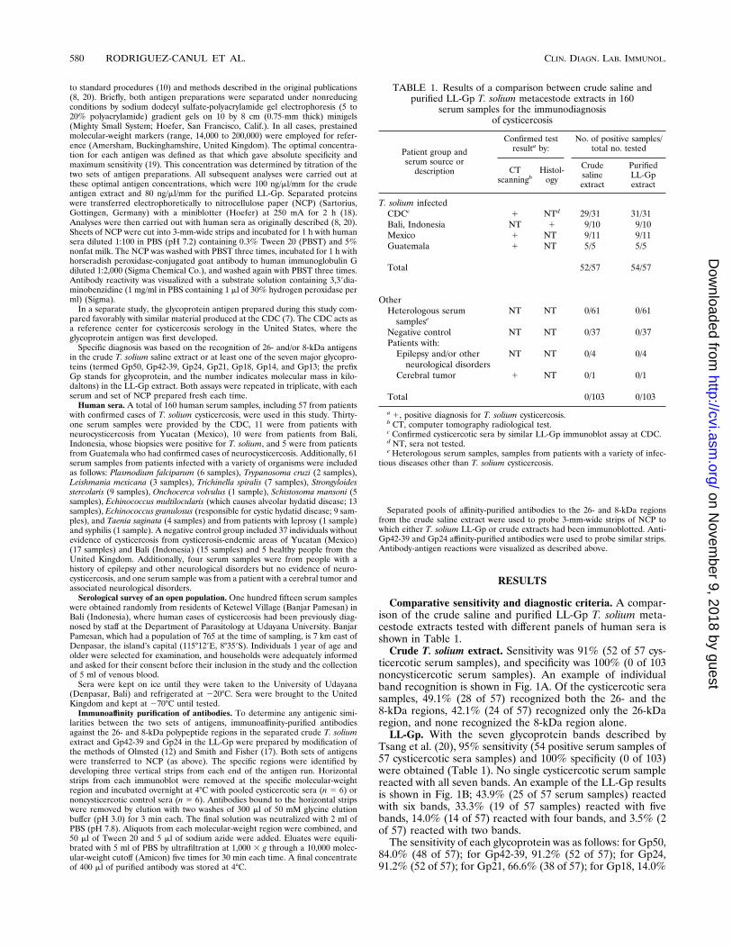
Comparative sensitivity and diagnostic criteria. A compar-ison of the crude saline and purified LL-Gp T. solium meta-cestode extracts tested with different panels of human sera isshown in Table 1.
Crude T. solium extract. Sensitivity was 91% (52 of 57 cys-ticercotic serum samples), and specificity was 100% (0 of 103noncysticercotic serum samples). An example of individualband recognition is shown in Fig. 1A. Of the cysticercotic serasamples, 49.1% (28 of 57) recognized both the 26- and the8-kDa regions, 42.1% (24 of 57) recognized only the 26-kDaregion, and none recognized the 8-kDa region alone.
LL-Gp. With the seven glycoprotein bands described byTsang et al. (20), 95% sensitivity (54 positive serum samples of57 cysticercotic sera samples) and 100% specificity (0 of 103)were obtained (Table 1). No single cysticercotic serum samplereacted with all seven bands. An example of the LL-Gp resultsis shown in Fig. 1B; 43.9% (25 of 57 serum samples) reactedwith six bands, 33.3% (19 of 57 samples) reacted with fivebands, 14.0% (14 of 57) reacted with four bands, and 3.5% (2of 57) reacted with two bands.
The sensitivity of each glycoprotein was as follows: for Gp50,84.0% (48 of 57); for Gp42-39, 91.2% (52 of 57); for Gp24,91.2% (52 of 57); for Gp21, 66.6% (38 of 57); for Gp18, 14.0%
TABLE 1. Results of a comparison between crude saline andpurified LL-Gp T. solium metacestode extracts in 160
serum samples for the immunodiagnosisof cysticercosis
Patient group andserum source or
description
Confirmed testresulta by:
No. of positive samples/total no. tested
CTscanningb
Histol-ogy
Crudesalineextract
PurifiedLL-Gpextract
T. solium infectedCDCc 1 NTd 29/31 31/31Bali, Indonesia NT 1 9/10 9/10Mexico 1 NT 9/11 9/11Guatemala 1 NT 5/5 5/5
Total 52/57 54/57
OtherHeterologous serum
sampleseNT NT 0/61 0/61
Negative control NT NT 0/37 0/37Patients with:
Epilepsy and/or otherneurological disorders
NT NT 0/4 0/4
Cerebral tumor 1 NT 0/1 0/1
Total 0/103 0/103
a 1, positive diagnosis for T. solium cysticercosis.b CT, computer tomography radiological test.c Confirmed cysticercotic sera by similar LL-Gp immunoblot assay at CDC.d NT, sera not tested.e Heterologous serum samples, samples from patients with a variety of infec-
tious diseases other than T. solium cysticercosis.
580 RODRIGUEZ-CANUL ET AL. CLIN. DIAGN. LAB. IMMUNOL.
on Novem
ber 9, 2018 by guesthttp://cvi.asm
.org/D
ownloaded from

(8 of 57); for Gp14, 50.8% (29 of 57); and for Gp13, 73.6% (42of 57).
Reliability. Both techniques proved to be highly reproduc-ible. There was little, if any, variation between each of thethree replicates. None of the sera from individuals with para-sitic infections other than cysticercosis or with neurologicaldisorders and none of the sera from the negative controlscross-reacted with either set of diagnostic antigens (Table 1).We were careful to measure the molecular weights of thediagnostic bands accurately, particularly with the crude extract,for which several sera samples from noncysticercotic individu-als reacted with molecules close to the diagnostic regions. Forinstance, one serum sample from an individual with E. mul-tilocularis recognized a 29-kDa region in the crude extract.
All cysticercotic individuals who were positive for the crudeantigen extract were also positive for the LL-Gp. Two individ-uals were positive for the LL-Gp but negative for the crudeantigen (these individuals recognized only Gp50, Gp18, Gp14,and Gp13).
Seroprevalence in a rural community. Results of seroepide-miological screening of 115 persons (the youngest subjecttested was $1 year old, and the oldest was 78 years old) showthat the rate of seropositivity measured by the LL-Gp washigher than that measured by the crude extract. Nine (7.8%)individuals were positive in both assays, and two cases werenegative with the crude antigen extract but positive with theLL-Gp, recognizing only the Gp13 band. One of the individu-als who were positive by both immunoblot assays was found tohave subcutaneous T. solium cystic infection.
Antigenic similarities by affinity-purified antibodies. Figure2 shows the antigenic similarities determined between bothsets of antigens. Affinity-purified antibodies to the 26-kDa re-gion of the crude saline extract recognized specifically theGp24 and Gp42-39 regions of the LL-Gp. Anti-8kDa affinity-purified antibodies recognized the 26-kDa region in the crudeextract but did not recognize the Gp24 region in the LL-Gpextract. Conversely, when affinity-purified antibodies to theGp42-39 and Gp24 regions from the LL-Gp were incubatedwith the crude antigen extract, the antibodies to Gp24 recog-nized epitopes from the 26-kDa region, but the antibodies tothe Gp42-39 region did not recognize any antigens in the crudesaline extract.
DISCUSSION
There have been several reports of comparisons of otherserodiagnostic tests for cysticercosis (enzyme-linked immu-nosorbent assays) with one or the other of these two immuno-blot assays (4, 6, 9, 16), but the present study describes the firstreported simultaneous evaluation of both of these immunoblotassays. In these two immunoblot assays, alternative methodswere used for the preparation of antigen for the immunodiag-nosis of T. solium cysticercosis. The results indicated that thesemipurified glycoprotein antigen (20) was slightly more sen-sitive than the simpler crude antigen extract described byGottstein et al. (8) (95% [54 of 57] compared to 91% [52 of57]).
The sensitivities and specificities of the immunoblot assaysin this study were similar to those originally reported (8, 9, 20,21). Both antigens were over 90% sensitive with sera frompatients with clinically confirmed cysticercosis. Both methodswere 100% specific, showing no cross-reaction with sera fromindividuals with a variety of parasitic and nonparasitic diseasesor neurological disorders and no cross-reaction with sera fromhealthy individuals.
Interpreting the results and measuring the diagnostic bands
FIG. 1. Western blot assays. Crude saline extract (A) and purified LL-Gp (B) from T. solium metacestodes. Lanes: 1 to 5, sera from patients with confirmedcysticercosis provided by CDC; 6, sera from negative control from the United Kingdom; 7, sera from negative control from cysticerosis-endemic area of Indonesia; 8,sera from patient with T. saginata infection; 9, sera from patient with E. multilocularis disease; 10, sera from patient with E. granulosus disease; 11 to 14, cysticercoticsera from patients from Mexico; 15, sera from negative control from cysticerosis-endemic areas of Mexico; 16, sera from patient with T. spiralis from Mexico; 17 and18, sera from treated cysticercotic patients from Mexico; 19 and 20, sera from patients from Guatemala; 21, sera from patient with epilepsy and other neurologicaldisorders from Indonesia; 22, sera from patient with cerebral tumor from Indonesia.
FIG. 2. Western blot assays. (A) crude saline extract and (B) purified LL-Gp.Lanes: 1, serum from a patient with cysticercosis; 2, anti-26-kDa affinity-purifiedantibodies; 3, anti-8-kDa affinity-purified antibodies; 4, anti-Gp24 affinity-puri-fied antibodies; 5, anti-Gp42-39 affinity-purified antibodies; 6, serum from neg-ative control.
VOL. 4, 1997 IMMUNODIAGNOSIS OF T. SOLIUM CYSTICERCOSIS 581
on Novem
ber 9, 2018 by guesthttp://cvi.asm
.org/D
ownloaded from

(molecular weight) had to be done carefully, particularly withthe crude antigen preparation. In one case, sera from an alve-olar hydatid patient reacted with a band at the 29-kDa region,which could, if mistaken for the 26-kDa band, have led to afalse-positive diagnosis for T. solium cysticercosis.
The results also indicated that two sets of molecules hadsome antigenic similarities. In particular, antibodies to the Gp24molecule recognized the 26-kDa molecule in the crude extractand vice versa. These antigens were the most sensitive mole-cules in their respective immunoblot assays, which might ex-plain the high level of similarity between the two assays. In allcases, individuals who were positive to the 26-kDa molecule fromthe crude saline extract were also positive to the Gp24 region.
Antibodies to the 8-kDa region in the crude extract recog-nized the 26-kDa region from the crude extract but not theGp24 region in the LL-Gp assay. One possible explanation isthat the 8-kDa molecule might be a small subunit of one ofseveral molecules in the 26-kDa region of the crude antigenpreparation, which was not present after glycoprotein purifi-cation. Alternatively, it may be that the 26- and the 8-kDamolecules share only some epitopes. Each molecule must haveits own, unique epitopes, since certain individuals were positivefor only one of the two. For instance, 1 person from thecommunity study recognized only the 8-kDa molecule, and 26individuals who were clinically cysticercotic recognized onlythe 26-kDa molecule.
Besides the routine diagnosis of cysticercosis in a clinicalsetting, the absolute specificity of both of these antigen prep-arations has allowed their use in studies on open populations.The performance of less-specific tests in such circumstances ispoor due to the high rate of false-positive diagnoses, resultingin poor positive predictive value. Both antigens, and in partic-ular the LL-Gp, have been used separately in studies of theepidemiology of T. solium in rural communities (6, 14, 15).Schantz et al. (16) demonstrated a correlation between theLL-Gp immunoblot assay result and a clinical finding of neu-rocysticercosis in a rural community in Mexico where no suchcorrelation was seen with a less-specific enzyme-linked immu-nosorbent assay.
The results of the current study from a T. solium-endemicarea of Indonesia indicated that the sensitivity of the LL-Gpappeared to be slightly higher than that of the crude antigenextract. In all but one case, however, it was impossible toclinically confirm infection. One individual, positive in bothassays, was subsequently diagnosed with subcutaneous T. so-lium cysticercosis. All individuals positive for the crude antigenwere positive by the LL-Gp assay.
In this study, the differences between the two antigenic prep-arations in terms of sensitivity were not, however, statisticallysignificant by chi-square (P . 0.05) (95% confidence), indicat-ing that which one of the two tests is chosen will depend onlaboratory criteria for antigen selection and other work prac-tices.
An extension of this study will be based on the screening ofan adult T. solium cDNA library with anti-26-kDa and anti-Gp24 affinity-purified antibodies for the identification of puta-tive clones for 26-kDa and/or Gp24 recombinant antigens.These antigens are important, since they are consistently rec-ognized by the cysticercotic sera of individuals in clinical andepidemiological studies (8, 15, 16, 20).
ACKNOWLEDGMENTS
We are grateful to the Consejo Nacional de Ciencia y Tecnologia(CONACYT, Mexico) for a scholarship to R. Rodriguez-Canul. J.Allan was supported by a Wellcome Trust Tropical fellowship. We arealso grateful for financial support provided by a project grant from the
European Commission (CI1 CT940081). The British Council fundedthe academic link between Salford and Udayana Universities.
Special thanks are conveyed to Peter Schantz of the CDC for pro-viding cysticercotic sera.
REFERENCES1. Bradford, M. M. 1976. A rapid and sensitive method for the quantitation of
microgram quantities of protein utilizing the principles of protein-dye bind-ing. Anal. Biochem. 72:248–254.
2. Chopra, J. S., U. Kaur, and R. C. Mahajan. 1981. Cysticercosis and epilepsy:a clinical and serological study. Trans. R. Soc. Trop. Med. Hyg. 75:518–520.
3. Craig, P. S., M. T. Rogan, and J. C. Allan. 1995. Hydatidosis and cysticer-cosis—larval cestodes, p. 209–237. In S. H. Guillespie and P. M. Hawkey(ed.), Medical parasitology: a practical approach. IRL Press, London, En-gland.
4. Diaz, F. J., M. Verastegui, R. H. Gilman, C. W. V. Tsang, B. J. Pilcher, C.Gallo, H. H. Garcia, P. Torres, T. Montenegro, E. Miranda, and the Cysti-cercosis Working Group in Peru. 1992. Immunodiagnosis of human cystic-ercosis (Taenia solium): a field comparison of an antibody-enzyme-linkedimmunosorbent assay (ELISA), an antigen-elisa, and an enzyme-linked im-munoelectrotransfer blot (EITB) assay in Peru. Am. J. Trop. Med. Hyg.46:610–615.
5. Flisser, A. 1994. Taeniasis and cysticercosis due to Taenia solium. p. 77–115.In T. Sun (ed.), Progress in clinical parasitology. CRC Press, Inc., BocaRaton, Fla.
6. Fritzche, M., B. Gottstein, M. C. Wigglesworth, and J. Eckert. 1990. Sero-logical survey of human cysticercosis in Irianese refugee camps in PapuaNew Guinea. Acta Trop. 47:69–77.
7. Garcia Noval, J., J. C. Allan, C. Fletes, E. Moreno, F. de Mata, R. Torres-Alvarez, H. Soto de Alfaro, P. Yurritia, H. Higueros-Morales, F. Mencos,and P. S. Craig. 1996. Epidemiology of Taenia solium taeniasis and cysti-cercosis in two rural Guatemalan communities. Am. J. Trop. Med. Hyg.55:282–289.
8. Gottstein, B., V. C. W. Tsang, and P. M. Schantz. 1986. Demonstration ofspecies-specific and cross-reactive components of Taenia solium metacestodeantigens. Am. J. Trop. Med. Hyg. 35:308–313.
9. Gottstein, B., D. Zini, and P. M. Schantz. 1987. Species specific immunodi-agnosis of Taenia solium cysticercosis by ELISA and immunoblotting. Trop.Med. Parasitol. 38:299–303.
10. Laemmli, U. K. 1970. Cleavage of structural proteins during the assembly ofthe head of bacteriophage T4. Nature 227:680–685.
11. Larralde, C., R. M. Montoya, E. Sciutto, M. L. Diaz, T. Govezensky, and E.Coltorti. 1989. Deciphering Western blots of tapeworm antigens (Taeniasolium, Echinococcus granulosus, and Taenia crassiceps) reacting with serafrom neurocysticercosis and hydatid disease patients. Am. J. Trop. Med.Hyg. 40:282–290.
12. Olmsted, J. B. 1981. Affinity purification of antibodies from diazotized paperblots of heterogeneous protein samples. J. Biol. Chem. 256:11955–11957.
13. Richard, F., and P. M. Schantz. 1991. Laboratory diagnosis of cysticercosis.Clin. Lab. Med. 4:1011–1028.
14. Sarti, E., P. M. Schantz, A. Plancarte, M. Wilson, I. O. Gutierrez, A. S.Lopez, J. Roberts, and A. Flisser. 1992. Prevalence and risk factor for Taeniasolium in humans and pigs in a village in Morelos, Mexico. Am. J. Trop. Med.Hyg. 46:677–684.
15. Sarti, E., P. M. Schantz, M. Wilson, O. Gutierrez, J. Aguilera, J. Roberts,and A. Flisser. 1994. Epidemiological investigation of Taenia solium taenia-sis and cysticerocisis in a rural village of Michoacan State, Mexico. Trans. R.Soc. Trop. Med. Hyg. 88:49–52.
16. Schantz, P. M., E. Sarti, A. Plancarte, M. Wilson, J. L. Criales, J. Roberts,and A. Flisser. 1994. Community-based epidemiological investigations ofcystsicercosis due to Taenia solium: comparison of serological screening testsand clinical findings in two populations in Mexico. Clin. Infect. Dis. 18:879–885.
17. Smith, D. E., and P. A. Fisher. 1984. Identification, developmental regula-tion, and response to heat shock of two antigenically related forms of a majornuclear envelope protein in Drosophila embryos: application of an improvedmethod for affinity purification of antibodies using polypeptides immobilizedon nitrocellulose blots. J. Cell Biol. 99:20–28.
18. Towbin, H., T. Staehelin, and J. Gordon. 1979. Electrophoresis transfer ofprotein from polyacrylamide gels to nitrocellulose sheets: procedure andsome applications. Proc. Natl. Acad. Sci. USA 76:14350–14354.
19. Tsang, V. C. W., J. M. Peralta, and A. R. Simons. 1983. Enzyme-linkedimmunoelectrotransfer blot techniques (EITB) for studying the specificitiesof antigens and antibodies separated by gel electrophoresis. Methods Enzy-mol. 92:377–391.
20. Tsang, V. C. W., J. A. Brand, and A. F. Boyer. 1989. An enzyme-linkedimmunoelectrotransfer blot assay and glycoprotein antigens for diagnosinghuman cysticercosis (Taenia solium). J. Infect. Dis. 159:50–58.
21. Wilson, M., R. Bryan, J. Fried, D. Ware, P. Schantz, J. B. Pilcher, andV. C. W. Tsang. 1991. Clinical evaluation of the cysticercosis enzyme-linkedimmunoelectrotransfer blot in patients with neurocysticercosis. J. Infect. Dis.164:1007–1009.
582 RODRIGUEZ-CANUL ET AL. CLIN. DIAGN. LAB. IMMUNOL.
on Novem
ber 9, 2018 by guesthttp://cvi.asm
.org/D
ownloaded from