comparative genomics and transcriptomics of … · comparative genomics and transcriptomics of...
TRANSCRIPT
Hain et al. BMC Genomics 2012, 13:144http://www.biomedcentral.com/1471-2164/13/144
RESEARCH ARTICLE Open Access
Comparative genomics and transcriptomics oflineages I, II, and III strains of ListeriamonocytogenesTorsten Hain1, Rohit Ghai1, André Billion1, Carsten Tobias Kuenne1, Christiane Steinweg1, Benjamin Izar1,Walid Mohamed1, Mobarak Abu Mraheil1, Eugen Domann1, Silke Schaffrath1, Uwe Kärst2, Alexander Goesmann3,Sebastian Oehm3, Alfred Pühler3, Rainer Merkl4, Sonja Vorwerk5, Philippe Glaser6, Patricia Garrido7,Christophe Rusniok7, Carmen Buchrieser7, Werner Goebel8 and Trinad Chakraborty1*
Abstract
Background: Listeria monocytogenes is a food-borne pathogen that causes infections with a high-mortality rate andhas served as an invaluable model for intracellular parasitism. Here, we report complete genome sequences for twoL. monocytogenes strains belonging to serotype 4a (L99) and 4b (CLIP80459), and transcriptomes of representativestrains from lineages I, II, and III, thereby permitting in-depth comparison of genome- and transcriptome -baseddata from three lineages of L. monocytogenes. Lineage III, represented by the 4a L99 genome is known to containstrains less virulent for humans.
Results: The genome analysis of the weakly pathogenic L99 serotype 4a provides extensive evidence of virulencegene decay, including loss of several important surface proteins. The 4b CLIP80459 genome, unlike the previouslysequenced 4b F2365 genome harbours an intact inlB invasion gene. These lineage I strains are characterized by thelack of prophage genes, as they share only a single prophage locus with other L. monocytogenes genomes 1/2aEGD-e and 4a L99. Comparative transcriptome analysis during intracellular growth uncovered adaptive expressionlevel differences in lineages I, II and III of Listeria, notable amongst which was a strong intracellular induction offlagellar genes in strain 4a L99 compared to the other lineages. Furthermore, extensive differences between strainsare manifest at levels of metabolic flux control and phosphorylated sugar uptake. Intriguingly, prophage geneexpression was found to be a hallmark of intracellular gene expression. Deletion mutants in the single sharedprophage locus of lineage II strain EGD-e 1/2a, the lma operon, revealed severe attenuation of virulence in a murineinfection model.
Conclusion: Comparative genomics and transcriptome analysis of L. monocytogenes strains from three lineagesimplicate prophage genes in intracellular adaptation and indicate that gene loss and decay may have led to theemergence of attenuated lineages.
Keywords: Listeria monocytogenes, Lineage, Comparative genomics, Gene decay, Comparative transcriptomics,Flagella, Prophage, Monocin, Isogenic deletion mutants, Murine infection
* Correspondence: [email protected] of Medical Microbiology, Justus-Liebig-University, Schubertstrasse81, Giessen, D-35392, GermanyFull list of author information is available at the end of the article
© 2012 Hain et al.; licensee BioMed Central Ltd. This is an Open Access article distributed under the terms of the CreativeCommons Attribution License (http://creativecommons.org/licenses/by/2.0), which permits unrestricted use, distribution, andreproduction in any medium, provided the original work is properly cited.
Hain et al. BMC Genomics 2012, 13:144 Page 2 of 17http://www.biomedcentral.com/1471-2164/13/144
BackgroundListeria monocytogenes is a Gram-positive, motile, non-sporulating, rod shaped bacterium. It is the causativeagent of listeriosis, a food-borne disease, which afflictsboth humans and animals. There are only eight speciesin the entire genus, L. monocytogenes, L. marthii, L.innocua, L. seeligeri, L. welshimeri, L. ivanovii, L. grayiand L. rocourtiae. L. monocytogenes and L. ivanovii arethe pathogenic species while the others are apathogenic[1,2]. In the genus Listeria, non-pathogenic species havebeen hypothesized to have evolved through genomereduction from pathogenic progenitor strains [3]. L.monocytogenes is able to invade and replicate in bothphagocytic and non-phagocytic cells. The infectious lifecycle has been elucidated in detail, and several virulencefactors, essential for each stage of infection have beenidentified [4,5]. Pathogenic listeriae encode severalvirulence factors that are localized in a virulence genecluster (vgc) or Listeria pathogenicity island-1 (LIPI-1) inthe genome. However, a number of genes required forvirulence are not localized in this cluster, including thetwo internalins inlA and inlB. These encode proteinsthat are expressed on the surface of the bacterium andfacilitate the entry of the bacterium into the eukaryoticcell and their incorporation into a membrane-boundvacuole [6,7]. Further pathogenicity islands present inthe genus Listeria code for multiple internalins andadditional hemolysin genes in species L. ivanovii (LIPI-2)[8] and a subset of strains of lineage I (LIPI-3) [9].Within the four lineages of L. monocytogenes, strains
are generally classified by serotyping or MLST [10,11], ofwhich 1/2a, 1/2b and 4b are most commonly associatedwith human listerial infections [2,12]. The first outbreakof L. monocytogenes was described for the strain EGD-e,a serotype 1/2a strain of lineage II, following an epidemicin rabbits and guinea pigs in 1926 by E.G.D. Murray[13]. This strain has become a model Listeria strain, andwas the first listerial strain to be completely sequenced,along with the non-pathogenic Listeria innocua 6aCLIP11262 [14]. Subsequently, the first genome of a 4bserotype strain (F2365) of lineage I was completelysequenced [14,15]. It was isolated from Jalisco cheeseduring a listeriosis outbreak in California in 1985 andmainly associated with pregnancy-related cases. However,it has been recently shown that this strain contains non-sense and frameshift mutations in several genes. Owing toa frameshift in inlB, F2365 is severely compromised inCaco-2 invasion assays [16].Here we report thus the genome sequence of a clinical
isolate of the 4b serotype of lineage I, the L. monocytogenes4b strain CLIP80459 that was isolated in a clinicaloutbreak of listeriosis in France affecting 42 persons [17].We also present the complete genome sequencing of L.monocytogenes strain 4a L99 of lineage III. L99 was
originally isolated from food by Kampelmacher in 1950s inthe Netherlands. This strain is attenuated in its virulenceproperties and exhibits a restricted ability to grow withinthe liver and spleen of infected mice [18]. The availabilityof the complete genome of L. monocytogenes EGD-e sero-type 1/2a has permitted analysis of the intracellular geneexpression profile of this strain [19-21].The genome sequences of strains 4a L99 and 4b
CLIP80459 presented in this work provide a unique oppor-tunity to delineate specific adaptations of these lineagerepresentives both at the genomic and at the transcrip-tional level.
ResultsGeneral features of complete genomes of three lineagesof L. MonocytogenesThe overall features of the completely sequenced circulargenomes of L. monocytogenes 4a L99, L. monocytogenes4b CLIP80459, L. monocytogenes 1/2a EGD-e, L. monocy-togenes 4b F2365 and L. innocua 6a CLIP11262 aregiven in Table 1. Computational multi-virulence-locussequence typing (MVLST) [22] analysis showed thatstrain 4b CLIP80459 belongs to epidemic clone ECII andstrain 4b F2365 to epidemic clone ECI as previouslyreported by Nelson and colleagues [15], respectively. TheL. monocytogenes genomes are remarkably syntenic: gen-ome size, G + C content, percentage coding and averagelength of protein-coding genes are similar among all fourstrains (which was previously reported for other listerialgenomes) [14,15]. All four L. monocytogenes genomesharbour 67 tRNA genes and contain six complete copiesof rRNA operons (16 S-23 S-5 S), of which two arelocated on the right and four on the left replichore. Thechromosomes of 4a L99 and 4b CLIP80459 are devoid ofmobile genetic elements and harbour no plasmid.We observed four different prophage regions in the
genome of the 4a L99 and only one in the 4b CLIP80459strain (see prophage region II). L. monocytogenes 4a L99prophage I is located at position 71438 bp (lmo4a_0064-lmo4a_0115), prophage II at (lmo4a_0148-lmo4a 0153,prophage-remnant: lmaDC; 4b ClIP80459 Lm4b_00117b-Lm4b00134 or monocin region), prophage III at1224779 bp (lmo4a_1221-lmo4a_1293) and prophage IVat 2668913 bp (lmo4a_2599-lmo4a_2658). Two prophageregions, I and III, are located adjacent to tRNAs. Prophageregion I is flanked by tRNALys and prophage region III isinserted within the region between the gene for tRNAArg
and ydeI compared to L. monocytogenes 1/2a EGD-e. Atthis very chromosomal location in L. welshimeri 6bSLCC5334 there is an insertion of a prophage [3,23,24],while L. ivanovii harbours the species-specific Listeriapathogenicity island 2 (LIPI-2), which contains a sphingo-myelinase C (SmcL) and also a cluster of internalin genes[8]. These findings confirm previous observations [3]
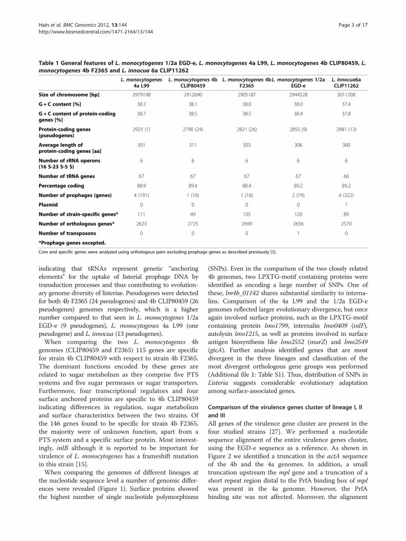
Table 1 General features of L. monocytogenes 1/2a EGD-e, L. monocytogenes 4a L99, L. monocytogenes 4b CLIP80459, L.monocytogenes 4b F2365 and L. innocua 6a CLIP11262
L. monocytogenes4a L99
L. monocytogenes 4bCLIP80459
L. monocytogenes 4bF2365
L. monocytogenes 1/2aEGD-e
L. innocua6aCLIP11262
Size of chromosome [bp] 2979198 2912690 2905187 2944528 3011208
G + C content [%] 38.2 38.1 38.0 38.0 37.4
G + C content of protein-codinggenes [%]
38.7 38.5 38.5 38.4 37.8
Protein-coding genes(pseudogenes)
2925 (1) 2790 (24) 2821 (26) 2855 (9) 2981 (13)
Average length ofprotein-coding genes [aa]
301 311 303 306 300
Number of rRNA operons(16 S-23 S-5 S)
6 6 6 6 6
Number of tRNA genes 67 67 67 67 66
Percentage coding 88.9 89.4 88.4 89.2 89.2
Number of prophages (genes) 4 (191) 1 (16) 1 (16) 2 (79) 6 (322)
Plasmid 0 0 0 0 1
Number of strain-specific genes* 111 49 105 120 89
Number of orthologous genes* 2623 2725 2699 2656 2570
Number of transposons 0 0 0 1 0
*Prophage genes excepted.
Core and specific genes were analyzed using orthologous pairs excluding prophage genes as described previously [3].
Hain et al. BMC Genomics 2012, 13:144 Page 3 of 17http://www.biomedcentral.com/1471-2164/13/144
indicating that tRNAs represent genetic “anchoringelements” for the uptake of listerial prophage DNA bytransduction processes and thus contributing to evolution-ary genome diversity of listeriae. Pseudogenes were detectedfor both 4b F2365 (24 pseudogenes) and 4b CLIP80459 (26pseudogenes) genomes respectively, which is a highernumber compared to that seen in L. monocytogenes 1/2aEGD-e (9 pseudogenes), L. monocytogenes 4a L99 (onepseudogene) and L. innocua (13 pseudogenes).When comparing the two L. monocytogenes 4b
genomes (CLIP80459 and F2365) 115 genes are specificfor strain 4b CLIP80459 with respect to strain 4b F2365.The dominant functions encoded by these genes arerelated to sugar metabolism as they comprise five PTSsystems and five sugar permeases or sugar transporters.Furthermore, four transcriptional regulators and foursurface anchored proteins are specific to 4b CLIP80459indicating differences in regulation, sugar metabolismand surface characteristics between the two strains. Ofthe 146 genes found to be specific for strain 4b F2365,the majority were of unknown function, apart from aPTS system and a specific surface protein. Most interest-ingly, inlB although it is reported to be important forvirulence of L. monocytogenes has a frameshift mutationin this strain [15].When comparing the genomes of different lineages at
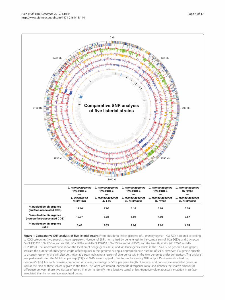
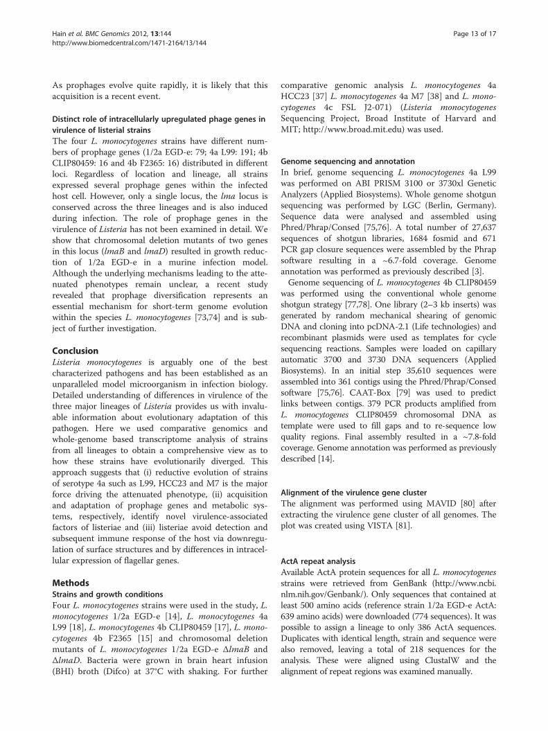
the nucleotide sequence level a number of genomic differ-ences were revealed (Figure 1). Surface proteins showedthe highest number of single nucleotide polymorphisms
(SNPs). Even in the comparison of the two closely related4b genomes, two LPXTG-motif containing proteins wereidentified as encoding a large number of SNPs. One ofthese, lm4b_01142 shares substantial similarity to interna-lins. Comparison of the 4a L99 and the 1/2a EGD-egenomes reflected larger evolutionary divergence, but onceagain involved surface proteins, such as the LPXTG-motifcontaining protein lmo1799, internalin lmo0409 (inlF),autolysin lmo1215, as well as proteins involved in surfaceantigen biosynthesis like lmo2552 (murZ) and lmo2549(gtcA). Further analysis identified genes that are mostdivergent in the three lineages and classification of themost divergent orthologous gene groups was performed(Additional file 1: Table S1). Thus, distribution of SNPs inListeria suggests considerable evolutionary adaptationamong surface-associated genes.
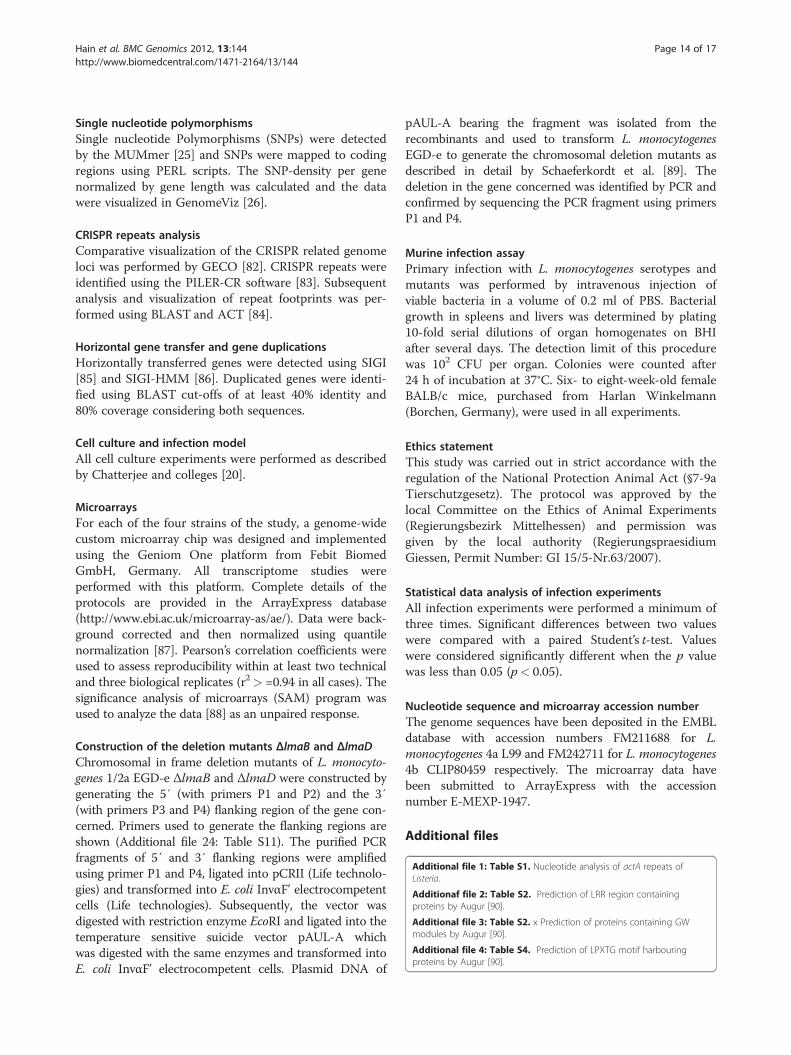
Comparison of the virulence genes cluster of lineage I, IIand IIIAll genes of the virulence gene cluster are present in thefour studied strains [27]. We performed a nucleotidesequence alignment of the entire virulence genes cluster,using the EGD-e sequence as a reference. As shown inFigure 2 we identified a truncation in the actA sequenceof the 4b and the 4a genomes. In addition, a smalltruncation upstream the mpl gene and a truncation of ashort repeat region distal to the PrfA binding box of mplwas present in the 4a genome. However, the PrfAbinding site was not affected. Moreover, the alignment
0 kb
Comparative SNP analysisof five listerial strains
350 kb
700 kb
1050 kb
1400 kb
1750 kb
2100 kb
2450 kb
L. monocytogenes 1/2a EGD-e
vs. L. innocua 6a
CLIP11262
L. monocytogenes 1/2a EGD-e
vs. L. monocytogenes
4a L99
L. monocytogenes 1/2a EGD-e
vs. L. monocytogenes
4b CLIP80459
L. monocytogenes 1/2a EGD-e
vs. L. monocytogenes
4b F2365
L. monocytogenes 4b F2365
vs. L. monocytogenes
4b CLIP80459
% nucleotide divergence (surface-associated CDS)
11.14 7.00 5.16 5.09 0.59
% nucleotide divergence(non-surface-associated CDS)
10.77 6.38 5.01 4.99 0.57
% nucleotide divergenceratio
3.46 9.79 2.96 2.02 4.55
Figure 1 Comparative SNP analysis of five listerial strains From outside to inside: genome of L. monocytogenes 1/2a EGD-e colored accordingto COG categories (two strands shown separately). Number of SNPs normalized by gene length in the comparison of 1/2a EGD-e and L. innocua6a CLIP11262, 1/2a EGD-e and 4a L99, 1/2a EGD-e and 4b CLIP80459, 1/2a EGD-e and 4b F2365, and the two 4b strains (4b F2365 and 4bCLIP80459). The innermost circle shows the location of phage genes (blue) and virulence genes (black) in the 1/2a EGD-e genome. Line graphsindicate the number of SNPs/gene length reflecting loci in the genome having a disproportionate number of SNPs. However, if a gene is specificto a certain genome, this will also be shown as a peak indicating a region of divergence within the two genomes under comparison. This analysiswas performed using the MUMmer package [25] and SNPs were mapped to coding regions using PERL scripts. Data were visualized byGenomeViz [26]. For each pairwise comparison of strains, percentage of SNPs per gene length of surface- and non-surface-associated genes, aswell as the ratio of these values is given in the table. The latter was named “nucleotide divergence ratio” and denotes the relative amount ofdifference between those two classes of genes, in order to identify more (positive value) or less (negative value) abundant mutation in surface-associated than in non-surface-associated genes.
Hain et al. BMC Genomics 2012, 13:144 Page 4 of 17http://www.biomedcentral.com/1471-2164/13/144
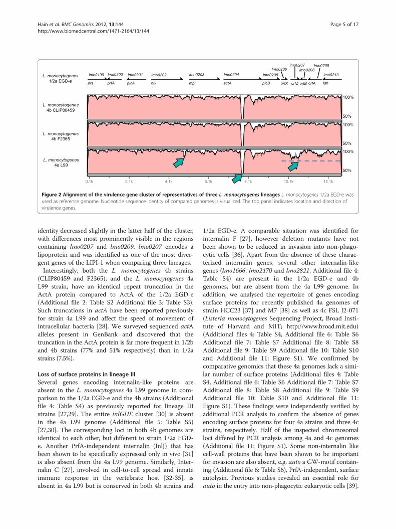
Figure 2 Alignment of the virulence gene cluster of representatives of three L. monocytogenes lineages L. monocytogenes 1/2a EGD-e wasused as reference genome. Nucleotide sequence identity of compared genomes is visualized. The top panel indicates location and direction ofvirulence genes.
Hain et al. BMC Genomics 2012, 13:144 Page 5 of 17http://www.biomedcentral.com/1471-2164/13/144
identity decreased slightly in the latter half of the cluster,with differences most prominently visible in the regionscontaining lmo0207 and lmo0209. lmo0207 encodes alipoprotein and was identified as one of the most diver-gent genes of the LIPI-1 when comparing three lineages.Interestingly, both the L. monocytogenes 4b strains
(CLIP80459 and F2365), and the L. monocytogenes 4aL99 strain, have an identical repeat truncation in theActA protein compared to ActA of the 1/2a EGD-e(Additional file 2: Table S2 Additional file 3: Table S3).Such truncations in actA have been reported previouslyfor strain 4a L99 and affect the speed of movement ofintracellular bacteria [28]. We surveyed sequenced actAalleles present in GenBank and discovered that thetruncation in the ActA protein is far more frequent in 1/2band 4b strains (77% and 51% respectively) than in 1/2astrains (7.5%).
Loss of surface proteins in lineage IIISeveral genes encoding internalin-like proteins areabsent in the L. monocytogenes 4a L99 genome in com-parison to the 1/2a EGD-e and the 4b strains (Additionalfile 4: Table S4) as previously reported for lineage IIIstrains [27,29]. The entire inlGHE cluster [30] is absentin the 4a L99 genome (Additional file 5: Table S5)[27,30]. The corresponding loci in both 4b genomes areidentical to each other, but different to strain 1/2a EGD-e. Another PrfA-independent internalin (InlJ) that hasbeen shown to be specifically expressed only in vivo [31]is also absent from the 4a L99 genome. Similarly, Inter-nalin C [27], involved in cell-to-cell spread and innateimmune response in the vertebrate host [32-35], isabsent in 4a L99 but is conserved in both 4b strains and
1/2a EGD-e. A comparable situation was identified forinternalin F [27], however deletion mutants have notbeen shown to be reduced in invasion into non-phago-cytic cells [36]. Apart from the absence of these charac-terized internalin genes, several other internalin-likegenes (lmo1666, lmo2470 and lmo2821, Additional file 4:Table S4) are present in the 1/2a EGD-e and 4bgenomes, but are absent from the 4a L99 genome. Inaddition, we analysed the repertoire of genes encodingsurface proteins for recently published 4a genomes ofstrain HCC23 [37] and M7 [38] as well as 4c FSL J2-071(Listeria monocytogenes Sequencing Project, Broad Insti-tute of Harvard and MIT; http://www.broad.mit.edu)(Additional files 4: Table S4, Additional file 6: Table S6Additional file 7: Table S7 Additional file 8: Table S8Additional file 9: Table S9 Additional file 10: Table S10and Additional file 11: Figure S1). We confirmed bycomparative genomics that these 4a genomes lack a simi-lar number of surface proteins (Additional files 4: TableS4, Additional file 6: Table S6 Additional file 7: Table S7Additional file 8: Table S8 Additional file 9: Table S9Additional file 10: Table S10 and Additional file 11:Figure S1). These findings were independently verified byadditional PCR analysis to confirm the absence of genesencoding surface proteins for four 4a strains and three 4cstrains, respectively. Half of the inspected chromosomalloci differed by PCR analysis among 4a and 4c genomes(Additional file 11: Figure S1). Some non-internalin likecell-wall proteins that have been shown to be importantfor invasion are also absent, e.g. auto a GW-motif contain-ing (Additional file 6: Table S6), PrfA-independent, surfaceautolysin. Previous studies revealed an essential role forauto in the entry into non-phagocytic eukaryotic cells [39].
Hain et al. BMC Genomics 2012, 13:144 Page 6 of 17http://www.biomedcentral.com/1471-2164/13/144
The vip gene product, a PrfA-dependent LPXTG protein(Additional file 7: Table S7), described as a receptor for theeukaryotic Gp96 surface protein and important for latestages of infection [40], is also absent from the 4a L99genome. In addition to these missing genes, InlI is slightlytruncated. However Ami (Additional file 6: Table S6), animportant listerial adhesion protein seems to be present ina shorter version in both 4b strains [41,42], whereasthe number of lipoproteins (Additional file 8: Table S8),LysM- and (Additional file 9: Table S9) NLPC/P60-motifcontaining proteins (Additional file 10: Table S10) wascomparable among the four strains under study.Overall, in comparison to 1/2a EGD-e and the two 4b
genomes, 4a L99 strain has lost a number of crucialdeterminants required for listerial invasion. The selectiveloss of genes primarily responsible for the first steps ofinfection may contribute to the poor invasion ability andthe attenuated nature of the 4a L99 strain.
Decay of phage genes in the L. Monocytogenes 4a L99strainThe 1/2a EGD-e genome contains 79 prophage genes intwo different loci, the 4a L99 genome includes 193 phagegenes at four loci, while the 4b genomes encode with 16,for the smallest number of prophage genes limited to asingle locus (also called the monocin-locus) at the sameposition in the chromosomes.This monocin locus, a cryptic prophage region, is
conserved in all L. monocytogenes lineages and includesthe lma genes [43]. Although previously thought to be spe-cific to L. monocytogenes, it was shown that lmaDCBA isalso present in several apathogenic L. innocua strains.However, not all genes of the operon are present in all L.monocytogenes strains. The 4a L99 genome lacks lmaAand lmaB (Additional file 12: Figure S2). The entire locusin 1/2a EGD-e and the two 4b genomes has 16 genes, butonly five of these genes are present in the 4a L99 genome.lmaA and lmaB are absent in L. welshimeri. Interestingly,the structure of this prophage locus in strain 4a L99 andother lineage III strains is more similar to L. welshimerithan to other pathogenic listeriae (Additional file 12:Figure S2).
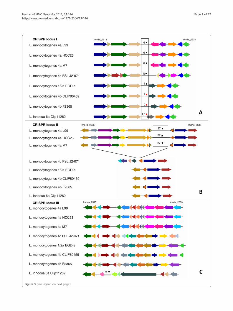
The CRISPR system of ListeriaThe L. monocytogenes 4a L99 genome was found to con-tain two adjacent CRISPR loci (I and II) with CRISPRrepeats (Figure 3A and 3B). Both loci contain sequencesof length 35 bp separated by repeat sequences of length29 bp. However, they differ considerably in the numberof repeat copies (6 in locus I, and 29 in locus II, respect-ively). While locus I is highly conserved in the 4b strains,1/2a EGD-e and L. innocua, locus II was exclusivelypresent in 4a genomes of L99, HCC23, M7, but notin another lineage III genome of 4c FSL J2-071 (Figure 3
A-C). It is not known whether the CRISPR system isfunctional in the 4a L99 genome. However, by sequencesimilarity searches using the spacers to detect possibleprophage DNA traces, we were able to identify the PSAprophage that is known to infect serotype 4 strains.Assuming a functional CRISPR system in 4a L99 suggestsa resistance to the PSA bacteriophage (Additional file 13:Figure S3).
Gene duplications in the Listeria genomes expandmetabolic systemsWe found substantial evidence for a minimum of 231 toa maximum of 296 gene duplications in the Listeriagenomes (Additional file 14: Figure S4 and Additionalfile 15: Figure S5). It is evident that the majority of theseduplications are ancient events as they are shared amongall species and the number of gene pairs with a very highpercentage identity is very low (1-12% per strain).Functional classification of the duplicated genes revealedthat many of these have important implications inmetabolic pathways, like the pentose phosphate pathway,fructose and mannose metabolism, carbon fixation,glycolysis and pyruvate metabolism.While several duplicated genes could be mapped to
central metabolic pathways from the KEGG database,this was not possible for horizontally transferred genes(Additional file 16 Figure S6 and Additional file 17:Figure S7). However, not all duplicated genes seem to havearisen from true duplications, but some may have beentransferred horizontally, like some PTS system genes thatare L. monocytogenes EGD-e strain-specific genes. Thenumber of genes classified into known metabolic pathwaysor systems was significantly higher for duplicated genes,while several horizontally transferred genes could not bemapped.
Comparative intracellular transcriptomics of four L.Monocytogenes strains of the three major lineagesComparative transcriptome analysis of Listeria monocy-togenes strains of the two major lineages revealed differ-ences in virulence, cell wall, and stress response [44].Here we performed intracellular gene expressionanalyses using whole genome microarrays between fourL. monocytogenes strains belonging to the three majorlineages to investigate eventual differences. P388D1murine macrophages were infected and total RNA wasisolated four hours post infection and hybridized tobioarrays.In order to determine the core intracellular response
of L. monocytogenes we created a dataset of core-syntenichomologous genes for all four genomes and the expres-sion data for these genes were compared. We found thatin all strains studied the entire virulence genes cluster,(prfA, plcA, hly, mpl, actA, plcB and orfX) was highly
L. monocytogenes 4a L99
L. monocytogenes 4a HCC23
L. monocytogenes 4a M7
L. monocytogenes 4b CLIP80459
L. monocytogenes 4b F2365
L. monocytogenes 1/2a EGD-e
L. innocua 6a Clip11262
L. monocytogenes 4c FSL J2-071
6
6
6
2
2
4
1,5
10
10
L. monocytogenes 4a L99
L. monocytogenes 4a HCC23
L. monocytogenes 4a M7
L. monocytogenes 4b CLIP80459
L. monocytogenes 4b F2365
L. monocytogenes 1/2a EGD-e
L. innocua 6a Clip11262
L. monocytogenes 4c FSL J2-071
27
27
27
lmo4a_0513 lmo4a_0521
lmo4a_0525 lmo4a_0535
L. monocytogenes 4a L99
L. monocytogenes 4a HCC23
L. monocytogenes 4a M7
L. monocytogenes 4b CLIP80459
L. monocytogenes 4b F2365
L. monocytogenes 1/2a EGD-e
L. innocua 6a Clip11262
lmo4a_2595 lmo4a_2609
L. monocytogenes 4c FSL J2-071
CRISPR locus I
CRISPR locus II
CRISPR locus III
Figure 3 (See legend on next page.)
Hain et al. BMC Genomics 2012, 13:144 Page 7 of 17http://www.biomedcentral.com/1471-2164/13/144
(See figure on previous page.)Figure 3 Overview of CRISPR (clustered regularly interspaced short palindromic repeats) loci in L monocytogenes 1/2a EGD-e, L.monocytogenes 4a L99, L. monocytogenes 4a HCC23, L. monocytogenes 4a M7, L. monocytogenes 4c FSL J2-071, L. monocytogenes 4b CLIP80459, L.monocytogenes 4b F2365 and L. innocua 6a CLIP11262. (A): CRISPR locus I is shown for all five listeriae, black boxes indicate complete CRISPRrepeats, red boxes represent incomplete or truncated (*) CRISPR repeats. No cas genes were found to be associated with this locus. Flankinggenes are conserved in 1/2a EGD-e and both 4b genomes. Comparison of the intergenic sequences with the 4a L99 genome revealed asequence footprint of decaying repeat elements (2 repeat copies in both 4b genomes, and 1 copy in L. innocua 6a CLIP11262), indicating loss ofthe CRISPR repeats. (B): Locus II shows 29 copies of repeats and is associated with several cas genes (cas2, cas3, cas5 and cas6. cas1 is partiallydetectable, but seems to be truncated. (C): L. innocua 6a CLIP11262 harbours the CRISPR locus III at position 2.77 Mb in the genome, which isneighboured by a single cas2 gene. No other CRISPR repeats nor any cas gene homologs were found in the 4b genomes.
Hain et al. BMC Genomics 2012, 13:144 Page 8 of 17http://www.biomedcentral.com/1471-2164/13/144
induced within the infected host cells. Furthermore genesknown to be important for bacterial survival, such as hpt,clpE, bilEA and two LRR domain-containing proteins(lmo0514 and lmo2445) were upregulated in all strains.Interestingly, three mannose transporting PTS systems
(lmo0021-lmo0024, lmo0781-lmo0784, lmo1997-lmo2002),two fructose specific systems (lmo2335 and lmo2733), twogalacitol specific systems (lmo0503, lmo0507, lmo0508 andlmo2665-lmo2667), two beta-glucoside systems (the partialsystem lmo0373-lmo0374 and lmo0874-lmo0876), and twocellobiose specific systems (the partial system lmo0901 andlmo0914-lmo0916) were commonly upregulated in allstrains. These possibly represent the most frequently usedsubstrates of listeriae in the cytosol. Only one mannosespecific PTS system, (lmo0096-lmo0098) is downregulatedby all studied strains (Additional file 18 Figure S8 andAdditional file 19: Text S1).Most surprisingly, all Listeria strains studied expressed
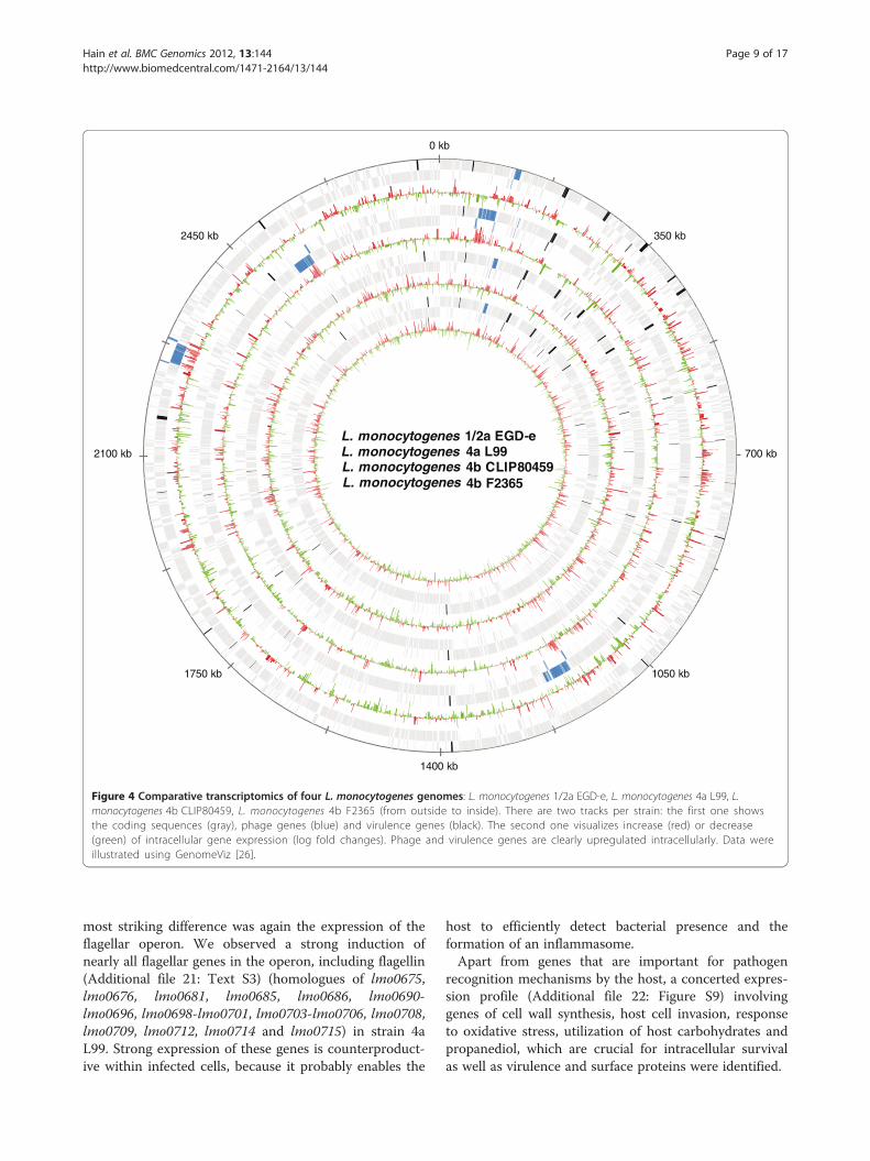
the genes of the lma operon and surrounding prophagegenes of the monocin locus, including a conserved holin(lmo0112, lmo0113, lmo0115, lmo0116, lmo0128) duringintracellular growth. However, the functions of several ofthese genes are not defined. The only locus that isconserved in all three lineages (albeit with somedeletions in 4a L99) is the monocin lma locus. The lmaAgene product has been shown to provoke a delayed typehypersensitivity reaction in mice immune to L. monocyto-genes. It is also secreted at 20°C but much less [45] at 37°C. The lma operon produces two transcripts, a 2100 bplmaDCBA transcript expressed both at 20°C and 37°C,and a 1050 bp lmaBA transcript induced at lowertemperatures [43]. Additional prophage genes were highlyexpressed in the individual strains (Figure 4). Takentogether, high intracellular prophage gene expression,despite several differences in prophage gene content, isone of the most striking observations across all Listerialineages.All strains showed induction of the eut operon
suggesting that ethanolamine may be used as a carbonand nitrogen source in intracellular conditions. The zinctransporters were also commonly upregulated indicatinga role of zinc in intracellular survival as well as thespermidine/putrescine ABC transporters (potB, potC andpotD). Furthermore, the non-oxidative branch of the
pentose phosphate pathway was utilized by all listeriae,possibly to generate NADPH for countering oxidativestress in intracellular conditions. The upregulation ofgenes of the pentose phosphate pathway has been shownpreviously [19,20,46] and it has been speculated that it isimportant for generation of erythrose-4-phosphate foraromatic amino acid biosynthesis or for generation ofpentose sugars. Accordingly; we observed a downregula-tion of several genes involved in pyrimidine and purinebiosynthesis from pentose sugars (e.g. lmo1463, lmo1497,lmo1565, lmo1832, lmo1836, lmo1856, lmo1929,lmo2154, lmo2155, lmo2390 and lmo2559).Downregulated genes included the agr locus (lmo0048-
lmo0051) as demonstrated previously [20,46] and severalgenes of the tryptophan biosynthesis operon (trpA, trpB,trpF and trpD), and some tRNA synthetase genes (ileS,valS, glyS and glyQ). Diminished energy generation wasindicated by decreased expression of the cytochromegenes cluster cytABCD. With respect to the pentosephosphate pathway, we detected downregulation of thephosphoribosyl pyrophosphate synthetase (prs, lmo0199)gene, which is required for the production of PRPP(phosphoribosyl pyrophosphate) that links the pentosephosphate pathway to the biosynthesis of purines andpyrimidines. While several genes of the glycolyticoperon, and several individual genes were downregulatedby 1/2a EGD-e, 4b CLIP80459 strain or 4a L99, the 4bF2365 strain showed increased expression (Additionalfile 20: Text S2).
Differences in flagellin expression are the most prominentdifferences among strainsTo address the observation that strain 4b CLIP80459grows more efficiently inside the host than strain 4bF2365, we performed a direct comparison of thetranscriptome data derived from these two strains. Mostimportant differences were found in the regulation offlagellar genes. While intracellular bacteria of strain 4bF2365 upregulated a substantial number of flagellargenes, including fliS, fliI, flhA, fliF, filE, flgB, flgC, flgG,fliD as well as the transcriptional regulator degU(lmo2515), in the 4b strain CLIP80459 only fliR wasupregulated. When comparing the intracellular tran-scriptome of strain 4a L99 to the 1/2a and 4b strains the
0 kb
L. monocytogenes 1/2a EGD-e 4a L99 4b CLIP80459 4b F2365
350 kb
700 kb
1050 kb
1400 kb
1750 kb
2100 kb
2450 kb
L. monocytogenesL. monocytogenesL. monocytogenes
Figure 4 Comparative transcriptomics of four L. monocytogenes genomes: L. monocytogenes 1/2a EGD-e, L. monocytogenes 4a L99, L.monocytogenes 4b CLIP80459, L. monocytogenes 4b F2365 (from outside to inside). There are two tracks per strain: the first one showsthe coding sequences (gray), phage genes (blue) and virulence genes (black). The second one visualizes increase (red) or decrease(green) of intracellular gene expression (log fold changes). Phage and virulence genes are clearly upregulated intracellularly. Data wereillustrated using GenomeViz [26].
Hain et al. BMC Genomics 2012, 13:144 Page 9 of 17http://www.biomedcentral.com/1471-2164/13/144
most striking difference was again the expression of theflagellar operon. We observed a strong induction ofnearly all flagellar genes in the operon, including flagellin(Additional file 21: Text S3) (homologues of lmo0675,lmo0676, lmo0681, lmo0685, lmo0686, lmo0690-lmo0696, lmo0698-lmo0701, lmo0703-lmo0706, lmo0708,lmo0709, lmo0712, lmo0714 and lmo0715) in strain 4aL99. Strong expression of these genes is counterproduct-ive within infected cells, because it probably enables the
host to efficiently detect bacterial presence and theformation of an inflammasome.Apart from genes that are important for pathogen
recognition mechanisms by the host, a concerted expres-sion profile (Additional file 22: Figure S9) involvinggenes of cell wall synthesis, host cell invasion, responseto oxidative stress, utilization of host carbohydrates andpropanediol, which are crucial for intracellular survivalas well as virulence and surface proteins were identified.
Hain et al. BMC Genomics 2012, 13:144 Page 10 of 17http://www.biomedcentral.com/1471-2164/13/144
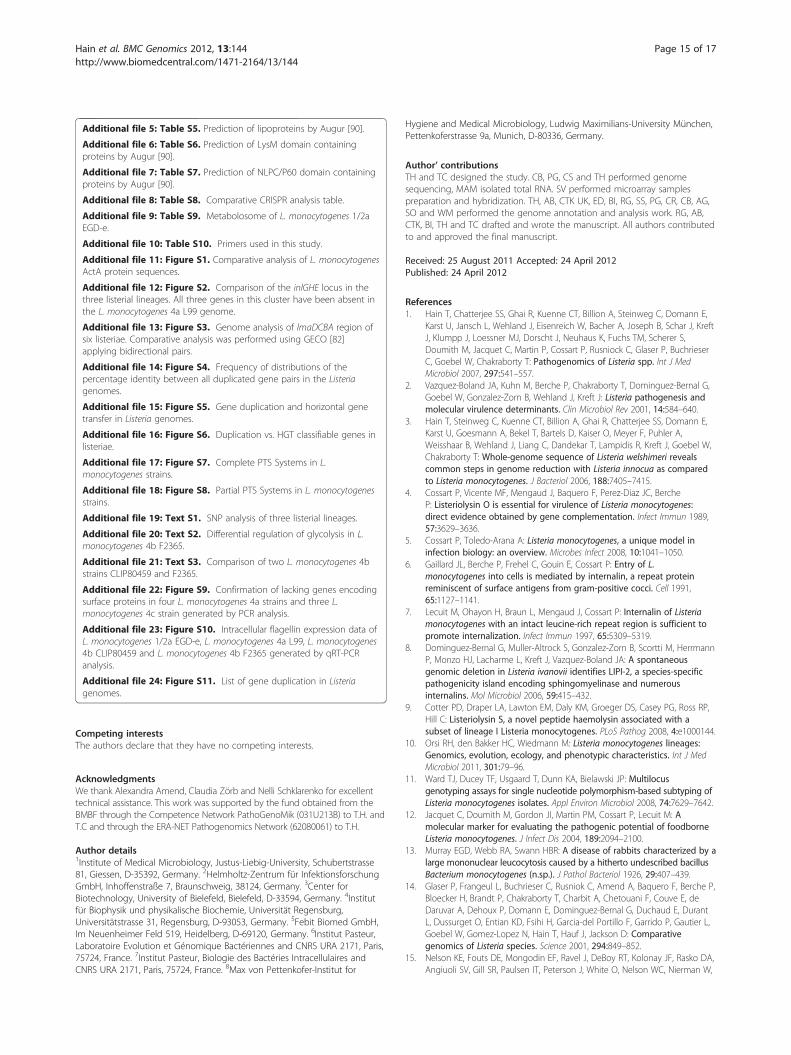
Differential growth of the three lineages and ΔlmaB andΔlmaD isogenic mutants in a mouse infection and cellinfection modelsWe observed a severe deficiency in entry of strain L99 inHeLa and Caco-2 cells as well as poor cell-to-cell trans-mission with macrophages and L929 fibroblasts whencompared to 1/2a EGD-e (data not shown). Impaired in-vasion ability of host cells may be due to lack of severalinternalin genes in the genome of strain 4a L99. It islikely that both, decreased invasive ability and strongintracellular expression of flagellar genes contribute to-wards the rapid clearance of the 4a L99 strain in in vivoexperiments in mice. Upregulation of several DNA repairgenes was also seen in strain 4a L99 compared to the
Days post-infection0 2 4 6 8 10
CF
U/s
ple
en
101
102
103
104
105
106
*
Days post-infection
0 2 4 6 8 10
CFU
/live
r
103
104
105
106
107
*
*
A
B
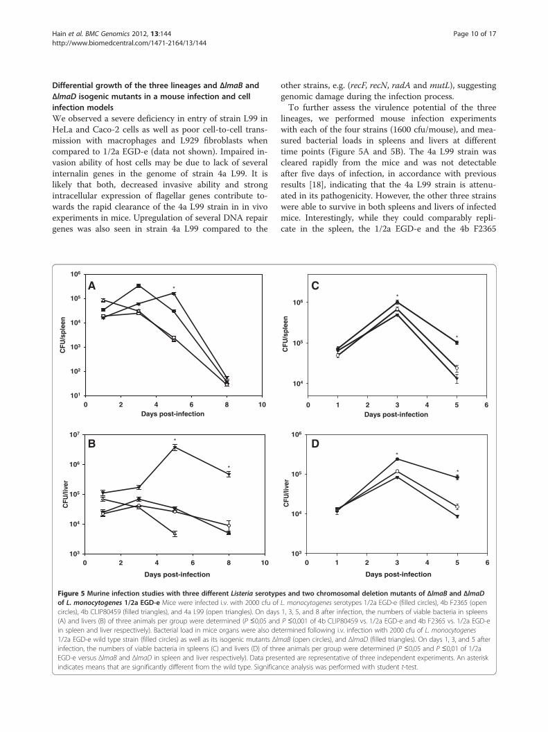
Figure 5 Murine infection studies with three different Listeria serotypof L. monocytogenes 1/2a EGD-e Mice were infected i.v. with 2000 cfu ofcircles), 4b CLIP80459 (filled triangles), and 4a L99 (open triangles). On days(A) and livers (B) of three animals per group were determined (P ≤0,05 andin spleen and liver respectively). Bacterial load in mice organs were also de1/2a EGD-e wild type strain (filled circles) as well as its isogenic mutants Δlminfection, the numbers of viable bacteria in spleens (C) and livers (D) of threEGD-e versus ΔlmaB and ΔlmaD in spleen and liver respectively). Data preseindicates means that are significantly different from the wild type. Significa
other strains, e.g. (recF, recN, radA and mutL), suggestinggenomic damage during the infection process.To further assess the virulence potential of the three
lineages, we performed mouse infection experimentswith each of the four strains (1600 cfu/mouse), and mea-sured bacterial loads in spleens and livers at differenttime points (Figure 5A and 5B). The 4a L99 strain wascleared rapidly from the mice and was not detectableafter five days of infection, in accordance with previousresults [18], indicating that the 4a L99 strain is attenu-ated in its pathogenicity. However, the other three strainswere able to survive in both spleens and livers of infectedmice. Interestingly, while they could comparably repli-cate in the spleen, the 1/2a EGD-e and the 4b F2365
Days post-infection0 1 2 3 4 5 6
CFU
/spl
een
104
105
106*
*
Days post-infection
0 1 2 3 4 5 6
CFU
/live
r
103
104
105
106
*
*
D
C
es and two chromosomal deletion mutants of ΔlmaB and ΔlmaDL. monocytogenes serotypes 1/2a EGD-e (filled circles), 4b F2365 (open1, 3, 5, and 8 after infection, the numbers of viable bacteria in spleensP ≤0,001 of 4b CLIP80459 vs. 1/2a EGD-e and 4b F2365 vs. 1/2a EGD-etermined following i.v. infection with 2000 cfu of L. monocytogenesaB (open circles), and ΔlmaD (filled triangles). On days 1, 3, and 5 aftere animals per group were determined (P ≤0,05 and P ≤0,01 of 1/2anted are representative of three independent experiments. An asterisknce analysis was performed with student t-test.
Hain et al. BMC Genomics 2012, 13:144 Page 11 of 17http://www.biomedcentral.com/1471-2164/13/144
bacterial loads in liver were significantly lower than the4b CLIP80459 strain whose counts remained signifi-cantly higher even on days five and eight post-infection.Isogenic mutants of ΔlmaB and ΔlmaD showed similarcounts in mice spleens and livers. However, bothmutants have shown a significantly lower level of growththan 1/2a EGD-e on days 3 and 5 post-infection(Figure 5C and 5D).
DiscussionWe sequenced and analysed the genomes of representa-tives of three major lineages of species L. monocytogenesto correlate gene content with (i) its wide spectrum ofpathogenic abilities, (ii) its differing properties forsurvival in the hosts, and (iii) its adaptive propertiesduring growth under extracellular conditions.
Decay of surface proteins in the virulence attenuated L.Monocytogenes 4a strainAnalysis of the 4a L99 genome revealed extensive loss ofa large number of internalins, internalin-like proteinsand other surface proteins important for invasive ability.For strain 4a L99, which was isolated from contaminatedfood in the 1950’s, it might be possible that mutationshave taken place over this lengthy time of storage underin vitro conditions. Surprisingly, a previously knownactA truncation in the 4a genomes of L99, HCC23 andM7, was also found in a higher number of lineages Istrains compared to lineage II, but not in the actA geneof another lineage III strain of 4c FSL J2-071 indicating aserotype-specific heterogeneity of ActA sequences withinthe genus Listeria. The loss of this proline-repeat inActA is correlated with lowered actin-based motility inthe cytosol. In addition, comparative nucleotide analysisindicated that the latter half of the LIPI-I pathogenicityisland in strain 4a L99 has diverged significantly fromthat of the 4b and 1/2a strain leading to a loss of theopen reading frames lmo0206 to lmo0209. Loss oflmo0206 (orfX) has been shown to confer a severegrowth effect on survival in macrophages, [20] while lossof lmo0207 has a small effect on growth in macrophagesand no data are presently available for lmo0208 andlmo0209 and their role in virulence.
Differential regulation of intracellular flagella geneexpression by strains of different lineagesHighly sensitive and widely distributed host microbe-associated microbial pattern receptors (TLRs and NLRs)continuously patrol the cell surface, endosomes and thecytosol for signs of microbial presence by sensing cellwall components, bacterial DNA, lipoproteins and flagel-lin. Ligands may be shared between the surface and thecytosolic receptors, e.g. cell wall components and flagel-lin may be sensed both by TLRs and also by cytosolic
receptors. We detected the intracellular expression ofthe flagellin gene in 1/2a EGD-e [20]. Recently, it hasbeen shown that cytosolic flagellin, expressed by L.monocytogenes strain 10403 S (serotype 1/2a) is detectedby multiple Nod-like receptors, including IPAF andNALP3, and also by a pathway involving the adaptorprotein ASC and the cytosolic DNA sensor AIM2,which is required for the formation of the inflammasome[47-49]. Detection of flagellin in the cytosol viathese pathways leads to caspase-1 mediated cleavage ofpro-IL-1B and release of active IL-1B. Mice lackingcaspase-1 or ASC are unable to mount active IL-1Bresponse to intracellular pathogens such as Shigella flex-neri and Francisella tularensis [50,51]. All strains investi-gated in this study were found to express flagellar genesin the cytosol, except for strain 4b CLIP80459. Theability to successfully downregulate flagellar (flaA) geneexpression is probably critical for evading host detectionand promoting bacterial intracellular growth. In line withthis observation, a 1/2a EGD-e chromosomal deletionmutant of the gene displayed increased survival in mouseinfection assays [52].In keeping with this finding, both strains 4b F2365 and
4a L99 displayed strong induction of several flagellargenes during intracellular growth and were more readilycleared from the host. This suggests strain-specific differ-ences in the ability to avoid host recognition can lead tolarge differences in virulence manifestation, despiteseveral commonalities in the adaptations of the lineagesto the intracellular lifestyle. Although all the strainsinvestigated in this study were able to induce all genes ofthe virulence genes cluster intracellularly, it is likely thatthere are a multitude of effects including differences invirulence gene expression, uptake of carbohydrates,membrane protein expression and flagellar biosynthesis,all of which contribute to the observed phenotypicproperties.
Effects of gene duplication events on metabolicadaptation and survival within the hostThe processes of gene duplications, horizontal genetransfer and gene loss influence the short- and long-termevolution of prokaryotic genomes. The benefits of geneduplications in the short term can be seen clearly in con-ditions of antibiotic treatment [53,54], toxin exposure[55], heavy metal stress [56,57], extreme temperatures[58], nutrient limitation [59,60] and even parasitic andsymbiotic lifestyles [54,61]. Duplications found in allListeria genomes seem to have been ancient i.e. precedespecies differentiation, with only the exception of therecent prophage duplication in L. innocua 6a CLIP11262.Classification of duplicated genes revealed several paralo-gous genes in metabolic pathways, while very fewhorizontally transferred genes could be classified at all.
Hain et al. BMC Genomics 2012, 13:144 Page 12 of 17http://www.biomedcentral.com/1471-2164/13/144
The highest numbers of gene duplications were identi-fied in the following categories: ABC transporters, PTSsystems, pentose phosphate pathway, starch and sucrosemetabolism, fructose and mannose metabolism, andcarbon fixation. Surprisingly, we found a high number ofduplicated gene paralogues involved in the regulation ofthe non-oxidative branch of the pentose phosphatepathway and in the generation of ribose-5-phospate fromribulose-5-phosphate. Under conditions of intracellulargrowth, we observed differences in the ability of thelineages to express horizontally transferred genes. 1/2aEGD-e was most successful in this regard (17 genes),followed by 4a L99 (10 genes), 4b F2365 (6 genes) and4b CLIP80459 (2 genes). Apart from the horizontallytransferred genes, differences in the expression of strain-specific genes in the cytosol were apparent (1/2a EGD-e:45; 4a L99: 49; 4b F2365 11; 4b CLIP80459: 3).PTS systems enable listeriae to utilize host carbohy-
drates, a mechanism that is essential for the intracellularsurvival. PTS systems (EII) for the utilization of fructoseand beta-glucosides, mannose and cellobiose were mostfrequently observed in the investigated Listeria genomes.Although the numbers of PTS systems are comparableamong the investigated genomes (Additional file 18:Figure S8), even a slight difference in presence/absenceof a PTS system available as an additional carbohydrateutilization mechanism may have dramatic effects onlisterial survival inside the host cytosol [61-63], specific-ally on the master regulator PrfA [61,62,64,65]. Forinstance, the pentitol PTS system in 1/2a EGD-e is notpresent in either the 4b or the 4a L99 genomes. A trans-poson insertion mutant of this system (lmo1971) hasbeen shown to have significantly attenuated growth inepithelial cells [46]. Several partial PTS systems are alsopresent in the genome (Additional file 19: Text S1).These are independently expressed intracellularly, andrepresent broadly shared and commonly regulatedsystems. In accordance, the pathogenic strain 4bCLIP80459 was found to upregulate more PTS systemsthan strain 4b F2365, which may contribute to betterintracellular survival of 4b CLIP80459.In addition to phosphorylated sugars, there are other
nitrogen and carbon sources available to intracellularbacteria, such as ethanolamine. Ethanolamine is used assubstrate and an energy supply by Salmonella entericagrown under anaerobic conditions and is suggested to beused by other bacteria [66]. A locus homologous to thatof the ethanolamine operon of S. enterica has also beendescribed in Listeria [67]. The gene organization of thelocus is not identical to the Salmonella cluster, but allthe genes of the cluster have homologous sequences inListeria (Additional file 23: Figure S10). Previous studiesidentified genes of the locus to be upregulated intracellu-larly during infection and were shown to play a critical
role for intracellular survival [46]. Our data support thisobservation and further demonstrate upregulation ofseveral genes of this locus across all three pathogeniclineages of Listeria, suggesting that the functions of thelocus are conserved. However, since the locus is alsopresent in the apathogenic L. innocua strain 6aCLIP11262, it may exemplify a general requirement ofListeria to cope with nutrient rather than a specificvirulence adaptation. Furthermore, degradation of thephagosomal membrane that traps intracellular listeriae,results in the release of ethanolamine as a byproduct andmay serve an energy source in the host cytosol.Not only the efficient recruitment of carbohydrate
substrates, but also the differential channeling throughdifferent pathways represents an important adaptionwithin the host cytosol. It has been shown that an essen-tial mechanism to counteract oxidative stress is toreroute carbohydrate flux via the pentose phosphatepathway, which is required for the biosynthesis of reduc-tive substrates rather than through glycolysis pathway[68]. Indeed, we observed that all lineages prefer to chan-nel carbohydrate flux via the pentose phosphate pathway,rather than glycolysis. In contrast to the other strains,only strain 4b F2365 was unable to downregulateglycolysis, suggesting that the inability to route sugarsefficiently via pentose phosphate contributes to the poorintracellular growth of this strain.
The CRISPR system in Listeria reveals expansion andatrophyA CRISPR (Clustered, regularly interspaced short palin-dromic repeats) locus, associated with several cas geneswas identified in the 4a L99 genome. CRISPRs are highlydivergent loci found in genomes of all archaea andseveral bacteria [69]. A CRISPR system is composed ofthe cas (CRISPR-associated) genes, a leader sequenceand arrays of direct repeats separated by non-repetitivespacer sequences resulting in a RNA-interference likeinnate phage-resistance mechanism [70]. A recent studyin Streptococcus thermophilus demonstrated howbacteria are able to integrate new spacer sequencesderived from infecting phages, directly into the CRISPRarrays, and that this ability confers phage-resistance[71]. The mechanism of resistance has also been eluci-dated [70]. Among the genomes compared in this study,only the 4a L99 genomes of L99, HCC23 and M7 pos-sesses cas genes and several CRISPR repeats. There areonly two repeats in each 4b genome, five in 1/2a EGD-ea single one in L. innocua 6a CLIP11262, but none ofthese strains harbour identifiable cas genes. In addition,a small sRNA rliB is located in the repeat region of 1/2aEGD-e and contributes to virulence in mice [72]. Wewere also able to detect a DNA sequence of a potentialprophage (PSA) using the spacers from the 4a genome.
Hain et al. BMC Genomics 2012, 13:144 Page 13 of 17http://www.biomedcentral.com/1471-2164/13/144
As prophages evolve quite rapidly, it is likely that thisacquisition is a recent event.
Distinct role of intracellularly upregulated phage genes invirulence of listerial strainsThe four L. monocytogenes strains have different num-bers of prophage genes (1/2a EGD-e: 79; 4a L99: 191; 4bCLIP80459: 16 and 4b F2365: 16) distributed in differentloci. Regardless of location and lineage, all strainsexpressed several prophage genes within the infectedhost cell. However, only a single locus, the lma locus isconserved across the three lineages and is also inducedduring infection. The role of prophage genes in thevirulence of Listeria has not been examined in detail. Weshow that chromosomal deletion mutants of two genesin this locus (lmaB and lmaD) resulted in growth reduc-tion of 1/2a EGD-e in a murine infection model.Although the underlying mechanisms leading to the atte-nuated phenotypes remain unclear, a recent studyrevealed that prophage diversification represents anessential mechanism for short-term genome evolutionwithin the species L. monocytogenes [73,74] and is sub-ject of further investigation.
ConclusionListeria monocytogenes is arguably one of the bestcharacterized pathogens and has been established as anunparalleled model microorganism in infection biology.Detailed understanding of differences in virulence of thethree major lineages of Listeria provides us with invalu-able information about evolutionary adaptation of thispathogen. Here we used comparative genomics andwhole-genome based transcriptome analysis of strainsfrom all lineages to obtain a comprehensive view as tohow these strains have evolutionarily diverged. Thisapproach suggests that (i) reductive evolution of strainsof serotype 4a such as L99, HCC23 and M7 is the majorforce driving the attenuated phenotype, (ii) acquisitionand adaptation of prophage genes and metabolic sys-tems, respectively, identify novel virulence-associatedfactors of listeriae and (iii) listeriae avoid detection andsubsequent immune response of the host via downregu-lation of surface structures and by differences in intracel-lular expression of flagellar genes.
MethodsStrains and growth conditionsFour L. monocytogenes strains were used in the study, L.monocytogenes 1/2a EGD-e [14], L. monocytogenes 4aL99 [18], L. monocytogenes 4b CLIP80459 [17], L. mono-cytogenes 4b F2365 [15] and chromosomal deletionmutants of L. monocytogenes 1/2a EGD-e ΔlmaB andΔlmaD. Bacteria were grown in brain heart infusion(BHI) broth (Difco) at 37°C with shaking. For further
comparative genomic analysis L. monocytogenes 4aHCC23 [37] L. monocytogenes 4a M7 [38] and L. mono-cytogenes 4c FSL J2-071) (Listeria monocytogenesSequencing Project, Broad Institute of Harvard andMIT; http://www.broad.mit.edu) was used.
Genome sequencing and annotationIn brief, genome sequencing L. monocytogenes 4a L99was performed on ABI PRISM 3100 or 3730xl GeneticAnalyzers (Applied Biosystems). Whole genome shotgunsequencing was performed by LGC (Berlin, Germany).Sequence data were analysed and assembled usingPhred/Phrap/Consed [75,76]. A total number of 27,637sequences of shotgun libraries, 1684 fosmid and 671PCR gap closure sequences were assembled by the Phrapsoftware resulting in a ~6.7-fold coverage. Genomeannotation was performed as previously described [3].Genome sequencing of L. monocytogenes 4b CLIP80459
was performed using the conventional whole genomeshotgun strategy [77,78]. One library (2–3 kb inserts) wasgenerated by random mechanical shearing of genomicDNA and cloning into pcDNA-2.1 (Life technologies) andrecombinant plasmids were used as templates for cyclesequencing reactions. Samples were loaded on capillaryautomatic 3700 and 3730 DNA sequencers (AppliedBiosystems). In an initial step 35,610 sequences wereassembled into 361 contigs using the Phred/Phrap/Consedsoftware [75,76]. CAAT-Box [79] was used to predictlinks between contigs. 379 PCR products amplified fromL. monocytogenes CLIP80459 chromosomal DNA astemplate were used to fill gaps and to re-sequence lowquality regions. Final assembly resulted in a ~7.8-foldcoverage. Genome annotation was performed as previouslydescribed [14].
Alignment of the virulence gene clusterThe alignment was performed using MAVID [80] afterextracting the virulence gene cluster of all genomes. Theplot was created using VISTA [81].
ActA repeat analysisAvailable ActA protein sequences for all L. monocytogenesstrains were retrieved from GenBank (http://www.ncbi.nlm.nih.gov/Genbank/). Only sequences that contained atleast 500 amino acids (reference strain 1/2a EGD-e ActA:639 amino acids) were downloaded (774 sequences). It waspossible to assign a lineage to only 386 ActA sequences.Duplicates with identical length, strain and sequence werealso removed, leaving a total of 218 sequences for theanalysis. These were aligned using ClustalW and thealignment of repeat regions was examined manually.
Hain et al. BMC Genomics 2012, 13:144 Page 14 of 17http://www.biomedcentral.com/1471-2164/13/144
Single nucleotide polymorphismsSingle nucleotide Polymorphisms (SNPs) were detectedby the MUMmer [25] and SNPs were mapped to codingregions using PERL scripts. The SNP-density per genenormalized by gene length was calculated and the datawere visualized in GenomeViz [26].
CRISPR repeats analysisComparative visualization of the CRISPR related genomeloci was performed by GECO [82]. CRISPR repeats wereidentified using the PILER-CR software [83]. Subsequentanalysis and visualization of repeat footprints was per-formed using BLAST and ACT [84].
Horizontal gene transfer and gene duplicationsHorizontally transferred genes were detected using SIGI[85] and SIGI-HMM [86]. Duplicated genes were identi-fied using BLAST cut-offs of at least 40% identity and80% coverage considering both sequences.
Cell culture and infection modelAll cell culture experiments were performed as describedby Chatterjee and colleges [20].
MicroarraysFor each of the four strains of the study, a genome-widecustom microarray chip was designed and implementedusing the Geniom One platform from Febit BiomedGmbH, Germany. All transcriptome studies wereperformed with this platform. Complete details of theprotocols are provided in the ArrayExpress database(http://www.ebi.ac.uk/microarray-as/ae/). Data were back-ground corrected and then normalized using quantilenormalization [87]. Pearson’s correlation coefficients wereused to assess reproducibility within at least two technicaland three biological replicates (r2> =0.94 in all cases). Thesignificance analysis of microarrays (SAM) program wasused to analyze the data [88] as an unpaired response.
Construction of the deletion mutants ΔlmaB and ΔlmaDChromosomal in frame deletion mutants of L. monocyto-genes 1/2a EGD-e ΔlmaB and ΔlmaD were constructed bygenerating the 5′ (with primers P1 and P2) and the 3′(with primers P3 and P4) flanking region of the gene con-cerned. Primers used to generate the flanking regions areshown (Additional file 24: Table S11). The purified PCRfragments of 5′ and 3′ flanking regions were amplifiedusing primer P1 and P4, ligated into pCRII (Life technolo-gies) and transformed into E. coli InvαF’ electrocompetentcells (Life technologies). Subsequently, the vector wasdigested with restriction enzyme EcoRI and ligated into thetemperature sensitive suicide vector pAUL-A whichwas digested with the same enzymes and transformed intoE. coli InvαF’ electrocompetent cells. Plasmid DNA of
pAUL-A bearing the fragment was isolated from therecombinants and used to transform L. monocytogenesEGD-e to generate the chromosomal deletion mutants asdescribed in detail by Schaeferkordt et al. [89]. Thedeletion in the gene concerned was identified by PCR andconfirmed by sequencing the PCR fragment using primersP1 and P4.
Murine infection assayPrimary infection with L. monocytogenes serotypes andmutants was performed by intravenous injection ofviable bacteria in a volume of 0.2 ml of PBS. Bacterialgrowth in spleens and livers was determined by plating10-fold serial dilutions of organ homogenates on BHIafter several days. The detection limit of this procedurewas 102 CFU per organ. Colonies were counted after24 h of incubation at 37°C. Six- to eight-week-old femaleBALB/c mice, purchased from Harlan Winkelmann(Borchen, Germany), were used in all experiments.
Ethics statementThis study was carried out in strict accordance with theregulation of the National Protection Animal Act (§7-9aTierschutzgesetz). The protocol was approved by thelocal Committee on the Ethics of Animal Experiments(Regierungsbezirk Mittelhessen) and permission wasgiven by the local authority (RegierungspraesidiumGiessen, Permit Number: GI 15/5-Nr.63/2007).
Statistical data analysis of infection experimentsAll infection experiments were performed a minimum ofthree times. Significant differences between two valueswere compared with a paired Student’s t-test. Valueswere considered significantly different when the p valuewas less than 0.05 (p< 0.05).
Nucleotide sequence and microarray accession numberThe genome sequences have been deposited in the EMBLdatabase with accession numbers FM211688 for L.monocytogenes 4a L99 and FM242711 for L. monocytogenes4b CLIP80459 respectively. The microarray data havebeen submitted to ArrayExpress with the accessionnumber E-MEXP-1947.
Additional files
Additional file 1: Table S1. Nucleotide analysis of actA repeats ofListeria.
Additionaf file 2: Table S2. Prediction of LRR region containingproteins by Augur [90].
Additional file 3: Table S2. x Prediction of proteins containing GWmodules by Augur [90].
Additional file 4: Table S4. Prediction of LPXTG motif harbouringproteins by Augur [90].
Hain et al. BMC Genomics 2012, 13:144 Page 15 of 17http://www.biomedcentral.com/1471-2164/13/144
Additional file 5: Table S5. Prediction of lipoproteins by Augur [90].
Additional file 6: Table S6. Prediction of LysM domain containingproteins by Augur [90].
Additional file 7: Table S7. Prediction of NLPC/P60 domain containingproteins by Augur [90].
Additional file 8: Table S8. Comparative CRISPR analysis table.
Additional file 9: Table S9. Metabolosome of L. monocytogenes 1/2aEGD-e.
Additional file 10: Table S10. Primers used in this study.
Additional file 11: Figure S1. Comparative analysis of L. monocytogenesActA protein sequences.
Additional file 12: Figure S2. Comparison of the inlGHE locus in thethree listerial lineages. All three genes in this cluster have been absent inthe L. monocytogenes 4a L99 genome.
Additional file 13: Figure S3. Genome analysis of lmaDCBA region ofsix listeriae. Comparative analysis was performed using GECO [82]applying bidirectional pairs.
Additional file 14: Figure S4. Frequency of distributions of thepercentage identity between all duplicated gene pairs in the Listeriagenomes.
Additional file 15: Figure S5. Gene duplication and horizontal genetransfer in Listeria genomes.
Additional file 16: Figure S6. Duplication vs. HGT classifiable genes inlisteriae.
Additional file 17: Figure S7. Complete PTS Systems in L.monocytogenes strains.
Additional file 18: Figure S8. Partial PTS Systems in L. monocytogenesstrains.
Additional file 19: Text S1. SNP analysis of three listerial lineages.
Additional file 20: Text S2. Differential regulation of glycolysis in L.monocytogenes 4b F2365.
Additional file 21: Text S3. Comparison of two L. monocytogenes 4bstrains CLIP80459 and F2365.
Additional file 22: Figure S9. Confirmation of lacking genes encodingsurface proteins in four L. monocytogenes 4a strains and three L.monocytogenes 4c strain generated by PCR analysis.
Additional file 23: Figure S10. Intracellular flagellin expression data ofL. monocytogenes 1/2a EGD-e, L. monocytogenes 4a L99, L. monocytogenes4b CLIP80459 and L. monocytogenes 4b F2365 generated by qRT-PCRanalysis.
Additional file 24: Figure S11. List of gene duplication in Listeriagenomes.
Competing interestsThe authors declare that they have no competing interests.
AcknowledgmentsWe thank Alexandra Amend, Claudia Zörb and Nelli Schklarenko for excellenttechnical assistance. This work was supported by the fund obtained from theBMBF through the Competence Network PathoGenoMik (031U213B) to T.H. andT.C and through the ERA-NET Pathogenomics Network (62080061) to T.H.
Author details1Institute of Medical Microbiology, Justus-Liebig-University, Schubertstrasse81, Giessen, D-35392, Germany. 2Helmholtz-Zentrum für InfektionsforschungGmbH, Inhoffenstraße 7, Braunschweig, 38124, Germany. 3Center forBiotechnology, University of Bielefeld, Bielefeld, D-33594, Germany. 4Institutfür Biophysik und physikalische Biochemie, Universität Regensburg,Universitätstrasse 31, Regensburg, D-93053, Germany. 5Febit Biomed GmbH,Im Neuenheimer Feld 519, Heidelberg, D-69120, Germany. 6Institut Pasteur,Laboratoire Evolution et Génomique Bactériennes and CNRS URA 2171, Paris,75724, France. 7Institut Pasteur, Biologie des Bactéries Intracellulaires andCNRS URA 2171, Paris, 75724, France. 8Max von Pettenkofer-Institut for
Hygiene and Medical Microbiology, Ludwig Maximilians-University München,Pettenkoferstrasse 9a, Munich, D-80336, Germany.
Author’ contributionsTH and TC designed the study. CB, PG, CS and TH performed genomesequencing, MAM isolated total RNA. SV performed microarray samplespreparation and hybridization. TH, AB, CTK UK, ED, BI, RG, SS, PG, CR, CB, AG,SO and WM performed the genome annotation and analysis work. RG, AB,CTK, BI, TH and TC drafted and wrote the manuscript. All authors contributedto and approved the final manuscript.
Received: 25 August 2011 Accepted: 24 April 2012Published: 24 April 2012
References1. Hain T, Chatterjee SS, Ghai R, Kuenne CT, Billion A, Steinweg C, Domann E,
Karst U, Jansch L, Wehland J, Eisenreich W, Bacher A, Joseph B, Schar J, KreftJ, Klumpp J, Loessner MJ, Dorscht J, Neuhaus K, Fuchs TM, Scherer S,Doumith M, Jacquet C, Martin P, Cossart P, Rusniock C, Glaser P, BuchrieserC, Goebel W, Chakraborty T: Pathogenomics of Listeria spp. Int J MedMicrobiol 2007, 297:541–557.
2. Vazquez-Boland JA, Kuhn M, Berche P, Chakraborty T, Dominguez-Bernal G,Goebel W, Gonzalez-Zorn B, Wehland J, Kreft J: Listeria pathogenesis andmolecular virulence determinants. Clin Microbiol Rev 2001, 14:584–640.
3. Hain T, Steinweg C, Kuenne CT, Billion A, Ghai R, Chatterjee SS, Domann E,Karst U, Goesmann A, Bekel T, Bartels D, Kaiser O, Meyer F, Puhler A,Weisshaar B, Wehland J, Liang C, Dandekar T, Lampidis R, Kreft J, Goebel W,Chakraborty T: Whole-genome sequence of Listeria welshimeri revealscommon steps in genome reduction with Listeria innocua as comparedto Listeria monocytogenes. J Bacteriol 2006, 188:7405–7415.
4. Cossart P, Vicente MF, Mengaud J, Baquero F, Perez-Diaz JC, BercheP: Listeriolysin O is essential for virulence of Listeria monocytogenes:direct evidence obtained by gene complementation. Infect Immun 1989,57:3629–3636.
5. Cossart P, Toledo-Arana A: Listeria monocytogenes, a unique model ininfection biology: an overview. Microbes Infect 2008, 10:1041–1050.
6. Gaillard JL, Berche P, Frehel C, Gouin E, Cossart P: Entry of L.monocytogenes into cells is mediated by internalin, a repeat proteinreminiscent of surface antigens from gram-positive cocci. Cell 1991,65:1127–1141.
7. Lecuit M, Ohayon H, Braun L, Mengaud J, Cossart P: Internalin of Listeriamonocytogenes with an intact leucine-rich repeat region is sufficient topromote internalization. Infect Immun 1997, 65:5309–5319.
8. Dominguez-Bernal G, Muller-Altrock S, Gonzalez-Zorn B, Scortti M, HerrmannP, Monzo HJ, Lacharme L, Kreft J, Vazquez-Boland JA: A spontaneousgenomic deletion in Listeria ivanovii identifies LIPI-2, a species-specificpathogenicity island encoding sphingomyelinase and numerousinternalins. Mol Microbiol 2006, 59:415–432.
9. Cotter PD, Draper LA, Lawton EM, Daly KM, Groeger DS, Casey PG, Ross RP,Hill C: Listeriolysin S, a novel peptide haemolysin associated with asubset of lineage I Listeria monocytogenes. PLoS Pathog 2008, 4:e1000144.
10. Orsi RH, den Bakker HC, Wiedmann M: Listeria monocytogenes lineages:Genomics, evolution, ecology, and phenotypic characteristics. Int J MedMicrobiol 2011, 301:79–96.
11. Ward TJ, Ducey TF, Usgaard T, Dunn KA, Bielawski JP: Multilocusgenotyping assays for single nucleotide polymorphism-based subtyping ofListeria monocytogenes isolates. Appl Environ Microbiol 2008, 74:7629–7642.
12. Jacquet C, Doumith M, Gordon JI, Martin PM, Cossart P, Lecuit M: Amolecular marker for evaluating the pathogenic potential of foodborneListeria monocytogenes. J Infect Dis 2004, 189:2094–2100.
13. Murray EGD, Webb RA, Swann HBR: A disease of rabbits characterized by alarge mononuclear leucocytosis caused by a hitherto undescribed bacillusBacterium monocytogenes (n.sp.). J Pathol Bacteriol 1926, 29:407–439.
14. Glaser P, Frangeul L, Buchrieser C, Rusniok C, Amend A, Baquero F, Berche P,Bloecker H, Brandt P, Chakraborty T, Charbit A, Chetouani F, Couve E, deDaruvar A, Dehoux P, Domann E, Dominguez-Bernal G, Duchaud E, DurantL, Dussurget O, Entian KD, Fsihi H, Garcia-del Portillo F, Garrido P, Gautier L,Goebel W, Gomez-Lopez N, Hain T, Hauf J, Jackson D: Comparativegenomics of Listeria species. Science 2001, 294:849–852.
15. Nelson KE, Fouts DE, Mongodin EF, Ravel J, DeBoy RT, Kolonay JF, Rasko DA,Angiuoli SV, Gill SR, Paulsen IT, Peterson J, White O, Nelson WC, Nierman W,

Hain et al. BMC Genomics 2012, 13:144 Page 16 of 17http://www.biomedcentral.com/1471-2164/13/144
Beanan MJ, Brinkac LM, Daugherty SC, Dodson RJ, Durkin AS, Madupu R,Haft DH, Selengut J, Van Aken S, Khouri H, Fedorova N, Forberger H, Tran B,Kathariou S, Wonderling LD, Uhlich GA, Bayles DO, Luchansky JB, Fraser CM:Whole genome comparisons of serotype 4b and 1/2a strains of the food-borne pathogen Listeria monocytogenes reveal new insights into the coregenome components of this species. Nucleic Acids Res 2004, 32:2386–2395.
16. Nightingale KK, Milillo SR, Ivy RA, Ho AJ, Oliver HF, Wiedmann M: Listeriamonocytogenes F2365 carries several authentic mutations potentiallyleading to truncated gene products, including inlB, and demonstratesatypical phenotypic characteristics. J Food Prot 2007, 70:482–488.
17. de Valk H, Vaillant V, Jacquet C, Rocourt J, Le Querrec F, Stainer F,Quelquejeu N, Pierre O, Pierre V, Desenclos JC, Goulet V: Two consecutivenationwide outbreaks of Listeriosis in France, October 1999-February2000. Am J Epidemiol 2001, 154:944–950.
18. Chakraborty T, Ebel F, Wehland J, Dufrenne J, Notermans S: Naturallyoccurring virulence-attenuated isolates of Listeria monocytogenes capableof inducing long term protection against infection by virulent strains ofhomologous and heterologous serotypes. FEMS Immunol Med Microbiol1994, 10:1–9.
19. Camejo A, Buchrieser C, Couve E, Carvalho F, Reis O, Ferreira P, Sousa S,Cossart P, Cabanes D: In vivo transcriptional profiling of Listeriamonocytogenes and mutagenesis identify new virulence factors involvedin infection. PLoS Pathog 2009, 5:e1000449.
20. Chatterjee SS, Hossain H, Otten S, Kuenne C, Kuchmina K, Machata S,Domann E, Chakraborty T, Hain T: Intracellular gene expression profile ofListeria monocytogenes. Infect Immun 2006, 74:1323–1338.
21. Joseph B, Goebel W: Life of Listeria monocytogenes in the host cells’cytosol. Microbes Infect 2007, 9:1188–1195.
22. Chen Y, Zhang W, Knabel SJ: Multi-virulence-locus sequence typingidentifies single nucleotide polymorphisms which differentiate epidemicclones and outbreak strains of Listeria monocytogenes. J Clin Microbiol2007, 45:835–846.
23. Canchaya C, Fournous G, Brussow H: The impact of prophages onbacterial chromosomes. Mol Microbiol 2004, 53:9–18.
24. Dorscht J, Klumpp J, Bielmann R, Schmelcher M, Born Y, Zimmer M,Calendar R, Loessner MJ: Comparative genome analysis of Listeriabacteriophages reveals extensive mosaicism, programmed translationalframeshifting, and a novel prophage insertion site. J Bacteriol 2009,191:7206–7215.
25. Kurtz S, Phillippy A, Delcher AL, Smoot M, Shumway M, Antonescu C,Salzberg SL: Versatile and open software for comparing large genomes.Genome Biol 2004, 5:R12.
26. Ghai R, Hain T, Chakraborty T: GenomeViz: visualizing microbial genomes.BMC Bioinforma 2004, 5:198.
27. Doumith M, Cazalet C, Simoes N, Frangeul L, Jacquet C, Kunst F, Martin P,Cossart P, Glaser P, Buchrieser C: New aspects regarding evolution andvirulence of Listeria monocytogenes revealed by comparative genomicsand DNA arrays. Infect Immun 2004, 72:1072–1083.
28. Sokolovic Z, Schuller S, Bohne J, Baur A, Rdest U, Dickneite C, Nichterlein T,Goebel W: Differences in virulence and in expression of PrfA and PrfA-regulated virulence genes of Listeria monocytogenes strains belonging toserogroup 4. Infect Immun 1996, 64:4008–4019.
29. Jia Y, Nightingale KK, Boor KJ, Ho A, Wiedmann M, McGann P: Distributionof internalin gene profiles of Listeria monocytogenes isolates fromdifferent sources associated with phylogenetic lineages. FoodbornePathog Dis 2007, 4:222–232.
30. Raffelsbauer D, Bubert A, Engelbrecht F, Scheinpflug J, Simm A, Hess J,Kaufmann SH, Goebel W: The gene cluster inlC2DE of Listeriamonocytogenes contains additional new internalin genes and isimportant for virulence in mice. Mol Gen Genet 1998, 260:144–158.
31. Linden SK, Bierne H, Sabet C, Png CW, Florin TH, McGuckin MA, Cossart P:Listeria monocytogenes internalins bind to the human intestinal mucinMUC2. Arch Microbiol 2008, 190:101–104.
32. Domann E, Zechel S, Lingnau A, Hain T, Darji A, Nichterlein T, Wehland J,Chakraborty T: Identification and characterization of a novel PrfA-regulated gene in Listeria monocytogenes whose product, IrpA, is highlyhomologous to internalin proteins, which contain leucine-rich repeats.Infect Immun 1997, 65:101–109.
33. Engelbrecht F, Chun SK, Ochs C, Hess J, Lottspeich F, Goebel W, Sokolovic Z:A new PrfA-regulated gene of Listeria monocytogenes encoding a small,secreted protein which belongs to the family of internalins. Mol Microbiol1996, 21:823–837.
34. Gouin E, dib-Conquy M, Balestrino D, Nahori MA, Villiers V, Colland F, DramsiS, Dussurget O, Cossart P: The Listeria monocytogenes InlC proteininterferes with innate immune responses by targeting the I{kappa}Bkinase subunit IKK{alpha}. Proc Natl Acad Sci U S A 2010, 107:17333–17338.
35. Rajabian T, Gavicherla B, Heisig M, Muller-Altrock S, Goebel W, Gray-OwenSD, Ireton K: The bacterial virulence factor InlC perturbs apical celljunctions and promotes cell-to-cell spread of Listeria. Nat Cell Biol 2009,11:1212–1218.
36. Dramsi S, Dehoux P, Lebrun M, Goossens PL, Cossart P: Identification offour new members of the internalin multigene family of Listeriamonocytogenes EGD. Infect Immun 1997, 65:1615–1625.
37. Steele CL, Donaldson JR, Paul D, Banes MM, Arick T, Bridges SM, LawrenceML: Genome sequence of lineage III Listeria monocytogenes strain HCC23.J Bacteriol 2011, 193:3679–3680.
38. Chen J, Xia Y, Cheng C, Fang C, Shan Y, Jin G, Fang W: Genome sequenceof the nonpathogenic Listeria monocytogenes serovar 4a strain M7. JBacteriol 2011, 193:5019–5020.
39. Cabanes D, Dussurget O, Dehoux P, Cossart P: Auto, a surface associatedautolysin of Listeria monocytogenes required for entry into eukaryoticcells and virulence. Mol Microbiol 2004, 51:1601–1614.
40. Cabanes D, Sousa S, Cebria A, Lecuit M, Garcia-del Portillo F, Cossart P: Gp96is a receptor for a novel Listeria monocytogenes virulence factor, Vip, asurface protein. EMBO J 2005, 24:2827–2838.
41. Milohanic E, Jonquieres R, Glaser P, Dehoux P, Jacquet C, Berche P, CossartP, Gaillard JL: Sequence and binding activity of the autolysin-adhesin Amifrom epidemic Listeria monocytogenes 4b. Infect Immun 2004, 72:4401–4409.
42. Milohanic E, Jonquieres R, Cossart P, Berche P, Gaillard JL: The autolysinAmi contributes to the adhesion of Listeria monocytogenes to eukaryoticcells via its cell wall anchor. Mol Microbiol 2001, 39:1212–1224.
43. Schaferkordt S, Chakraborty T: Identification, cloning, and characterizationof the Ima operon, whose gene products are unique to Listeriamonocytogenes. J Bacteriol 1997, 179:2707–2716.
44. Severino P, Dussurget O, Vencio RZ, Dumas E, Garrido P, Padilla G, PiveteauP, Lemaitre JP, Kunst F, Glaser P, Buchrieser C: Comparative transcriptomeanalysis of Listeria monocytogenes strains of the two major lineagesreveals differences in virulence, cell wall, and stress response. ApplEnviron Microbiol 2007, 73:6078–6088.
45. Gohmann S, Leimeister-Wachter M, Schiltz E, Goebel W, Chakraborty T:Characterization of a Listeria monocytogenes-specific protein capable ofinducing delayed hypersensitivity in Listeria-immune mice. Mol Microbiol1990, 4:1091–1099.
46. Joseph B, Przybilla K, Stuhler C, Schauer K, Slaghuis J, Fuchs TM, Goebel W:Identification of Listeria monocytogenes genes contributing tointracellular replication by expression profiling and mutant screening. JBacteriol 2006, 188:556–568.
47. Sauer JD, Witte CE, Zemansky J, Hanson B, Lauer P, Portnoy DA: Listeriamonocytogenes triggers AIM2-mediated pyroptosis upon infrequentbacteriolysis in the macrophage cytosol. Cell Host Microbe 2010, 7:412–419.
48. Warren SE, Mao DP, Rodriguez AE, Miao EA, Aderem A: Multiple Nod-likereceptors activate caspase 1 during Listeria monocytogenes infection. JImmunol 2008, 180:7558–7564.
49. Wu J, Fernandes-Alnemri T, Alnemri ES: Involvement of the AIM2, NLRC4,and NLRP3 inflammasomes in caspase-1 activation by Listeriamonocytogenes. J Clin Immunol 2010, 30:693–702.
50. Mariathasan S, Weiss DS, Dixit VM, Monack DM: Innate immunity againstFrancisella tularensis is dependent on the ASC/caspase-1 axis. J Exp Med2005, 202:1043–1049.
51. Sansonetti PJ, Phalipon A, Arondel J, Thirumalai K, Banerjee S, Akira S,Takeda K, Zychlinsky A: Caspase-1 activation of IL-1beta and IL-18 areessential for Shigella flexneri-induced inflammation. Immunity 2000,12:581–590.
52. Bigot A, Pagniez H, Botton E, Frehel C, Dubail I, Jacquet C, Charbit A,Raynaud C: Role of FliF and FliI of Listeria monocytogenes in flagellarassembly and pathogenicity. Infect Immun 2005, 73:5530–5539.
53. Koch AL: Evolution of antibiotic resistance gene function. Microbiol Rev1981, 45:355–378.
54. Romero D, Palacios R: Gene amplification and genomic plasticity inprokaryotes. Annu Rev Genet 1997, 31:91–111.
55. Reinbothe S, Ortel B, Parthier B: Overproduction by gene amplification ofthe multifunctional arom protein confers glyphosate tolerance to aplastid-free mutant of Euglena gracilis. Mol Gen Genet 1993, 239:416–424.

Hain et al. BMC Genomics 2012, 13:144 Page 17 of 17http://www.biomedcentral.com/1471-2164/13/144
56. Kondrashov FA, Rogozin IB, Wolf YI, Koonin EV: Selection in the evolutionof gene duplications. Genome Biol 2002, 3:RESEARCH0008.
57. van Hoof NA, Hassinen VH, Hakvoort HW, Ballintijn KF, Schat H, Verkleij JA,Ernst WH, Karenlampi SO, Tervahauta AI: Enhanced copper tolerance inSilene vulgaris (Moench) Garcke populations from copper mines isassociated with increased transcript levels of a 2b-type metallothioneingene. Plant Physiol 2001, 126:1519–1526.
58. Riehle MM, Bennett AF, Long AD: Genetic architecture of thermaladaptation in Escherichia coli. Proc Natl Acad Sci U S A 2001, 98:525–530.
59. Brown CJ, Todd KM, Rosenzweig RF: Multiple duplications of yeast hexosetransport genes in response to selection in a glucose-limitedenvironment. Mol Biol Evol 1998, 15:931–942.
60. Sonti RV, Roth JR: Role of gene duplications in the adaptation ofSalmonella typhimurium to growth on limiting carbon sources. Genetics1989, 123:19–28.
61. Lai CY, Baumann L, Baumann P: Amplification of trpEG: adaptation ofBuchnera aphidicola to an endosymbiotic association with aphids. ProcNatl Acad Sci U S A 1994, 91:3819–3823.
62. Stoll R, Mertins S, Joseph B, Muller-Altrock S, Goebel W: Modulation of PrfAactivity in Listeria monocytogenes upon growth in different culturemedia. Microbiology 2008, 154:3856–3876.
63. Stoll R, Goebel W: The major PEP-phosphotransferase systems (PTSs) forglucose, mannose and cellobiose of Listeria monocytogenes, and theirsignificance for extra- and intracellular growth. Microbiology 2010,156:1069–1083.
64. Ake FM, Joyet P, Deutscher J, Milohanic E: Mutational analysis of glucosetransport regulation and glucose-mediated virulence gene repression inListeria monocytogenes. Mol Microbiol 2011, .
65. Joseph B, Mertins S, Stoll R, Schar J, Umesha KR, Luo Q, Muller-Altrock S,Goebel W: Glycerol metabolism and PrfA activity in Listeriamonocytogenes. J Bacteriol 2008, 190:5412–5430.
66. Cannon GC, Bradburne CE, Aldrich HC, Baker SH, Heinhorst S, Shively JM:Microcompartments in prokaryotes: carboxysomes and relatedpolyhedra. Appl Environ Microbiol 2001, 67:5351–5361.
67. Buchrieser C, Rusniok C, Kunst F, Cossart P, Glaser P: Comparison of thegenome sequences of Listeria monocytogenes and Listeria innocua: clues forevolution and pathogenicity. FEMS Immunol Med Microbiol 2003, 35:207–213.
68. Ralser M, Wamelink MM, Kowald A, Gerisch B, Heeren G, Struys EA, Klipp E,Jakobs C, Breitenbach M, Lehrach H, Krobitsch S: Dynamic rerouting of thecarbohydrate flux is key to counteracting oxidative stress. J Biol 2007, 6:10.
69. Makarova KS, Haft DH, Barrangou R, Brouns SJ, Charpentier E, Horvath P,Moineau S, Mojica FJ, Wolf YI, Yakunin AF, van der OJ, Koonin EV: Evolutionand classification of the CRISPR-Cas systems. Nat Rev Microbiol 2011,9:467–477.
70. Tang TH, Polacek N, Zywicki M, Huber H, Brugger K, Garrett R, Bachellerie JP,Huttenhofer A: Identification of novel non-coding RNAs as potentialantisense regulators in the archaeon Sulfolobus solfataricus. Mol Microbiol2005, 55:469–481.
71. Barrangou R, Fremaux C, Deveau H, Richards M, Boyaval P, Moineau S,Romero DA, Horvath P: CRISPR provides acquired resistance againstviruses in prokaryotes. Science 2007, 315:1709–1712.
72. Toledo-Arana A, Dussurget O, Nikitas G, Sesto N, Guet-Revillet H, BalestrinoD, Loh E, Gripenland J, Tiensuu T, Vaitkevicius K, Barthelemy M, VergassolaM, Nahori MA, Soubigou G, Regnault B, Coppee JY, Lecuit M, Johansson J,Cossart P: The Listeria transcriptional landscape from saprophytism tovirulence. Nature 2009, 459:950–956.
73. Orsi RH, Borowsky ML, Lauer P, Young SK, Nusbaum C, Galagan JE, BirrenBW, Ivy RA, Sun Q, Graves LM, Swaminathan B, Wiedmann M: Short-termgenome evolution of Listeria monocytogenes in a non-controlledenvironment. BMC Genomics 2008, 9:539.
74. Verghese B, Lok M, Wen J, Alessandria V, Chen Y, Kathariou S, Knabel S:comK prophage junction fragments as markers for Listeriamonocytogenes genotypes unique to individual meat and poultryprocessing plants and a model for rapid niche-specific adaptation, biofilmformation, and persistence. Appl Environ Microbiol 2011, 77:3279–3292.
75. Ewing B, Hillier L, Wendl MC, Green P: Base-calling of automatedsequencer traces using phred. I. Accuracy assessment. Genome Res 1998,8:175–185.
76. Gordon D, Abajian C, Green P: Consed: a graphical tool for sequencefinishing. Genome Res 1998, 8:195–202.
77. Fleischmann RD, Adams MD, White O, Clayton RA, Kirkness EF, Kerlavage AR,Bult CJ, Tomb JF, Dougherty BA, Merrick JM: Whole-genome random
sequencing and assembly of Haemophilus influenzae Rd. Science 1995,269:496–512.
78. Frangeul L, Nelson KE, Buchrieser C, Danchin A, Glaser P, Kunst F: Cloningand assembly strategies in microbial genome projects. Microbiology 1999,145(Pt 10):2625–2634.
79. Frangeul L, Glaser P, Rusniok C, Buchrieser C, Duchaud E, Dehoux P, Kunst F:CAAT-Box, Contigs-Assembly and Annotation Tool-Box for genomesequencing projects. Bioinformatics 2004, 20:790–797.
80. Bray N, Pachter L: MAVID: constrained ancestral alignment of multiplesequences. Genome Res 2004, 14:693–699.
81. Frazer KA, Pachter L, Poliakov A, Rubin EM, Dubchak I: VISTA: computationaltools for comparative genomics. Nucleic Acids Res 2004, 32:W273–W279.
82. Kuenne CT, Ghai R, Chakraborty T, Hain T: GECO–linear visualization forcomparative genomics. Bioinformatics 2007, 23:125–126.
83. Edgar RC: PILER-CR: fast and accurate identification of CRISPR repeats.BMC Bioinforma 2007, 8:18.
84. Carver TJ, Rutherford KM, Berriman M, Rajandream MA, Barrell BG, Parkhill J:ACT: the Artemis Comparison Tool. Bioinformatics 2005, 21:3422–3423.
85. Merkl R: SIGI: score-based identification of genomic islands. BMCBioinforma 2004, 5:22.
86. Waack S, Keller O, Asper R, Brodag T, Damm C, Fricke WF, Surovcik K, MeinickeP, Merkl R: Score-based prediction of genomic islands in prokaryoticgenomes using hidden Markov models. BMC Bioinforma 2006, 7:142.
87. Bolstad BM, Irizarry RA, Astrand M, Speed TP: A comparison ofnormalization methods for high density oligonucleotide array databased on variance and bias. Bioinformatics 2003, 19:185–193.
88. Tusher VG, Tibshirani R, Chu G: Significance analysis of microarrays applied tothe ionizing radiation response. Proc Natl Acad Sci U S A 2001, 98:5116–5121.
89. Schaferkordt S, Chakraborty T: Vector plasmid for insertional mutagenesisand directional cloning in Listeria spp. Biotechniques 1995, 19:720–725.
90. Billion A, Ghai R, Chakraborty T, Hain T: Augur–a computational pipelinefor whole genome microbial surface protein prediction and classification.Bioinformatics 2006, 22:2819–2820.
doi:10.1186/1471-2164-13-144Cite this article as: Hain et al.: Comparative genomics andtranscriptomics of lineages I, II, and III strains of Listeria monocytogenes.BMC Genomics 2012 13:144.
Submit your next manuscript to BioMed Centraland take full advantage of:
• Convenient online submission
• Thorough peer review
• No space constraints or color figure charges
• Immediate publication on acceptance
• Inclusion in PubMed, CAS, Scopus and Google Scholar
• Research which is freely available for redistribution
Submit your manuscript at www.biomedcentral.com/submit