comparative histological and ultrastructural studies on
TRANSCRIPT

Journal of American Science, 2011;7(8) http://www.americanscience.org
251
Comparative Histological and Ultrastructural Studies on the Stomach of Schilbe mystus and the Intestinal Swelling of Labeo niloticus
S. A. A. Naguib; H. A. EI-Shabaka and F. Ashour
Department of Zoology, Faculty of Science, Ain Shams University
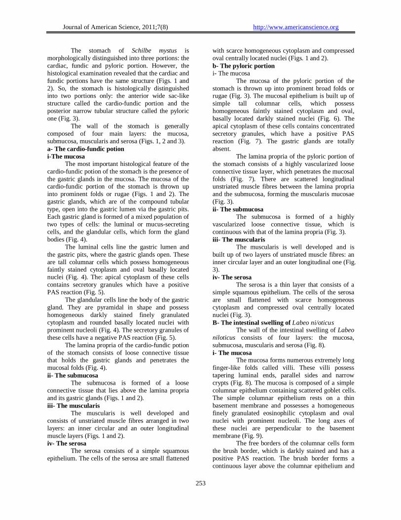
[email protected] Abstract: The present investigation aims to illustrate the histological and ultrastructural differences of the stomach of a carnivorous fish, Schilbe mystus, and the intestinal swelling of a herbivorous fish, Labeo niloticus. The stomach of Schilbe mystus is morphologically divided into three portions: the cardiac, pyloric and fundic portions. However, the histological examination revealed that the stomach is actually divided into two portions: the cardio-fundic portion and the pyloric one. Moreover, the mucosa of the cardiofundic portion of the stomach of Schilbe mystus revealed that it consists of two types of cells: the luminal and glandular cells. The luminal cells, which line the gastric lumen and gastric pits, are tall columnar with apically concentrated secretory granules that have a positive PAS reaction. In addition, the glandular cells, which line the bodies of the gastric glands, are pyramidal in shape and possess scattered secretory granules. The pyloric portion of the stomach has the same structure as the cardio-fundic portion, but the gastric glands are completely absent. The histological examination of the mucosa of the intestinal swelling of Labeo niloticus revealed that it is built up of a simple columnar epithelium that contains goblet cells. The ultrastructural examination of the gastric mucosa of the stomach of Schilbe mystus revealed three types of cells: the luminal, endocrine and exocrine (oxynticopeptic) cells. The luminal cells are tall columnar and are characterized by the presence of apically aggregated secretory granules and a prominent vesiculotubular system. The gastric endocrine cells are mainly pyramidal in shape and are characterized by numerous secretory granules, which exhibit various sizes and shapes. According to the dominant size of the secretory granules, the endocrine cells are differentiated into three types: type-I, type-2 and type-3 cells. Moreover, the gastric glands of Schilbe mystus are made up of a single type of exocrine cells, the oxynticopeptic cells. The oxynticopeptic cells are characterized by the combined features of the mammalian oxyntic and peptic cells. The oxynticopeptic cells have numerous apical microvilli, a well-developed vesiculotubular system, numerous mitochondria, a rough endoplasmic reticulum and numerous secretory granules.The ultrastructural examination of the mucosa of the intestinal swelling of Labeo ni/oticus revealed that the enterocytes are tall columnar and possess welldeveloped microvilli, numerous mitochondria, microtubules, a few endocytotic channels and aggregated chylomicrons. [S. A. A. Naguib; H. A. EI-Shabaka and F. Ashour. Comparative Histological and Ultrastructural Studies on the Stomach of Schilbe mystus and the Intestinal Swelling of Labeo niloticus. Journal of American Science 2011;7(8):251-263]. (ISSN: 1545-1003). http://www.americanscience.org. Keywords: Stomach histology, Stomach ultrastructure, Labeo nilotieus, Schilbe mystus, Teleost. 1. Introduction
AI-Hussaini and Kholy (1953) found that the stomach of Tilapia nilotiea is divided into a wide caecal region and a narrow pyloric one. They believed that the oxyntic and peptic cells are not found in the gastric mucosa of some omnivorous teleosts. In addition, Kapoor et al. (1975) and Grau et al. (1992) stated that the main features of the carnivorous fish digestive tract are the presence of a large stomach and a short intestine.
Reifel and Travill (1978) stated that the shape of the teleostean stomach is variable. They reported that the stomach shape may be rectilinear as in Esox amerieanus and Esox lucius or siphonal as in Ambloplites rupestris, Lepomis maerochirus and Pomoxis nigromaculatus or caecal as in Ictalurus nebulosus, Mieropterus salmoides and Pereajlaveseens. They also added that the rectilinear
and caecal types are associated with piscivorous species.
Noaillac-Depeyre and Gas (1982) stated that the gastric mucosa of the perch, Perea jluviatilis, and the catfish, Ameiurus nebulosus, contains three and four different types of endocrine cells, respectively. Each endocrine cell type possesses characteristic secretory granules. They also demonstrated that these cells are mainly found among the mucoid cells and rarely among the gastric glandular cells.
Klumpp and Nichols (1983) found that the absence of the stomach in the alimentary tract of the herbivore southern sea garfish, Hyporhamphus melanoehir, indicate that acid hydrolysis does not have a role in the breakdown of the plant cell walls. Therefore, they suggested that this fish rely on the action of its pharyngeal mill to rupture the plant cell walls.

Journal of American Science, 2011;7(8) http://www.americanscience.org
252
Martin and Blaber (1984) stated that the stomach of a caecal-type in some Ambassidae fish is described in fish with predatory habits and might serve as a storage site for irregular intake of large quantities of food.
Cataldi et al. (1987) mentioned that the gastric glands, which contain one type f cells, are present in the cardiac region and increase in number in the fundic one, but they are completely absent in the pyloric region.
MacDonald (1987), in the juvenile and adult Dover sole, Solea solea, and Caceci and Hrubec (1989 & 1990), in the black mollie, Poeeilia spp., stated that the apical microvilli characterize the epithelial cells of all regions of the gastrointestinal tract. Also, Osman and Caceci (1991) observed that the gastric epithelium of Tilapia nilotiea has numerous short apical microvilli.
Tibbetts (1997) stated that the digestive system of the snub-nosed garfish, Arrhamphus sclerolepis krefftii, lacks many of the alimentary modifications common in the alimentary tract of the herbivorous fishes such as the acidic stomach, long intestine and pyloric caeca.
Arellano et al. (2001) stated that the gastric glands of Solea senegalensis are numerous in the pyloric and fundic regions, but they are absent in the cardiac portion. These glands are formed of two types of cells: the light and dark cells. Moreover, Gallagher et al. (2001) found that the gastric glands of the carnivorous pinfish, Lagodon rhomboides, have both mucous and secretory cells. They stated that the secretory cells are of a single type, which has both acid- and enzymesecreting properties.
Unal et al. (2001) stated that the true stomach of Chalealburnus tarichi is absent histologically, and instead there is an expansion at the anterior part of the intestine called the post-oesophageal swelling.
Diaz et al. (2003) stated that the stomach of Engraulis anchoita consists of two parts, the cranial and pyloric parts, both of which are lined by a simple columnar epithelium. They also added that the gastric glands, which are of the branched tubular type, are located along the stomach and are formed of a single type of glandular cells. On the contrary, Carrasson et al. (2006) stated that the gastric glands of Dentex dentex, which are of the simple tubular type, are found in the cardiac portion only and are formed of two glandular cell types.
Petri nee et al. (2005) stated that the presence of mucosubstances in the mucosa of the digestive tract of Esox lucius and Silurus glanis are involved in lubrication and protection processes of the mucosa against acidity and enzymatic actions. The present investigation aims to illustrate the histological and ultrastructural differences pf the
stomach of a carnivorous fish, Schilbe mystus, and the intestinal swellingofa herbivorous fish, Labeo niloticus. 2. Material and Methods
The freshwater Nile silver catfish, Schilbe mystus (Linnaeus, 1758), (Order: Siluriformes, Family: Schilbeidae) and the freshwater Nile carp, Labeo niloticus (Forsski'll, 1775), (Order: Cypriniformes, Family: Cyprinidae) were used in the present investigation. 1-Collection of the samples The adult silver catfish, Schilbe mystus, ranging from 15 to 28 cm in length, and the Nile carp, Labeo niloticus, ranging from 20 to 40 cm in length, were caught alive from the River Nile at different localities between Beni Suef and Giza. They were killed and dissected immediately in the field. 2- Histological and ultrastructural studies Fresh adult specimens were carefully dissected and small pieces of the stomach and intestinal swelling of Schilbe mystus and Labeo niloticus, respectively, were fixed in aqueous Bouin's fluid. Sections of 5-7 µm thick were cut and were stained with Harris' haematoxylin and counterstained with eosin. Some sections were stained with periodic acid-Schiff reagent (PAS) and counterstained with haematoxylin to demonstrate the mucus-secreting cells. Small pieces of the stomach and intestinal swelling of Schilbe mystus and Labeo niloticus, respectively, were immediately fixed in cold 3% glutaraldehyde solution in phosphate buffer (pH 7.2 to 7.4) or in a mixture of glutaraldehyde and formaldehyde for three hours to overnight at 4°C, then immersed in the phosphate buffer for 24 hrs.
Resin capsules were prepared and cut with the ultramicrotome into semithin sections (1µm thick) and stained with toluidine blue. The sections were examined with the light microscope to select the suitable areas representing the desired observations. Silver to pale golden ultrathin sections (600A in thickness) were mounted on copper grids. The sections were stained with 5% uranyle acetate and lead citrate according to Reynolds (1963). Finally, the grids were examined with a JOEL 1200 EX 11 electron microscope at the Electron Microscope Unit in the Central Laboratory, Faculty of Science, Ain Shams University and a JOEL 100S electron microscope at the Electron Microscope Unit at the National Cancer Institute. 3. Results 1-Histological studies A-The stomach of Schilbe mysuls
Journal of American Science, 2011;7(8) http://www.americanscience.org
253
The stomach of Schilbe mystus is morphologically distinguished into three portions: the cardiac, fundic and pyloric portion. However, the histological examination revealed that the cardiac and fundic portions have the same structure (Figs. 1 and 2). So, the stomach is histologically distinguished into two portions only: the anterior wide sac-like structure called the cardio-fundic portion and the posterior narrow tubular structure called the pyloric one (Fig. 3).
The wall of the stomach is generally composed of four main layers: the mucosa, submucosa, muscularis and serosa (Figs. 1, 2 and 3). a- The cardio-fundic potion i-The mucosa
The most important histological feature of the cardio-fundic potion of the stomach is the presence of the gastric glands in the mucosa. The mucosa of the cardio-fundic portion of the stomach is thrown up into prominent folds or rugae (Figs. 1 and 2). The gastric glands, which are of the compound tubular type, open into the gastric lumen via the gastric pits. Each gastric gland is formed of a mixed population of two types of cells: the luminal or mucus-secreting cells, and the glandular cells, which form the gland bodies (Fig. 4).
The luminal cells line the gastric lumen and the gastric pits, where the gastric glands open. These are tall columnar cells which possess homogeneous faintly stained cytoplasm and oval basally located nuclei (Fig. 4). The: apical cytoplasm of these cells contains secretory granules which have a positive PAS reaction (Fig. 5).
The glandular cells line the body of the gastric gland. They are pyramidal in shape and possess homogeneous darkly stained finely granulated cytoplasm and rounded basally located nuclei with prominent nucleoli (Fig. 4). The secretory granules of these cells have a negative PAS reaction (Fig. 5).
The lamina propria of the cardio-fundic potion of the stomach consists of loose connective tissue that holds the gastric glands and penetrates the mucosal folds (Fig. 4). ii- The submucosa The submucosa is formed of a loose connective tissue that lies above the lamina propria and its gastric glands (Figs. 1 and 2). iii- The muscularis The muscularis is well developed and consists of unstriated muscle fibres arranged in two layers: an inner circular and an outer longitudinal muscle layers (Figs. 1 and 2). iv- The serosa The serosa consists of a simple squamous epithelium. The cells of the serosa are small flattened
with scarce homogeneous cytoplasm and compressed oval centrally located nuclei (Figs. 1 and 2). b- The pyloric portion i- The mucosa The mucosa of the pyloric portion of the stomach is thrown up into prominent broad folds or rugae (Fig. 3). The mucosal epithelium is built up of simple tall columnar cells, which possess homogeneous faintly stained cytoplasm and oval, basally located darkly stained nuclei (Fig. 6). The apical cytoplasm of these cells contains concentrated secretory granules, which have a positive PAS reaction (Fig. 7). The gastric glands are totally absent. The lamina propria of the pyloric portion of the stomach consists of a highly vascularized loose connective tissue layer, which penetrates the mucosal folds (Fig. 7). There are scattered longitudinal unstriated muscle fibres between the lamina propria and the submucosa, forming the muscularis mucosae (Fig. 3). ii- The submucosa The submucosa is formed of a highly vascularized loose connective tissue, which is continuous with that of the lamina propria (Fig. 3). iii- The muscularis The muscularis is well developed and is built up of two layers of unstriated muscle fibres: an inner circular layer and an outer longitudinal one (Fig. 3). iv- The serosa The serosa is a thin layer that consists of a simple squamous epithelium. The cells of the serosa are small flattened with scarce homogeneous cytoplasm and compressed oval centrally located nuclei (Fig. 3). B- The intestinal swelling of Labeo ni/oticus The wall of the intestinal swelling of Labeo niloticus consists of four layers: the mucosa, submucosa, muscularis and serosa (Fig. 8). i- The mucosa The mucosa forms numerous extremely long finger-like folds called villi. These villi possess tapering luminal ends, parallel sides and narrow crypts (Fig. 8). The mucosa is composed of a simple columnar epithelium containing scattered goblet cells. The simple columnar epithelium rests on a thin basement membrane and possesses a homogeneous finely granulated eosinophilic cytoplasm and oval nuclei with prominent nucleoli. The long axes of these nuclei are perpendicular to the basement membrane (Fig. 9). The free borders of the columnar cells form the brush border, which is darkly stained and has a positive PAS reaction. The brush border forms a continuous layer above the columnar epithelium and

Journal of American Science, 2011;7(8) http://www.americanscience.org
254
is interrupted only by the openings of the goblet cells (Fig. 9). The goblet cells are flask-shaped with two prominent parts: the swollen apical part and the elongated basal one. Each cell opens on the luminal surface by a single pore situated at the top of the swollen part. These cells have a homogeneous faintly stained eosinophilic cytoplasm and small basally located nuclei (Fig. 9). The lymphocytes are numerous and usually aggregate at the bases of the columnar epithelium and some of them frequently appear in the free ends of the cells. The lymphocytes are easily detected by their scanty amount of cytoplasm and rounded darkly stained nuclei (Fig. 9). The lamina propria consists of a loose connective tissue that penetrates and supports the villi (Fig. 8). ii- The submucosa
The submucosa is extremely thin 'and is formed of a vascularized loose connective tissue that lies immediately above the lamina propria without a separating line (Fig. 8). iii- The muscularis
The muscularis of the intestinal swelling consists of two muscle layers: an inner circular layer and an outer longitudinal one (Fig. 8). The muscularis of the most anterior region of the intestinal swelling is built up of striated muscle fibres (Fig. 10), while the muscularis of the posterior region is built up of unstriated muscle fibres, whose circular layer is more developed than the longitudinal one (Fig. 11). iv- The serosa
The serosa is an extremely thin layer of simple squamous epithelium. These cells are flattened with small amount of cytoplasm and compressed oval nuclei, which are located at the widest parts of the cells (Figs. 10 and 11). 2-Ultrastructural STUDIES A-The stomach of Schilbe mystus The ultrastructural examination of the gastric mucosa of Schilbe mystus revealed three types of cells: the luminal (the mucus-secreting), basal endocrine and basal exocrine (the oxynticopeptic) cells. The first two types of these cells are found throughout the gastric mucosa, whereas the second type is restricted only to the cardio-fundic portion of the stomach. i- The luminal cells
The gastric luminal cells are tall columnar (about 27 µm in average height), electron-dense and rest on a prominent basement membrane. There are numerous apically aggregated secretory granules, which are homogeneous, electron-dense and
elongated in shape with variable sizes ranging from 0.16 to 0.7 µm in the long axis. Some secretory granules are frequently fused with the apical cell membrane, emptying their contents into the gastric lumen (Fig. 12).
The vesiculotubular system is made up of numerous small smooth vesicles with dense outlines. They are scattered throughout the cell cytoplasm especially at the apical region in between the secretory granules (Fig. 12). The mean diameter of these vesicles is about 0.06 µm.
The endoplasmic reticulum is differentiated into two types: the rough and smooth endoplasmic reticula. The rough endoplasmic reticulum is well developed and consists of long tubules studded with numerous ribosomes. On the other hand, the smooth endoplasmic reticulum consists of short smooth tubules. The free ribosomes are abundant and scattered throughout the cell cytoplasm (Fig. 13).
There are numerous microfilaments scattered throughout the cell cytoplasm (Fig. 13).
The nucleus is large, oval (the long axis is about 11 µm) or rounded (the diameter is about 4 µm) in shape, basally located and contains a prominent centrally located nucleolus (Fig. 12). The nuclear envelope is a double membrane consisting of an outer and an inner nuclear membrane that encompass the nucle,oplasm. The nuclear pores are found at intervals in the nuclear envelope. The chromatin is found mainly as euchromatin, which is represented by the pale granular areas of the nucleoplasm. Moreover, the heterochromatin is mainly represented by small dense granular aggregations scattered throughout the nucleus and on the inner nuclear membrane (Fig. 13).
The lateral cell membranes form numerous complex interdigitations (Fig12). Moreover, the apical ends of the lateral cell membranes of the adjacent cells join together by junctional complexes (Fig. 14). ii- The endocrine cells The gastric endocrine cells are electron-lucent and are scattered throughout the gastric mucosal epithelium. They are pyramidal, cuboidal or rounded in shape. These cells are seen in direct contact with the basement membrane (Fig. 15). The rough endoplasmic reticulum consists of a network of membranous tubules, which ramify throughout the cytoplasm (Fig. 15). The mitochondria are few in number and rounded or elongated in shape. The diameter of the rounded mitochondria ranges from 0.4 to 0.6 µm. The inner mitochondrial membrane forms numerous lamellar cristae (Fig. 16). The nuclei of these cells are more or less rounded (the mean diameter is about 4 µm) or oval in
Journal of American Science, 2011;7(8) http://www.americanscience.org
255
shape (the mean long axis is about 5 µm) and sometimes they appear indented. The nuclear envelope, which encloses. the nucleoplasm, consists as usual of two membranes. The chromatin is represerited by the euchromatin and heterochromatin. The euchromatin is represented by the electron-lucent nuclear material. On the other- hand, .the heterochromatin is represented by small electron-dense clumps on the inner aspect of the nuclear envelope and small dark patches scattered in the nucleoplasm. The nuclear envelope possesses nuclear pores, at areas where the heterochromatin is absent (Figs. 15 and 16). The most prominent and dominant feature of the endocrine cells is the presence of secretory granules, which are numerous and are scattered in the cell cytoplasm. These secretory granules exhibit variable sizes and shapes in different cells. According to the dominant size of the secretory granules, the endocrine cells are differentiated into'three types: type-1, type-2 and type-3 cells. Type-l of the endocrine cells The type-l cells possess large, rounded secretory granules, which vary in diameter from 0.1 µm to 0.3µm (with a mean diameter of about 0.18 µ1m). These secretory granules are scattered in the cell cytoplasm and are usually aggregated at the basal region of the cell. These secretory granules consist of a homogeneous, electron-dense substance with variable densities. Each secretory granule is tightly surrounded by a membrane, but in some cases an electron lucent halo is interposed between the granule core and its surrounding or limiting. membrane (Fig. 17). Type-2 of the endocrine cells The type-2 cells possess small, rounded secretory granules, which vary in diameter from 0.05 µm to 0.1 µm (with a mean diameter of about 0.07 µm). These secretory granules are usually basally concentrated in the cell cytoplasm and, contain a homogeneous, electron-dense substance. Each secretory granule contains an electron-dense core which is sometimes separated from the surrounding membrane by an electron-translucent space, the halo (Fig; 18). Type-3 of the endocrine cells.
The type-3 cells possess large secretory granules, which vary in diameter
from 0.13 µm to 0.45µm (with a mean diameter of about 0.27 µm). These secretory granules are scattered throughout the cell cytoplasm and each granule consists of a small eccentric electron-dense core inside a rounded or irregular ni.embrane-limited vacuole. Some vacuoles appear empty without the electron opaque core (Fig. 19). iii- The exocrine cells
The gastric glands are made up of a single type of cells, the exocrine cells, which are referred to as the oxynticopeptic cells. These cells secrete both the hydrochloric acid and gastric enzymes.
The oxynticopeptic cells, which possess electron-dense figures, are more or less cuboidal in shape (the mean length is about 11µm) and their luminal surfaces are either smooth or have numerous thin long microvilli (Figs. 20 and 21) The diameter of these microvilli is about 0.1 µm and their length varies from 0.5 to 2.9 µm. These cells possess a large number of spherical secretory zymogenous granules, which vary in diameter from 0.4 to 2µm. These granules are scattered throughout the cytoplasm and are aggregated at the apical surface of the cell among the numerous smooth tubules and vesicles. Each secretory granule contains an electron-dense core, which is tightly surrounded by a limiting membrane (Fig. 22).
The apical cell cytoplasm contains an extensive and extremely complex vesiculotubular system, which is composed of smooth membranous tubules (with a uniform diameter of 0.04 µm) and vesicles (with a mean diameter of about 0.1µm) (Fig. 22).
The rough endoplasmic reticulum is found intermingled with the mitochondria and consists of interconnected tubules and cisternae, which are studded with large numbers of ribosomes. On the other hand, the smooth endoplasmic reticulum consists of a few smooth tubules that lack the ribosomes. The free ribosomes are abundant and scattered throughout the cytoplasm (Fig. 22).
The mitochondria are large, numerous and are scattered throughout the cell cytoplasm. They are round, oval or elongated in shape with a diameter varying from 0.6 to 1.8µm (with a mean diameter of about 1.2 µm). The cristae are numerous and lamellar in shape (Fig. 22).
The nuclei are basally located in the cell and are usually rounded (with a mean diameter of about 4.5 µm) or oval (with a mean long axis of about 6 µm) in shape, and sometimes they appear indented (Figs. 20 and 21).
The lateral cell membranes form smooth contacts with the adjacent cells and lack interdigitations. The apical ends of the lateral cell membranes of the adjacent cells join together by the junctional complexes (Figs. 21 and 22). B- The intestinal swelling of Labeo niloticlIs
The mucosa of the intestinal swelling of Labeo ni/oticus consists mainly of two types of cells: the columnar cells or enterocytes and goblet cells. i- The enterocytes The enterocytes, the most dominant cell type, are tall columnar cells (about 66µm in average

Journal of American Science, 2011;7(8) http://www.americanscience.org
256
height), which rest on a prominent basement membrane. The apical cell membrane bears numerous regularly arranged cytoplasmic projections, the microvilli, which form the brush border (Fig. 23). These microvilli are covered by the glycocalyx and the upper parts of some of them are dense and form spherical bleb, called the capitulum (Krementz and Chapman, 1974) (Fig. 24). The mean length of the microvilli is about 0.77µm and the mean diameter is about 0.09µm. The apical cytoplasm, just below the microvilli, has no cellular organelles and forms the terminal web or net (Figs. 23 and 24). Invaginations of the apical cell membrane are frequently seen among the microvilli, forming the endocytotic channels (Fig. 24). The enterocytes cytoplasm contains aggregated small lipid particles, the chylomicrons (Fig. 25).
The Golgi apparatus is well developed and is formed of one or two sets of parallel stacks, which are located near the nucleus in the laterobasal region of the cell (Fig. 26).
The endoplasmic reticulum is differentiated into two types, the rough and smooth endoplasmic reticula. The rough endoplasmic reticulum is well developed and consists of numerous long parallel tubules, which are mainly concentrated around the nucleus. On the other hand, the smooth endoplasmic reticulum is poorly developed and consists of a few short smooth tubules. The free ribosomes are scattered throughout the cell cytoplasm (Fig. 26).
The mitochondria are numerous, electron-dense, polymorphic (rounded, oval, elongated and ring-like in shape) and with variable sizes (the mean diameter is about 0.3 µm and the mean long axis is about 0.9 µm). The mitochondria are mainly aggregated at both the apical and basal regions of the cell (Figs. 23 and 24). The inner mitochondrial membrane forms numerous lamellar cristae (Fig. 24). The cytoplasm is characterized by the presence of numerous lysosomes which are surrounded by distinct membranes (Fig. 25).
The enterocytes contain numerous and well developed microtubules, which are scattered in the cytoplasm and run parallel to the lateral cell membrane (Fig. 23). The mean diameter of these microtubules is about 0.02 µm (22 nm).
The nuclei of the enterocytes are large, oval in shape (with a long axis of about 10µm) and each one contains a prominent eccejjtric nucleolus (Fig. 23). The nuclear envelope is a double membrane consisting of the outer and inner nuclear membranes that encompass the nucleoplasm. The nuclear pores are found at intervals in the nuclear envelope. The euchromatin is represented by the pale granular areas of the nucleoplasm. On the other hand, the heterochromatin is represented by small dark
granular patches scattered throughout the nucleoplasm and attached to the inner side of the inner nuclear membrane (Fig. 26).
The lateral walls of the cell membrane form smooth contact with the adjacent cells and lack interdigitations. The apical ends of these lateral walls of the adjacent cells join together by the junctional complexes (Figs. 23 and 24). ii- The goblet cells The goblet cells are flask-shaped and each cell is differenti"ated into two parts: a large swollen apical part and an elongated basal one (Fig. 27).
The swollen apical part is filled with large, oval or rounded homogeneous secretory granules. These secretory granules coalesce with each other, vary in both electron density and size (the mean diameter is about 1.2 µm) (Fig. 27). The cytoplasmic organelles are scarce, poorly developed and are aggregated in the elongated basal part. The Golgi apparatus is well developed, found in the supranuclear position and is formed of one or more sets of numerous parallel stacks (Fig. 28). The rough endoplasmic reticulum is found as scattered parallel tubules and cisternae, which are studded with the ribosomes. On the other hand, the smooth endoplasmic reticulum consists of a few parallel tubules that lack the ribosomes (Fig. 28). The mitochondria are oval in shape, small in size (the long axis is about 0.7 µm) and possess few lamellar cristae (Fig. 28). The nuclei of the goblet cells are oval in shape, (the long axis is about 4µm), basally located and each one possesses a prominent nucleolus and a well developed chromatin (Figs. 27 and 28). 4. Discussions
The wall of the stomach of Schilbe mystus and the intestinal swelling of Labeo niloticus consists mainly of four layers: the mucosa, submucosa, muscularis and serosa. This result conforms to that of Albrecht et al. (2001) in the omnivorous Leporinus friderici and Leporinus taeniofasciatus, Park and Kim (2001) in the carnivorous Misgurnus mizolepis, Diaz et al. (2003) in the carnivorous Engraulis anchoita, and Carrasson et al. (2006) in the carnivorous Dentex dentex.
The digestive system of the herbivorous Labeo niloticus lacks a true stomach and consequently lacks acid hydrolysis, so this fish may rely on the action of its strong pharyngeal mill and pharyngeal teeth to rupture the plant cell walls. These results agree with those of Klumpp and Nichols (1983) and Tibbetts (1997) in the herbivorous stomachless Hyporhamphus melanochir and Arrhamphus sclerolepis krefftii, respectively. Moreover, Logothetis et al. (2001) and Cinar and Senol (2006)

Journal of American Science, 2011;7(8) http://www.americanscience.org
257
stated that the gut pH of the herbivorous stomachless Atherinops affinis and the omnivorous stomachless Pseudophoxinus antalyae, respectively, is around neutral or slightly basic. In addition, Logothetis et al. (2001) postulated that the pepsin activity is not present in the gut of Atherinops affinis.
The absence of a true stomach in the digestive system of the herbivorous Labeo niloticus, which belongs to the family Cyprinidae, may be related to phylogeny rather than to a character associated to the diet. This observation agrees with that of Weisel (! 962) who demonstrated that the carnivorous Ptychocheilus oregonense, which belongs to the family Cyprinidae, also lacks the true stomach. In addition, Reifel and Travill (1978) found that the digestive system of the cyprinid Notemigonus crysoleucas and Pimephales promelas also lacks the true stomach.
The digestive system of the carnivorous Schilbe mystus possesses a caecaltype stomach with a highly distensible caecum, which may be used as a reservoir. This observation agrees with that of Reifel and Travill (1978) in the piscivorous Micropterus salmoides and Perca flavescens. Moreover, Grau et al. (1992), in Seriola dumerili, and Carrasson et al. (2006), in Dentex dentex, stated that the carnivorous fishes with a predatory habit and irregular intake of large quantities of food possess a caecal-type stomach. The stomach of Schilbe mystus is morphologically divided into three regions: the cardiac, fundic and pyloric regions, but histologically it is divided into two regions only: the anterior wide sac-like cardio-fundic region and the posterior narrow tube-like pyloric one. These observations are in accordance with those of Al-Hussaini and Kholy (1953), in Clarias lazera, Tilapia nilotica and Sargus vulgaris, Grau et al. (1992), in Seriola dumerili, and Carrasson et al. (2006) in Dentex dentex. However, these observations contradict with those of Caceci et al. (1997) who showed that the stomach of Oreochromis niloticus is divided histologically into three regions: the initial, middle and terminal regions.
The gastric muscularis of the carnivorous Schilbe mystus is well-developed and consists of unstriated muscle fibres arranged in two layers: an inner circular layer and an outer longitudinal one. The powerful gastric muscularis may act as a triturating device. These results agree with those of Grau et al. (1992), in the carnivorous Seriola dumerili, and Ostos Garrido et al. (1996), in the carnivorous Anguilla anguilla.
The cardio-fundic portion of the stomach of Schilbe mystus is characterized by the presence of gastric glands of the compound tubular type, while these glands are totally absent from the pyloric portion. These results agree with those of AI-Hussaini
and Kholy (1953), in Clarias lazera and Sargus vulgaris, Cataldi et al. (1987), in Sparus aurata, Grau et al. (1992), in Seriola dumerili, Ostos Garrido et al. (1993), in Oncorhynchus mykiss, Satora (1998), in Ancistrus multispinnis, Albrecht et al. (2001), in Leporinus friderici and Leporinus taeniofasciatus, and Mai et al. (2005), in Pseudosciaena crocea. However, these results contradict with those of Osman and Caceci (1991), in Tilapia niJotica, Diaz et al. (2003), in Engraulis anchoita, and Carrasson et al. (2006), in Dentex dentex, who stated that the gastric glands are formed of the simple tubular type and are found in both the cardiac and pyloric portions.
The gastric mucosa of Schilbe mystus contains three different types of endocrine cells, which contain characteristic granules and show an extensive contact with the basement membrane. These cells may be involved in the secretion of various -kinds of hormones and play an important role in some physiological functions of the gut. These results agree with those of NoaillacDepeyre and Gas (1982), Podkowa and Goniakowska- Witalinska (2003), Lee et al. (2004) and Khidr (2006) who described three types of endocrine cells in the gastric mucosa of Perca jluviatilis. Hypostomus plecostomus, Coreoperca herzi and Chrysichthys auratus, respectively.
The gastric luminal cells of Schilbe mystus, which line the gastric lumen and gastric pits, are tall columnar cells with numerous apically concentrated granules. These secretory granules have a positive PAS reaction and may serve to protect the stomach epithelium from the auto-digestion processes caused by the gastric enzymes. These results confirm the findings of Gniu et al. (1992), in Seriola dumerili, Morrison and Wright (1999), in Oreochromis niloticus, Petrinec et al. (2005), in Esox lucius and Silurus glanis, and Carrasson et al. (2006), in Dentex dentex.
The secretory granules of the gastric luminal cells of Schilbe mystus are of variable shapes and sizes and are frequently seen in contact with the apical cell membranes to empty their contents into the gastric lumen via exocytosis. These results confirm what was shown by Ostos Garrido et al. (1993), in Oncorhynchus mykiss and Ostos Garrido et al. (1996), in Anguilla anguilla.
The lateral cell membranes of the gastric luminal cells of Schilbe mystus possess numerous complex interdigitations and the cohesion of these cells is ensured by the junctional complexes in the apical zones. These observations agree with those of Ostos Garrido et al. (1993), in Oncorhynchus mykiss and Ostos Garrido et al. (1996), in Anguilla anguilla.
The gastric gland bodies of the carnivorous Schilbe mystus are built up of a ingle exocrine cell

Journal of American Science, 2011;7(8) http://www.americanscience.org
258
type, the oxynticopeptic cells, which probably perform the functions of the mammalian oxyntic and peptic cells related to the synthesis of both hydrochloric acid and pepsinogen, respectively. This result is in accordance with that of Grau et al. (1992), in the carnivorous Seriola dumerili, Ostos Garrido et al. (1996), in the carnivorous Anguilla anguilla and Petrinec et al. (2005), in the carnivorous Esox lucius and Silurus glanis. However, this result differs from that of Podkowa and Goniakowska-Witaliilska (2003) and Carrasson et al. (2006) who stated that the gastric gland bodies of Hypostomus plecostomus and Dentex dentex, respectively, are built up of two cell types: the light and dark cells.
In Schilbe my!;tits stomach, the oxynticopeptic cells posse~s combined features of both the oxyntic and peptic cells of the mammalian stomach. The apical cell membrane of the oxynticopeptic cell possesses numerous microvilli and the apical region of the cytoplasm contains a well-developed vesiculotubular system and numerous scattered mitochondria, which may be involved in the hydrochloric acid secretion. In addition, these cells possess a well-developed rough endoplasmic reticulum and numerous secretory granules, which may be involved in the pepsinogen secretion. These observations are in accordance with those of Ostos Garrido et al. (1993), in Oncorhynchus mykiss, and Ostos Garrido et al. (1996), in Anguilla anguilla. Moreover, Gallagher et al. (2001) stated that the vesiculotubular system of the gastric oxynticopeptic cells of Logodon rhomboides is typical of that found in the gastric oxyntic cells of the mammalian animals.
The luminal surfaces of the gland cells of Schilbe mystus are either smooth or have numerous thin and long microvilli. This observation agrees with that of Coetzee et al. (1991) who reported that the presence or absence of the microvilli of the gastric gland cells of Hydrocynus forskahlii may be ascribed to the secretion cyclic activity of these cells.
The muscularis mucosae is found in the pyloric portion of the stomach of Schilbe mystus, whereas it is absent from the cardio-fundic portion of the stomach. Reifel and Travill (1978) stated that the combination of a well developed musculature and the absence of the gastric glands of seven teleost species may indicate that the primary function of the pyloric region of the stomach is the mixing and pushing the food distally.
The enterocytes of the intestinal swelling of Labeo nilotieus contain a few numbers of endocytotic channels. Noaillac-Oepeyre and Gas (1974 and 1976), in Cyprinus earpio and Tinea
tinea, respectively, and Olsen et al. (1999), in Salvelinus alpinus, suggested that the endocytosis of lipid is not a major route of lipid absorption in the anterior intestine enterocytes. Moreover, Stroband and Oebets (1978) showed that the anterior and middle segments of the intestine of the herbivorous Ctenopharyngodon idella possess endocytotic vesicles to uptake protein macromolecules.
The tips of some microvilli of the enterocytes of the intestinal swelling of Labeo nilotieus possess blebs or capitula, which may increase the apical surface and intensify penetration. Similar results were reported by Kuperman and Kuz'mina (1994) in Esox lucius, Lota Iota and Abramis brama.
The enterocytes of the intestinal swelling of Labeo nilotieus contain small chylomicron particles. This observation suggests that the enterocytes of the intestinal swelling are involved in lipid absorption. This suggestion is in accordance with that of Stroband and Oebets (1978), in Ctenopharyngodon idella, Anderson (1986), in Gire/la trieuspidata, Oeplano et al. (1989), in Dicentrarehus labrax, Olsen et al. (1999), in Salvelinus alpinus, Gallagher et al. (2001), in Lagodon rhomboids, Caballero et al. (2003), in Sparusaurata, Kozaric et al. (2004), in Merluceius merlueeius,and Carrasson et al. (2006), in Dentex dentex. Moreover, Fontagne et al. (1998), in Cyprinus carpio stated that the intestinal fat droplets can be interpreted as a temporary storage form of the reesterified fatty acids.
The enterocytes of the intestinal swelling of Labeo nilotieus contain a few numbers of endocytotic channels. Noaillac-Oepeyre and Gas (1974 and 1976), in Cyprinus earpio and Tinea tinea, respectively, and Olsen et al. (1999), in Salvelinus alpinus, suggested that the endocytosis of lipid is not a major route of lipid absorption in the anterior intestine enterocytes. Moreover, Stroband and Debets (1978) stated that the anterior and middle segments of the intestine of the herbivorous Ctenopharyngodon idella possess endocytotic vesicles to uptake protein macromolecules.
The mucosa of the stomach of Sehilbe mystus and the intestinal swelling of Labeo nilotieus contains numerous lymphocytes, which may play a role in protecting the fish from pathogenic organisms. This result agrees with that of Osman and Caceci (1991), in Tilapia nilotiea and Park and Kim (2001), in Misgurnus m izolepis. Moreover, Temkin and McMillan (1986) stated that the gut of Carassius auratus lacks the discrete aggregations of lymphoid tissue, such as the Peyer's patches of mammals, but possesses a diffused lymphoid tissue as numerous lymphocytes.
Journal of American Science, 2011;7(8) http://www.americanscience.org
259
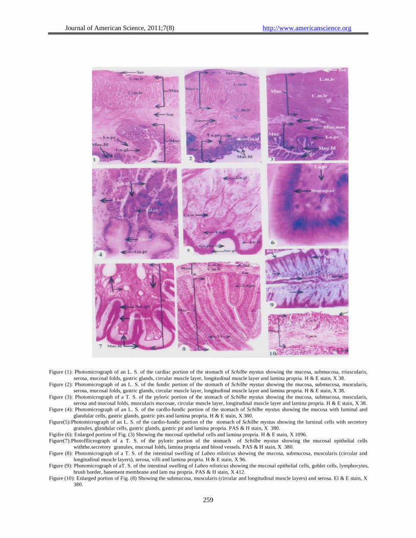
Figure (1): Photomicrograph of an L. S. of the cardiac portion of the stomach of Schilbe mystus showing the mucosa, submucosa, rriuscularis,
serosa, mucosal folds, gastric glands, circular muscle layer, longitudinal muscle layer and lamina propria. H & E stain, X 38. Figure (2): Photomicrograph of an L. S. of the fundic portion of the stomach of Schilbe mystus showing the mucosa, submucosa, muscularis,
serosa, mucosal folds, gastric glands, circular muscle layer, longitudinal muscle layer and lamina propria. H & E stain, X 38. Figure (3): Photomicrograph of a T. S. of the pyloric portion of the stomach of Schilbe mystus showing the mucosa, submucosa, muscularis,
serosa and mucosal folds, muscularis mucosae, circular muscle layer, longitudinal muscle layer and lamina propria. H & E stain, X 38. Figure (4): Photomicrograph of an L. S. of the cardlo-fundic portion of the stomach of Schilbe mystus showing the mucosa with luminal and
glandular cells, gastric glands, gastric pits and lamina propria. H & E stain, X 380. Figure(5):Photomicrograph of an L. S. of the cardio-fundic portion of the stomach of Schilbe mystus showing the lurninal cells with secretory
granules, glandular cells, gastric glands, gastric pit and lamina propria. PAS & H stain, X 380. Figifre (6): Enlarged portion of Fig. (3) Showing the mucosal epithelial cells and lamina propria. H & E stain, X 1096. Figure(7):Photofflicrograph of a T. S. of the pyloric portion of the stomach of Schilbe mystus showing the mucosal epithelial cells
withthe.secretory granules, mucosal folds, lamina propria and blood vessels. PAS & H stain, X 380. Figure (8): Photomicrograph of a T. S. of the intestinal swelling of Labeo niloticus showing the mucosa, submucosa, muscularis (circular and
longitudinal muscle layers), serosa, villi and lamina propria. H & E stain, X 96. Figure (9): Photomicrograph of aT. S. of the intestinal swelling of Labeo niloticus showing the mucosal epithelial cells, goblet cells, lymphocytes,
brush border, basement membrane and lam ma propria. PAS & H stain, X 412. Figure (10): Enlarged portion of Fig. (8) Showing the submucosa, muscularis (circular and longitudinal muscle layers) and serosa. El & E stain, X
380.

Journal of American Science, 2011;7(8) http://www.americanscience.org
260
Figure (11): Photomicrograph of a T. S. of the intestinal swelling of Labeo niloticus showing the mucosa, submucosa, muscularis
(circular and longitudinal muscle layers), serosa, ncosal epithelial cells, goblet cell, lymphocytes, brush border, basement membrane and blood vessel. H & E stain, X 375.
Figure (12): Electron micrograph of the gastric mucosa of Schilbe mystus showing the luminal cells with their secretory granules, vesiculotubular system. nucleus with a prominent nucleolus and lateral interdigitations.
Figure (13): Electron micrograph of a gastric luminal cell of Schilbe inystus showing the rough and smooth endoplasmic reticula, free ribosomes, microfilaments, nucleus, euchromatin, heterochromatin, nuclear envelope and a nuclear pore.
Figure (14): Electron micrograph of the gastric luminal cells of Schi/be mystus showing the secretory granules and junctional complexes.
Figure (15): Electron micrograph of an endocrine cell of Schilbe mystus showing the nucleus, nuclear envelope, nuclear pore, euchromatin, heterochromatin, secretory granules, rough endoplasmic reticul urn and basement membrane.
Figure (16): Electron micrograph of an endocrine cell, between two exocrine cells, of Schilbe mystus showing the nucleus, euchromatin, heterochromatin, nuclear envelope, nuclear pore, rnitochondria and rough endoplasmic reticulum.
Figure (17): Electron micrograph of type-i of the endocrine cells of Schilbe mystus showing the nucleus, euchromatin, heterochromatin, nuclear envelope, rough endoplasmic reticulum, secretory granules and halo.
Figure (18): Electron micrograph of type-2 of the endocrine cells of Schilbe mystus showing the nucleus, nucleolus, rough endoplasrnic reticulum, secretory granules and halo.
Figure (19): Electron micrograph of type-3 of the endocrine cells of Schilbe mystus showing the nucleus, rough endoplasmic reticulum, secretory granules Golgi apparatus.

Journal of American Science, 2011;7(8) http://www.americanscience.org
261
Figure (20): Electron micrograph of the gastric gland of Schilbe mystus showing the exocrine cells and their smooth apical cell membrane
(arrows), nucleus, mitochondria and secretory granules. Figure (21): Electron micrograph of the gastric gland of Schilbe mystus showing the exocrine cells with long microvilli on the apical cell
membrane (arrows), vesiculotubular system, nucleus, mitochondria, secretory granules and junctional complex. Figure (22): Electron micrograph of the exocrine cells of Schilbe mystus showing the vesiculotubular system, nucleus, nuclear pore, mitochondria,
rough smooth endoplasmic reticula, Free ribosomes, secretory granules and junctional complex. Figure (23): Electron micrograph of the intestinal swelling mucosa of Labeo niloticus showing the enterocytes, microvilli, terminal web,
mitochondria, microtubules, nucleus, nucleolus and junctional complexes. Figure (24): Electromicrograph of the apical part of the enterocytes of the intestinal swelling of Labeo niloticus showing the microvilli, terminal
web, capitulum, endocytotic channel, junctional complex and mitochondria. Figure (25): Electron micrograph of -the enteroc.ytes of the intestinal swelling of Labeo niloticus showing the lysosomes and chylomicrons inside
the enterocyte. Figure (26): Electron micrograph of the enterocytes of Labeo niloticus showing the Golgi apparatus, rough and smooth endoplasmic reticula, free
ribosomes, mitochoñdria, nucleus, nucleolus, euchromatin, hêterochromatin, nuclear envelope and nuclear pores. Figure (27): Electron micrograph of the intestinal swelling mucos of Labeo niloticus showing the enterocytes, goblet cells with the swollen apical
parts (arrow heads) and elongated basal parts (asterisks), nucleus, nucleolus and secretory granules. Figure (28): Electron micrograph of the elongaled basal part of the goblet cell of Labeo niloticus showing the secretory granules, Golgi apparatus,
rough and smOoth endoplasmic reticula, free ribosomes, mitochondia, heterochromatin, euchromatin and nucleus

Journal of American Science, 2011;7(8) http://www.americanscience.org
262
LIST OF ABBREVIATIONS
Ba.mb Basement membrane Bl.v Blood vessel Br.bo Brush border C.m.lr Circular muscle layer Cap Capitulum Chy Chylomicrons Fe Euchromatin En.ce Endocrine cell Enc.ch Endocytotic channels Ent Enterocyte Ex.ce Exocrine cell Fr.rb Free ribosomes G.ce Goblet cell Ga.gl Gastric gland Ga.pt Gastric pit GLee Glandular cell Go.ap Golgi apparatus Ha Halo He Heterochromatin md Interdigitations Jn.comp Junctional complex L.m.lr Longitudinal muscle layer La.pr Lamina propria Ls Lysosomes Lu.ce Luminal cell
Ly Lymphocyte Mit Microuilaments Mit Microtuhules Mivi Microvilli Mt Mitochondria Muc Mucosa Muc.ep.ce Mucosal epithelial cells Muc.fd Mucosal ftlcls Mus Muscularis Mus.muc Muscularis mucosae N Nucleus N.env Nuclear envelope N.po Nuclear pore Nu Nucleolus R.e.r Rough endoplasmic reticulum S.e.r Smooth endoplasmic reticulum Sec.gr Secretory granules Ser Serosa Sm Submucosa Te.w Terminal web Vi Villi Vt.sy Vesiculotubular system
References Albrecht, M, p.; FelTeira, M, F, N, and Caramaschi, E. P. (2001).
Anatomical features and histology of the digestive tract of two related neotropical omnivorous fishes (Characiformes; Anostomidae). J. Fish Biol., 58: 419- 430.
AI-Hussaini, A. H. and Kholy, A. A. (1953). On the functional morphology of the alimentary tract of some omnivorous teleost fish. Proc. Egypt. Acad. Sci., 9: 17-39.
Anderson, T. A. (1986). Histological and cytological structure of the gastrointestinal tract of the luderick, Gire/la tricuspidata (Pisces, Kyphosidae), in relation to diet. J. Morphol., 190: 109-119.
Arellano, J. M.; Storch, V. and Sarasquete, C. (2001). Histological and histochemical observations in the stomach of the Senegal sole, Solea senegalensis. Histol. Histopathol., 16 (2): 511-521.
Caballero, M. J.; Izquierdo, M. S.; Kjorsvik, E.; Montero, D.; Socorro, J.; Fermindez, A. J. and Rosenlund, G. (2003). Morphological aspects of intestinal cells from gilthead seabream (Sparus aurata) fed diets containing different lipid sources. Aquaculture, 225: 325-340.
Caceci, T. and Hrubec, T. C. (1989). Ultrastructure of the digestive tube of the black mollie. Proc. Elect. Micro. Soc. Am., 47: 914-915.
Caceci, T. and Hrubec, T. C. (1990): Histology and ultrastructure of the gut of the black mollie (Poecillia spp.), a hybrid teleost. J. Morphol., 204: 265-280.
Caceci, T.; EI-Habback, H. A.; Smith, S. A. and Smith, B. J. (1997). The stomach of Oreochromis niloticus has three regions. J. Fish Biol., 50: 939-952.
Carrasson, M.; Grau, A.; Dopazo, L. R. and Crespo, S. (2006). A histological histochemical and ultrastructural study of the
digestive tract of Dentex dentex (Pisces, Sparidae). Histol. Histopathol., 21(6): 579-593.
Cataldi, E.; Cataudella, S.; Monaco, G.; Rossi, A. and Tancioni, L. (1987). A study of the histology and morphology of the digestive tract of the sea bream, Sparus aurata. J. Fish Biol., 30: 135-145.
Cinar, K. and Senol, N. (2006). Histological and histochemical characterization of the mucosa of the digestive tract in tlower fish (Pselluophoxinlls antalyae). Anat. Histol. Embryol., 35(3): 147-151.
Coetzee, H. L.; Nel, M. M.; Swanepoel, J. H. and Aswegen, G. V. (1991). Light, electron microscopical and immunocytochemical investigation of the stomach wall of the tigerfish f(ydroL)lnllS forskahlii. J. Morphol., 208: 311-321.
Deplano, M.; Connes, R.; Diaz, J. P. and Paris, J. (1989). Intestinal steatosis in the farm-reared sea bass Dicentrarchus labrax. Dis. I\quat. Org., 6: 121-130.
Diaz, A. O.; Garcia, A. M.; Devincenti, C. V. and Goldemberg, A. L. (2003). Morphological and histochemical characterization of the mucosa of the digestive tract in Engrolllis anchoita (Hubbs and Marini, 1935) Anat HistoL EmbryoL, 32(6): 341-346.
Fontagne, S.; Geurden, I.; Escaffre, A. M. and Bergot, P. (1998). Histological changes induced by dietary phospholipids in intestine and liver of common carp (Cyprinus earpio L.) larvae. Aquaculture, 161: 213-223.
Gallagher, M. L.; Luczkovich, J, J. and Stellwag, E. J. (2001). Characterization of the ultrastructure of the gastrointestinal tract mucosa, stomach contents and liver enzyme activity of the pinfish during development J. Fish Biol., 58:1704-1713.
Grau, A.; Crespo, S.; Sarasquete, M.C. and Gonzidez De Canales, M. L. (1992).The digestive tract of the amberjack Seriola

Journal of American Science, 2011;7(8) http://www.americanscience.org
263
dllrnerili Risso: a light and scanning electron microscope study. J. Fish Biol., 41: 287-303.
Kapoor, B. G.; Smit, H. and Verighina, r. A. (1975). The alimentary canal and digestion in teleosts. In: Advances in Marine Biology, Vol. 13. F.S. Russell and M. Young (eds.). Academic Press, London. 109-239.
Khidr, B. M. (2006). Electron microscopic studies on the intestinal mucosa of the Nile catfish, Chrysiehthys auratus (Geoffroy) with special interest in its absorptive function. Egypt. 1. Zool., 47: 143-165.
Klumpp, D. W. and Nichols, P. D. (1983). Nutrition of the Southern Sea garfish Hyporhamphus rnelanoehir: gut passage rate and daily consumption of two food types and assimilation of seagrass components. Mar. Ecol. Prog. Series, 12: 207-216.
Kozaric, Z.; Kuzir, S.; Nejedli, S.; Petrinec, Z. and Srebocan, E. (2004). Histochemical distribution of dige,stive enzymes in hake, Merlllccills rnerlueeius L., 1758. Vet. Arhiv., 74(4): 299-308.
Krementz, A. B. and Chapman, C. B. (1974). Ultrastructure of the posterior half of the intestine of the channel catfish, Ictaulrlls punctatlls. J. Morphol., 145: 441-482.
Kuperman, B. I. and Kuz'mina, V. V. (1994). The ultrastructure of the intestinal epithelium in fishes with different types of feeding. J. Fish Biol. 44: 181-193.
Lee, J.H.; Ku, S.K.; Park, K.D. and Lee, H.S. (2004). Immunohistochernical study of the gastrointestinal endocrine cells in the Korean aucha perch. J. Fish Biol., 65: 170-181.
Logothetis, E. A.; Horn, M. 1-1. and Dickson, K. A. (2001). Gut morphology and function in Atherinops affinis (Teleostei: Atherinopsidae), stomachless omnivore feeding on macroalgae. J. Fish Biol., 59: 1298-13 12.
MacDonald, N. L. (l7). An electron microscopic examination of the gastrointestinal epithelium of the Dover sole, Solea .s’olea C. J. Fish Biol., 31: 27-36.
Mai, K.; Yu, H.; Ma, H.; Duan, Q.; Gisbert, E.; Zambonino Infante, J. C. and Cahu, C. L. (2005). A histological study on the development of the digestive system of Pseudosciaena crocea larvae and juveniles. J. Fish Biol., 67:1094-1106.
Martin, T. J. and Blaber, S. J. M. (1984). Morphology and histology of the alimentary tract of Ambassidae (Cuvier) (Teleostei) in relation to feeding. J. Morphol., 182: 295-305.
Morrison, C. M. and Wright Jr, J. R. (1999). A study of the histology of the digestive tract of the Nile tilapia. J. Fish Biol., 54: 597-606.
Noaillac-Depeyre, J. and Gas, N. (1974). Fat absorption by the enterocytes of the carp (Cyprinus carpio L.). Cell Tiss. Res., 155: 353-365.
Noaillac-Depeyre, J. and Gas, N. (1976). Electron microscopic study on gut epithelium of the tench (Tinca tinca L.) with respect to its absorptive functions. Tiss. Cell, 8: 511-530.
Noaillac-Depeyre, J. and Gas, N. (1982). Ultrastructure of endocrine cells in the stomach of two teleost fish, Percafluviatilis L. and Ameiurus nebulosus L. Cell Tiss. Res., 221: 65 7-678.
Olsen, R. E.; Myklebust, R.; Kaino,T. and Ringo, E. (1999). Lipid digestibility and ultrastructural changes in the enterocytes of Arctic char (Salvelinus alpinus L.) fed linseed oil and soybean lecithin. Fish Physiol. Biochem., 21:35-44.
Osman, A. H. K. and Caceci, T. (1991). Histology of the stomach of Tilapia nilotica (Linnaeus, 1758) from the River Nile. J. Fish Biol., 38: 211-223.
Ostos Garrido, M. V.; Gonzalez Oiler, C. and Abaurrea Equisoain, M. A. (1996). Effect of diet on gastric mucosa cells in the European eel (Anguilla anguilla L.). F-[istochemical and ultrastructural study. Micron, 27(1): 25-34.
Ostos Garrido, M. V.; Nunez Torres, M. I. and Abaurrea Equisoain, M. A. (1993). Histological, histochemical and
uitrastructurai analysis of the gastric mucosa in Oncorhynchus mykiss. Aquaculture, 115: 121-132.
Park, J. Y. and Kim, I. S. (2001). Histology and mucin histochemistry of the gastrointestinal tract of the mud bach, in relation to respiration. J. Fish Biol., 58: 861-872.
Petrinec, Z.; Nejedli, S.; Kuir, S. and Opaëak, A. (2005). Mucosubstances of the digestive tract mucosa in northern pike (Esox lucius L.) and European catfish (Silurus glanis L.). Vet. Arhiv., 75(4): 317-327.
Podkowa, D. and Goniakowska-Witaliñska, L. (2003). Morphology of the air- breathing stomach of the catfish Hypostomus plecostomus. J. Morphol., 257: 147- 163.
Reifel, C. W. and Travill, A. A. (1978). Structure and carbohydrate histochemistry of th. stomach in eight species of teleosts. J. Morphol., 158(2): 155-168.
Reynolds, E. S. (1963). The use of lead citrate at high pH as an eIectron-opaqu stain in electron microscopy. J. Cell Biol., 17: 208-2 12.
Satora, L. (l998). Histological and ultrastructural study of the stomach of the air- breathing Ancistrus multispinnis (Siluriformes, Teleostei). Can. J. Zool., 76: 83-86.
Stroband, H. W. J. and Debets, F. M. H. (1978). The ultrastructure and renewal of the intestinal epithelium of the Juvenile grasscarp, Ctenopharyngodon idella (Val.). Cell Tiss. Res., 187: 18 1-200.
Temkin, R. J. and McMillan, D. B. (1986). Gut-associated lymphoid tissue (GALT) of the goldfish, Carassius auratus. J. Morphol., 190: 9-26.
Tibbetts, I. R. (1997). The distribution and function of mucous cells and their secretions in the alimentary tract of Arrhamphus scierolepis kreffiui. J. Fish Biol., 50: 809-820.
Unal, G.; cetinkaya, O.; Kankaya, E. and Elp, M. (2001). Histological study of the organogenesis of the digestive system and swim bladder of the Chalcalburnus tarichi Pallas, 1811 (Cyprinidae). Turk. J. Zool., 25: 217-228.
Weisel, G. F. (1962). Comparative study of the digestive tract of a sucker, Catostomus catostomus, and a predaceous minnow, Ptvchocheilus oregonense. Am. Midland Naturalist, 68(2): 334-346.
7/3/2011