comparative morphological interpretations on the bones of...
TRANSCRIPT

http://bdvets.org/javar/ El-Ghazali et al./ J. Adv. Vet. Anim. Res., 5(4): 410–419, December 2018 410
JOURNALOFADVANCEDVETERINARYANDANIMALRESEARCHISSN2311-7710(Electronic)http://doi.org/10.5455/javar.2018.e292 December 2018A periodical of the Network for the Veterinarians of Bangladesh (BDvetNET) VOL5,NO.4,PAGES410–419
ORIGINALARTICLE
Comparative morphological interpretations on the bones of the pelvic limb of New Zealand rabbit (Oryctolagus cuniculus) and domestic cat (Felis domestica)
HanaaMohamedEl-Ghazali,EmanIsmailEl-beheryDepartmentofAnatomyandEmbryology,FacultyofVeterinaryMedicine,ZagazigUniversity,Zagazig44511,Egypt
Correspondence Hanaa Mohamed El-Ghazali [email protected] Department of Anatomy and Embryology, Faculty ofVeterinaryMedicine,ZagazigUniversity,Zagazig44511,Egypt.
How to cite:El-GhazaliHM,El-beheryEI.Comparativemorphological interpretationsonthebonesofthepelvic limbofNewZealandrabbit(Oryctolagus cuniculus)anddomesticcat(Felis domestica).JAdvVetAnimRes2018;5(4):410–9.
ABSTRACT
Objective: Regardingthedisplayingofthemaindifferencesbetweenthepelviclimbofrabbitandcat.Materials and methods:Ourworkwasperformedon10NewZealandrabbits(Oryctolagus cunic-ulus)anddomesticcats(Felis domestica)withvariableagesandofbothsexes.Afterweighingoftheanimals,sedation,andanesthesia,theanimalswereexaminedradiographically.Thebonesofthepelviclimbwereprepared,measuredforitslength/cmthendescribedandcompared.Results:Theiliactuberosityandtheconversionoftheacetabularnotchintoforamenwerechar-acteristicsofOscoxaeoftherabbit.Theintertrochantericcrestwasdetectedonthefemurofthecat.Intherabbit,theleginterosseousspacewaslocatedintheproximalthirdofthisregionwhileinthecat,itwasextendedalongitslength.Thefirstmetatarsalwasundevelopedinthecatbutwasabsentintherabbitsometatarsalwerefourintherabbitandfiveinthecat.Thedigitsofthepelvic limbs inbothanimalswerefour innumber.Thedistalsesamoidwassingle,transverselysituated,andshuttle-shapedinrabbitbutitwasabsentincat.Conclusion: So, the chief points of variationbetween thepelvic limbbones of rabbit and catenabledustokeepawaythecommercialfraudandfacilitatedtheiruseasananimalmodelforeducationpurposes.
ARTICLE HISTORY
ReceivedJune06,2018RevisedAugust10,2018AcceptedAugust12,2018PublishedNovember09,2018
KEYWORDS
Acetabulum;fabellae;intertrochantericcrest;unguicularprocess
Introduction
Rabbit is efficiently feasible to be used as a source of meat and for the fur industry. Due to the high fertility, short generation period, little size, low cost of rabbit, and it is being used in the production of antibodies, Okerman [1] considered rabbit as a perfect laboratory animal. The rab-bit is used for recognition of some anomalies and diseases as an excellent experimental clinico-anatomical example of humans and animals [2].
In mole-rat (Spalax leucodon Nordmann), the acetab-ulum is deep and there is a little foramen instead of the acetabular notch. The greater trochanter of the femur is separated from the head by a notch [3]. The fibula and tibia of rabbit are fused but in the cat they are separated [4]. In marmoset (Callithrix jacchus), the tarsus contains seven tarsal bones and no distal sesamoid bones are present at the plantar sides of the distal interphalangeal joints [5]. In
the cat, there are seven tarsal bones, four complete meta-tarsals with four digits [6]. In the rabbit, the plantar sur-face of the hind limb from the tarsus distally was in contact with the ground at rest [7].
For the above-mentioned reasons, our research focuses on the most important differences between the bones of the pelvic limb of rabbits and cats to keep away from the commercial fraud where rabbits are used as human food in our country. Also, we hope to facilitate the process of anatomy education by using rabbits and cats as animal models.
Materials and methods
Ethical statement
All obtained animals in this paper were managed accord-ing to the Institutional Animal Care and the Research
ThisisanOpenAccessarticledistributedunderthetermsoftheCreativeCommonsAttribution4.0Licence(http://creativecommons.org/licenses/by/4.0)

http://bdvets.org/javar/ El-Ghazali et al./ J. Adv. Vet. Anim. Res., 5(4): 410–419, December 2018 411
Ethics Committee of the Zagazig University, Egypt (ZU-IACUC/2/F/12/2018).
Number of animals and site of collection
The current research was accomplished on 10 healthy adults New Zealand rabbits (Oryctolagus cuniculus) and domestic cats (Felis domestica) with variable ages and of both sexes. The weight of animals was about 2.5–3.5 kg. The rabbits were obtained from the animal laboratory house in Faculty of Veterinary Medicine, Zagazig University, Egypt. The cats were bought from pet house in Zagazig city, Sharkia Governorate, Egypt.
Procedures
The sedation and anesthesia occurred with 3 mg/kg xylazine i.v followed by 3 mg/kg ketamine i.v injection through the ear vein and with 1 mg/kg of i.m. xylazine then followed by 5 mg/kg of i.m. ketamine for rabbits and cats, respectively [8]. By using the digital scale, the animals were weighted. For the radiographical exam-ination, two animals of both species were prepared and inspected in the Department of Surgery, Anesthesiology and Radiation, Faculty of Veterinary Medicine, Zagazig University. The animal exsanguinations were carried out
through the common carotid artery under complete anes-thesia. After evisceration and dissection of pelvic limbs from the trunk and removal of the skin and muscles care-fully, the bony preparations were performed according to Onwuama et al. [9].
The comparison between the same bones of the pelvic limb in both species was applied and focused on several points as gross morphology, length/cm, and numbers. The measurements were obtained using graduated tape. By using a digital camera with resolution (16.1 megapixels, Sony DSC-W690, 36v and 10x optical zoom), the bones were photographed.
Data analysis
The mean ± standard deviation (SD) of the bones mea-sured data were estimated by using the SPSS software pro-gram (version 16.0; Chicago, USA).
The used anatomical nomenclature was based on Nomina Anatomica Veterinaria [10] whenever possible.
Results
The pelvic limbs of both animals were formed of Os coxae, femur, tibia and fibula, tarsal and metatarsal bones and
Figure 1. Radiographs of rabbit’s (A) and cat’s (B) pelvic limbs (lateral view) showing Osc: Os coxae, Fe: Femur, P: Patella, Fb: Fabellae, Ti: Tibia, Fi: Fibula, Tb: Tarsal bones, Mt: Metatarsal bones and D: Digit.

http://bdvets.org/javar/ El-Ghazali et al./ J. Adv. Vet. Anim. Res., 5(4): 410–419, December 2018 412
digits associated with the patella, fabellae, and sesamoid bones (Fig. 1A and B).
Os coxae
In rabbit and cat, it was formed from the union of three bones; ilium, ischium, and pubis which they shared in the formation of the acetabulum. The latter was formed of two portions; articular and non-articular parts [acetabular fossa (Fossa acetabuli)] (Fig. 2A and B). The articular part was crescentic in shape in rabbit and cat. In the rabbit, the ends of the articular part met each other caudomedially above the acetabular notch (Incisura acetabuli) converting it into a foramen (Fig. 2A).
In the rabbit, the wing of the ilium was shovel-like while in the cat it was spatula shape (deep concave gluteal sur-face and flat pelvic one). The gluteal surface (Facies glutea) in rabbit was marked by a thick ridge [gluteal line (Lineae gluteae)] (Fig. 2A) while in cat it was ventrolateral and began from the coxal tuber and extended parallel to the lateral border (Fig. 2B). In the rabbit, the line ended at the iliac tuberosity (Fig. 2A). The pelvic surface had a C-shaped auricular surface (Facies auricularis) for articulation with the wing of the sacrum in cat and rabbit (Fig. 2C and D). The iliac crest (Crista iliaca) was thin in rabbit and thick in the cat (Fig. 2C and D). The lateral and medial borders were thin edges and transparent in rabbit while in the cat the medial one was thick. The sacral tuber (Tuber sacrale) was thick while the coxal (Tuber coxae) one was thin in rabbit and cat (Fig. 2A–D). The body had medial, lateral, and ventrolateral surfaces and medial, lateral and ventral borders in the rabbit. On the other hand, the body in the cat had medial and lateral surfaces and lateral and medial borders. The pelvic surface carried faint obturator groove (Sulcus obturatorius) in both animals (Fig. 2C and D). The lateral border of the rabbit bears truncated iliac tuberosity (Fig. 2A). The medial border shared in the formation of a large portion of the greater ischiatic notch (Fig. 2C). The ventral border in rabbit had an iliopectinal line while the lateral one of cat continued with this line and born a psoas tubercle (Fig. 2D).
The ischium was quadrilateral in shape with pubic and acetabular branches. The pubic branch met its fellow at the pelvic symphysis and reached the symphyseal branch of the pubis (Fig. 2C and D). The acetabular branch was shared in the formation of the acetabulum and dorsal ischiatic spine (Spina ischiadica). The latter spine was thin beak-like in rabbit and low everted in the cat (Fig. 2C and D).
The surfaces of the ischium were pelvic and ventral which were thin and smooth. The ischial borders were medial (symphyseal), cranial, caudal, and lateral. The latter borders formed the pelvic symphysis, the caudal boundary of the obturator foramen (Foramen obtura-tum), ischial arch, and lesser ischiatic notch, respectively
(Fig. 2A–D). The ischial arch was deeply inverted v-shape in rabbit whereas it was a shallow concave one in the cat (Fig. 2C and D). The ischium had four angles, the caudola-teral one formed the ischiatic tuber (Tuber ischiadicum) which it had lateral tuberosity in rabbit (Fig. 2C).
The pubis was inverted L-shape. It had acetabular and symphyseal branches. The pubis had pelvic and ventral surfaces and cranial, caudal, and medial borders. The pec-tin of the pubis carried clear pubic tubercle in rabbit and indistinct one in cat medially. The iliopectineal eminence in rabbit was clear and connected laterally to the iliopecte-neal line but in cat, it was indistinct (Fig. 2C and D). In both animals, the caudal border was formed the cranial bound-ary of the obturator foramen and in rabbit, it carried small projection medially (Fig. 2C). The obturator foramen in the rabbit was large oval with a thin sharp medial boundary while in cat it was elliptical with small thick medial bound-ary (Fig. 2A and B).
Femur
There was a great difference in the position of the femur between the two animals as it was located horizontally in rabbit and oblique cranioventral in the cat (Fig. 1A and B). The relative length of the femur was 9.040 ± 0.7427 cm in rabbit and 11.440 ± 0.3169 cm in the cat. In the rabbit, the body had two surfaces and two borders while in cat, it was cylindrical with four surfaces and two bor-ders (Fig. 3A and B). In the rabbit, the dorsal surface was convex and the ventral one was concave longitu-dinally (Fig. 3A and C). The two surfaces were smooth and rounded from side to side. In the cat, the nutrient foramen was present just above the middle of the caudal surface (Fig. 3D).
The lesser trochanter (Trochanteric minor) in rabbit (Fig. 3A, C, and E) was in the proximal third of the medial border and just distal to it, the nutrient foramen was pres-ent (Fig. 3E). The former was a triangle in shape with the tooth-like apex (Fig. 3C and E) while in cat, it was large rounded in shape (Fig. 3D). In the rabbit, the third trochan-ter (Trochanteric tertius) (Fig. 3A, C, E, and F) was present at the same level of the lesser trochanter even as it was absent in cat (Fig. 3B, D, and G).
The proximal extremity had a hemispherical head and rounded one in rabbit and cat, respectively (Fig. 3F and G). Fovea capitis was a small circular pit in rabbit (Fig. 3F) while it was large in the cat (Fig. 3G). Laterally, there was the greater trochanter (Trochanteric major) which was divided in both animals. In the rabbit, it was elevated than the level of the head and its caudal part was bent medially but in cat, it was at the same level (Fig. 3F and G). The trochanteric ridge was bounded the trochanteric fossa (Fig. 3C). In the cat, the intertrochanteric crest was connected between the greater trochanter and the lesser one (Fig. 3D).

http://bdvets.org/javar/ El-Ghazali et al./ J. Adv. Vet. Anim. Res., 5(4): 410–419, December 2018 413
Figure 2. Photomacrographs of Os coxae of rabbit (A) and cat (B) (ventral view), Os coxae of rabbit (C) and cat (D) (caudodorsal view) with the insert showing pelvic surface of wing of rabbit’s and cat’s ilium showing Gs: Gluteal surface, Gl: Gluteal line, It: Iliac tuberosity, ILc: Iliac crest, As: Acetabular articular surface, Acf: Acetabular fossa, Acn: Acetabular notch converted into foramen, Cot: Coxal tuber, St: Sacral tuber, Ps: Pelvic surface, Aus: Auricular surface, Pt: Pubic tubercle, Ipe: Iliopectineal eminence, Pst: Psoas tuber-cle, Of: Obturator foramen, Og: Obturator groove, Dis: Dorsal ischiatic spine, Blue line: Greater ischiatic notch, purple line: Lesser ischiatic notch, Ia: Ischial arch, Ist: Ischiatic tuber, Lt: Lateral tuberosity, and Psy: Pelvic symphysis.

http://bdvets.org/javar/ El-Ghazali et al./ J. Adv. Vet. Anim. Res., 5(4): 410–419, December 2018 414
Figure 3. Photomacrographs of femur of rabbit (A) (dorsal view), femur of cat (B) (cranial view), femur of rabbit (C) (ventral view), femur of cat (D) (caudal view), femur of rabbit (E) (Medial view), proximal extremity of femur of rabbit (F) and cat (G) (cranial view) with the insert showing fovea capitis on the head (medial view), distal extremity of femur of rabbit (H) (cranial), (I) (lateral) and (J) (caudodorsal) views, distal extremity of femur of cat (K)(caudal view), lateral fabella of rabbit (L) (dorsal and ven-tral views), lateral (1) and medial (2) fabella of cat (M) (dorsal) and (N) (ventral) views, patella of rabbit (O) and cat (P) (cranial and caudal views) showing Gt: Greater trochanter, Let: Lesser trochanter, Tht: Third trochanter, H: Head, N: Neck, Crs: Cranial, Cds: Caudal, Ds: Dorsal and Vs: Ventral surfaces, Mb: Medial and Lb: lateral borders, P: Patella articulated with trochlea, Tr: Trochlea, Tri: Trochanteric ridge, Tf: Trochanteric fossa, Itc: Intertrochanteric crest, C: Condyles, Icf: Intercondyloid fossa, Nf: Nutrient fora-men, F: Facets for fabellae, Fc: Fovea capitis, Mec: Medial epicondyle, Crp and Cdp: Cranial and caudal parts of the greater trochan-ter, Ef: Extensor fossa, Pf: Popliteal fossa, Lf: Lateral and Mf: Medial fabellae, Ct: Caudoventral tubercle and n: notch on the lateral border of patella.
http://bdvets.org/javar/ El-Ghazali et al./ J. Adv. Vet. Anim. Res., 5(4): 410–419, December 2018 415
The distal extremity was composed of the trochlea dor-sally in rabbit and cranially in the cat. Also, two condyles and epicondyles (lateral and medial) present ventrally in rabbit and caudally in the cat (Fig. 3B–E). There was deeply intercondyloid fossa (Fossa intercondylaris) (Fig. 3C and D) and faint extensor (Fossa extensoria), one in rabbit (Fig. 3H) whilst the extensor fossa in the cat was indistinct. The popliteal fossa (Fossa m. poplitei) (Fig. 3I) was present on lateral epicondyle. There was a faint smooth area above the two condyles for articulation with lateral and medial fabellae in both animals (Fig. 3D, E, J, and K).
Patella and fabellae
In the rabbit, the lateral fabella was elongated, quadri-lateral with large, broad medial part, and small tuberous lateral one. It had two surfaces; dorsal irregular and ven-tral smooth saddle shaped (Fig. 3L). In the cat, the lateral fabella was an irregular triangle in shape with convex dor-sal surface and flat ventral one (Fig. 3M and N). The medial fabella in rabbit and cat was rounded in shape. It had a tubercle caudoventrally in rabbit while in cat had a convex dorsal surface and concave smooth ventral one (Fig. 3J, M, and N).
In rabbit and cat, the patella was pyramidal in shape. Its distal apex was more rounded in rabbit and pointed in cat while the proximal base was broad in rabbit and rounded in the cat. It had a cranial convex rough non-articular sur-face and smooth caudal articular one. The latter surface was divided by a ridge into a small lateral part and large medial one in rabbit but in cat it was concave. The lateral border had a small notch distally in rabbit even as in cat, the two borders were rounded (Fig. 3O and P).
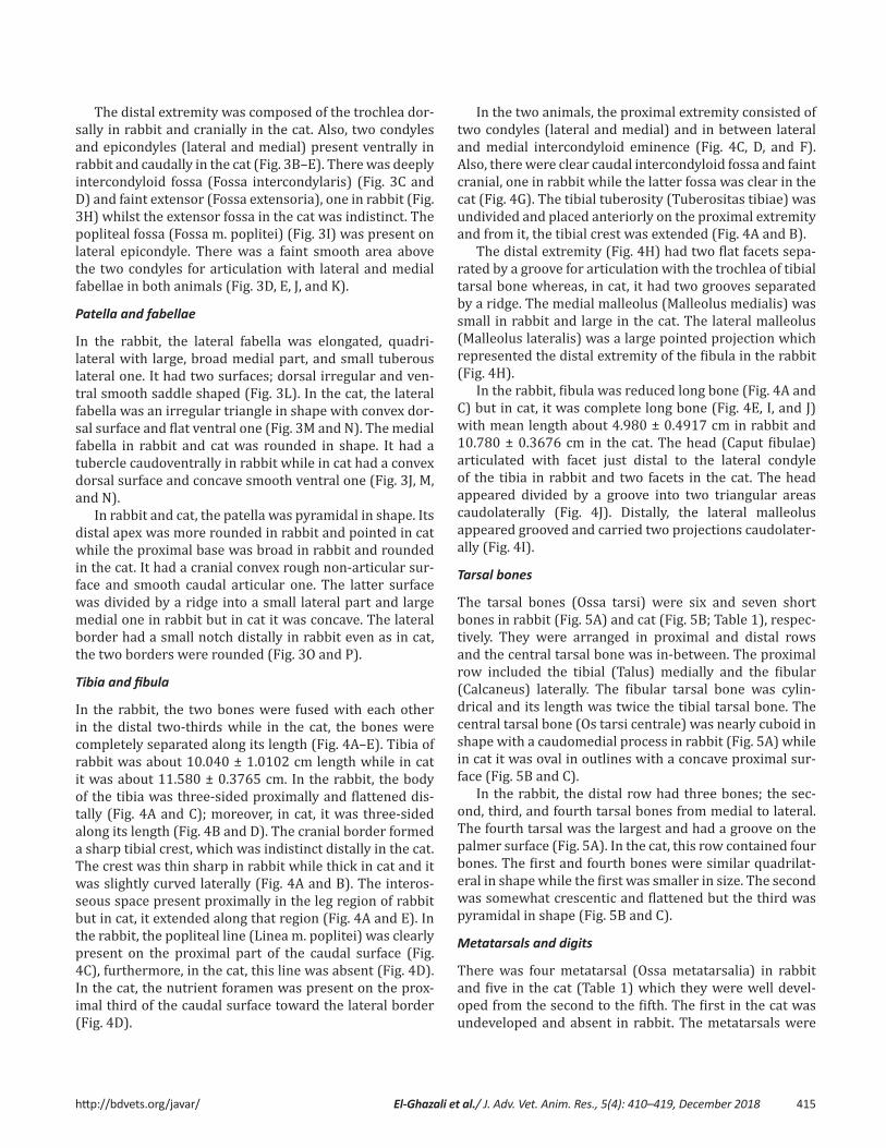
Tibia and fibula
In the rabbit, the two bones were fused with each other in the distal two-thirds while in the cat, the bones were completely separated along its length (Fig. 4A–E). Tibia of rabbit was about 10.040 ± 1.0102 cm length while in cat it was about 11.580 ± 0.3765 cm. In the rabbit, the body of the tibia was three-sided proximally and flattened dis-tally (Fig. 4A and C); moreover, in cat, it was three-sided along its length (Fig. 4B and D). The cranial border formed a sharp tibial crest, which was indistinct distally in the cat. The crest was thin sharp in rabbit while thick in cat and it was slightly curved laterally (Fig. 4A and B). The interos-seous space present proximally in the leg region of rabbit but in cat, it extended along that region (Fig. 4A and E). In the rabbit, the popliteal line (Linea m. poplitei) was clearly present on the proximal part of the caudal surface (Fig. 4C), furthermore, in the cat, this line was absent (Fig. 4D). In the cat, the nutrient foramen was present on the prox-imal third of the caudal surface toward the lateral border (Fig. 4D).
In the two animals, the proximal extremity consisted of two condyles (lateral and medial) and in between lateral and medial intercondyloid eminence (Fig. 4C, D, and F). Also, there were clear caudal intercondyloid fossa and faint cranial, one in rabbit while the latter fossa was clear in the cat (Fig. 4G). The tibial tuberosity (Tuberositas tibiae) was undivided and placed anteriorly on the proximal extremity and from it, the tibial crest was extended (Fig. 4A and B).
The distal extremity (Fig. 4H) had two flat facets sepa-rated by a groove for articulation with the trochlea of tibial tarsal bone whereas, in cat, it had two grooves separated by a ridge. The medial malleolus (Malleolus medialis) was small in rabbit and large in the cat. The lateral malleolus (Malleolus lateralis) was a large pointed projection which represented the distal extremity of the fibula in the rabbit (Fig. 4H).
In the rabbit, fibula was reduced long bone (Fig. 4A and C) but in cat, it was complete long bone (Fig. 4E, I, and J) with mean length about 4.980 ± 0.4917 cm in rabbit and 10.780 ± 0.3676 cm in the cat. The head (Caput fibulae) articulated with facet just distal to the lateral condyle of the tibia in rabbit and two facets in the cat. The head appeared divided by a groove into two triangular areas caudolaterally (Fig. 4J). Distally, the lateral malleolus appeared grooved and carried two projections caudolater-ally (Fig. 4I).
Tarsal bones
The tarsal bones (Ossa tarsi) were six and seven short bones in rabbit (Fig. 5A) and cat (Fig. 5B; Table 1), respec-tively. They were arranged in proximal and distal rows and the central tarsal bone was in-between. The proximal row included the tibial (Talus) medially and the fibular (Calcaneus) laterally. The fibular tarsal bone was cylin-drical and its length was twice the tibial tarsal bone. The central tarsal bone (Os tarsi centrale) was nearly cuboid in shape with a caudomedial process in rabbit (Fig. 5A) while in cat it was oval in outlines with a concave proximal sur-face (Fig. 5B and C).
In the rabbit, the distal row had three bones; the sec-ond, third, and fourth tarsal bones from medial to lateral. The fourth tarsal was the largest and had a groove on the palmer surface (Fig. 5A). In the cat, this row contained four bones. The first and fourth bones were similar quadrilat-eral in shape while the first was smaller in size. The second was somewhat crescentic and flattened but the third was pyramidal in shape (Fig. 5B and C).
Metatarsals and digits
There was four metatarsal (Ossa metatarsalia) in rabbit and five in the cat (Table 1) which they were well devel-oped from the second to the fifth. The first in the cat was undeveloped and absent in rabbit. The metatarsals were
http://bdvets.org/javar/ El-Ghazali et al./ J. Adv. Vet. Anim. Res., 5(4): 410–419, December 2018 416
Figure 4. Photomacrographs of tibia and fibula of rabbit (A) and tibia of cat (B) (cranial view), tibia and fibula of rabbit (C) and tibia of cat (D) (caudal view), tibia and fibula of cat (E) (lateral view), proximal extremities of tibia of rabbit (F) (caudal view) and cat (G) (dorsal view), distal extremities of tibia of (1): rabbit and (2): cat (H) (ventral view), lateral malleolus of cat (I) (1:cranial and 2: cau-dal views) and fibula of cat (J) (1:cranial and 2:caudal views) showing Tt: Tibial tuberosity, Tc: Tibial crest, Ios: Interosseous space, Ti: Tibia, Fi: Fibula, Lm: Lateral and Mm: Medial malleolae, C: Condyles, Pl: Popliteal line, Ls: Lateral, Ms: Medial, Crs: Cranial and Cds: Caudal surfaces, Lb: Lateral, Mb: Medial and Crb: cranial borders, Nf: Nutrient foramen, E: Eminences, Fo: Two fossae, G: Groove, Ri: Ridge, H: Head, Pj: Two projections and Aft: Two facets for articulation with tibia.

http://bdvets.org/javar/ El-Ghazali et al./ J. Adv. Vet. Anim. Res., 5(4): 410–419, December 2018 417
Figure 5. Photomacrographs of tarsal bones of rabbit (A) and cat (B) (craniodorsal view) with the insert showing the palmar sur-face of the 4th tarsal bone, tarsal and metatarsal bones of cat (C) (cranial view), pes region of rabbit (D) and cat (E) (dorsal view), metatarsal and digits of rabbit (F) and cat (G) (dorsolateral view), digits and sessamoid bones of rabbit (H) (palmar view), proximal extremity of metatarsal bones of cat (I) (dorsal view), distal phalanx of rabbit (J) and cat (K) (lateral view) showing L: lateral and M: Medial sides, Ft: Fibular, Tt: Tibial, Ct: Central, 1st: First, 2nd: Second, 3rd: Third and 4th: Fourth tarsal bones, Gr: groove on the palmar surface of 4th, Mt: Metatarsal bones, 1’, 2’, 3’, 4’, and 5’: First, second, third, fourth, and fifth metatarsal bones, II, III, IV, and V: Second, third, fourth, and fifth digits, Cl: Claw, Flt: Flexor tubercle, Uc: Unguicular crest, Up: Unguicular process, Pp: Proximal, Mp: Middle and Dp: Distal phalanges, Pss: Proximal, and Dss: Distal sesamoid bones.

http://bdvets.org/javar/ El-Ghazali et al./ J. Adv. Vet. Anim. Res., 5(4): 410–419, December 2018 418
similar to the metacarpals as long bones, had two extremi-ties and shaft but extremely exceed in their size (Fig. 5D–I).
In the two animals, the digits (Ossa digitorum pedis) were four in number (Table 1). In the rabbit, the fifth digit was shortest (Fig. 5F). The second and fifth digits were the shortest and the third and fourth digits were the longest in the cat (Fig. 5G). In two animals, the third phalanx was enclosed in claw and triangular in shape with base prox-imally and apex distally (unguicular process). The latter process was broad in rabbit (Fig. 5J) and more pointed in the cat (Fig. 5K). The relative length of the pes region was about 10.680 ± 1.1924 cm and 11.160 ± 0.4377 cm in rab-bit and cat, respectively.
Sesamoid bones
There were three sesamoids in rabbit and two in the cat for each digit (Fig. 5G and H; Table 1). The proximal sesamoids (Ossa sesamoidea proximalia) were paired and vertically situated on the palmar surface of the fetlock joint. It was pyramidal in shape with notched base in rabbit while in cat it was elongated. The distal sesamoid (Os sesamoideum distal) was single and transversely situated on the coffin joint. It was shuttle-shaped in rabbit, whereas in cat it was absent (Fig. 5G and H).
Discussion
Aspinall et al. [4] in cat reported that the pubis was not involved in the formation of the acetabulum of the hip joint. Moreover, Cruise and Nathan [11] recorded that, the pres-ence of a small accessory bone in rabbit, osacetabulum,
that form the acetabulum, by way of the ischium and ilium. These previous observations were in disagreement with our study in both species and we added that the acetabular notch in rabbit was converted into a foramen. On the other hand, Casteleyn et al. [5] in common marmoset (Callithrix jacchus) clarified that the acetabulum was deep and con-tained a lunate articular surface which was intermittent by an acetabular notch and he added that the dorsal ischiatic spine was distinct. But we observed that this spine was thin elevated in rabbit and low everted one in the cat.
Concerning the different shapes of the obturator fora-men, we observed that it was large oval in rabbit as well as [11] but it was very large in common marmoset [5] and elliptical in the cat.
The position of the femur was horizontal in rabbit which adapted their standing position while in the cat it was oblique cranioventral as other domestic animals [12]. The femur had marked major and minor trochanters and the third trochanter was absent in common marmoset [5] as our work in the cat. The third trochanter was present in mole-rats on the proximal third of the femur [3] simi-lar to in our observations in the rabbit. Furthermore, the occurrence of three trochanters on the femur was stated by Salami et al. [13] in the African giant pouched rat (Cricetomys gambianus). Presence of femoral condyles and the patellar groove on the femur leads to complexity of its distal epiphysis [14]. The presence of the fabellae in both species in this study was in agreement with Casteleyn et al. [5] in common marmoset. Humans had a round, triangular, or rectangular ossified lateral fabella [15]. Patella varied in its shape as it was ovoid in common marmoset [5] but it was triangular in rabbit and pyramidal in the cat in our research.
Tibia and fibula were separated by an interosseous space and were not fused [5] in common marmoset similar to the present study in the cat. But the fibula was fused with the tibia for over half its length [1,11] in rabbit as our result.
Regarding tarsal bones, they were six in rabbit go hand with [11]. While Casteleyn et al. [5] in marmoset men-tioned that, the tarsal bones were seven arranged in three rows; talus and calcaneus in the crural row, a reduced mid-dle central tarsal one, and four bones in the metatarsal row. Rodentia order had similarly in the number of tarsal bones as eight in two rows [3,13]. But their arrangement was differed as in two rows, three proximally and four dis-tally and a central one was in the distal of the talus in mole-rats [3]. Even as in the African giant pouched rat, Salami et al. [13] revealed that tarsal bones were four proximally and four distally.
Cruise and Nathan [11] clarified that, there were four well-developed metatarsals (II–III–IV–V) with an imma-ture metatarsal I unlike the rabbit in this paper, metatarsal
Table 1. Elucidatethenumbersofthebonesinpesregionofrabbitsandcats.
Species
Bones
Rabbit Cat
Tarsal(fromMedial.tolateral)1strow→
central2ndrow→
62(TibialandFibular.T.B)CentralT.BII,III,andIV
72(TibialandFibular.T.B)CentralT.BI,II,III,andIV
Metatarsal(fromMedialtolateral)
4
II,III,IV,andVI→absent
5
II,III,IV,andVI→reduced
Digits 4I→absentII,III,IV,andV→eachcarrythreephalanges
4I→absentII,III,IV,andV→eachcarrythreephalanges
Sesamoid bonesProximal
Distal
82foreachdigit41foreachdigit
82foreachdigitAbsent
http://bdvets.org/javar/ El-Ghazali et al./ J. Adv. Vet. Anim. Res., 5(4): 410–419, December 2018 419
one was absent but in cat there was a rudimentary meta-tarsal I. They added that the digits were four each with three phalanges as our observation in both animals. Although Ozkan [3] in mole rat and Casteleyn et al. [5] in marmoset stated that, the digits were five in number. The latter author added that the medial digit had two pha-langes, its distal phalanx was flat, blunt, and covered by a flat nail. Whereas the claws of the other four digits were curved and sharp.
There were axial and abaxial ovoid sesamoid bones on the plantar sides of the distal extremities of the metatarsal bones and there were no distal sesamoid bones [5] as our result in cat but differed from that of rabbit which it pos-sessed distal sesamoids.
Concerning the shape of the distal phalanx which was triangular with broad or more pointed unguicular pro-cess in rabbit and cat, respectively that similar to the attribution of [16] in African giant rat to the burrowing habit.
Conclusion
There were a lot of variations between the bones of the pelvic limbs of rabbits and cats, which inspired us an idea for the later research by comparing their musculature in both species.
Conflict of Interests
The authors affirm that they have no conflict of interest.
Authors’ contribution
Both authors have done all the practical steps together as well as writing the paper and reviewing English, also read-ing and approving the final manuscript.
References[1] Okerman L. Anatomical peculiarities. In: Diseases of domestic rab-
bits. 2nd edition, Blackwell Scientific Publications, Oxford, London, Edinburgh Boston, Melbourne, Paris, Berlin, Vienna, pp 10–14, 1994.
[2] Abidu-Figueiredo M, Xavier-Silva B, Cardinot T, Babinski M, Chagas M. Celiac artery in New Zealand rabbit: anatomical study of its origin and arrangement for experimental research
and surgical practice. Pesquisa Vet Brasil 2008; 28:237–40. https://doi.org/10.1590/S0100-736X2008000500002
[3] Özkan ZE. Macro-anatomical investigations on the hind limb skel-eton of mole-rat (Spalax leucodon Nordmann). Veterinarski Arhiv 2002; 72(3):159–66.
[4] Aspinall V, Cappello M, Phillips C. Introduction to veterinary anat-omy and physiology textbook. 3rd edition, Elsevier, Edinburgh, pp 166–70, 2015.
[5] Casteleyn C, Bakker J, Breugelmans S, Kondova I, Saunders J, Langermans JAM, et al. Anatomical description and morphometry of the skeleton of the common marmoset (Callithrix jacchus). Lab Anim 2012; 46:152–63. https://doi.org/10.1258/la.2012.011167
[6] Hudson LC, Hamilton WP. Atlas of the feline anatomy for veteri-narians. W.B. Saunders Company, Phalidelphia, London, Toronto, Montreal, Sydney, Tokyo, pp 24–39, 1993.
[7] McCracken TO, Kainer RA, Carlson D. Color atlas of small animal anatomy: the essentials. 1st edition, Blackwell Publishing, Ames, IA, 2008.
[8] Hall LW, Clarke KW, Trim CM. Veterinary anesthesia. 10th edition, WB Saunders. Harcourt Publishers Limited, New York, pp 441–66, 2001.
[9] Onwuama KT, Salami SO, Ali O, Nzalak, JO. Effect of different meth-ods of bone preparation on the skeleton of the african giant pouched rat (Cricetomys gambianus). Int J Morphol 2012; 30(2):425–7. https://doi.org/10.4067/S0717-95022012000200011
[10] Nomina Anatomica Veterinaria. 5th edition, prepared by the International Committe on Veterinary Gross Anatomical Nomenclature (I.C.V.G.A.N.) and authorized by the General assembly of the World Association of Veterinary Anatomists (W.A.V.A.), konxville,T.N (USA). Published by the Editorial Committee, Hannover, Columbia, Ghent and Sapporo, pp 18–23, 2012.
[11] Cruise JL, Nathan RB. Anatomy. In: Manning PJ, Ringler DH, Newcomer CE (eds.). The biology of the laboratory rabbit, 2nd edi-tion, Academic Press, London, pp 47–61, 1994.
https://doi.org/10.1016/B978-0-12-469235-0.50009-9[12] Dyce KM, Sack WO, Wensing CJG. Textbook of veterinary anatomy.
4th edition, WB Saunders Company, Philadelphia, 2010.[13] Salami SO, Onwuama KT, Byanet O, Ibe SC, Ojo SA. Morphological
studies of the appendicular skeleton of the African giant pouched rat (Cricetomys gambianus) part (ii) pelvic limb. J Vet Med Anim Health 2011;3(7):88–93.
[14] Pazzaglia UE, Sibilia V, Congiu T, Pagani F, Ravanelli M, Zarattini G. Setup of a bone aging experimental model in the rabbit compar-ing changes in cortical and trabecular bone: morphological and morphometric study in the femur. J Morphol 2015; 276:733–47. https://doi.org/10.1002/jmor.20374
[15] Corvalan C, Tang C, Robinson M. Fabella and cyamella of the human knee joint: discovery by dissection and ultrasound examination. Eur J Anat 2018; 22(2):103–9.
[16] Olude MA, Olopade JO, Mustapha OA. Macro-anatomical investiga-tions of the skeletons of the African giant rat (Cricetomys gambi-anus Waterhouse): Pelvic limb. Eur J Anat 2009; 13(3):127–31.