comparative neuroanatomy: integrating classic and modern
TRANSCRIPT
P
Ctc
Ha
Jb
c
Sd
e
a
ARRAA
KCVDWV
C
1
h00
Neuroscience Research 146 (2019) 1–12
Contents lists available at ScienceDirect
Neuroscience Research
jo ur nal homepage: www.elsev ier .com/ locate /neures
erspective
omparative neuroanatomy: Integrating classic and modern methodso understand association fibers connecting dorsal and ventral visualortex
iromasa Takemuraa,b,∗, Franco Pestilli c, Kevin S. Weinerd,e
Center for Information and Neural Networks (CiNet), National Institute of Information and Communications Technology, and Osaka University, Suita,apanGraduate School of Frontier Biosciences, Osaka University, Suita, JapanDepartments of Psychological and Brain Sciences, Computer Science and Intelligent Systems Engineering, Programs in Neuroscience and Cognitive Science,chool of Optometry, Indiana University, Bloomington, IN, USADepartment of Psychology, University of California, Berkeley, CA, USAHelen Wills Neuroscience Institute, University of California, Berkeley, CA, USA
r t i c l e i n f o
rticle history:eceived 15 June 2018eceived in revised form 19 October 2018ccepted 25 October 2018vailable online 30 October 2018
eywords:omparative anatomyisual cortexiffusion MRIhite matter
a b s t r a c t
Comparative neuroanatomy studies improve understanding of brain structure and function and provideinsight regarding brain development, evolution, and also what features of the brain are uniquely human.With modern methods such as diffusion MRI (dMRI) and quantitative MRI (qMRI), we are able to measurestructural features of the brain with the same methods across human and non-human primates. In thisreview article, we discuss how recent dMRI measurements of vertical occipital connections in humansand macaques can be compared with previous findings from invasive anatomical studies that examinedconnectivity, including relatively forgotten classic strychnine neuronography studies. We then reviewrecent progress in understanding the neuroanatomy of vertical connections within the occipitotemporalcortex by combining modern quantitative MRI and classical histological measurements in human andmacaque. Finally, we a) discuss current limitations of dMRI and tractography and b) consider potential
ertical occipital fasciculus paths for future investigations using dMRI and tractography for comparative neuroanatomical studies ofwhite matter tracts between species. While we focus on vertical association connections in visual cortexin the present paper, this same approach can be applied to other white matter tracts. Similar efforts arelikely to continue to advance our understanding of the neuroanatomical features of the brain that are
shared across species, as well as to distinguish the features that are uniquely human.© 2018 The Authors. Published by Elsevier B.V. This is an open access article under the CC BY-NC-NDlicense (http://creativecommons.org/licenses/by-nc-nd/4.0/).
ontents
1. Introduction . . . . . . . . . . . . . . . . . . . . . . . . . . . . . . . . . . . . . . . . . . . . . . . . . . . . . . . . . . . . . . . . . . . . . . . . . . . . . . . . . . . . . . . . . . . . . . . . . . . . . . . . . . . . . . . . . . . . . . . . . . . . . . . . . . . . . . . . . . . . . . 22. General approach: comparative studies of white matter across species using diffusion MRI and tractography . . . . . . . . . . . . . . . . . . . . . . . . . . . . . . . . . . . . . . . 33. Vertical association fibers in the occipito-temporal lobe: comparative studies of white matter across species using dMRI and tractography . . . . . . . 34. Diffusion MRI measurements of the VOF are consistent with invasive connectivity studies in macaque: from classic strychnine neuronography tochemical tracers . . . . . . . . . . . . . . . . . . . . . . . . . . . . . . . . . . . . . . . . . . . . . . . . . . . . . . . . . . . . . . . . . . . . . . . . . . . . . . . . . . . . . . . . . . . . . . . . . . . . . . . . . . . . . . . . . . . . . . . . . . . . . . . . . . . . . . . . . . . . . . . 4
4.1. Strychnine neuronography . . . . . . . . . . . . . . . . . . . . . . . . . . . . . . . . . . . . . . . . . . . . . . . . . . . . . . . . . . . . . . . . . . . . . . . . . . . . . . . . . . . . . . . . . . . . . . . . . . . . . . . . . . . . . . . . . . . . . . . 44.2. Nauta method. . . . . . . . . . . . . . . . . . . . . . . . . . . . . . . . . . . . . . . . . . . . . . . . . . . . . . . . . . . . . . . . . . . . . . . . . . . . . . . . . . . . . . . . . . . . . . . . . . . . . . . . . . . . . . . . . . . . . . . . . . . . . . . . . . . . .5
4.3. Chemical tracers . . . . . . . . . . . . . . . . . . . . . . . . . . . . . . . . . . . . . . . . . . . . . . . . . . . .4.4. Inconsistencies and moving forward . . . . . . . . . . . . . . . . . . . . . . . . . . . . . . .5. Histological and quantitative MRI measurements elucidate microstructu
∗ Corresponding author at: Center for Information and Neural Networks (CiNet), Nation-4 Yamadaoka, Suita-shi, Osaka 565-0871, Japan.
E-mail address: [email protected] (H. Takemura).
ttps://doi.org/10.1016/j.neures.2018.10.011168-0102/© 2018 The Authors. Published by Elsevier B.V. This is an open access article
/).
. . . . . . . . . . . . . . . . . . . . . . . . . . . . . . . . . . . . . . . . . . . . . . . . . . . . . . . . . . . . . . . . . . . . . . . . . . . . . . 5 . . . . . . . . . . . . . . . . . . . . . . . . . . . . . . . . . . . . . . . . . . . . . . . . . . . . . . . . . . . . . . . . . . . . . . . . . . . . . . 6ral inter-species similarities of white matter tracts . . . . . . . . . . . . . . . . . . . . . . . 6
al Institute of Information and Communications Technology, and Osaka University,
under the CC BY-NC-ND license (http://creativecommons.org/licenses/by-nc-nd/4.
2 H. Takemura et al. / Neuroscience Research 146 (2019) 1–12
6. A note on some limitations of diffusion MRI and tractography . . . . . . . . . . . . . . . . . . . . . . . . . . . . . . . . . . . . . . . . . . . . . . . . . . . . . . . . . . . . . . . . . . . . . . . . . . . . . . . . . . . . . . . . . 77. Concluding remarks . . . . . . . . . . . . . . . . . . . . . . . . . . . . . . . . . . . . . . . . . . . . . . . . . . . . . . . . . . . . . . . . . . . . . . . . . . . . . . . . . . . . . . . . . . . . . . . . . . . . . . . . . . . . . . . . . . . . . . . . . . . . . . . . . . . . . . 8
Competing financial interests . . . . . . . . . . . . . . . . . . . . . . . . . . . . . . . . . . . . . . . . . . . . . . . . . . . . . . . . . . . . . . . . . . . . . . . . . . . . . . . . . . . . . . . . . . . . . . . . . . . . . . . . . . . . . . . . . . . . . . . . . . . . . . 8Acknowledgements . . . . . . . . . . . . . . . . . . . . . . . . . . . . . . . . . . . . . . . . . . . . . . . . . . . . . . . . . . . . . . . . . . . . . . . . . . . . . . . . . . . . . . . . . . . . . . . . . . . . . . . . . . . . . . . . . . . . . . . . . . . . . . . . . . . . . . 8
. . . . . .
1
usvttgcbp2
wttb1arwwvta22T2
pmtDcnaoiDVKaR2cbcf2H22nlh
References . . . . . . . . . . . . . . . . . . . . . . . . . . . . . . . . . . . . . . . . . . . . . . . . . . . . . . . . . . . .
. Introduction
To date, the field of neuroscience has made great progress innderstanding the anatomical organization of the networks thatupport visual processing. Specifically, despite its complexity, theisual system is quite orderly across spatial scales spanning micronso several centimeters - from the retina to subcortical and cor-ical brain networks. In cortex, insight into how this orderlinessives rise to neural functions contributing to the complexity of per-eption has been gleaned from comparative studies across speciesoth classically (reviewed in Gross, 1998, pgs. 65–90) and withresent methods (Glasser et al., 2014; Lyon, 2009; Tootell et al.,003; Vanduffel et al., 2014).
For example, whereas classic anatomical and experimentalork used lesions in dogs and monkeys to localize a ‘visual cen-
er’ (Gross, 1998), work in humans used case studies in patientso map out relationships between visual deficits and locations ofrain damage (Henschen, 1893; Holmes and Lister, 1916; Inouye,909). The combination of findings from the two approaches led ton understanding that the occipital cortex contained a systematicepresentation of features distributed across space in our visualorld, which was crucial for visual perception. Of course, nowe know that we do not have just one, but instead dozens, of
isual areas. Presently, comparative studies have generated sys-ematic criteria for proposing analogous and homologous areasmong the visual systems of different species (de Sousa et al.,010; Felleman and Van Essen, 1991; Kaas, 2013; Leopold et al.,017; Nieuwenhuys, 1998; Orban et al., 2004; Takahata et al., 2014;ootell et al., 2003; Zilles, 2005; Zilles and Palomero-Gallagher,017).
Perhaps the clearest criteria have been established for com-arisons between the structural-functional organization of theacaque, an old-world monkey, and humans, which are evolu-
ionarily separated by about 35 million years (Zilles et al., 2013).espite the evolutionary separation, the macaque shares manyommonalities in brain structure and function with humans. Visualeuroscientists have successfully used the macaque visual systems a comparative model for the human visual system for decadesf research spanning rigorous neuroanatomical, electrophysiolog-cal, as well as behavioral studies (DeAngelis and Newsome, 1999;e Valois et al., 1974; De Valois and Jacobs, 1968; Felleman andan Essen, 1991; Fujita et al., 1992; Hubel and Livingstone, 1987;omatsu and Wurtz, 1988; Kriegeskorte et al., 2008; Livingstonend Hubel, 1988; Luppino et al., 2005; Newsome et al., 1989;ockland and Pandya, 1979; Tanaka et al., 1986; Tootell et al.,003; Tsao et al., 2008; Vanduffel et al., 2014; Zeki, 1974; andountless others). In addition to the wide usage of the macaquerain as a model for the structure and function of many humanortical systems, this comparative approach has also been success-ul using many other mammals - for example, apes (Hecht et al.,015; Rilling et al., 2008), marmosets (Mitchell and Leopold, 2015;ung et al., 2015), dolphins (Berns et al., 2015), dogs (Cuaya et al.,016; Datta et al., 2012), and sheep (Kendrick, 1991; Kendrick et al.,001). Nevertheless, because of the long, successful history of visual
euroscience research in macaque, this review largely focuses oninking studies of vertical connections within the occipital lobe inuman and macaque.
. . . . . . . . . . . . . . . . . . . . . . . . . . . . . . . . . . . . . . . . . . . . . . . . . . . . . . . . . . . . . . . . . . . . . . . . . . . . . . 9
It should be noted that human-macaque comparisons can bechallenging largely because very different methods are often usedin the two species. For example, while it is possible to collectmicron-scale measurements in the living macaque brain, it isvery rare to collect similar measurements in living human brains(Rilling, 2014). Non-invasive measurements in humans are oftenonly possible at resolutions ranging from the mm to the cm scale(Logothetis and Wandell, 2004; Shi and Toga, 2017; Wandell, 2016).Indeed, between-species comparisons of the functional organi-zation of visual cortex are commonly made from measurementsthat differ in a variety of ways. For instance, if we consider elec-trophysiology and functional magnetic resonance imaging (fMRI),these measurements have vastly different origins (e.g. neuronalvs. hemodynamic) and spatial scales (e.g. microns vs. centime-ters). Due to these differences in scale and origin, substantial effortis required to model the relationship between single unit elec-trophysiology and fMRI measurements (Logothetis et al., 2001;Logothetis and Wandell, 2004). Likewise, there are limitationswhen attempting to compare anatomical tracer measurements inmacaque with MR-based neuroimaging approaches, such as dif-fusion MRI (dMRI) in humans (Crick and Jones, 1993; Jbabdi andJohansen-Berg, 2011; Rokem et al., 2017; Wandell, 2016). Specifi-cally, tracer studies aim to identify precise details of axon terminals,while dMRI aims to estimate large-scale white matter tracts bymeasuring orientation of water diffusion in white matter tissue.
A promising approach that ameliorates these differences usesthe same non-invasive methodology in both species. The benefit ofthis approach is that it allows a ‘like vs. like’ comparison in termsof methodology and signals being measured before delving furtherinto the origins of the organization and signal, but at a cost forresolution. Successful examples of this approach are a number ofneuroimaging studies that have examined the large-scale spatiallayout of functional maps and functionally-specialized clusters invisual cortex across species using fMRI (Arcaro and Livingstone,2017; Brewer et al., 2002; Cottereau et al., 2017; Goda et al., 2014;Kolster et al., 2014; Tsao et al., 2008; Vanduffel et al., 2014; Wadeet al., 2008). Additional successful examples implement dMRI andcompare the organization of major white matter fasciculi, as wellas the cortical regions that these fasciculi connect, between species(Catani et al., 2017a; Croxson et al., 2005; Jbabdi et al., 2013; Marset al., 2016a; Ramnani et al., 2006; Rilling et al., 2008; Schmahmannet al., 2007; Thiebaut de Schotten et al., 2011). However, fewer com-parative studies have examined the similarities and differences ofassociation fibers in visual cortex between human and macaque.Due to this gap in knowledge, the measurements of the white mat-ter tracts in visual cortex are the focus of the present review. Morespecifically, this review focuses on measurements of the recentlyre-discovered vertical occipital fasciculus (VOF; Takemura et al.,2016b, 2017; Yeatman et al., 2014, 2013), which connects the dorsaland ventral visual streams in the occipital lobe across species.
The remainder of this review can be divided into five mainsections: (1) a brief review of methods for analyzing white mat-ter tracts from non-invasive dMRI data, (2) a human-macaquecomparison of the macroanatomy of vertical association fibers
in visual cortex using present, non-invasive dMRI methods andtractography, (3) a comparison between dMRI findings and inva-sive anatomical connectivity measurements, including relativelyforgotten strychnine neuronography findings in macaque, (4) a
cience
sbada
2a
t2B2tmer2IttbepCeSfnf2ttatt
fni‘aacmcid
ol(Daeue2RTamt
H. Takemura et al. / Neuros
ummary of recent findings supporting microstructural similaritiesetween the VOF and surrounding white matter tracts in humannd macaque, and (5) discussion regarding current limitations ofMRI-based tractography as a tool for neuroscience studies, as wells possible solutions.
. General approach: comparative studies of white mattercross species using diffusion MRI and tractography
A primary non-invasive technology to measure white matterracts is the combination of dMRI and tractography (Assaf et al.,017; Catani and Thiebaut de Schotten, 2012; Jbabdi and Johansen-erg, 2011; Mori and Zhang, 2006; Rokem et al., 2017; Shi and Toga,017; Thomason and Thompson, 2011; Wandell, 2016). dMRI aimso measure the diffusion of water molecules restricted by white
atter tissue organization to estimate the underlying fiber ori-ntation distribution. There is an enormous effort to improve theesolution and quality of the dMRI measurement itself (Jones et al.,018; McNab et al., 2013; Setsompop et al., 2012; Vu et al., 2015).n standard practice, dMRI measurements are first acquired, thenractography algorithms are used to exploit these measurementso model the trajectory of the long-range neuronal fibers wrappedy myelin sheaths (Behrens et al., 2003; Catani et al., 2002; Conturot al., 1999; Mori et al., 1999). Over the last two decades, tractogra-hy algorithms have improved substantially (Behrens et al., 2003;onturo et al., 1999; Daducci et al., 2015; Girard et al., 2017; Mangint al., 2002; Mori et al., 1999; Pestilli et al., 2014; Reisert et al., 2011;herbondy et al., 2009, 2008a; Smith et al., 2013; see Wandell, 2016or a review). Yet, we understand that additional limitations willeed to be overcome especially a) to eliminate what are potentially
alse connections that tractography can generate (Maier-Hein et al.,017) and perhaps more importantly, b) to identify connectionshat tractography routinely misses (Jbabdi et al., 2015). Indeed,ractography often faces a difficult trade-off between sensitivitynd specificity (Thomas et al., 2014), as we discuss in a later sec-ion (see the section titled, “A note on some limitations of dMRI andractography”).
Despite its limitation, dMRI measurements have been beneficialor a variety of studies comparing properties of anatomical con-ections between human and macaque. Specifically, just as fMRI
n awake behaving macaques has been proposed to serve as themissing link’ between single unit electrophysiology in macaquend fMRI in humans (Orban, 2002), we believe that dMRI has served,nd will continue to serve, as the ‘missing link’ between invasiveonnectivity methods in macaque and dMRI in humans. Further-ore, dMRI measurements in macaque open avenues to directly
ompare dMRI-based tractography findings with various types ofnvasive anatomical connectivity measurements in macaque, as weiscuss in the next section.
Comparative dMRI studies typically implement one of two typesf approaches: they either compare whole brain cortical parcel-ations to look at networks of brain connections across speciesAzadbakht et al., 2015; Betzel et al., 2018; Calabrese et al., 2015;onahue et al., 2016; Li et al., 2013; Mars et al., 2016b; Sotiropoulosnd Zalesky, 2017; van den Heuvel et al., 2016, 2015; Van Essent al., 2016) or they perform direct comparisons between individ-al white matter tracts and the regions that they connect (Croxsont al., 2005; Hecht et al., 2015; Hofer et al., 2008; Jbabdi et al.,013; Mars et al., 2016a; Oishi et al., 2011; Parker et al., 2002;amnani et al., 2006; Rilling et al., 2008; Schmahmann et al., 2007;
akemura et al., 2017; Thiebaut de Schotten et al., 2012, 2011). Bothpproaches complement one another in that the former affordsore theoretical questions about global brain network organiza-ion, while the latter is geared toward testing specific anatomical
Research 146 (2019) 1–12 3
hypotheses. In the sections below, we highlight our recent progressin the latter comparative approach.
3. Vertical association fibers in the occipito-temporal lobe:comparative studies of white matter across species usingdMRI and tractography
While there is a growing trend to use dMRI and tractography forcomparative studies, a paucity of comparative studies have exam-ined the relatively shorter association fibers within the occipitallobe. Here, we consider how the integration of classical anatomi-cal work in macaque with modern dMRI approaches can improveour understanding of comparative anatomy of vertical associationfibers in the occipito-temporal lobe (Takemura et al., 2016b, 2017;Yeatman et al., 2014, 2013).
Vertical white matter fibers connecting ventral portions of theoccipital and temporal lobes with dorsal portions of the occipi-tal and parietal lobes have been widely examined and argued formore than a century (Yeatman et al., 2014). Contrary to the 1890sand early 1900s, it is now commonly accepted that vertical whitematter fibers (a) exist (Fig. 1A; Güngör et al., 2017; Wu et al.,2016; Yeatman et al., 2014), (b) are located posterior to the arcu-ate fasciculus (Fig. 1A; Güngör et al., 2017; Panesar et al., 2018;Takemura et al., 2016b; Yeatman et al., 2014), and (c) are impor-tant for understanding disease and cognitive skills (Budisavljevicet al., 2018; Duan et al., 2015; Lee Masson et al., 2017; Oishi et al.,2018).
These vertical white matter fibers are often referred to asthe vertical occipital fasciculus (VOF). Indeed, recent approachescan identify the VOF using three-dimensional, reproducible dMRImeasurements and tractography algorithms (Duan et al., 2015;Lee Masson et al., 2017; Oishi et al., 2018; Panesar et al., 2018;Takemura et al., 2016b, 2017; Weiner et al., 2016; Wu et al., 2016;Yeatman et al., 2014). Importantly, the VOF is not only identifiablewith dMRI and tractography, but is also identifiable using differentpost-mortem dissection techniques (Güngör et al., 2017; Verganiet al., 2014; Wu et al., 2016). While the VOF can be reproduciblyidentified in living and post-mortem brains using different meth-ods, contentions regarding the structural definition and functionalrole of the human VOF still remain (Bartsch et al., 2013; Catani et al.,2017b; Martino and Garcia-Porrero, 2013; Panesar et al., 2018;Weiner et al., 2017).
Interestingly, in both post-mortem and living macaque brains,as well as in living human brains, tractography is not even neededto identify the VOF. More specifically, the VOF is visually identi-fiable from the principal diffusion direction (PDD) map (Pajevicand Pierpaoli, 1999) produced by fitting the diffusion tensor modelto dMRI data (Fig. 1A; left panel, in vivo human dMRI; middlepanel, ex vivo macaque dMRI; right panel, in vivo macaque dMRI;adapted from Takemura et al., 2017). The three-dimensional tra-jectory of the VOF is then also consistently reconstructed usingtractography methods (Fig. 1B; adapted from Takemura et al.,2017).
The comparison of human and macaque dMRI results revealssome commonalities in gross anatomical organization betweenthe VOF in the two species. Specifically, in both species, the VOFis located within the occipital lobe, adjacent and lateral to theoptic radiation (Takemura et al., 2017). The human-macaque com-monalities also extend to the visual areas within which the VOFstreamlines terminate. For example, the VOF streamlines termi-nate near V3A dorsally and V4 ventrally across species (Takemura
et al., 2017). Thus, while contentions remain regarding homolo-gies of cortical visual field maps (Tootell et al., 2003; Wandell andWinawer, 2011; Winawer et al., 2010), these findings demonstratea high-degree of VOF homology between humans and macaques.
4 H. Takemura et al. / Neuroscience Research 146 (2019) 1–12
Fig. 1. Evidence supporting the identification of the human and macaque VOF with diffusion MRI. (A) The macaque VOF is visible in PDD map obtained by fitting thediffusion-tensor model to dMRI data. The VOF (dotted yellow outline) depicted using the PDD map in a coronal section, from one representative human and two representativemacaque dMRI datasets (left: in vivo human dMRI data at 1.25 mm isotropic resolution, measured by WU-Minn Human Connectome Project; Van Essen et al., 2013; middle:ex vivo macaque dMRI data at 0.25 mm isotropic resolution, measured at National Institute of Health and provided by F. Q. Ye and D. A. Leopold; right: in vivo macaquedMRI data at 0.75 mm isotropic resolution, measured at Max Planck Institute and provided by G. A. Keliris and N. K. Logothetis). The color (see inset, upper left) indicatest periorS cipita( x vivoT
4ws
cd2Ymtftalncvbfto
he principal diffusion direction (red: left-right; green: anterior-posterior; blue: suulcus, POS: Parieto-occipital sulcus, OTS: Occipitotemporal sulcus; IOS: Inferior OcTakemura et al., 2016a). VOF reconstructions are shown for in vivo human (left), eakemura et al. (2017) with permission.
. Diffusion MRI measurements of the VOF are consistentith invasive connectivity studies in macaque: from classic
trychnine neuronography to chemical tracers
In both macaques and humans, dMRI findings of the VOF areonsistent with descriptions of the VOF in classic and modern fiberissection work (Güngör et al., 2017; Martino and Garcia-Porrero,013; Takemura et al., 2017; Wernicke, 1881; Wu et al., 2016;eatman et al., 2014). When definitions of white matter tractsatch between dMRI studies and classical dissection methods,
here is an increased sense of confidence for the existence of theascicles among researchers. However, we note that gross dissec-ion methods may describe anatomy of white matter tracts that aret a scale which may not provide enough detail to resolve particu-ar questions or debates on cortico-cortical connections in systemseuroscience. Instead, additional invasive methods - such as chemi-al tracers - do enable what could be considered a more fine-grainediew of cortico-cortical connections. Here, we summarize a num-
er of different invasive anatomical measurements - from relativelyorgotten strychnine neuronography to modern chemical tracers -hat are consistent with the dMRI and tractography identificationf the VOF.-inferior). STS: Superior Temporal Sulcus; IPS: Intraparietal Sulcus, Calc: Calcarinel Sulcus. (B) Macaque and human VOF reconstructed using ensemble tractography
macaque (middle) and in vivo macaque (right) data. Figures are reproduced from
4.1. Strychnine neuronography
Decades before dMRI was invented, evidence for vertical con-nections within the occipital lobe were identified using invasivestrychnine neuronography methods (Bailey et al., 1944, 1943;McCulloch, 1944; Petr et al., 1949). Strychnine neuronography isa chemical stimulation method used to examine connections ofcortical foci. Specifically, applying strychnine (which is a glycineantagonist) to one cortical focus causes electrical activity (e.g.strychnine spikes) to propagate to other cortical foci (de Barenneand McCulloch, 1939). Using these methods, connections can beexamined among cortical areas. Bailey, Bonin, and McCulloch con-ducted a series of studies in the 1940s that identified connectionsconsistent with the VOF in macaque and chimpanzee (Fig. 2A). Inthe late 40 s, Petr et al. (1949) replicated these findings of verti-cal connections in the occipital lobe of macaque with strychnineneuronography and further related them to the cytoarchitectonicparcellations of the occipital, temporal, and parietal lobes. Inter-
estingly, while Bonin and Bailey (1947) are commonly credited fordifferentiating area TEO from TE based on cytoarchitecture, it wasactually findings from strychnine neuronography that served as thefirst evidence to parcellate these areas from one another (Takemura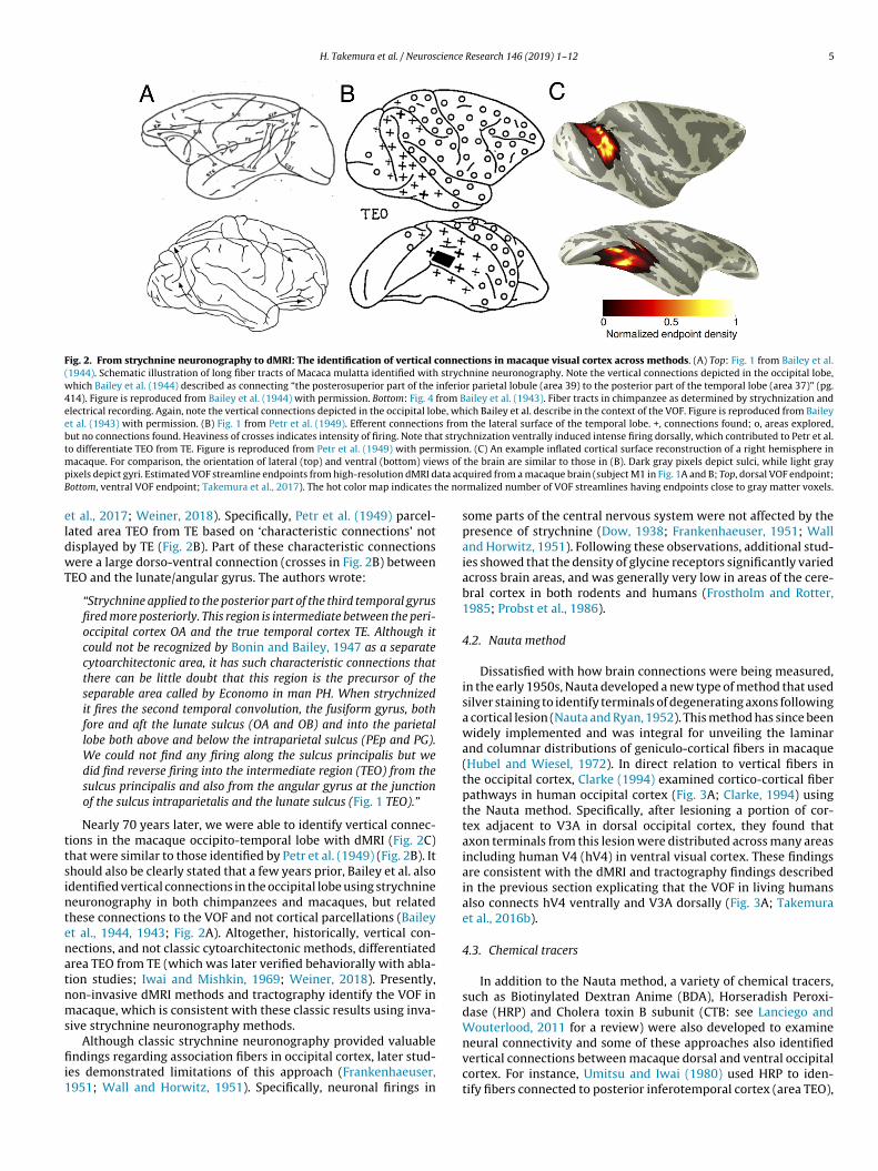
H. Takemura et al. / Neuroscience Research 146 (2019) 1–12 5
Fig. 2. From strychnine neuronography to dMRI: The identification of vertical connections in macaque visual cortex across methods. (A) Top: Fig. 1 from Bailey et al.(1944). Schematic illustration of long fiber tracts of Macaca mulatta identified with strychnine neuronography. Note the vertical connections depicted in the occipital lobe,which Bailey et al. (1944) described as connecting “the posterosuperior part of the inferior parietal lobule (area 39) to the posterior part of the temporal lobe (area 37)” (pg.414). Figure is reproduced from Bailey et al. (1944) with permission. Bottom: Fig. 4 from Bailey et al. (1943). Fiber tracts in chimpanzee as determined by strychnization andelectrical recording. Again, note the vertical connections depicted in the occipital lobe, which Bailey et al. describe in the context of the VOF. Figure is reproduced from Baileyet al. (1943) with permission. (B) Fig. 1 from Petr et al. (1949). Efferent connections from the lateral surface of the temporal lobe. +, connections found; o, areas explored,but no connections found. Heaviness of crosses indicates intensity of firing. Note that strychnization ventrally induced intense firing dorsally, which contributed to Petr et al.to differentiate TEO from TE. Figure is reproduced from Petr et al. (1949) with permission. (C) An example inflated cortical surface reconstruction of a right hemisphere inm ws of
p ata acB he no
eldwT
ttsintenatnms
fii1
acaque. For comparison, the orientation of lateral (top) and ventral (bottom) vieixels depict gyri. Estimated VOF streamline endpoints from high-resolution dMRI dottom, ventral VOF endpoint; Takemura et al., 2017). The hot color map indicates t
t al., 2017; Weiner, 2018). Specifically, Petr et al. (1949) parcel-ated area TEO from TE based on ‘characteristic connections’ notisplayed by TE (Fig. 2B). Part of these characteristic connectionsere a large dorso-ventral connection (crosses in Fig. 2B) between
EO and the lunate/angular gyrus. The authors wrote:
“Strychnine applied to the posterior part of the third temporal gyrusfired more posteriorly. This region is intermediate between the peri-occipital cortex OA and the true temporal cortex TE. Although itcould not be recognized by Bonin and Bailey, 1947 as a separatecytoarchitectonic area, it has such characteristic connections thatthere can be little doubt that this region is the precursor of theseparable area called by Economo in man PH. When strychnizedit fires the second temporal convolution, the fusiform gyrus, bothfore and aft the lunate sulcus (OA and OB) and into the parietallobe both above and below the intraparietal sulcus (PEp and PG).We could not find any firing along the sulcus principalis but wedid find reverse firing into the intermediate region (TEO) from thesulcus principalis and also from the angular gyrus at the junctionof the sulcus intraparietalis and the lunate sulcus (Fig. 1 TEO).”
Nearly 70 years later, we were able to identify vertical connec-ions in the macaque occipito-temporal lobe with dMRI (Fig. 2C)hat were similar to those identified by Petr et al. (1949) (Fig. 2B). Ithould also be clearly stated that a few years prior, Bailey et al. alsodentified vertical connections in the occipital lobe using strychnineeuronography in both chimpanzees and macaques, but relatedhese connections to the VOF and not cortical parcellations (Baileyt al., 1944, 1943; Fig. 2A). Altogether, historically, vertical con-ections, and not classic cytoarchitectonic methods, differentiatedrea TEO from TE (which was later verified behaviorally with abla-ion studies; Iwai and Mishkin, 1969; Weiner, 2018). Presently,on-invasive dMRI methods and tractography identify the VOF inacaque, which is consistent with these classic results using inva-
ive strychnine neuronography methods.
Although classic strychnine neuronography provided valuablendings regarding association fibers in occipital cortex, later stud-es demonstrated limitations of this approach (Frankenhaeuser,951; Wall and Horwitz, 1951). Specifically, neuronal firings in
the brain are similar to those in (B). Dark gray pixels depict sulci, while light grayquired from a macaque brain (subject M1 in Fig. 1A and B; Top, dorsal VOF endpoint;rmalized number of VOF streamlines having endpoints close to gray matter voxels.
some parts of the central nervous system were not affected by thepresence of strychnine (Dow, 1938; Frankenhaeuser, 1951; Walland Horwitz, 1951). Following these observations, additional stud-ies showed that the density of glycine receptors significantly variedacross brain areas, and was generally very low in areas of the cere-bral cortex in both rodents and humans (Frostholm and Rotter,1985; Probst et al., 1986).
4.2. Nauta method
Dissatisfied with how brain connections were being measured,in the early 1950s, Nauta developed a new type of method that usedsilver staining to identify terminals of degenerating axons followinga cortical lesion (Nauta and Ryan, 1952). This method has since beenwidely implemented and was integral for unveiling the laminarand columnar distributions of geniculo-cortical fibers in macaque(Hubel and Wiesel, 1972). In direct relation to vertical fibers inthe occipital cortex, Clarke (1994) examined cortico-cortical fiberpathways in human occipital cortex (Fig. 3A; Clarke, 1994) usingthe Nauta method. Specifically, after lesioning a portion of cor-tex adjacent to V3A in dorsal occipital cortex, they found thataxon terminals from this lesion were distributed across many areasincluding human V4 (hV4) in ventral visual cortex. These findingsare consistent with the dMRI and tractography findings describedin the previous section explicating that the VOF in living humansalso connects hV4 ventrally and V3A dorsally (Fig. 3A; Takemuraet al., 2016b).
4.3. Chemical tracers
In addition to the Nauta method, a variety of chemical tracers,such as Biotinylated Dextran Anime (BDA), Horseradish Peroxi-dase (HRP) and Cholera toxin B subunit (CTB: see Lanciego andWouterlood, 2011 for a review) were also developed to examine
neural connectivity and some of these approaches also identifiedvertical connections between macaque dorsal and ventral occipitalcortex. For instance, Umitsu and Iwai (1980) used HRP to iden-tify fibers connected to posterior inferotemporal cortex (area TEO),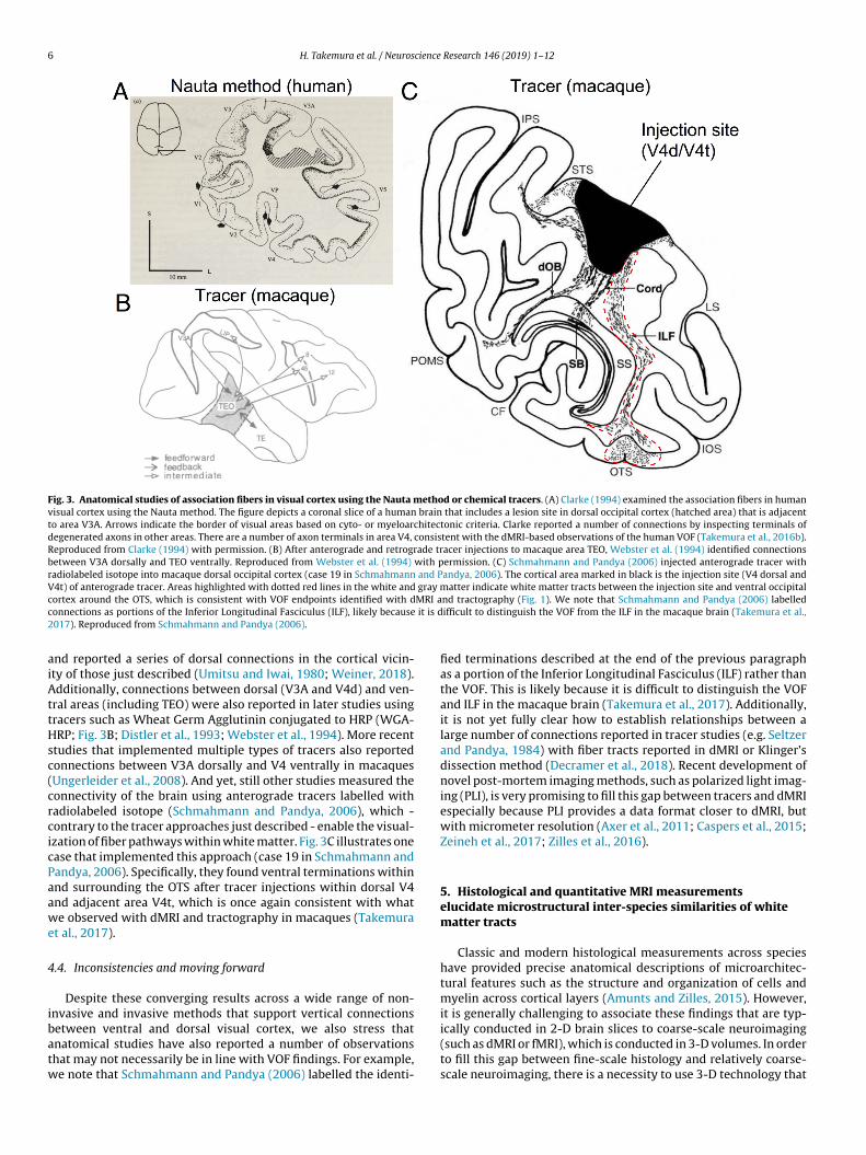
6 H. Takemura et al. / Neuroscience Research 146 (2019) 1–12
Fig. 3. Anatomical studies of association fibers in visual cortex using the Nauta method or chemical tracers. (A) Clarke (1994) examined the association fibers in humanvisual cortex using the Nauta method. The figure depicts a coronal slice of a human brain that includes a lesion site in dorsal occipital cortex (hatched area) that is adjacentto area V3A. Arrows indicate the border of visual areas based on cyto- or myeloarchitectonic criteria. Clarke reported a number of connections by inspecting terminals ofdegenerated axons in other areas. There are a number of axon terminals in area V4, consistent with the dMRI-based observations of the human VOF (Takemura et al., 2016b).Reproduced from Clarke (1994) with permission. (B) After anterograde and retrograde tracer injections to macaque area TEO, Webster et al. (1994) identified connectionsbetween V3A dorsally and TEO ventrally. Reproduced from Webster et al. (1994) with permission. (C) Schmahmann and Pandya (2006) injected anterograde tracer withradiolabeled isotope into macaque dorsal occipital cortex (case 19 in Schmahmann and Pandya, 2006). The cortical area marked in black is the injection site (V4 dorsal andV4t) of anterograde tracer. Areas highlighted with dotted red lines in the white and gray matter indicate white matter tracts between the injection site and ventral occipitalc MRI ac it is d2
aiAttHsc(crcicPaawe
4
ibatw
ortex around the OTS, which is consistent with VOF endpoints identified with donnections as portions of the Inferior Longitudinal Fasciculus (ILF), likely because017). Reproduced from Schmahmann and Pandya (2006).
nd reported a series of dorsal connections in the cortical vicin-ty of those just described (Umitsu and Iwai, 1980; Weiner, 2018).dditionally, connections between dorsal (V3A and V4d) and ven-
ral areas (including TEO) were also reported in later studies usingracers such as Wheat Germ Agglutinin conjugated to HRP (WGA-RP; Fig. 3B; Distler et al., 1993; Webster et al., 1994). More recent
tudies that implemented multiple types of tracers also reportedonnections between V3A dorsally and V4 ventrally in macaquesUngerleider et al., 2008). And yet, still other studies measured theonnectivity of the brain using anterograde tracers labelled withadiolabeled isotope (Schmahmann and Pandya, 2006), which -ontrary to the tracer approaches just described - enable the visual-zation of fiber pathways within white matter. Fig. 3C illustrates onease that implemented this approach (case 19 in Schmahmann andandya, 2006). Specifically, they found ventral terminations withinnd surrounding the OTS after tracer injections within dorsal V4nd adjacent area V4t, which is once again consistent with whate observed with dMRI and tractography in macaques (Takemura
t al., 2017).
.4. Inconsistencies and moving forward
Despite these converging results across a wide range of non-nvasive and invasive methods that support vertical connections
etween ventral and dorsal visual cortex, we also stress thatnatomical studies have also reported a number of observationshat may not necessarily be in line with VOF findings. For example,e note that Schmahmann and Pandya (2006) labelled the identi-nd tractography (Fig. 1). We note that Schmahmann and Pandya (2006) labelledifficult to distinguish the VOF from the ILF in the macaque brain (Takemura et al.,
fied terminations described at the end of the previous paragraphas a portion of the Inferior Longitudinal Fasciculus (ILF) rather thanthe VOF. This is likely because it is difficult to distinguish the VOFand ILF in the macaque brain (Takemura et al., 2017). Additionally,it is not yet fully clear how to establish relationships between alarge number of connections reported in tracer studies (e.g. Seltzerand Pandya, 1984) with fiber tracts reported in dMRI or Klinger’sdissection method (Decramer et al., 2018). Recent development ofnovel post-mortem imaging methods, such as polarized light imag-ing (PLI), is very promising to fill this gap between tracers and dMRIespecially because PLI provides a data format closer to dMRI, butwith micrometer resolution (Axer et al., 2011; Caspers et al., 2015;Zeineh et al., 2017; Zilles et al., 2016).
5. Histological and quantitative MRI measurementselucidate microstructural inter-species similarities of whitematter tracts
Classic and modern histological measurements across specieshave provided precise anatomical descriptions of microarchitec-tural features such as the structure and organization of cells andmyelin across cortical layers (Amunts and Zilles, 2015). However,it is generally challenging to associate these findings that are typ-
ically conducted in 2-D brain slices to coarse-scale neuroimaging(such as dMRI or fMRI), which is conducted in 3-D volumes. In orderto fill this gap between fine-scale histology and relatively coarse-scale neuroimaging, there is a necessity to use 3-D technology that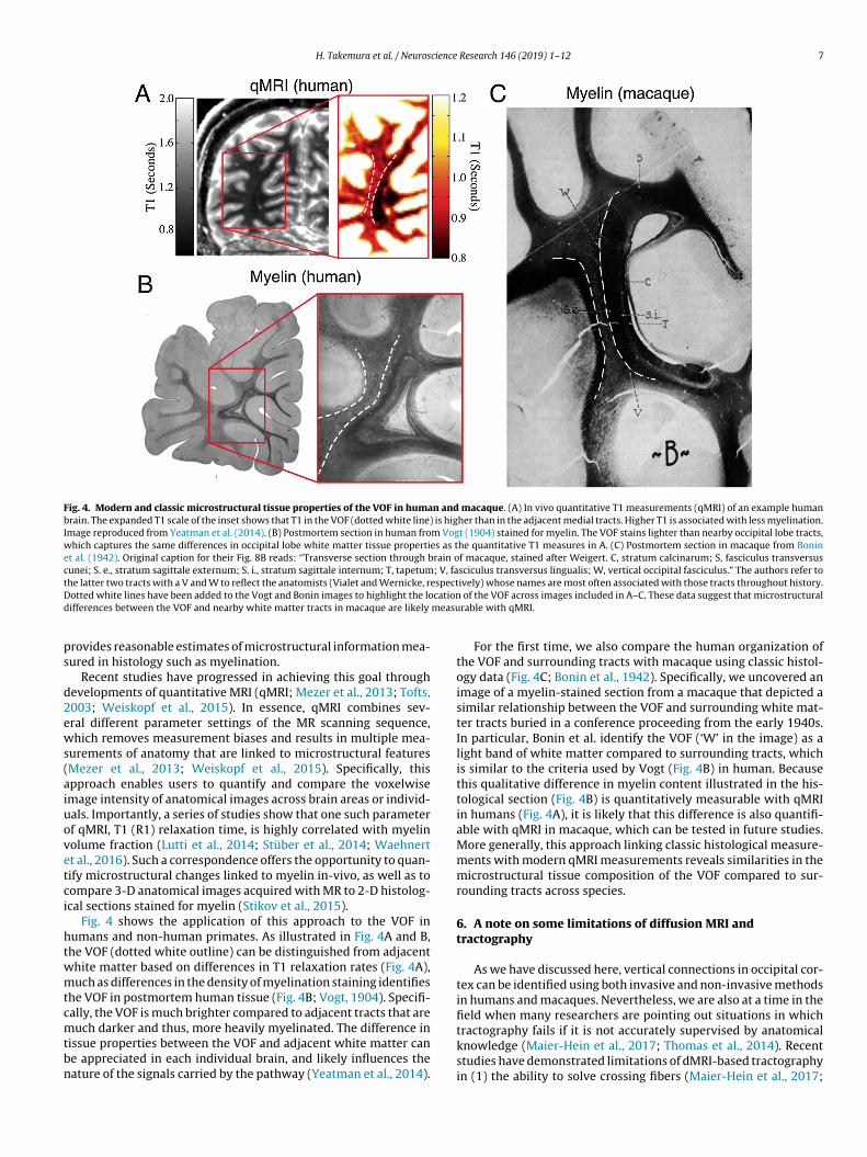
H. Takemura et al. / Neuroscience Research 146 (2019) 1–12 7
Fig. 4. Modern and classic microstructural tissue properties of the VOF in human and macaque. (A) In vivo quantitative T1 measurements (qMRI) of an example humanbrain. The expanded T1 scale of the inset shows that T1 in the VOF (dotted white line) is higher than in the adjacent medial tracts. Higher T1 is associated with less myelination.Image reproduced from Yeatman et al. (2014). (B) Postmortem section in human from Vogt (1904) stained for myelin. The VOF stains lighter than nearby occipital lobe tracts,which captures the same differences in occipital lobe white matter tissue properties as the quantitative T1 measures in A. (C) Postmortem section in macaque from Boninet al. (1942). Original caption for their Fig. 8B reads: ‘’Transverse section through brain of macaque, stained after Weigert. C, stratum calcinarum; S, fasciculus transversuscunei; S. e., stratum sagittale externum; S. i., stratum sagittale internum; T, tapetum; V, fasciculus transversus lingualis; W, vertical occipital fasciculus.” The authors refer tot espectD cationd measu
ps
d2ews(aiuovetci
htwmtcmtbn
he latter two tracts with a V and W to reflect the anatomists (Vialet and Wernicke, rotted white lines have been added to the Vogt and Bonin images to highlight the loifferences between the VOF and nearby white matter tracts in macaque are likely
rovides reasonable estimates of microstructural information mea-ured in histology such as myelination.
Recent studies have progressed in achieving this goal throughevelopments of quantitative MRI (qMRI; Mezer et al., 2013; Tofts,003; Weiskopf et al., 2015). In essence, qMRI combines sev-ral different parameter settings of the MR scanning sequence,hich removes measurement biases and results in multiple mea-
urements of anatomy that are linked to microstructural featuresMezer et al., 2013; Weiskopf et al., 2015). Specifically, thispproach enables users to quantify and compare the voxelwisemage intensity of anatomical images across brain areas or individ-als. Importantly, a series of studies show that one such parameterf qMRI, T1 (R1) relaxation time, is highly correlated with myelinolume fraction (Lutti et al., 2014; Stüber et al., 2014; Waehnertt al., 2016). Such a correspondence offers the opportunity to quan-ify microstructural changes linked to myelin in-vivo, as well as toompare 3-D anatomical images acquired with MR to 2-D histolog-cal sections stained for myelin (Stikov et al., 2015).
Fig. 4 shows the application of this approach to the VOF inumans and non-human primates. As illustrated in Fig. 4A and B,he VOF (dotted white outline) can be distinguished from adjacenthite matter based on differences in T1 relaxation rates (Fig. 4A),uch as differences in the density of myelination staining identifies
he VOF in postmortem human tissue (Fig. 4B; Vogt, 1904). Specifi-ally, the VOF is much brighter compared to adjacent tracts that are
uch darker and thus, more heavily myelinated. The difference inissue properties between the VOF and adjacent white matter cane appreciated in each individual brain, and likely influences theature of the signals carried by the pathway (Yeatman et al., 2014).
ively) whose names are most often associated with those tracts throughout history. of the VOF across images included in A–C. These data suggest that microstructuralrable with qMRI.
For the first time, we also compare the human organization ofthe VOF and surrounding tracts with macaque using classic histol-ogy data (Fig. 4C; Bonin et al., 1942). Specifically, we uncovered animage of a myelin-stained section from a macaque that depicted asimilar relationship between the VOF and surrounding white mat-ter tracts buried in a conference proceeding from the early 1940s.In particular, Bonin et al. identify the VOF (‘W’ in the image) as alight band of white matter compared to surrounding tracts, whichis similar to the criteria used by Vogt (Fig. 4B) in human. Becausethis qualitative difference in myelin content illustrated in the his-tological section (Fig. 4B) is quantitatively measurable with qMRIin humans (Fig. 4A), it is likely that this difference is also quantifi-able with qMRI in macaque, which can be tested in future studies.More generally, this approach linking classic histological measure-ments with modern qMRI measurements reveals similarities in themicrostructural tissue composition of the VOF compared to sur-rounding tracts across species.
6. A note on some limitations of diffusion MRI andtractography
As we have discussed here, vertical connections in occipital cor-tex can be identified using both invasive and non-invasive methodsin humans and macaques. Nevertheless, we are also at a time in thefield when many researchers are pointing out situations in which
tractography fails if it is not accurately supervised by anatomicalknowledge (Maier-Hein et al., 2017; Thomas et al., 2014). Recentstudies have demonstrated limitations of dMRI-based tractographyin (1) the ability to solve crossing fibers (Maier-Hein et al., 2017;
8 cience
Rre2fesfisaticwt
dpatm2au2eoie(te2iee
em2eadteemotnP2att
daeitour
a
H. Takemura et al. / Neuros
oebroeck et al., 2008), (2) its dependency on tractography algo-ithms or parameter settings (Bastiani et al., 2012; Chamberlandt al., 2014; Domin et al., 2014; Kunimatsu et al., 2004; Parizel et al.,007; Taoka et al., 2009; Thomas et al., 2014), and (3) its accuracyor estimating fiber projections into cortical gray matter (Reveleyt al., 2015). Importantly, Thomas et al. (2014) pointed out that oneignificant challenge of tractography is a trade-off between speci-city and sensitivity: methods with higher sensitivity are moreusceptible to false positives, while methods with higher specificityre more susceptible to false negatives. We fully acknowledge thatractography has limitations and should be used with great caren order to draw accurate and reproducible neuroanatomical con-lusions. After all, no method is perfect and we remain optimistichen considering the many potential paths forward for reducing
hese limitations.For example, several methodical approaches are currently being
eveloped to solve these established problems. The first - anderhaps the most intuitive approach - is to incorporate priornatomical knowledge when identifying specific white matterracts with tractography that are known to exist from invasive
ethods (Catani et al., 2002; Conturo et al., 1999; Wakana et al.,004). For instance, anatomical knowledge regarding the optic radi-tion was often used to improve the accuracy of the algorithmssed to identify this pathway using tractography (Benjamin et al.,014; Chamberland et al., 2017; Kammen et al., 2016; Sherbondyt al., 2008b). This approach (1) substantially reduced the problemf false positives in tractography, (2) is now widely implementedn several different types of tractography software (Wassermannt al., 2016; Yeatman et al., 2018, 2012; Yendiki et al., 2011), and3) effectively identifies reproducible connections using dMRI (andractography), as well as tracers (Jbabdi et al., 2013; Schmahmannt al., 2007; Takemura et al., 2017; Thiebaut de Schotten et al.,011). However, this approach is only applicable when identify-
ng major white matter tracts that are known to exist and arestablished as accepted fascicles in the wide neuroanatomical lit-rature.
The second approach is to statistically evaluate tractography bystimating how well a set of streamline trajectories predicts theeasured dMRI signal (Caiafa and Pestilli, 2017; Daducci et al.,
015; Pestilli et al., 2014; Sherbondy et al., 2009, 2008a; Smitht al., 2013). For example, Linear Fascicle Evaluation (LiFE; Caiafand Pestilli, 2017; Pestilli et al., 2014) is a method that first predictsMRI signals from connections (streamlines) that are generated byractography algorithms, and then removes streamlines that do notxplain dMRI signals. This approach increases the specificity of tractstimates (Schurr et al., 2018). Furthermore, by also testing howuch the removal of a specific tract reduces the prediction accuracy
f dMRI signals, this approach has provided methods to evaluatehe degree of statistical evidence supporting the identification ofovel white matter tracts identified in living brains (Caiafa andestilli, 2017; Gomez et al., 2015; Leong et al., 2016; Pestilli et al.,014; Takemura et al., 2016a; Uesaki et al., 2018). Of course, welso acknowledge that a statistically significant model only meanshat the model can explain data, but does not fully guarantee thathe model is anatomically correct (Daducci et al., 2016).
In our view, these two approaches are complementary. MacaqueMRI is an ideal case for integrating the strength of these twopproaches to draw careful conclusions in tractography studies. Forxample, as described in the first approach (and as we reviewedn this paper), we collected established anatomical evidence ofhe macaque VOF from classical and modern studies using vari-us anatomical methods. As described in the second approach, we
sed LiFE to statistically evaluate the evidence of macaque VOF inelation to dMRI signals (Takemura et al., 2017).Finally, we note that a comparison between anatomical studiesnd tractography itself still has significant challenges. Studies com-
Research 146 (2019) 1–12
paring tractography and anatomical tracers often draw completelydifferent conclusions regarding the degree of correlation betweenthe two measurements (Aydogan et al., 2018; Azadbakht et al.,2015; Donahue et al., 2016; Thomas et al., 2014; van den Heuvelet al., 2015). We believe that these apparently conflicting conclu-sions are largely derived from the fact that different groups aretesting different tractography methods, while also using differenttypes of tracer data with respect to (a) tracer types, (b) selectionsof injection sites, and (c) quantification methods. We stress thatdata sharing and open science will continue to enable independentlaboratories to test different methods and the scientific communitycan carefully evaluate problems arising from method selections aswe have done with the VOF and the connections with visual fieldmaps in our previous work (Takemura et al., 2017).
7. Concluding remarks
In this paper, we have reviewed how recent advanced neu-roimaging methods of white matter and classical neuroanatomicalstudies of connectivity can be incorporated together to provideinsights regarding the comparative anatomy of the VOF. While wefocus on the human-macaque comparison of the VOF, the sameapproach can be applied to other white matter tracts across species.In this comparative approach, we also stress the importance of theclassical literature in providing additional support for similaritiesand differences in microstructural and macroanatomical features ofwhite matter across species. Additionally, despite the fact that dMRIand tractography may have some limitations, comparative dMRIstudies with the combination of (a) careful understanding of thehistorical and modern literature of invasive anatomical methodsin humans and macaques, (b) novel statistical evaluation meth-ods, and (c) open sharing of data and methods (Glasser et al.,2016; Majka et al., 2016; Milham et al., 2018; Reveley et al., 2017;Woodward et al., 2018) has the potential to help overcome theselimitations. We believe this combination will continue to advancedMRI-comparative approaches moving forward beyond verticalassociation connections in visual cortex. Future studies implement-ing a comparative dMRI approach as we review here will continueto advance our knowledge regarding what neuroanatomical fea-tures of the brain are shared with other species, as well as whatfeatures are uniquely human.
Competing financial interests
The authors declare no competing financial interests associatedwith this article.
Acknowledgements
H.T. was supported by Japan Society for the Promotion of Science(JSPS) KAKENHI (JP17H04684) and Grant-in-Aid for JSPS Fellows(JP15J00412). F.P. was supported by NSFIIS-1636893, NSFBCS-1734853, NIH NIMHULTTR001108, a Microsoft Research Award, theIndiana University Areas of Emergent Research initiative “Learning:Brains, Machines, Children,” and the Indiana University PervasiveTechnology Institute and Research Technologies Division. K.S.W.was supported by start-up funds from UC Berkeley. We thank DavidA. Leopold, Frank Q. Ye, Georgios A. Keliris and Nikos K. Logo-thetis for providing diffusion MRI dataset from macaque brainsto produce the figures in this article. Human dMRI data used to
produce the figure in this article were provided by Human Connec-tome Project, WU-Minn Consortium (Van Essen, D. and Ugurbil, K.,1U54MH091657). We also thank Kendrick N. Kay for comments onan earlier version of the manuscript.
cience
R
A
A
A
A
A
A
B
B
B
B
B
B
B
B
B
B
B
B
C
C
C
C
C
C
C
C
C
C
C
C
H. Takemura et al. / Neuros
eferences
munts, K., Zilles, K., 2015. Architectonic mapping of the human brain beyond brod-mann. Neuron 88, 1086–1107.
rcaro, M.J., Livingstone, M.S., 2017. Retinotopic organization of scene areas inmacaque inferior temporal cortex. J. Neurosci. 37, 7373–7389.
ssaf, Y., Johansen-Berg, H., Thiebaut de Schotten, M., 2017. The role of diffusionMRI in neuroscience. NMR Biomed., http://dx.doi.org/10.1002/nbm.3762 [Epubahead of print].
xer, M., Amunts, K., Grässel, D., Palm, C., Dammers, J., Axer, H., Pietrzyk, U., Zilles,K., 2011. A novel approach to the human connectome: ultra-high resolutionmapping of fiber tracts in the brain. Neuroimage 54, 1091–1101.
ydogan, D.B., Jacobs, R., Dulawa, S., Thompson, S.L., Francois, M.C., Toga, A.W., Dong,H., Knowles, J.A., Shi, Y., 2018. When tractography meets tracer injections: asystematic study of trends and variation sources of diffusion-based connectivity.Brain Struct. Funct. 223, 2841–2858.
zadbakht, H., Parkes, L.M., Haroon, H.A., Augath, M., Logothetis, N.K., de Crespigny,A., D’Arceuil, H.E., Parker, G.J.M., 2015. Validation of high-resolution tractog-raphy against in vivo tracing in the macaque visual cortex. Cereb. Cortex 25,4299–4309.
ailey, P., Bonin, V.G., Davis, E.W., Garol, H.W., Mcculloch, W.S., 1944. Further obser-vations on associational pathways in the brain of Macaca mulatta. J. Neuropathol.Exp. Neurol. 3, 413.
ailey, P., Bonin, V.G., Garol, H.W., McCulloch, W.S., 1943. Long association fibers incerebral hemispheres of monkey and chimpanzee. J. Neurophysiol. 6, 129–134.
artsch, A.J., Geletneky, K., Jbabdi, S., 2013. The temporoparietal fiber intersectionarea and wernicke perpendicular fasciculus. Neurosurgery 73, E381–E382.
astiani, M., Shah, N.J., Goebel, R., Roebroeck, A., 2012. Human cortical connec-tome reconstruction from diffusion weighted MRI: the effect of tractographyalgorithm. Neuroimage 62, 1732–1749.
ehrens, T.E.J., Woolrich, M.W., Jenkinson, M., Johansen-Berg, H., Nunes, R.G., Clare,S., Matthews, P.M., Brady, J.M., Smith, S.M., 2003. Characterization and propa-gation of uncertainty in diffusion-weighted MR imaging. Magn. Reson. Med. 50,1077–1088.
enjamin, C.F., Singh, J.M., Prabhu, S.P., Warfield, S.K., 2014. Optimization of trac-tography of the optic radiations. Hum. Brain Mapp. 35, 683–697.
erns, G.S., Cook, P.F., Foxley, S., Jbabdi, S., Miller, K.L., Marino, L., 2015. Diffusiontensor imaging of dolphin brains reveals direct auditory pathway to temporallobe. Proc. R. Soc. B 282, 1203.
etzel, R.F., Medaglia, J.D., Bassett, D.S., 2018. Diversity of meso-scale architecturein human and non-human connectomes. Nat. Commun. 9, 346.
onin, V.G., Bailey, P., 1947. The Neocortex of Macaca Mulatta. University of IllinoisPress.
onin, V.G., Garol, H.W., McCulloch, W.S., 1942. The functional organization of theoccipital lobe. In: Klüver, H. (Ed.), Biological Symposia, Vol. VII, Visual Mecha-nisms. The Jaques Cattell Press, Lancaster, Pennsylvania, pp. 165–192.
rewer, A.A., Press, W.A., Logothetis, N.K., Wandell, B.A., 2002. Visual areas inmacaque cortex measured using functional magnetic resonance imaging. J. Neu-rosci. 22, 10416–10426.
udisavljevic, S., Dell’Acqua, F., Castiello, U., 2018. Cross-talk connections underlyingdorsal and ventral stream integration during hand actions. Cortex 103, 224–239.
aiafa, C.F., Pestilli, F., 2017. Multidimensional encoding of brain connectomes. Sci.Rep. 7, 11491.
alabrese, E., Badea, A., Cofer, G., Qi, Y., Johnson, G.A., 2015. A diffusion mri trac-tography connectome of the mouse brain and comparison with neuronal tracerdata. Cereb. Cortex 25, 4628–4637.
aspers, S., Axer, M., Caspers, J., Jockwitz, C., Jütten, K., Reckfort, J., Grässel, D.,Amunts, K., Zilles, K., 2015. Target sites for transcallosal fibers in human visualcortex—a combined diffusion and polarized light imaging study. Cortex 72,40–53.
atani, M., Thiebaut de Schotten, M., 2012. Atlas of Human Brain Connections. OxfordUniversity Press.
atani, M., Howard, R.J., Pajevic, S., Jones, D.K., 2002. Virtual in vivo interactivedissection of white matter fasciculi in the human brain. Neuroimage 17, 77–94.
atani, M., Robertsson, N., Beyh, A., Huynh, V., de Santiago Requejo, F., Howells, H.,Barrett, R.L.C., Aiello, M., Cavaliere, C., Dyrby, T.B., Krug, K., Ptito, M., D’Arceuil, H.,Forkel, S.J., Dell’Acqua, F., 2017a. Short parietal lobe connections of the humanand monkey brain. Cortex 97, 339–357.
atani, M., Yaghi, Z., Jo, Y., Beyh, A., De Santiago Requejo, F., Forkel, S., Ffytche, D.,2017b. The anatomy of the vertical occipital system. In: Organization for HumanBrain Mapping, Vancouver.
hamberland, M., Scherrer, B., Prabhu, S.P., Madsen, J., Fortin, D., Whittingstall, K.,Descoteaux, M., Warfield, S.K., 2017. Active delineation of Meyer’s loop usingoriented priors through MAGNEtic tractography (MAGNET). Hum. Brain Mapp.38, 509–527.
hamberland, M., Whittingstall, K., Fortin, D., Mathieu, D., Descoteaux, M., 2014.Real-time multi-peak tractography for instantaneous connectivity display.Front. Neuroinform. 8, 59.
larke, S., 1994. Association and intrinsic connections of human extrastriate visualcortex. Proc. Biol. Sci. 257, 87–92.
onturo, T.E., Lori, N.F., Cull, T.S., Akbudak, E., Snyder, A.Z., Shimony, J.S., McKinstry,
R.C., Burton, H., Raichle, M.E., 1999. Tracking neuronal fiber pathways in theliving human brain. Proc. Natl. Acad. Sci. U. S. A. 96, 10422–10427.ottereau, B.R., Smith, A.T., Rima, S., Fize, D., Héjja-Brichard, Y., Renaud, L., Lejards, C.,Vayssière, N., Trotter, Y., Durand, J.-B., 2017. Processing of egomotion-consistentoptic flow in the rhesus macaque Cortex. Cereb. Cortex 27, 330–343.
Research 146 (2019) 1–12 9
Crick, F., Jones, E., 1993. Backwardness of human neuroanatomy. Nature 361,109–110.
Croxson, P.L., Johansen-Berg, H., Behrens, T.E.J., Robson, M.D., Pinsk, M.A., Gross,C.G., Richter, W., Richter, M.C., Kastner, S., Rushworth, M.F.S., 2005. Quantitativeinvestigation of connections of the prefrontal cortex in the human and macaqueusing probabilistic diffusion tractography. J. Neurosci. 25, 8854–8866.
Cuaya, L.V., Hernández-Pérez, R., Concha, L., 2016. Our faces in the dog’s brain: func-tional imaging reveals temporal cortex activation during perception of humanfaces. PLoS One 11, e0149431.
Daducci, A., Dal Palú, A., Descoteaux, M., Thiran, J.-P., 2016. Microstructure informedtractography: pitfalls and open challenges. Front. Neurosci. 10, 247.
Daducci, A., Dal Palù, A., Lemkaddem, A., Thiran, J.-P., 2015. COMMIT: convex opti-mization modeling for microstructure informed tractography. IEEE Trans. Med.Imaging 34, 246–257.
Datta, R., Lee, J., Duda, J., Avants, B.B., Vite, C.H., Tseng, B., Gee, J.C., Aguirre, G.D.,Aguirre, G.K., 2012. A digital atlas of the dog brain. PLoS One 7, e52140.
de Barenne, J.G.D., McCulloch, W.S., 1939. Physiological delimitation of neurones inthe central nervous system. Am. J. Physiol. 127, 620–628.
de Sousa, A.A., Sherwood, C.C., Schleicher, A., Amunts, K., MacLeod, C.E., Hof, P.R.,Zilles, K., 2010. Comparative cytoarchitectural analyses of striate and extrastri-ate areas in hominoids. Cereb. Cortex 20, 966–981.
De Valois, R.L., Jacobs, G.H., 1968. Primate color vision. Science 162, 533–540.De Valois, R.L., Morgan, H.C., Polson, M.C., Mead, W.R., Hull, E.M., 1974. Psychophys-
ical studies of monkey vision—I. Macaque luminosity and color vision tests. Vis.Res. 14, 53–67.
DeAngelis, G.C., Newsome, W.T., 1999. Organization of disparity-selective neuronsin macaque area MT. J. Neurosci. 19, 1398–1415.
Decramer, T., Swinnen, S., van Loon, J., Janssen, P., Theys, T., 2018. White matter tractanatomy in the rhesus monkey: a fiber dissection study. Brain Struct. Funct. 223,3681–3688.
Distler, C., Boussaoud, D., Desimone, R., Ungerleider, L.G., 1993. Cortical connec-tions of inferior temporal area TEO in macaque monkeys. J. Comp. Neurol. 334,125–150.
Domin, M., Langner, S., Hosten, N., Lotze, M., 2014. Comparison of parameter thresh-old combinations for diffusion tensor tractography in chronic stroke patients andhealthy subjects. PLoS One 9, e98211.
Donahue, C.J., Sotiropoulos, S.N., Jbabdi, S., Hernandez-Fernandez, M., Behrens, T.E.,Dyrby, T.B., Coalson, T., Kennedy, H., Knoblauch, K., Van Essen, D.C., Glasser, M.F.,2016. Using diffusion tractography to predict cortical connection strength anddistance: a quantitative comparison with tracers in the monkey. J. Neurosci. 36,6758–6770.
Dow, R.S., 1938. The electrical activity of the cerebellum and its functional signifi-cance. J. Physiol. 94, 67–86.
Duan, Y., Norcia, A.M., Yeatman, J.D., Mezer, A., 2015. The structural properties ofmajor white matter tracts in strabismic amblyopia. Invest. Ophthalmol. Vis. Sci.56, 5152–5160.
Felleman, D.J., Van Essen, D.C., 1991. Distributed hierarchical processing in the pri-mate cerebral cortex. Cereb. Cortex 1, 1–47.
Frankenhaeuser, B., 1951. Limitations of method of strychnine neuronography. J.Neurophysiol. 14, 73–79.
Frostholm, A., Rotter, A., 1985. Glycine receptor distribution in mouse CNS: autora-diographic localization of [3H]strychnine binding sites. Brain Res. Bull. 15,473–486.
Fujita, I., Tanaka, K., Ito, M., Cheng, K., 1992. Columns for visual features of objectsin monkey inferotemporal cortex. Nature 360, 343–346.
Girard, G., Daducci, A., Petit, L., Thiran, J.-P., Whittingstall, K., Deriche, R., Wasser-mann, D., Descoteaux, M., 2017. AxTract: toward microstructure informedtractography. Hum. Brain Mapp. 38, 5485–5500.
Glasser, M.F., Goyal, M.S., Preuss, T.M., Raichle, M.E., Van Essen, D.C., 2014. Trends andproperties of human cerebral cortex: correlations with cortical myelin content.Neuroimage 93 (Pt. 2), 165–175.
Glasser, M.F., Smith, S.M., Marcus, D.S., Andersson, J.L.R., Auerbach, E.J., Behrens,T.E.J., Coalson, T.S., Harms, M.P., Jenkinson, M., Moeller, S., Robinson, E.C.,Sotiropoulos, S.N., Xu, J., Yacoub, E., Ugurbil, K., Van Essen, D.C., 2016. The humanconnectome project’s neuroimaging approach. Nat. Neurosci. 19, 1175–1187.
Goda, N., Tachibana, A., Okazawa, G., Komatsu, H., 2014. Representation of the mate-rial properties of objects in the visual cortex of nonhuman primates. J. Neurosci.34, 2660–2673.
Gomez, J., Pestilli, F., Witthoft, N., Golarai, G., Liberman, A., Poltoratski, S., Yoon, J.,Grill-Spector, K., 2015. Functionally defined white matter reveals segregatedpathways in human ventral temporal cortex associated with category-specificprocessing. Neuron 85, 216–227.
Gross, C.G., 1998. Brain, Vision, Memory: Tales in the History of Neuroscience. MITPress, Cambridge, MA.
Güngör, A., Baydin, S., Middlebrooks, E.H., Tanriover, N., Isler, C., Rhoton Jr., A.L.,2017. The white matter tracts of the cerebrum in ventricular surgery and hydro-cephalus. J. Neurosurg. 126, 945–971.
Hecht, E.E., Gutman, D.A., Bradley, B.A., Preuss, T.M., Stout, D., 2015. Virtual dis-section and comparative connectivity of the superior longitudinal fasciculus inchimpanzees and humans. Neuroimage 108, 124–137.
Henschen, S.E., 1893. On the visual path and centre. Brain 16, 170–180.
Hofer, S., Merboldt, K.-D., Tammer, R., Frahm, J., 2008. Rhesus monkey and humanshare a similar topography of the corpus callosum as revealed by diffusion tensorMRI in vivo. Cereb. Cortex 18, 1079–1084.
Holmes, G., Lister, W.T., 1916. Disturbances of vision from cerebral lesions, withspecial reference to the cortical representation of the macula. Brain 39, 34–73.

1 cience
H
H
H
I
I
J
J
J
J
K
K
K
K
K
K
K
K
L
L
L
L
L
L
L
L
L
L
L
M
0 H. Takemura et al. / Neuros
ubel, D.H., Livingstone, M.S., 1987. Segregation of form, color, and stereopsis inprimate area 18. J. Neurosci. 7, 3378–3415.
ubel, D.H., Wiesel, T.N., 1972. Laminar and columnar distribution of geniculo-cortical fibers in the macaque monkey. J. Comp. Neurol. 146, 421–450.
ung, C.-C., Yen, C.C., Ciuchta, J.L., Papoti, D., Bock, N.A., Leopold, D.A., Silva, A.C.,2015. Functional mapping of face-selective regions in the extrastriate visualcortex of the marmoset. J. Neurosci. 35, 1160–1172.
nouye, T., 1909. Die Sehstroungen bei Schussverietzungen der kortikalenSehsphare. W. Engelmann, Leipzig, Germany.
wai, E., Mishkin, M., 1969. Further evidence on the locus of the visual area in thetemporal lobe of the monkey. Exp. Neurol. 25, 585–594.
babdi, S., Johansen-Berg, H., 2011. Tractography: where do we go from here? BrainConnect. 1, 169–183.
babdi, S., Lehman, J.F., Haber, S.N., Behrens, T.E., 2013. Human and monkey ventralprefrontal fibers use the same organizational principles to reach their targets:tracing versus tractography. J. Neurosci. 33, 3190–3201.
babdi, S., Sotiropoulos, S.N., Haber, S.N., Van Essen, D.C., Behrens, T.E., 2015. Mea-suring macroscopic brain connections in vivo. Nat. Neurosci. 18, 1546–1555.
ones, D.K., Alexander, D.C., Bowtell, R., Cercignani, M., Dell’Acqua, F., McHugh, D.J.,Miller, K.L., Palombo, M., Parker, G.J.M., Rudrapatna, U.S., Tax, C.M.W., 2018.Microstructural imaging of the human brain with a “super-scanner”: 10 keyadvantages of ultra-strong gradients for diffusion MRI. Neuroimage 182, 8–38.
aas, J.H., 2013. The evolution of brains from early mammals to humans. WIREs CognSci 4, 33–45.
ammen, A., Law, M., Tjan, B.S., Toga, A.W., Shi, Y., 2016. Automated retinofugalvisual pathway reconstruction with multi-shell HARDI and FOD-based analysis.Neuroimage 125, 767–779.
endrick, K.M., 1991. How the sheep’s brain controls the visual recognition of ani-mals and humans. J. Anim. Sci. 69, 5008–5016.
endrick, K.M., da Costa, A.P., Leigh, A.E., Hinton, M.R., Peirce, J.W., 2001. Sheep don’tforget a face. Nature 414, 165.
olster, H., Janssens, T., Orban, G.A., Vanduffel, W., 2014. The retinotopic organiza-tion of macaque occipitotemporal cortex anterior to V4 and caudoventral to themiddle temporal (MT) cluster. J. Neurosci. 34, 10168–10191.
omatsu, H., Wurtz, R.H., 1988. Relation of cortical areas MT and MST to pursuit eyemovements. I. Localization and visual properties of neurons. J. Neurophysiol. 60,580–603.
riegeskorte, N., Mur, M., Ruff, D.A., Kiani, R., Bodurka, J., Esteky, H., Tanaka, K.,Bandettini, P.A., 2008. Matching categorical object representations in inferiortemporal cortex of man and monkey. Neuron 60, 1126–1141.
unimatsu, A., Aoki, S., Masutani, Y., Abe, O., Hayashi, N., Mori, H., Masumoto, T.,Ohtomo, K., 2004. The optimal trackability threshold of fractional anisotropy fordiffusion tensor tractography of the corticospinal tract. Magn. Reson. Med. Sci.3, 11–17.
anciego, J.L., Wouterlood, F.G., 2011. A half century of experimental neuroanatom-ical tracing. J. Chem. Neuroanat. 42, 157–183.
ee Masson, H., Wallraven, C., Petit, L., 2017. “Can touch this”: cross-modal shapecategorization performance is associated with microstructural characteristics ofwhite matter association pathways. Hum. Brain Mapp. 38, 842–854.
eong, J.K., Pestilli, F., Wu, C.C., Samanez-Larkin, G.R., Knutson, B., 2016. White-matter tract connecting anterior insula to nucleus accumbens correlates withreduced preference for positively skewed gambles. Neuron 89, 63–69.
eopold, D.A., Mitchell, J.F., Freiwald, W.A., 2017. Evolved mechanisms of high-levelvisual perception in primates. In: Kaas, J. (Ed.), Evolution of Nervous System. ,2nd edition, pp. 203–235.
i, L., Hu, X., Preuss, T.M., Glasser, M.F., Damen, F.W., Qiu, Y., Rilling, J., 2013. Map-ping putative hubs in human, chimpanzee and rhesus macaque connectomesvia diffusion tractography. Neuroimage 80, 462–474.
ivingstone, M., Hubel, D., 1988. Segregation of form, color, movement, and depth:anatomy, physiology, and perception. Science 240, 740–749.
ogothetis, N.K., Wandell, B.A., 2004. Interpreting the BOLD signal. Annu. Rev. Phys-iol. 66, 735–769.
ogothetis, N.K., Pauls, J., Augath, M., Trinath, T., Oeltermann, A., 2001. Neurophysi-ological investigation of the basis of the fMRI signal. Nature 412, 150–157.
uppino, G., Ben Hamed, S., Gamberini, M., Matelli, M., Galletti, C., 2005. Occipital(V6) and parietal (V6A) areas in the anterior wall of the parieto-occipital sulcusof the macaque: a cytoarchitectonic study. Eur. J. Neurosci. 21, 3056–3076.
utti, A., Dick, F., Sereno, M.I., Weiskopf, N., 2014. Using high-resolution quantita-tive mapping of R1 as an index of cortical myelination. Neuroimage 93 (Pt. 2),176–188.
yon, D.C., 2009. The evolution of visual cortex and visual systems. In: Kaas, J.H. (Ed.),Evolutionary Neuroscience. , pp. 757–792.
aier-Hein, K.H., Neher, P.F., Houde, J.-C., Côté, M.-A., Garyfallidis, E., Zhong, J.,Chamberland, M., Yeh, F.-C., Lin, Y.-C., Ji, Q., Reddick, W.E., Glass, J.O., Chen,D.Q., Feng, Y., Gao, C., Wu, Y., Ma, J., Renjie, H., Li, Q., Westin, C.-F., Deslauriers-Gauthier, S., Omar Ocegueda González, J., Paquette, M., St-Jean, S., Girard, G.,Rheault, F., Sidhu, J., Tax, C.M.W., Guo, F., Mesri, H.Y., Dávid, S., Froeling, M.,Heemskerk, A.M., Leemans, A., Boré, A., Pinsard, B., Bedetti, C., Desrosiers, M.,Brambati, S., Doyon, J., Sarica, A., Vasta, R., Cerasa, A., Quattrone, A., Yeatman,J., Khan, A.R., Hodges, W., Alexander, S., Romascano, D., Barakovic, M., Auría, A.,Esteban, O., Lemkaddem, A., Thiran, J.-P., Ertan Cetingul, H., Odry, B.L., Mailhe,
B., Nadar, M.S., Pizzagalli, F., Prasad, G., Villalon-Reina, J.E., Galvis, J., Thompson,P.M., De Santiago Requejo, F., Laguna, P.L., Lacerda, L.M., Barrett, R., Dell’Acqua,F., Catani, M., Petit, L., Caruyer, E., Daducci, A., Dyrby, T.B., Holland-Letz, T., Hilge-tag, C.C., Stieltjes, B., Descoteaux, M., 2017. The challenge of mapping the humanconnectome based on diffusion tractography. Nat. Commun. 8, 1349.Research 146 (2019) 1–12
Majka, P., Chaplin, T.A., Yu, H.-H., Tolpygo, A., Mitra, P.P., Wójcik, D.K., Rosa, M.G.P.,2016. Towards a comprehensive atlas of cortical connections in a primate brain:mapping tracer injection studies of the common marmoset into a referencedigital template. J. Comp. Neurol. 524, 2161–2181.
Mangin, J.F., Poupon, C., Cointepas, Y., Riviere, D., Papadopoulos-Orfanos, D., Clark,C.A., Regis, J., Le Bihan, D., 2002. A framework based on spin glass models forthe inference of anatomical connectivity from diffusion-weighted MR data—atechnical review. NMR Biomed. 15, 481–492.
Mars, R.B., Foxley, S., Verhagen, L., Jbabdi, S., Sallet, J., Noonan, M.P., Neubert, F.-X., Andersson, J.L., Croxson, P.L., Dunbar, R.I.M., Khrapitchev, A.A., Sibson, N.R.,Miller, K.L., Rushworth, M.F.S., 2016a. The extreme capsule fiber complex inhumans and macaque monkeys: a comparative diffusion MRI tractographystudy. Brain Struct. Funct. 221, 4059–4071.
Mars, R.B., Verhagen, L., Gladwin, T.E., Neubert, F.-X., Sallet, J., Rushworth, M.F.S.,2016b. Comparing brains by matching connectivity profiles. Neurosci. Biobehav.Rev. 60, 90–97.
Martino, J., Garcia-Porrero, J.A., 2013. In reply: wernicke’s perpendicular fasciculusand vertical portion of the superior longitudinal fasciculus. Neurosurgery 73,E382–E383.
McCulloch, W.S., 1944. The functional organization of the cerebral cortex. Physiol.Rev. 24, 390–407.
McNab, J.A., Edlow, B.L., Witzel, T., Huang, S.Y., Bhat, H., Heberlein, K., Feiweier, T.,Liu, K., Keil, B., Cohen-Adad, J., Tisdall, M.D., Folkerth, R.D., Kinney, H.C., Wald,L.L., 2013. The Human Connectome Project and beyond: initial applications of300 mT/m gradients. Neuroimage 80, 234–245.
Mezer, A., Yeatman, J.D., Stikov, N., Kay, K.N., Cho, N.J., Dougherty, R.F., Perry, M.L.,Parvizi, J., Hua le, H., Butts-Pauly, K., Wandell, B.A., 2013. Quantifying the localtissue volume and composition in individual brains with magnetic resonanceimaging. Nat. Med. 19, 1667–1672.
Milham, M.P., Ai, L., Koo, B., Xu, T., Amiez, C., Balezeau, F., Baxter, M.G., Blezer, E.L.A.,Brochier, T., Chen, A., Croxson, P.L., Damatac, C.G., Dehaene, S., Everling, S., Fair,D.A., Fleysher, L., Freiwald, W., Froudist-Walsh, S., Griffiths, T.D., Guedj, C., Hadj-Bouziane, F., Ben Hamed, S., Harel, N., Hiba, B., Jarraya, B., Jung, B., Kastner,S., Klink, P.C., Kwok, S.C., Laland, K.N., Leopold, D.A., Lindenfors, P., Mars, R.B.,Menon, R.S., Messinger, A., Meunier, M., Mok, K., Morrison, J.H., Nacef, J., Nagy,J., Rios, M.O., Petkov, C.I., Pinsk, M., Poirier, C., Procyk, E., Rajimehr, R., Reader,S.M., Roelfsema, P.R., Rudko, D.A., Rushworth, M.F.S., Russ, B.E., Sallet, J., Schmid,M.C., Schwiedrzik, C.M., Seidlitz, J., Sein, J., Shmuel, A., Sullivan, E.L., Ungerleider,L., Thiele, A., Todorov, O.S., Tsao, D., Wang, Z., Wilson, C.R.E., Yacoub, E., Ye, F.Q.,Zarco, W., Zhou, Y.D., Margulies, D.S., Schroeder, C.E., 2018. An open resource fornon-human primate imaging. Neuron 100, 61–74.
Mitchell, J.F., Leopold, D.A., 2015. The marmoset monkey as a model for visual neu-roscience. Neurosci. Res. 93, 20–46.
Mori, S., Crain, B.J., Chacko, V.P., van Zijl, P.C., 1999. Three-dimensional tracking ofaxonal projections in the brain by magnetic resonance imaging. Ann. Neurol. 45,265–269.
Mori, S., Zhang, J., 2006. Principles of diffusion tensor imaging and its applicationsto basic neuroscience research. Neuron 51, 527–539.
Nauta, W.J.H., Ryan, L.F., 1952. Selective silver impregnation of degenerating axonsin the central nervous system. Stain Technol. 27, 175–179.
Newsome, W.T., Britten, K.H., Movshon, J.A., 1989. Neuronal correlates of a percep-tual decision. Nature 341, 52–54.
Nieuwenhuys, R., 1998. Comparative neuroanatomy: place, principles and pro-gramme. In: The Central Nervous System of Vertebrates. Springer, Berlin,Heidelberg, pp. 273–326.
Oishi, H., Takemura, H., Aoki, S.C., Fujita, I., Amano, K., 2018. Microstructuralproperties of the vertical occipital fasciculus explain the variability in humanstereoacuity. Proc. Natl. Acad. Sci. U. S. A. [Epub ahead of print] https://doi.org/10.1073/pnas.1804741115.
Oishi, K., Huang, H., Yoshioka, T., Ying, S.H., Zee, D.S., Zilles, K., Amunts, K., Woods,R., Toga, A.W., Pike, G.B., Rosa-Neto, P., Evans, A.C., van Zijl, P.C., Mazziotta, J.C.,Mori, S., 2011. Superficially located white matter structures commonly seen inthe human and the macaque brain with diffusion tensor imaging. Brain Connect.1, 37–47.
Orban, G.A., 2002. Functional MRI in the awake monkey: the missing link. J. Cogn.Neurosci. 14, 965–969.
Orban, G.A., Van Essen, D., Vanduffel, W., 2004. Comparative mapping of highervisual areas in monkeys and humans. Trends Cogn. Sci. 8, 315–324.
Pajevic, S., Pierpaoli, C., 1999. Color schemes to represent the orientation ofanisotropic tissues from diffusion tensor data: application to white matter fibertract mapping in the human brain. Magn. Reson. Med. 42, 526–540.
Panesar, S.S., Alves, J., Yeh, F.C., Fernandez-Miranda, J.C., 2018. Structure, asymmetryand segmentation of the human parietal aslant and vertical occipital fasciculi.bioRxiv, 252825.
Parizel, P.M., Van Rompaey, V., Van Loock, R., Van Hecke, W., Van Goethem,J.W., Leemans, A., Sijbers, J., 2007. Influence of user-defined parameters ondiffusion tensor tractography of the corticospinal tract. Neuroradiol. J. 20,139–147.
Parker, G.J.M., Stephan, K.E., Barker, G.J., Rowe, J.B., MacManus, D.G., Wheeler-Kingshott, C.A.M., Ciccarelli, O., Passingham, R.E., Spinks, R.L., Lemon, R.N.,Turner, R., 2002. Initial demonstration of in vivo tracing of axonal projections in
the macaque brain and comparison with the human brain using diffusion tensorimaging and fast marching tractography. Neuroimage 15, 797–809.Pestilli, F., Yeatman, J.D., Rokem, A., Kay, K.N., Wandell, B.A., 2014. Evalua-tion and statistical inference for human connectomes. Nat. Methods 11,1058–1063.
cience
P
P
R
R
R
R
R
R
R
R
R
S
S
S
S
S
S
S
S
S
S
S
S
S
T
T
T
T
T
H. Takemura et al. / Neuros
etr, R., Holden, L.B., Jirout, J., 1949. The efferent intercortical connections of thesuperficial cortex of the temporal lobe (Macaca mulatta). J. Neuropathol. Exp.Neurol. 8, 100–103.
robst, A., Cortés, R., Palacios, J.M., 1986. The distribution of glycine receptors in thehuman brain. A light microscopic autoradiographic study using [3H]strychnine.Neuroscience 17, 11–35.
amnani, N., Behrens, T.E.J., Johansen-Berg, H., Richter, M.C., Pinsk, M.A., Andersson,J.L.R., Rudebeck, P., Ciccarelli, O., Richter, W., Thompson, A.J., Gross, C.G., Robson,M.D., Kastner, S., Matthews, P.M., 2006. The evolution of prefrontal inputs tothe cortico-pontine system: diffusion imaging evidence from macaque monkeysand humans. Cereb. Cortex 16, 811–818.
eisert, M., Mader, I., Anastasopoulos, C., Weigel, M., Schnell, S., Kiselev, V., 2011.Global fiber reconstruction becomes practical. Neuroimage 54, 955–962.
eveley, C., Gruslys, A., Ye, F.Q., Glen, D., Samaha, J., E Russ, B., Saad, Z., K Seth, A.,Leopold, D.A., Saleem, K.S., 2017. Three-dimensional digital template atlas of themacaque brain. Cereb. Cortex 27, 4463–4477.
eveley, C., Seth, A.K., Pierpaoli, C., Silva, A.C., Yu, D., Saunders, R.C., Leopold, D.A.,Ye, F.Q., 2015. Superficial white matter fiber systems impede detection of long-range cortical connections in diffusion MR tractography. Proc. Natl. Acad. Sci. U.S. A. 112, E2820–E2828.
illing, J.K., 2014. Comparative primate neuroimaging: insights into human brainevolution. Trends Cogn. Sci. 18, 46–55.
illing, J.K., Glasser, M.F., Preuss, T.M., Ma, X., Zhao, T., Hu, X., Behrens, T.E.J., 2008. Theevolution of the arcuate fasciculus revealed with comparative DTI. Nat. Neurosci.11, 426–428.
ockland, K.S., Pandya, D.N., 1979. Laminar origins and terminations of cortical con-nections of the occipital lobe in the rhesus monkey. Brain Res. 179, 3–20.
oebroeck, A., Galuske, R., Formisano, E., Chiry, O., Bratzke, H., Ronen, I., Kim, D.-S.,Goebel, R., 2008. High-resolution diffusion tensor imaging and tractography ofthe human optic chiasm at 9.4 T. Neuroimage 39, 157–168.
okem, A., Takemura, H., Bock, A.S., Scherf, K.S., Behrmann, M., Wandell, B.A., Fine, I.,Bridge, H., Pestilli, F., 2017. The visual white matter: the application of diffusionMRI and fiber tractography to vision science. J. Vis. 17 (2), 4.
chmahmann, J.D., Pandya, D., 2006. Fiber Pathways of the Brain. Oxford Univ. Press,New York.
chmahmann, J.D., Pandya, D.N., Wang, R., Dai, G., D’Arceuil, H.E., de Crespigny,A.J., Wedeen, V.J., 2007. Association fibre pathways of the brain: parallel obser-vations from diffusion spectrum imaging and autoradiography. Brain 130,630–653.
churr, R., Duan, Y., Norcia, A.M., Ogawa, S., Yeatman, J.D., Mezer, A.A., 2018. Tractog-raphy optimization using quantitative T1 mapping in the human optic radiation.Neuroimage 181, 645–658.
eltzer, B., Pandya, D.N., 1984. Further observations on parieto-temporal connec-tions in the rhesus monkey. Exp. Brain Res. 55, 301–312.
etsompop, K., Cohen-Adad, J., Gagoski, B.A., Raij, T., Yendiki, A., Keil, B., Wedeen,V.J., Wald, L.L., 2012. Improving diffusion MRI using simultaneous multi-sliceecho planar imaging. Neuroimage 63, 569–580.
herbondy, A.J., Dougherty, R.F., Ananthanarayanan, R., Modha, D.S., Wandell, B.A.,2009. Think global, act local; projectome estimation with BlueMatter. Med.Image Comput. Comput. Assist. Interv. 12, 861–868.
herbondy, A.J., Dougherty, R.F., Ben-Shachar, M., Napel, S., Wandell, B.A., 2008a.ConTrack: finding the most likely pathways between brain regions using diffu-sion tractography. J. Vis. 8 (9), 15.
herbondy, A.J., Dougherty, R.F., Napel, S., Wandell, B.A., 2008b. Identifying thehuman optic radiation using diffusion imaging and fiber tractography. J. Vis.8 (10), 12.
hi, Y., Toga, A.W., 2017. Connectome imaging for mapping human brain pathways.Mol. Psychiatry 22, 1230–1240.
mith, R.E., Tournier, J.D., Calamante, F., Connelly, A., 2013. SIFT: spherical-deconvolution informed filtering of tractograms. Neuroimage 67, 298–312.
otiropoulos, S.N., Zalesky, A., 2017. Building connectomes using diffusion MRI: why,how and but. NMR Biomed., http://dx.doi.org/10.1002/nbm.3752.
tikov, N., Campbell, J.S.W., Stroh, T., Lavelée, M., Frey, S., Novek, J., Nuara, S., Ho, M.-K., Bedell, B.J., Dougherty, R.F., Leppert, I.R., Boudreau, M., Narayanan, S., Duval,T., Cohen-Adad, J., Picard, P.-A., Gasecka, A., Côté, D., Pike, G.B., 2015. In vivohistology of the myelin g-ratio with magnetic resonance imaging. Neuroimage118, 397–405.
tüber, C., Morawski, M., Schafer, A., Labadie, C., Wahnert, M., Leuze, C., Streicher,M., Barapatre, N., Reimann, K., Geyer, S., Spemann, D., Turner, R., 2014. Myelinand iron concentration in the human brain: a quantitative study of MRI contrast.Neuroimage 93 (Pt. 1), 95–106.
akahata, T., Miyashita, M., Tanaka, S., Kaas, J.H., 2014. Identification of oculardominance domains in New World owl monkeys by immediate-early geneexpression. Proc. Natl. Acad. Sci. U. S. A. 111, 4297–4302.
akemura, H., Pestilli, F., Weiner, K.S., Keliris, G.A., Landi, S.M., Sliwa, J., Ye, F.Q.,Barnett, M.A., Leopold, D.A., Freiwald, W.A., Logothetis, N.K., Wandell, B.A.,2017. Occipital white matter tracts in human and macaque. Cereb. Cortex 27,3346–3359.
akemura, H., Caiafa, C.F., Wandell, B.A., Pestilli, F., 2016a. Ensemble tractography.PLoS Comput. Biol. 12, e1004692.
akemura, H., Rokem, A., Winawer, J., Yeatman, J.D., Wandell, B.A., Pestilli, F., 2016b.
A major human white-matter pathway between dorsal and ventral visual cortex.Cereb. Cortex 26, 2205–2214.anaka, K., Hikosaka, K., Saito, H., Yukie, M., Fukada, Y., Iwai, E., 1986. Analysis oflocal and wide-field movements in the superior temporal visual areas of themacaque monkey. J. Neurosci. 6, 134–144.
Research 146 (2019) 1–12 11
Taoka, T., Morikawa, M., Akashi, T., Miyasaka, T., Nakagawa, H., Kiuchi, K., Kishi-moto, T., Kichikawa, K., 2009. Fractional anisotropy-threshold dependence intract-based diffusion tensor analysis: evaluation of the uncinate fasciculus inAlzheimer disease. AJNR Am. J. Neuroradiol. 30, 1700–1703.
Thiebaut de Schotten, M., Dell’Acqua, F., Forkel, S.J., Simmons, A., Vergani, F., Murphy,D.G., Catani, M., 2011. A lateralized brain network for visuospatial attention. Nat.Neurosci. 14, 1245–1246.
Thiebaut de Schotten, M., Dell’Acqua, F., Valabregue, R., Catani, M., 2012. Monkey tohuman comparative anatomy of the frontal lobe association tracts. Cortex 48,82–96.
Thomas, C., Ye, F.Q., Irfanoglu, M.O., Modi, P., Saleem, K.S., Leopold, D.A., Pierpaoli,C., 2014. Anatomical accuracy of brain connections derived from diffusion MRItractography is inherently limited. Proc. Natl. Acad. Sci. U. S. A. 111, 46.
Thomason, M.E., Thompson, P.M., 2011. Diffusion imaging, white matter, and psy-chopathology. Annu. Rev. Clin. Psychol. 7, 63–85.
Tofts, P.S., 2003. Quantitative MRI of the Brain: Measuring Changes Caused by Dis-ease. John Wiley and Sons, Chichester.
Tootell, R.B., Tsao, D., Vanduffel, W., 2003. Neuroimaging weighs in: humans meetmacaques in “primate” visual cortex. J. Neurosci. 23, 3981–3989.
Tsao, D.Y., Moeller, S., Freiwald, W.A., 2008. Comparing face patch systems inmacaques and humans. Proc. Natl. Acad. Sci. U. S. A. 105, 19514–19519.
Uesaki, M., Takemura, H., Ashida, H., 2018. Computational neuroanatomy ofhuman stratum proprium of interparietal sulcus. Brain Struct. Funct. 223,489–507.
Umitsu, Y., Iwai, E., 1980. The posterior inferotemporal cortex as an anatomicallydistinguishable area from adjacent cortical areas of the anterior inferotempo-ral cortex and the prestriate cortex. In: Ito, M., Kubota, K., Tsukahara, N., Yagi,K. (Eds.), Integrative Control Functions of the Brain. Elsevier, New York, pp.384–386.
Ungerleider, L.G., Galkin, T.W., Desimone, R., Gattass, R., 2008. Cortical connectionsof area V4 in the macaque. Cereb. Cortex 18, 477–499.
van den Heuvel, M.P., Bullmore, E.T., Sporns, O., 2016. Comparative connectomics.Trends Cogn. Sci. 20, 345–361.
van den Heuvel, M.P., de Reus, M.A., Feldman Barrett, L., Scholtens, L.H., Coopmans,F.M.T., Schmidt, R., Preuss, T.M., Rilling, J.K., Li, L., 2015. Comparison of diffu-sion tractography and tract-tracing measures of connectivity strength in rhesusmacaque connectome. Hum. Brain Mapp. 36, 3064–3075.
Van Essen, D.C., Donahue, C., Dierker, D.L., Glasser, M.F., 2016. Parcellations andconnectivity patterns in human and macaque cerebral cortex. In: Kennedy, H.,Van Essen, D.C., Christen, Y. (Eds.), Micro-, Meso- and Macro-Connectomics ofthe Brain. Springer, Cham (CH).
Van Essen, D.C., Smith, S.M., Barch, D.M., Behrens, T.E.J., Yacoub, E., Ugurbil, K., WU-Minn HCP Consortium, 2013. The WU-Minn human connectome project: anoverview. Neuroimage 80, 62–79.
Vanduffel, W., Zhu, Q., Orban, G.A., 2014. Monkey cortex through fMRI glasses. Neu-ron 83, 533–550.
Vergani, F., Mahmood, S., Morris, C.M., Mitchell, P., Forkel, S.J., 2014. Intralobar fibresof the occipital lobe: a post mortem dissection study. Cortex 56, 145–156.
Vogt, O., 1904. Neurobiologische Arbeiten. Fischer.Vu, A.T., Auerbach, E., Lenglet, C., Moeller, S., Sotiropoulos, S.N., Jbabdi, S., Andersson,
J., Yacoub, E., Ugurbil, K., 2015. High resolution whole brain diffusion imaging at7T for the Human Connectome Project. Neuroimage 122, 318–331.
Wade, A., Augath, M., Logothetis, N., Wandell, B., 2008. fMRI measurements of colorin macaque and human. J. Vis. 8 (10), 6.
Waehnert, M.D., Dinse, J., Schäfer, A., Geyer, S., Bazin, P.-L., Turner, R., Tardif, C.L.,2016. A subject-specific framework for in vivo myeloarchitectonic analysis usinghigh resolution quantitative MRI. Neuroimage 125, 94–107.
Wakana, S., Jiang, H., Nagae-Poetscher, L.M., van Zijl, P.C.M., Mori, S., 2004. Fibertract-based atlas of human white matter anatomy. Radiology 230, 77–87.
Wall, P.D., Horwitz, N.H., 1951. Observations on the physiological action of strych-nine. J. Neurophysiol. 14, 257–263.
Wandell, B.A., 2016. Clarifying human white matter. Annu. Rev. Neurosci. 39,103–128.
Wandell, B.A., Winawer, J., 2011. Imaging retinotopic maps in the human brain.Vision Res. 51, 718–737.
Wassermann, D., Makris, N., Rathi, Y., Shenton, M., Kikinis, R., Kubicki, M., Westin,C.-F., 2016. The white matter query language: a novel approach for describinghuman white matter anatomy. Brain Struct. Funct. 221, 4705–4721.
Webster, M.J., Bachevalier, J., Ungerleider, L.G., 1994. Connections of inferior tem-poral areas TEO and TE with parietal and frontal cortex in macaque monkeys.Cereb. Cortex 4, 470–483.
Weiner, K.S., 2018. Area TEO and “Area?” : cytoarchitectonic confusion corrected byconnectivity and cortical ablation. Brain Struct. Funct. 223, 3515–3529.
Weiner, K.S., Jonas, J., Gomez, J., Maillard, L., Brissart, H., Hossu, G., Jacques, C., Loftus,D., Colnat-Coulbois, S., Stigliani, A., Barnett, M.A., Grill-Spector, K., Rossion, B.,2016. The face-processing network is resilient to focal resection of human visualcortex. J. Neurosci. 36, 8425–8440.
Weiner, K.S., Yeatman, J.D., Wandell, B.A., 2017. The posterior arcuate fasciculus andthe vertical occipital fasciculus. Cortex 97, 274–276.
Weiskopf, N., Mohammadi, S., Lutti, A., Callaghan, M.F., 2015. Advances in MRI-basedcomputational neuroanatomy: from morphometry to in-vivo histology. Curr.
Opin. Neurol. 28, 313–322.Wernicke, C., 1881. Lehrbuch der Gehirnkrankheiten für Aerzte und Studirende.Kassel Theodor Fischer.
Winawer, J., Horiguchi, H., Sayres, R.A., Amano, K., Wandell, B.A., 2010. Mapping hV4and ventral occipital cortex: the venous eclipse. J. Vis. 10 (5), 1.
1 cience
W
W
Y
Y
Y
Y
Y
2 H. Takemura et al. / Neuros
oodward, A., Hashikawa, T., Maeda, M., Kaneko, T., Hikishima, K., Iriki, A., Okano,H., Yamaguchi, Y., 2018. The brain/MINDS 3D digital marmoset brain atlas. Sci.Data 5, 180009.
u, Y., Sun, D., Wang, Y., Wang, Y., Wang, Y., 2016. Tracing short connections of thetemporo-parieto-occipital region in the human brain using diffusion spectrumimaging and fiber dissection. Brain Res. 1646, 152–159.
eatman, J.D., Dougherty, R.F., Myall, N.J., Wandell, B.A., Feldman, H.M., 2012. Tractprofiles of white matter properties: automating fiber-tract quantification. PLoSOne 7, e49790.
eatman, J.D., Rauschecker, A.M., Wandell, B.A., 2013. Anatomy of the visual wordform area: adjacent cortical circuits and long-range white matter connections.Brain Lang. 125, 146–155.
eatman, J.D., Richie-Halford, A., Smith, J.K., Keshavan, A., Rokem, A., 2018. Abrowser-based tool for visualization and analysis of diffusion MRI data. Nat.Commun. 9, 940.
eatman, J.D., Weiner, K.S., Pestilli, F., Rokem, A., Mezer, A., Wandell, B.A., 2014.The vertical occipital fasciculus: a century of controversy resolved by in vivomeasurements. Proc. Natl. Acad. Sci. U. S. A. 111, E5214–E5223.
endiki, A., Panneck, P., Srinivasan, P., Stevens, A., Zöllei, L., Augustinack, J., Wang, R.,Salat, D., Ehrlich, S., Behrens, T., Jbabdi, S., Gollub, R., Fischl, B., 2011. Automated
Research 146 (2019) 1–12
probabilistic reconstruction of white-matter pathways in health and diseaseusing an atlas of the underlying anatomy. Front. Neuroinform. 5, 23.
Zeineh, M.M., Palomero-Gallagher, N., Axer, M., Grä�el, D., Goubran, M., Wree, A.,Woods, R., Amunts, K., Zilles, K., 2017. Direct visualization and mapping of thespatial course of fiber tracts at microscopic resolution in the human hippocam-pus. Cereb. Cortex 27, 1779–1794.
Zeki, S.M., 1974. Functional organization of a visual area in the posterior bank of thesuperior temporal sulcus of the rhesus monkey. J. Physiol. 236, 549–573.
Zilles, K., 2005. Evolution of the human brain and comparative cyto-and receptorarchitecture. In: From Monkey Brain to Human Brain., pp. 41–56.
Zilles, K., Palomero-Gallagher, N., 2017. Comparative analysis of receptor types thatidentify primary cortical sensory areas. In: Kaas, J. (Ed.), Evolution of NervousSystems. , second edition. Academic Press, pp. 225–245.
Zilles, K., Palomero-Gallagher, N., Amunts, K., 2013. Development of cortical foldingduring evolution and ontogeny. Trends Neurosci. 36, 275–284.
Zilles, K., Palomero-Gallagher, N., Gräßel, D., Schlömer, P., Cremer, M., Woods, R.,Amunts, K., Axer, M., 2016. Chapter 18 - High-resolution fiber and fiber tractimaging using polarized light microscopy in the human, monkey, rat, and mousebrain. In: Rockland, K.S. (Ed.), Axons and Brain Architecture. Academic Press, SanDiego, pp. 369–389.