comparative solution equilibrium studies on
DESCRIPTION
comparative solution equilibrium studiesTRANSCRIPT
Journal of Inorganic Biochemistry 152 (2015) 93–103
Contents lists available at ScienceDirect
Journal of Inorganic Biochemistry
j ourna l homepage: www.e lsev ie r .com/ locate / j inorgb io
Comparative solution equilibrium studies onpentamethylcyclopentadienyl rhodium complexes of 2,2ʹ-bipyridine andethylenediamine and their interaction with human serum albumin
Éva A. Enyedy a,⁎, János P. Mészáros a, Orsolya Dömötör a,b, Carmen M. Hackl c, Alexander Roller c,Bernhard K. Keppler c,d, Wolfgang Kandioller c,d
a Department of Inorganic and Analytical Chemistry, University of Szeged, Dóm tér 7, H-6720 Szeged, Hungaryb MTA-SZTE Bioinorganic Chemistry Research Group, University of Szeged, Dóm tér 7, H-6720 Szeged, Hungaryc Institute of Inorganic Chemistry, Faculty of Chemistry, University of Vienna, Waehringer Str. 42, A-1090 Vienna, Austriad University of Vienna, Research Platform Translational Cancer Therapy Research, Waehringer Str. 42, A-1090 Vienna, Austria
Abbreviations: Cp*, pentamethylcyclopentadienydansylglycine; dhp, 1,2-dimethyl-3-hydroxy-pyrid-4(1Hdimethyl-4-silapentane-1-sulfonic acid; en, ethylenedmass; HSA, human serum albumin; LMM, low molecemission wavelength; λEX, fluorescence excitation wavmethyl-pyran-4(1H)-one; N-ACMe, N-acetyl-l-cysteine mmethionine; N-MeIm, 1-methylimidazole; PBS, phosmodified phosphate buffered saline; phen, 1,10-phecarboxylic acid, 2-picolinic acid; pp, polypyridyl; PTA, 1[3.3.1.1]decane; RhCp*, (η5-pentamethylcyclopentadienylble; WF, warfarin.⁎ Corresponding author.
E-mail address: [email protected] (É.A. Enye
http://dx.doi.org/10.1016/j.jinorgbio.2015.08.0250162-0134/© 2015 Elsevier Inc. All rights reserved.
a b s t r a c t
a r t i c l e i n f oArticle history:Received 1 June 2015Received in revised form 25 August 2015Accepted 26 August 2015Available online 2 September 2015
Keywords:Stability constantsX-ray crystal structureHalf-sandwich complexesRhodiumAlbuminDeferiprone
Complex formation equilibrium processes of the (N,N) donor containing 2,2ʹ-bipyridine (bpy) andethylenediamine (en) with (η5-pentamethylcyclopentadienyl)rhodium(III) were investigated in aqueous solu-tion via pH-potentiometry, 1H NMR spectroscopy, and UV–vis spectrophotometry in the absence and presenceof chloride ions. The structure of [RhCp*(en)Cl]ClO4 (Cp*, pentamethylcyclopentadienyl) was also studied bysingle-crystal X-ray diffraction. pKa values of 8.56 and 9.58 were determined for [RhCp*(bpy)(H2O)]
2+ and[RhCp*(en)(H2O)]2+, respectively resulting in the formation of negligible amount ofmixed hydroxido complexesat pH 7.4. Stability and the H2O/Cl− co-ligand exchange constants of bpy and en complexes considerably exceedthose of the bidentate O-donor deferiprone. The strong affinity of the bpy and en complexes to chloride ionsmostprobably contributes to their low antiproliferative effect. Interactions between human serum albumin (HSA)and [RhCp*(H2O)3]2+, its complexes formedwith deferiprone, bpy and enwere alsomonitored by 1HNMR spec-troscopy, ultrafiltration/UV–vis and spectrofluorometry. Numerous binding sites (≥8) are available for[RhCp*(H2O)3]
2+; and the interaction takes placemost probably via covalent bonds through the imidazole nitro-gen of His. According to the various fluorescence studies [RhCp*(H2O)3]2+ binds on sites I and II, and coordina-tion of surface side chain donor atoms of the protein is also feasible. The binding of the bpy and en complex isweaker and slower compared to that of [RhCp*(H2O)3]
2+, and formation of ternary HSA-RhCp*-ligand adductswas proved. In the case of the deferiprone complex, the RhCp* fragment is cleaved off when HSA is loadedwith low equivalents of the compound.
© 2015 Elsevier Inc. All rights reserved.
1. Introduction
Organometallic compounds possess great structural variety andhave been used to a large extent in homogenous catalysis and theinterest in their potential application as anticancer agents is currently
l; bpy, 2,2ʹ-bipyridine; DG,)-one (deferiprone); DSS, 4,4-iamine; HMM, high molecularular mass; λEM, fluorescenceelength; maltol, 3-hydroxy-2-ethyl ester; N-AM, N-acetyl-l-phate buffered saline; PBSʹ,nanthroline; pic, pyridine-2-,3,5-triaza-7-phosphatricyclo-) rhodium(III); UV–vis, UV-visi-
dy).
increasing. Organoruthenium complexes have been identified aspromising alternatives to anticancer Pt compounds since theyshow less severe side effects and are less toxic in general [1–3].Ru(III) containing compounds, namely imidazolium trans-[tetrachlorido(DMSO)(imidazole)ruthenate(III)] (NAMI-A) [4],indazolium trans-[tetrachloridobis(1H-indazole)ruthenate(III)](KP1019) [5] and its sodium salt (NKP-1339/IT-139) [6] are under clinicalevaluations with promising results. It has been recently reported that thecatalytic activity of the [Ru(II)(η6-p-cymene)] complexes of sulfonamidoethyleneamine ligands can be transferred in cancer cells, which has influ-ence on the extent of intracellular conversion of NAD+ to NADH. This ex-citing finding offers a novel drug design concept [7]. In all, these resultsstrongly motivate the development of novel metallodrugs with antitu-mor activity.
Rh(III) is isoelectronic with Ru(II), and the kinetic inertness of the[Rh(H2O)6]3+ complex can be overcome by coordination of neutralarene or especially by anionic pentamethylcyclopentadienyl ligands
94 É.A. Enyedy et al. / Journal of Inorganic Biochemistry 152 (2015) 93–103
[8,9]. Among half-sandwich organorhodium compounds foremost (η5-pentamethylcyclopentadienyl)rhodium(III) (RhCp*) complexes of1,3,5-triaza-7-phosphatricyclo-[3.3.1.1]decane (PTA) were tested forin vitro antiproliferative activity, but their efficacy was negligible [10].Recently, we reported RhCp* complexes of (O,O) donor containing3-hydroxy-4-pyrone ligands [11] and 1,2-dimethyl-3-hydroxy-pyridin-4(1H)-one (deferiprone, dhp) [12], which showed cytotoxicitywith IC50 values in the range of 50–300 μM in human cancer cell lines.Whereas RhCp* complexes formed with 2-picolinic acid (pic) [12] and3-hydroxy(thio)pyrones with pendant morpholine, piperidine orN-methylpyrazine moieties [13] exhibited only poor or no anticanceractivity. Promising in vitro antiproliferative activities have been report-ed for the RhCp* complexes of (N,N) polypyridyl (pp) ligands bySheldrick et al. [14–17] possessing IC50 values in the low micromolarconcentration range. However, the RhCp* complexes of the simplestbidentate alkylamino and aromatic N-donor ligands, namelyethylenediamine (en) and 2,2ʹ-bipyridine (bpy) did not show signifi-cant cytotoxicity (IC50 N100 μM measured in MCF-7 and HT-29 cells)[17]. On the contrary, the antiproliferative activity of [Ru(II)(η6-p-cymene)(en)Cl]PF6 and [Ru(II)(η6-biphenyl)(en)Cl]PF6 complexes de-veloped in the laboratory of P.J. Sadler is comparable to carboplatin(IC50 6–9 μM measured in A2780 cells) [18].
In addition, [RhCp*(bpy)(H2O)]2+ is known as one of the most suc-cessful non-enzymatic catalysts and used to regenerate nicotinamidecofactors (e.g. NAD(P)H) associated to dehydrogenase enzymes inaqueous solution [19–22]. Itwas pointed out that the electrocatalytic ef-ficiency of the complex is strongly influenced by the experimental con-ditions such as pH, the presence of chloride ions, peptides or aminoacids [19,20]. Interaction of [RhCp*(bpy)(H2O)]2+ with bovine serumalbumin and His, Cys, Trp and Met amino acids decreased the activityin formate-driven reduction of NAD+. Notably the activity was alsodiminished in the presence of halide ions [20]. Although [RhCp*(bpy)(H2O)]2+ has been extensively studied in solution [23,24], and itssolid phase structure was also published [25], no solution stability dataare available in the literature, only the pKa value for the deprotonationof the complex was reported [8,20].
Therefore, one of the aims of the present study was to characterizethe solution speciation of the RhCp* complex of bpy in aqueous solutioninvolving the influence of the presence of chloride ions. On the otherhand, the organometallic complexes are considered as prodrugs andcan undergo ligand exchange under physiological conditions, thus thestability of the metal-leaving groups, and metal-chelate bonds is vitalfor understanding and controlling their bioactivity. Detailed solutionequilibrium studies on RhCp* complexes are rare in the literature[26–29], especially which provide stability constants. In our previousworks complex formation equilibrium processes of RhCp* complexeswere investigated in the case of (O,O) donor hydroxypyr(idin)one li-gands and the (O,N) donor pic [11,12]. In the work reported here, wedetermined the stability constants of the RhCp* complexes of bpy anden (Chart 1) formed in aqueous solution to provide comparable thermo-dynamic data and specify the solution speciation under physiologicallymore relevant conditions. Bpy serves as simplified model for pp ligandsas well. Additionally, interaction between the most important non-specific blood transfer protein, human serum albumin (HSA) and theen, bpy and the biologically more active deferiprone complexes was in-vestigated by 1H NMR, ultrafiltration, fluorometry involving well-
Chart 1. Chemical structures of [RhCp*(H2O)3]2+ (a), en (b) and bpy (c).
established site markers for sites I and II and amino acid side chainmodels.
2. Experimental
2.1. Chemicals
All solvents were of analytical grade and used without furtherpurification. En, bpy, dhp, KCl, KNO3, NaCl, AgNO3, AgClO4, HCl,HNO3, KOH, 4,4-dimethyl-4-silapentane-1-sulfonic acid (DSS), 1-methylimidazole (N-MeIm), N-acetyl-L-cysteine methyl ester (N-ACMe), N-acetyl-L-methionine (N-AM), dansylglycine (DG),warfarin (WF), HSA (as lyophilized powder with fatty acids,A1653), KH2PO4, NaH2PO4, Na2HPO4 were purchased from Sigma-Aldrich in puriss quality. Doubly distilled Milli-Q water wasused for preparation of samples. Dimeric rhodium precursor[rhodium(III)(η5-1,2,3,4,5-pentamethylcyclopentadienyl)(μ-Cl)Cl]2 ([RhCp*(μ-Cl)Cl]2) and chlorido[ethylenediamine-κ2N](η5-pentamethylcyclopentadienyl)rhodium(III) perchlorate ([RhCp*(en)Cl]ClO4) were prepared according to literature procedures[17,30], although AgClO4 was used instead of AgCF3SO3 in the lat-ter case. Elemental analyses were carried out with a PerkinElmer2400 CHN Elemental Analyzer by the Microanalytical Laboratoryof the University of Vienna. The exact concentration of the ligandstock solutions together with the proton dissociation constantswere determined by pH-potentiometric titrations using the soft-ware HYPERQUAD [31]. RhCp* stock solutions were obtained bydissolving a known amount of [RhCp*(μ-Cl)Cl]2 in water, whilethe stock solution of [RhCp*(H2O)3](NO3)2 was obtained froman aqueous solution of [RhCp*(μ-Cl)Cl]2 after removal of chlorideions using equivalent amounts of AgNO3. The exact concentra-tions of the RhCp* stock solutions (with or without chloride)were checked by pH-potentiometric titrations employing stabili-ty constants for [(RhCp*)2(hydroxido)i] (i = 2 or 3) complexes[11].
HSA solution was freshly prepared before the experiments and itsconcentration was estimated from its UV absorption: ε280 nm(HSA) =36,850 M−1 cm−1 [32]. Solutions of WF and DG were prepared priorto the analyses with one equivalent of NaOH and their concentrationswere calculated on the basis of their UV–vis spectra: ε308 nm(WF) =14,475 M−1 cm−1, ε327 nm(DG) = 5068 M−1 cm−1. Stock solutions ofN-MeIm, N-ACMe and N-AM were prepared on a weight-in-volumebasis in a modified phosphate buffered saline (PBSʹ) at pH 7.40. PBSʹcontains 12 mM Na2HPO4, 3 mM KH2PO4, 1.5 mM KCl and 100.5 mMNaCl; and the concentration of the K+, Na+ and Cl− ions correspondsto that of the human blood serum.
2.2. pH-Potentiometric measurements
The pH-potentiometric measurements for the determination of theproton dissociation constants of the ligands and the overall stabilityconstants of the RhCp* complexes were carried out at 25.0 ± 0.1 °C inwater and at an ionic strength of 0.20 M KCl or KNO3 used for keepingthe activity coefficients constant. The titrations were performed withcarbonate-free KOH solution (0.20 M). The exact concentrations ofHCl, HNO3 and KOH solutions were determined by pH-potentiometrictitrations. An Orion 710A pH-meter equipped with a Metrohm com-bined electrode (type 6.0234.100) and a Metrohm 665 Dosimat burettewere used for the pH-potentiometric measurements. The electrodesystem was calibrated to the pH = − log[H+] scale by means of blanktitrations (strong acid vs. strong base: HCl/HNO3 vs. KOH), as suggestedby Irving et al. [33]. The average water ionization constant, pKw, wasdetermined as 13.76 ± 0.01 at 25.0 °C, I = 0.20 M (KCl, KNO3), whichcorresponds well to the literature [34]. The reproducibility of the titra-tion points included in the calculations was within 0.005 pH units. ThepH-potentiometric titrations were performed in the pH range

95É.A. Enyedy et al. / Journal of Inorganic Biochemistry 152 (2015) 93–103
2.0−11.5. The initial volume of the samples was 10.0 mL. The ligandconcentration was 1.0−2.0 mM and metal ion-to-ligand ratios of 1:1,1:1.5 and 1:2 were used. The accepted fitting of the titration curveswas always less than 10 μL. Sampleswere degassed by bubbling purifiedargon through them for ca. 10min prior to themeasurements and itwasalso passed over the solutions during the titrations.
The computer program PSEQUAD [35] was utilized to establish thestoichiometry of the complexes and to calculate the overall stabilityconstants. β (MpLqHr) is defined for the general equilibrium:
pMþ qL þ rH⇌ MpLqHr as β MpLqHr� � ¼ MpLqHr
� �= M½ �p L½ �q H½ �r ð1Þ
where M denotes the metal moiety RhCp* and L the completelydeprotonated ligand. Logβ values of the various [(RhCp*)2(hydroxido)i](i = 2 or 3) complexes were calculated based on the pH-potentiometrictitration data in the absence and presence of chloride ions and werefound to be in a good agreement with our previously published data [11].
2.3. UV–vis spectrophotometric and fluorometric measurements
A Hewlett Packard 8452A diode array spectrophotometer was usedto record the UV–vis spectra in the interval 200–800 nm. The pathlength (l) was 1 or 0.5 cm. A tandem cuvette (Hellma Tandem Cell,238-QS, l = 1 cm) was used to study the rate of complex formation ofRhCp* with en and bpy, and the reactants were separated until thereaction was triggered. UV–vis spectra were recorded before andimmediately after the mixing and changes were followed till no fur-ther absorbance change was observed. One of the isolated pocketscontained the ligand and its concentration was 100 μM (bpy) or200 μM (en); and the other contained the equivalent amount ofRhCp*. The pH values of the samples in both pockets were identicaland adjusted to 1.9 or 3.8 by HCl, 7.4 by 20 mM phosphate buffer,and 11.0 by KOH solutions. Constant ionic strength of 0.2 M (KNO3
or KCl) was applied. Prior to the measurements the effect of variousbuffers (phosphate, 4-(2-hydroxyethyl)-1-piperazineethanesulfonicacid (HEPES), tris(hydroxymethyl)methylamine (Tris) and 3-(N-morpholino)propanesulfonic acid (MOPS)) on the UV–vis spectra ofthe RhCp*–dhp (1:1) system was tested at pH 7.4, and phosphatecaused minimum alterations compared to the others. The spectropho-tometric titrations were performed with samples containing only bpy(0.1 mM), RhCp* (0.1 mM) or a 1:1 molar ratio of RhCp* and ligand(0.1 mM bpy or 0.2 mM en) over the pH range 2.0–11.5 at an ionicstrength of 0.20 M (KCl or KNO3) and at 25.0 ± 0.1 °C. Measurementson the RhCp* complexes of bpy were also carried out in the absenceand presence of chloride ions by preparing individual samples, inwhich the 0.20 M KCl or KNO3 was partially or completely replacedby HCl or HNO3 and pH values, varying in the range of approximately0.7–2.0, were calculated from the strong acid content. The overall sta-bility constant of the complex [RhCp*(bpy)(Z)] was determined spec-trophotometrically from competition titrations of the [RhCp*(en)Z]complex by bpy at pH 7.4 (20 mM phosphate buffer) and at anionic strength of 0.20 M (KCl). Samples contained 100 μM RhCp*and 100 μM en, and the concentration of bpy was varied between0–170 μM. Absorbance data recorded after 24 h incubation time inthe wavelength interval from 240 to 500 nm were used for the calcu-lations. UV–vis spectra were also recorded to investigate the H2O/Cl−
exchange processes in the [RhCp*(L)(H2O)]2+ complexes at pH 7.4(20 mM phosphate buffer) in dependence of the Cl− concentration(0–200 mM). Stability constants were calculated with the computerprogram PSEQUAD [35].
Fluorescence spectra were recorded on a Hitachi-F4500 fluorometerin 1 cm quartz cell at 25.0 ± 0.1 °C. All solutions were prepared in PBSʹ(pH 7.4) andwere incubated for 1 or 24 h. Samples contained 1 μMHSAand various HSA-to-ligand or RhCp* or [RhCp*(L)Z] ratios (from 1:0 to1:10) were used. In the site marker displacement experiments theHSA-to-site marker (WF or DG) ratio was 1:1 and the concentration of
the compounds was varied. The excitation wavelengths were 295, 310or 335 nm depending on the type of the experiment and the emissionwas read in the range of 310–650 nm. The quenching (KQʹ) or displace-ment constants (KWFʹ andKDGʹ) were calculatedwith the computer pro-gram PSEQUAD [35] based on the equilibrium processes and mass-balance equations for the components detailed in the Supplementarydata using the same approach applied in our previous works [36,37].
2.4. 1H and 13C NMR measurements
1H NMR studies were carried out on a Bruker Ultrashield 500 Plus in-strument. All the 1H NMR spectra were recorded with the WATERGATEwater suppression pulse scheme using DSS internal standard. Ligandsbpy and enwere dissolved in a 10% (v/v) D2O/H2Omixture to yield a con-centration of 1 or 2 mM and were titrated at 25 °C, at I= 0.20 M (KCl orKNO3) in absence or presence of RhCp* at 1:1 metal-to-ligand ratio. Sta-bility constants for the complexes were calculated by the computer pro-gram PSEQUAD [35]. To study the interaction with HSA and the bindingsite model compounds (N-MeIm, N-ACMe, N-AM) 1H NMR spectrawere recorded for samples containing RhCp* (1 mM or 0.75 mM), the li-gands (en, bpy, dhp) at 1:1 metal-to-ligand ratio with or without half-equivalent of HSA or one equivalent of the binding sitemodel, respective-ly. Samples were prepared in PBSʹ buffer and incubated for 24 h at 25 °C.
1H and 13C NMR spectra of complex [RhCp*(en)Cl]ClO4 dissolved inCDCl3 were recorded at 25 °C using a Bruker FT-NMR spectrometerAvance III™ 500 MHz.
2.5. Membrane ultrafiltration/UV–vis measurements
Samples were separated by ultrafiltration through 10 kDa mem-brane filters (Microcon YM-10 centrifugal filter unit, Millipore) in low(LMM) and high molecular mass (HMM) fractions using a temperaturecontrolled centrifuge (Sanyo, 10,000/s, 10 min). Samples (0.50 mL)contained 50 μM HSA and RhCp* or its en, bpy and dhp complexes(50,150, 450 μM) in PBSʹ buffer (pH 7.4) at 25.0± 0.1 °C andwere incu-bated for 1 h or 24 h. The LMM fraction containing the non-boundmetalcomplex was separated from the protein and its adducts in the HMMfraction. The concentration of the non-bound compounds in the LMMfractions was determined by UV–vis spectrophotometry by comparingthe recorded spectra to those of reference samples without the protein.When the complex partly decomposed due to the protein binding, freeligand was also detected in the LMM fraction. In this case the recordedspectrumwas deconvoluted with the aid of themolar absorbance spec-tra of the ligand and metal complex by Excel Solver (Microsoft Office2007).
2.6. Synthesis and crystallographic structure determination of complex[RhCp*(en)Cl]ClO4·2H2O (1)
Two equivalents of silver perchlorate (41 mg, 0.2 mmol, 2 eq) wereadded in one portion to a solution of [RhCp*(μ-Cl)Cl]2 (62mg, 0.1mmol,1 eq) in acetone (10 mL) and stirred at ambient temperature protectedfrom light irradiation for 0.5 h. The formed AgCl precipitate was filteredoff and the solvent was removed under reduced pressure resulting in[RhCp*(acetone)2Cl]ClO4 which was stirred with en (12 mg, 0.2 mmol,2 eq) dissolved in a mixture of CH3OH and CH2Cl2 (1:1, 15 mL) atroom temperature for 2 h. The color of the solution changed from darkorange to yellow. Removal of the solvent mixture under reduced pres-sure afforded a yellow solid which was dissolved in 2 mL water. Yellowneedle-like crystals appeared after 12 h which were suitable for X-raydata collection. 1H NMR (500.10 MHz, CDCl3): δ = 1.69 (s, 15H,CH3,Cp⁎), 2.31–2.42 (m, 2H, CH2,en), 2.45–2.56 (m, 2H, CH2,en),4.21–4.32 (m, 2H, NH2,en), 5.03–5.15 (m, 2H, NH2,en); 13C NMR(125.75 MHz, CDCl3): δ = 8.5 (CH3,Cp⁎), 44.2 (CH2,en), 93.6 (CCp⁎).Elemental analysis (%) for C12H23Cl2N2O4Rh·2H2O calc. C: 30.72; H:5.80; N: 5.97; found: C: 30.49; H: 5.91; N: 5.88.
Table 1Proton dissociation constants (pKa) of en and bpy and overall stability constants (logβ) and pKa values of their [RhCp*(L)Z] complexes in chloride-free and chloride-containing aqueoussolutions determined by various methods, and H2O/Cl− exchange constants (logK) for the [RhCp*(L)(H2O)]2+ complexes {T = 25 °C; I = 0.20 M; Z = H2O or Cl−}.a
en bpy
0.2 M KNO3 0.2 M KCl 0.2 M KNO3 0.2 M KCl
pK1 (ligand) 7.25(1) pH-metry 7.19(1)7.21(1)
pH-metry1H NMR
4.41(2) pH-metry 4.52(1)4.53(1)
pH-metry1H NMR
pK2 (ligand) 10.01(1) pH-metry 9.98(1)10.04(1)
pH-metry1H NMR
– –
logβ [RhCp*(L)Z] 15.04(5) pH-metry 14.48(1) pH-metry N12.95b Estimated 12.95(6)c UV–vispKa [RhCp*(L)Z] 9.58(2) 1H NMR 11.05(1) UV–vis 8.66(3)
8.61(2)8.41(3)
UV–vis1H NMRpH-metry
10.39(2)10.39(5)10.25(5)
UV–vis1H NMRpH-metry
Species at pH 7.4d 99% [RhCp*(L)(H2O)]2+
1% [RhCp*(L)(OH)]+97% [RhCp*(L)(Cl)]+
3% [RhCp*(L)(H2O)]2+94% [RhCp*(L)(H2O)]2+
6% [RhCp*(L)(OH)]+99% [RhCp*(L)(Cl)]+
1% [RhCp*(L)(H2O)]2+
logK (H2O/Cl−)e 2.14(1) 2.58(1)
a Charges of the complexes are omitted for simplicity. Standard deviations (SD) are in parenthesis. Z = H2O or Cl− for chloride-containing samples; Z = H2O for chloride-free media. Hy-drolysis products of the organometallic cation: logβ [(RhCp*)2H−2] =−11.12, logβ [(RhCp*)2H−3] =−19.01 at I= 0.20 M (KCl) and logβ [(RhCp*)2H−2] =−8.53, logβ [(RhCp*)2H−3] =−14.26 at I= 0.20 M (KNO3) taken from Ref. [11].
b The strong absorption of the NO3− hinders its determination by UV–vis spectrophotometry.
c Determined by UV–vis via competition studies.d Calculated at pH= 7.4, cL = cRhCp⁎ = 1 μM.e For the [RhCp*(L)(H2O)]2+ + Cl− ⇌ [RhCp*(L)(Cl)]+ + H2O equilibrium determined by UV–vis at pH= 7.4 and at various chloride ion total concentrations.
Chart 2. Complexation and co-ligand exchange equilibrium processes for the[RhCp*(L)(H2O)]2+ species.
96 É.A. Enyedy et al. / Journal of Inorganic Biochemistry 152 (2015) 93–103
Single crystals of [RhCp*(en)Cl]ClO4·2H2O (1) were analyzed on aBruker D8 Venture diffractometer at 100 K. The single crystal was posi-tioned at 34 mm from the detector and 1698 frames for 4 s exposuretime over 0.4° scan width were measured. The data were processedusing the SAINT software package [38]. The structures were solved bydirect methods and refined by full-matrix least-squares techniques.Non-hydrogen atoms were refined with anisotropic displacement pa-rameters. Secondary CH2 and NH2 hydrogen atoms were inserted witha riding model. Idealized methyl hydrogen atoms were refined as rotat-ing groups. The following computer programswere used: structure solu-tion, SHELXS-97 [39]; refinement, SHELXL-2013 [39]; OLEX2 [40];SHELXLE [41]; molecular diagrams, ORTEP-3 [42]; scattering factors[43]. The crystallographic data files for complex 1 have been depositedwith the Cambridge Crystallographic Database as CCDC 1062657. Crystaldata and structure refinement details for complex 1 are given in Table S1.
3. Results and discussion
3.1. Acid–base properties of the studied ligands and the [RhCp*(H2O)3]2+
cation
Proton dissociation constants of en and bpy (Chart 1) determined bypH-potentiometry and 1H NMR titrations (Table 1, Fig. S1) are in rea-sonably good agreement with data reported in the literature underidentical conditions as used in this study (I = 0.2 M KNO3 or 0.2 MKCl) [44–47]. Hydrolytic behavior of the [RhCp*(H2O)3]2+ organome-tallic cation was already studied [11,26], and the structure of themajor hydrolysis product, [(RhCp*)2(μ-OH)3]+, was characterized bysingle-crystal X-ray analysis [29]. Overall stability constants for the μ-hydroxido-bridged dinuclear species ([(RhCp*)2(μ-OH)3]+ and[(RhCp*)2(μ-OH)2]) were reported at various ionic strengths in our pre-vious work [11], and these data were utilized for our calculations.
3.2. Complex formation equilibria of [RhCp*(H2O)3]2+ with en and bpy
Complex formation equilibrium processes of [RhCp*(H2O)3]2+ withen and bpy were studied in aqueous solution by the combined use ofpH-potentiometric, 1H NMR and UV–vis titrations in the absence andpresence of chloride ions. The stoichiometries and overall stability con-stants of the complexes providing the best fits to the experimental dataare listed in Table 1. Complex formation and co-ligand exchange equi-librium processes are represented in Chart 2.
Since chloride ions can partly replace the aqua ligands, the deter-mined stability constants in the chloride-containing medium are
regarded as conditional stability constants and are valid only underthe given conditions. It is known that the rate of the ligand substitutionreactions in the half-sandwich cation [RhCp*(H2O)3]2+ is strongly de-pending on the nature of the chelating ligands [8]. Therefore, complexformation of RhCp* with the respective ligand scaffold (en and bpy)was followed spectrophotometrically at three different pH values inorder to monitor the reaction rates (Fig. S2). It was found that the equi-librium can be reached relatively fast in all cases (b10 min) except theen containing samples at pHbetween ~2 and ~4where unusual long re-action times (up to 60 min) were observed.
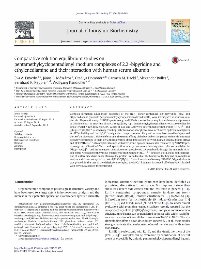
According to the pH-potentiometric titration curves (Fig. S3) record-ed at two experimental setups (I = 0.2 M KCl or KNO3) complexationtook place already at the starting pH (~2) with en, while proton dis-placement by the metal ion is almost complete at this pH in the caseof bpy. Consequently, overall stability constants could be determinedonly for the mono-ligand complex [RhCp*(en)Z] (Z = H2O and/or Cl−;charges are omitted for simplicity). In this latter complex en coordinatesvia the neutral bidentate (N,N) donor set confirmed by the X-ray struc-ture of complex 1 (as described in Section 3.3); and the coordinationsphere is completed with an aqua or chlorido ligand depending on thechloride ion content of the solution. Based on the pH-potentiometric
Fig. 1. High field region of the 1H NMR spectra of the [RhCp*(H2O)3]2+–en (1:1) system in chloride-containing aqueous solution recorded at indicated pH values (a). Concentration dis-tribution curves (solid lines) for the same system calculated on the basis of the stability constants determined andmolar fractions of the bound RhCp* (×) based on the 1H NMR peak in-tegrals of the Cp* methyl protons (b). {cRhCp⁎ = cen = 1 mM; T = 25 °C; I = 0.20 M (KCl); 10% D2O; Z = H2O or Cl−}.
97É.A. Enyedy et al. / Journal of Inorganic Biochemistry 152 (2015) 93–103
titrations (Fig. S3.a) pKa values of the [RhCp*(en)Z] complexes could bedetermined only with large uncertainties, thus pH-dependent 1H NMRspectra were recorded (Figs. 1, S4). Due to the slow ligand-exchange pro-cesses on the NMR time scale (t1/2(obs) ~ 1 ms) peaks belonging to thefree or bound ligand and to the bound or non-bound metal ion could bedetected separately. The integrated peak areas of the Cp* methyl protonswere converted to molar fractions of the bound RhCp*, which were alsocalculated with the aid of the logβ values of species [RhCp*(en)Z](Fig. 1.b). Fairly good correlation between the data of both methods wasobserved. Additionally, in the chloride-free medium an upfield shift ofthe peak belonging to [RhCp*(en)(H2O)]2+ was observed in the basicpH range (Fig. S4.a) strongly indicating the formation of the mixedhydroxido species, [RhCp*(en)(OH)]+ (Chart 2). Thus, pKa of the aquacomplex could be determined on the basis of the pH-dependent δ values(Fig. S4.b, Table 1). Notably, in the presence of chloride ions the signals ofspecies [RhCp*(en)Z] and [RhCp*(en)(OH)]+ were observed more sepa-rately (Fig. 1.a); although the pKa value could be not calculated due tothe non-satisfactory peak separation and was determined by thedeconvolution of the pH-dependent UV–vis spectra (Fig. S5).
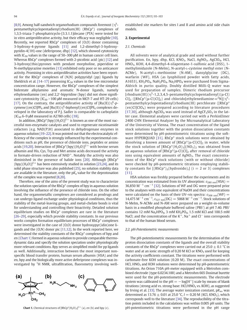
The 1HNMR spectra recorded for the RhCp*–bpy system undoubtedlyreveal the predominant formation of the complex [RhCp*(bpy)Z] in thepH ranges 1.9–~7 and 1.9–~8.5 at ionic strengths of 0.2 M KNO3 andKCl, respectively (Fig. S6). Since neither free metal ion nor ligand couldbe detected at the starting pH values at mM concentrations, UV–vis spec-tra were recorded under more diluted conditions and at strongly acidicpH values. The unaltered spectra at pH between 0.7–8.5 (Fig. 2) revealedthat the complex [RhCp*(bpy)Z] does not decompose in this pH range
Fig. 2. UV–vis spectra of the [RhCp*(H2O)3]2+–bpy (1:1) system (solid lines) in chloride-containing aqueous solution recorded at various pH values. Spectra of [RhCp*(H2O)3]2+
(dashed black line) and bpy (dashed grey line) are shown for comparison recorded atpH= 0.7. {cRhCp⁎ = cbpy = 100 μM; T = 25 °C; I = 0.20 M (KCl)}.
due to its outstandinghigh stability. Therefore the logβ value for this com-plex was determined spectrophotometrically by competition reactionswith en in the presence of chloride ions at pH 7.4 (Fig. S7). At this pHRhCp* forms [RhCp*(L)Z] species with both ligands predominantly. Ab-sorption bands of themetal-bound and free bpy were significantly differ-ent in thewavelength (λ) range 240 and 340 nm allowing the calculationof the stability constant of the bpy complex (Table 1).
The pKa values of the [RhCp*(bpy)Z] complexeswere determined bypH-potentiometry, by the deconvolution of pH-dependent UV–vis spec-tra (representative spectra are shown in Fig. 2 recorded in the chloride-containing medium), and by 1H NMR spectroscopy (Fig. S6). pKa valuesdetermined by the differentmethods are in reasonably good agreementwith each other (Table 1). The constant obtained in the absence of chlo-ride ions is comparable to previously published data (pKa = 8.5 [20];pKa = 8.2 [8]).
Representative concentration distribution curves were calculatedwith the aid of stability constants determined for the [RhCp*(H2O)3]2+–en system in both media studied (Fig. 3). These curves and the stabilitydata (Table 1) clearly reveal the significant effect of the chloride ions onthe solution speciation. Namely, the complex formation starts at some-what lower pH values in the absence of the competitor chloride ions;thus higher logβ [RhCp*(L)Z] values were obtained. On the other hand,chloride ions suppress the hydrolysis, as itwas documented for analogoushalf-sandwichorganometallic complexes [18,48,49]. The complete or par-tial displacement of the aqua ligand by chloride ions or vice versa at thethird coordination site of RhCp* (Chart 2)may have an impact on the bio-activity, since aquation of the chlorido complexes is considered as an
Fig. 3. Concentration distribution curves of the [RhCp*(H2O)3]2+–en (1:1) system in chlo-ride-free (black lines) and chloride-containing (grey lines) aqueous solutions as a functionof pH calculated with the aid of the stability constants determined. {cRhCp⁎ = cen= 1 mM;T = 25 °C; I = 0.20 M (KNO3 or KCl); Z = H2O or Cl−}.
Fig. 4.ORTEP view of [RhCp*(en)Cl]ClO4·2H2O (1) with ellipsoids drawn at 50% probabil-ity level. Counter ion and solvent molecules are omitted for clarity. Selected bond dis-tances (Å) and angles (deg): Rh1-N1: 2.1453(15); Rh1-N2: 2.1235(16); Rh1-Cl1:2.4339(5); Rh1-ring centroid: 1.76303(8); N2-Rh1-N1: 80.23(6)°; N1-Rh1-Cl1:88.09(4)°; N2-Rh1-Cl1: 85.41(4)°.
Fig. 5. pH-dependence of the negative logarithm of the summed equilibrium concentra-tions of the free RhCp* cation and its hydroxido species (pM) calculated for the[RhCp*(H2O)3]2+–bpy/en / pic/dhp systems under identical conditions. {cRhCp⁎ = cL =1 mM; T= 25 °C; I= 0.20 M (KCl)}. Calculations for the dhp- and pic-containing systemsare based on data from Ref. [12]. a pM=−log ([RhCp*] + [(RhCp*)2(OH)i]), where i = 2or 3.
98 É.A. Enyedy et al. / Journal of Inorganic Biochemistry 152 (2015) 93–103
important step in themechanismof action as in the case ofmany antican-cer metallodrugs [50–53]. Therefore the [RhCp*(L)(H2O)]2+ + Cl− ⇄[RhCp*(L)(Cl)]+ + H2O (L = en or bpy) equilibrium was also studiedspectrophotometrically. The exchange process was found to be fast andlogK (H2O/Cl−) constants (Table 1) were estimated with thedeconvolution of UV–vis spectra of the [RhCp*(L)(H2O)]2+ complexes re-corded at various chloride ion concentrations (see Fig. S8 for the en com-plex). The obtained logK (H2O/Cl−) constants are significantly high, thusrepresent a strong affinity of the en and bpy complexes towards the chlo-ride ions.
3.3. Crystal structure of complex [RhCp*(en)Cl]ClO4 (1)
The en complex of RhCp* as [RhCp*(en)Cl]Cl was prepared andcharacterized previously [54], although no crystal structure was provid-ed. The molecular structure of complex cation [RhCp*(en)Cl]+ has beenestablished in thiswork by single-crystal X-ray analysis as its perchloratesalt (Fig. 4). Crystallographic data are collected in Table S1, and selectedbond lengths and angles are listed in the legend of Fig. 4. Complex 1adopts the expected piano-stool geometry with the rhodium(III) centerbeing coordinated by a pentamethylcyclopentadienyl ring, a chlorido li-gand and the (N,N) chelate en. Themetal ion to ring centroid distance of1.7630 Å is somewhat shorter than the reported value for the[RhCp*(bpy)Cl]ClO4 complex (1.776 Å), while the Rh-N,Nʹ (2.1452 Åand 2.1234 Å) and Rh-Cl (2.4339 Å) bond lengths are longer in the encomplex. Distances of 2.100 and 2.385 Å were obtained for the Rh-N,Nʹand Rh-Cl bonds, respectively in the [RhCp*(bpy)Cl]+ cation [8] andbond lengths of 2.093 and 2.115 Å were reported for the Rh-N(1) andRh-N(2) bonds in the [RhCp*(bpy)(H2O)](CF3SO3)2 complex [19]. Thestructure of the analogous iridium(III) complex [IrCp*(en)Cl]CF3SO3
has been also published representing a close structural similarity to com-plex 1, although slightly shorter distances betweenmetal ion and the co-ordinating donor atoms (N, Cl) or the Cp* ring centroid are reported [55].
3.4. Comparison of solution stability and bioactivity of RhCp* complexesformed with en and bpy, (O,N) and (O,O) bidentate ligands
The biological activity of an organorhodium compound most likelydepends on numerous factors such as lipophilicity, geometry, charge,solution stability, kinetic inertness/lability. Our aim in this work was
to reveal the differences in the solution behavior of RhCp* complexesformed with bidentate alkylamine and aromatic N-donor ligands andto compare the obtained thermodynamic stability data to those of com-plexes containing (N,O) and (O,O) donor sets with respect to theirin vitro antiproliferative effects. In order to demonstrate the differencein the RhCp* binding ability of the chosen ligands the negative loga-rithm of the summed equilibrium concentrations of the non-boundmetal ion in its [RhCp*(H2O)3]2+ and μ-hydroxido bridged dinuclear([(RhCp*)2(OH)i], i = 2 or 3) forms were computed under identicalconditions (Fig. 5). This is a similar way as pM values are calculatedfor chelating agents [56]; however, using this approach the fraction ofthe hydrolyzed metal ion species, which becomes higher and higherwith increasing pH, is also taken into consideration. The higher pMvalue indicates a stronger metal ion binding ability. Pic and dhpwere chosen as representatives of the (O,N) and (O,O) donor ligands[11,12]. The calculations reveal the following stability trend at physio-logical pH: bpy N en N pic N dhp.
Based on the stability constants determined it can also be concludedthat the [RhCp*(L)Z] complexes of bpy and en predominate even at1 μM concentration at pH 7.4 (Table 1). On the other hand, the forma-tion of themixed hydroxido complexes [RhCp*(L)(OH)]+ is completelysuppressed in the presence of 0.2 M chloride ions. The pKa value of theen complex is found to be higher than that of the bpy under both stud-ied conditions as it was also found for the analogous IrCp* complexes[57].
For a deeper insight into the relationships between solutionthermodynamic data such as stability (pM), chloride ion affinity (logK(H2O/Cl−)), acidity (pKa of [RhCp*(L)Z]) and antiproliferative effects(IC50) bar charts are shown for the RhCp* complexes of en and bpy as(N,N), pic as (N,O), dhp and 3-hydroxy-2-methyl-pyran-4(1H)-one(maltol) as (O,O) donor containing ligands (Fig. 6). Analysis of a fewdata points cannot give an overview about themultifactorial correlationbetween physicochemical properties and cytotoxicity; however, someconclusions can be still drawn based on this figure. Namely, no correla-tion is seen between the IC50 and pM values: the cytotoxicity does notfollow the solution stability order. The pKa values of these complexesare relatively high (≥8.56) resulting in negligible amount of mixedhydroxido species at pH 7.4, which are usually considered to be less re-active [51]. This can be a possible explanation for the lack of correlationbetween pKa and IC50 values. No correlation was found for the aquatedIrCp* complexes of 1,10-phenanthroline (phen), bpy and en ligands aswell [57]. On the other hand, complexes of bpy, en and pic show fairlypoor or no activity against various human cancer cell lines [12,17],while their logK (H2O/Cl−) values are ~1 order of magnitude higherthan those of the hydroxypyr(idin)one complexes with (O,O) donor
Fig. 6.pKa [RhCp*(L)(H2O)], pM, logK (H2O/Cl−) values and cytotoxicity data for theRhCp*complexes of maltol, dhp, pic, en and bpy. pKa, pM, logK and IC50 (against CH1/PA-1 ovar-ian adenocarcinoma cell lines) values for maltol, dhp and pic complexes are taken fromRef. [11,12]. IC50 values (against MCF-7 breast cancer cell lines) for complexes of en andbpy are taken from Ref. [17]. a pM= −log ([RhCp*] + [(RhCp*)2(OH)i]), where i = 2 or3, and values are calculated under identical conditions: {cRhCp⁎ = cL = 1 mM; pH = 7.4;T = 25 °C; I = 0.20 M (KCl)}.
Fig. 7.High field regions of the 1H NMR spectra recorded for the HSA–[RhCp*(en)Z] (a), HSA–[RPBSʹ buffer with the corresponding spectra of HSA and the RhCp* complexes. {cHSA = 0.5 mM;
99É.A. Enyedy et al. / Journal of Inorganic Biochemistry 152 (2015) 93–103
sets [11,12]. Thus, the stronger affinity of the complexes for retainingthe chlorido ligand at the third coordination sitemay explain the dimin-ished activity. However, no comparable H2O/Cl− exchange constants forRhCp* complexes are published in the literature to the best of ourknowledge, similar findings were found for IrCp* complexes of bpy, enand phen [57], and for someOs and Ru arene complexes as well [49,58].
3.5. Interaction of RhCp* complexes of en, bpy and dhp with human serumalbumin
Binding of anticancermetallodrugs to HSA is of considerable interestas it has a profound effect on the biodistribution, toxicity and side ef-fects. Moreover, HSA and HSA-bound drugs are known to accumulatein solid tumors as a consequence of the enhanced permeability and re-tention effect, which can be an operative way of selective tumortargeting [59]. HSAhas nonspecific bindingpockets and the principal re-gions of these sites are located in subdomains IIA and IIIA called as site Iand II, respectively [60,61]. Additionally, HSA contains metal bindingsites such as the N-terminal site, the reduced Cys34 residue, the multi-metal binding site, and certain side chain donor atoms are also able tocoordinate to the metal centers [60,62]. Thus, fairly diversified bindingevents should be taken into consideration for potential metallodrugswith regard on the binding modes and rates.
Interaction of some [RhCp*(L)Cl)] complexes towardsHSAwas stud-ied by 1H NMR spectroscopy, ultrafiltration/UV–vis and spectro-fluorometry in order to investigate differences or similarities in thestrength and nature of binding in the case of a biologically active(dhp) and non-active (en and bpy) organorhodium compounds. Nota-bly, the coordinated chlorido ligands in these RhCp* complexes are part-ly substituted by water molecules in the aqueous solution; therefore Z(where Z = H2O/Cl−) is used in their general formulae ([RhCp*(L)Z)]).All measurements in this work were performed at pH 7.4 using a mod-ified phosphate buffered saline (PBSʹ) in which the concentration of thechloride ions corresponds to that of the human blood serum (102mM).Based on the logK (H2O/Cl−) constants (Table 1) it can be estimated that97%, 93% and 38% of the bpy, en and dhp [12] complexes respectively arechlorinated at this chloride ion concentration. In addition, free[RhCp*(H2O)3]2+ (without any chelating ligand) was also included forcomparison. This aqua complex can hydrolyze at pH 7.4, e.g. at 1 mMconcentration 11% [RhCp*(H2O)3]2+, 35% [(RhCp*)2(μ-OH)3]+ and 54%[(RhCp*)2(μ-OH)2(H2O)2]2+ are present in the solution, however theformer two species are possibly partly chlorinated revealing the co-existence of various species in the solution of [RhCp*(H2O)3]2+.. Thusall protein binding constants determined here are regarded as condi-tional stability constants and valid only under the given conditions.Samples were incubated at 25 °C since the thermodynamic stability
hCp*(bpy)Z] (b), HSA–[RhCp*(dhp)Z] and HSA–[RhCp*(H2O)3]2+ systems (c) at pH 7.4 incRhCp⁎ = cL = 1 mM; T = 25 °C; incubation time = 1 or 24 h; 10% D2O; Z = H2O or Cl−}.
Fig. 8. Ratio of the bound compounds and HSA plotted against the ratio of the total concentrations of the complexes and HSA calculated from the UV–vis spectra recorded for the LMMfractions of the ultrafiltered samples (a). Fractions (%) of bound compounds at 1:1 complex-to-HSA ratio (b). Symbols: ♦: bound [RhCp*(en)Z] in the [RhCp*(en)Z]–HSA, ▲: bound[RhCp*(bpy)Z] in the [RhCp*(bpy)Z]–HSA,○: bound [RhCp*(H2O)3]2+ in the [RhCp*(H2O)3]2+–HSA, and □: bound [RhCp*(H2O)3]2+ and ■: bound dhp in the [RhCp*(dhp)Z]–HSA sys-tems. Values are themeans of two parallelmeasurements (relative standarddeviations: ≤ 4%). {cHSA= 50 μM; pH= 7.4 (PBSʹ buffer); T= 25 °C; incubation time= 24 h (and 1 h in the case of[RhCp*(en)Z]–HSA (9:1) and [RhCp*(bpy)Z]–HSA (9:1) systems); Z = H2O or Cl−}.
Fig. 9. Changes of fluorescence emission intensities at 340 nm plotted against the RhCp*-to-HSA ratios for the [RhCp*(H2O)3]2+–HSA (○,●) and [RhCp*(dhp)Z]–HSA (Δ,▲) sys-tems measured after 1 h (empty symbols) and 24 h (filled symbols) incubation periodsusing 295 nm excitation wavelength. {cHSA = 1 μM; pH = 7.4 (PBSʹ buffer); T = 25 °C;Z = H2O or Cl−}.
100 É.A. Enyedy et al. / Journal of Inorganic Biochemistry 152 (2015) 93–103
constants for the RhCp* complexes were determined at thistemperature.
First 1H NMR spectra of [RhCp*(L)Z] complexes in the absenceor in the presence of HSA were recorded (Fig. 7). The binding of[RhCp*(H2O)3]2+ and [RhCp*(dhp)Z] to HSA was found to be relativelyfast as the spectra recorded after 1 and 24 h incubation periods wereidentical (not shown here). Signals of the Cp* methyl protons showthat the original peaks of species [RhCp*(H2O)3]2+ or [RhCp*(dhp)Z]disappear upon interaction with HSA and the new peaks indicate thepresence of several different chemical environments around the metalcenter (Fig. 7.c). On the other hand, complex [RhCp*(dhp)Z] decom-poses under the condition used since the liberation of the free ligandis clearly seen in the spectrum (Fig. S9). On the contrary, the interactionbetween HSA and RhCp* complexes of en and bpy was found to bemuch slower (see the differences between 1 and 24 h incubation inFig. 7.a and b). (It is noteworthy that the equilibrium might not bereached after 24 h, but longer incubation timemight have no physiolog-ical relevance.) No ligand displacement by the protein was found, andthe en complex retained partly its original entity without binding(Fig. 7.a). The new Cp* methyl signals detected for the en and bpy com-plexes in the presence of HSA are different and display significant alter-ations from the peaks obtained in the case of the dhp complex or[RhCp*(H2O)3]2+ as well (Fig. S10). These findings strongly suggestthe formation of ternary adducts of the RhCp* complexes of en, bpywith the protein.
The direct interaction towards HSAwas also followed bymembraneultrafiltration. The unbound LMM fractions after separation were ana-lyzed by UV–vis quantification and the spectrawere comparedwith ref-erence spectra and/or were deconvoluted yielding the concentration ofthe unbound RhCp* complex and/or free ligand. The ratio of the boundcompounds and HSA was calculated and plotted against the ratio of thetotal concentrations of the complexes and the protein (Fig. 8.a). Analysisof these data show that the binding of [RhCp*(H2O)3]2+ is practicallyquantitative up to the applied ~9-fold excess, so true saturation wasnot achieved. The amount of bound RhCp* is almost the same in thecase of the dhp complex; however we could detect free ligand in theLMM fractions. At lower c(complex) / c(HSA) ratios dhp was notbound to the protein indicating that the complex decomposes as the1H NMR spectra also showed in Fig. S9. At a large complex excess the li-gand becomes bound, but always much less dhp is bound than the or-ganometallic fragment. Since there is no direct interaction betweendhp and HSA [36], its binding is possible only via the formation of a ter-nary complex in which the RhCp*–dhp bond is not cleaved. The bindingof complexes of en and bpy to HSA was found to be slow, which isreflected in the increased fractions of the bound compounds measuredafter 24 h incubation (Fig. 8a). The bpy complex does not decomposeupon binding to the protein based on the UV–vis spectra recorded
after separation, and the amount of the bound metal species is muchlower compared with the case of [RhCp*(H2O)3]2+ alone. (Since liganden has no absorption its concentration in the LMM fraction could not bedetermined by UV–vis spectrometry.) The binding of the en complex issomewhat weaker compared to the bpy complex (Fig. 8.b); however4–5 binding sites are feasible for both complexes.
The interaction of the complexes of en and bpy to HSA representsnon-dissociative characteristics, while the dhp complex suffers decom-position at the lower c(complex) / c(HSA) ratios and the protein re-places the original ligand. The latter finding strongly suggests thecoordination of the RhCp* fragment to HSA. In the complexes en andbpy the chlorido/aqua ligand at the third coordination site may besubstituted by a donor atomof the protein via the formation of a ternaryadduct; however formation of complex adducts via non-covalent inter-actions at the hydrophobic cavities of HSA such as sites I and II are alsopossible. Therefore, the binding of [RhCp*(H2O)3]2+ and [RhCp*(L)Z](L = dhp, en, bpy) at these sites were monitored by fluorometry. HSAcontains a single Trp (214) residue near site I that is responsible forthemajority of the intrinsic fluorescence of the protein. Upon excitationat 295 nm its emission can be attenuated by a binding event at or closeto Trp214. Addition of [RhCp*(H2O)3]2+ or [RhCp*(dhp)Z] to HSAquenches the Trp214 fluorescence emission (see data points obtainedafter 1 and 24 h in Fig. 9) indicating that the conformation of the hydro-phobic binding pocket is significantly affected by the binding of RhCp*.Based on the emission intensity changes quenching constantswere computed. LogKQʹ values of 5.8(1) and 5.9(1) obtained for[RhCp*(H2O)3]2+ and [RhCp*(dhp)Z], respectively represent quite
Chart 3. Chemical structures of protein binding site model compounds.
Table 2Distribution (%) of RhCp* in the [RhCp*(H2O)3]2+–ligand A–ligand B (1:1:1) ternary systemson thebasis of the 1HNMRpeak integrals of theCp*methyl protons. LigandA: en, bpyordhp;ligand B as protein binding site model (N-MeIm, N-ACMe or N-AM). {cRhCp⁎ = cligand A =cligand B = 0.75 mM; pH= 7.4 (PBSʹ buffer); T= 25 °C; incubation time= 24 h}.
A B [RhCp*(A)] [RhCp*(B)] [RhCp*(A)(B)]
en N-MeIm 22 5 73N-ACMe 100 0 0N-AM 100 0 0
bpy N-MeIm 14 0 86N-ACMe 92 0 8N-AM 67 0 33
dhp N-MeIm 0 9 91N-ACMe 100 0 0N-AM 0 10 90
101É.A. Enyedy et al. / Journal of Inorganic Biochemistry 152 (2015) 93–103
strong and similar ability to bind on site I. This similarity also suggeststhe dissociative feature of the binding of the dhp complex at least onthis site. In the case of the en and bpy complexes no quenchingwas ob-served even at 10-fold excess of complex after an incubation period of24 h.
Additionally, displacement reactions with WF and DG, which areknown site marker fluorescence probes for the binding sites I and II ofHSA [37,60], were carried out. Displacement of the site marker from itsbinding pocket is accompanied by a considerable decrease in the emis-sion intensity. In our case no measurable intensity changes were ob-served upon addition of en and bpy complexes up to 10-fold excessindicating no or fairly weak binding on these sites. On the contrary,[RhCp*(H2O)3]2+ and [RhCp*(dhp)Z] showed similar and significantcompetition with the site markers (Figs. S11, S12). TheWF displacementconstants logKWFʹ of 6.1(1) and 6.2(1) obtained for [RhCp*(H2O)3]2+ and[RhCp*(dhp)Z]were somewhat higher than those calculated for the com-petition with DG (logKDGʹ= 5.8(1) for both species.)
The binding in hydrophobic pockets of HSA in the case of[RhCp*(H2O)3]2+ and [RhCp*(dhp)Z] was found to be rapid, which istypical for the non-covalent interactions. On the other hand, coordina-tion of protein side chains such as His (His242 at site I) or Tyr(Tyr411, possibly His 464 at site II) [63], located nearby these sites, tothe RhCp* fragment is quite probable. Additionally coordination ofmore accessible e.g. surface donors of the protein may be responsiblefor the formation of the ternary RhCp*–ligand–HSA complexes. Themetal ion in the RhCp*–HSA system can be also bound by otherexposed residues considering the high number of binding sites.Coordination preferences for His, Met and Cys residues are suggestedfor Ru(II/III) complexes [64,65]. Tyr residue of peptides has beenrecently shown to coordinate to RhCp* via η6 bonding mode [66].Interactions of [Ru(II)(η6-p-cymene)(en)Cl]+ with HSA were investi-gated by W. Hu et al. and binding to surface His (128, 247, 510), Met(298) and to the free thiol Cys34moietieswas described [64]. Therefore,studies on interactions with some selected model compounds, namelyN-MeIm, N-ACMe, N-AM (Chart 3), representing the functional groups
Fig. 10. Selected regions of the 1H NMR spectra of [RhCp*(bpy)Z]–N-MeIm (1:1), [RhCp*(H2O)7.4. Regions of bpy and N-MeIm protons (a), methyl protons of N-MeIm (b), and Cp* methyl[RhCp*(bpy)Z], B: N-MeIm. {cN-MeIm = cRhCp⁎ = cbpy = 0.75 mM; T = 25 °C; pH= 7.4 (PBSʹ buf
of potential protein residues (His, Cys and Met, respectively) were per-formed by 1H NMR spectroscopy. Reference spectra were recorded forthe RhCp* complexes of dhp, en, bpy, for the model compounds in theabsence and in the presence of [RhCp*(H2O)3]2+ to identify the speciesformed in the RhCp*–bidentate ligand–model compound (1:1:1) terna-ry systems. Representative 1HNMR spectra for the RhCp*–bpy–N-MeImsystem are shown in Fig. 10.
Interaction between [RhCp*(H2O)3]2+ and themodel compounds at1:1 molar ratio took place fast, and the equilibrium was reached within1 h. 100%, 16% and 80% of RhCp* were bound to N-MeIm, N-ACMe, N-AM, respectively (Fig. S13). Notably, RhCp* is in at least three kinds ofchemical environment in the presence of equimolar N-MeIm(Fig. 10.c), while two different signals are assigned to the methyl pro-tons of the model compound suggesting the formation of varioustypes of binary complexes most probably as in the case of [Ru(II)(η6-p-cymene)] [67]. Surprisingly, only a low fraction of binary complexeswas formed with the Cys model, N-ACMe, although strictly anaerobicconditions were used to exclude atmospheric oxidation. Interaction be-tween the dhp complex and the amino acidmodelswas found to be fast,while the equilibrium was reached much slower with the (N,N) donorcontaining complexes, thus 24 h incubation period was generallyused. Formation of ternary complexes could be detected undoubtedlywith the aid of the reference spectra in most cases besides the binaryspecies.When the original ligandwas replaced by themodel compoundthe liberation of the free ligand could be observed. The integrated areasof the CH3 (Cp*) protons belonging to the various ternary and binarycomplexes (Fig. S13) were calculated to obtain the distribution ofRhCp* species (Table 2). The distribution in the ternary systems varieswith the type of the original ligand and the model compound. N-MeIm forms the highest amount of ternary adducts and also efficiently
3]2+ – N-MeIm (1:1) systems, [RhCp*(bpy)Z] and [RhCp*(H2O)3]2+ for comparison at pHprotons (c). Symbols for the identified compounds: MAB: [RhCp*(bpy)(N-MeIm)], MA:fer); incubation time = 24 h; 10% D2O; Z = H2O or Cl−}.
102 É.A. Enyedy et al. / Journal of Inorganic Biochemistry 152 (2015) 93–103
replaces the original ligands. Notably, the dhp complex could not retainits original entity in the presence of N-MeIm and N-AM. Based on thesefindings the RhCp* affinity order is the following: N-MeIm≫N-AM NN-ACMe, thus His(N) ≫ Met(S) N Cys(SH).
4. Conclusions
The rational design of metallodrugs requires detailed studies on theirsolution behavior, physicochemical properties affecting the pharmacoki-netics including their affinity towards endogenous bioligands. This kindof information may help to predict and control the fate of pharmaceuti-cals in biofluids. The main objective of this work was to characterizethe solution speciation and albumin binding properties of RhCp*complexes of two (N,N) donor ligands (bpy and en) in comparisonwith biologically more active complexes containing (O,O) donor set.Stoichiometry and stability of RhCp* complexes of en and bpy were de-termined in aqueous solution via a combined approach using pH-potentiometry, 1H NMR spectroscopy, and UV–vis spectrophotometryin the absence and presence of chloride ions. RhCp* forms prominentlyhigh stability complexes with bpy and en. [RhCp*(L)Z] (Z=H2O/Cl−)species predominate at physiological pH, and based on the stability con-stants their decomposition cannot occur even at low micromolar con-centrations. Moreover, mixed hydroxido species [RhCp*(L)(OH)]+ areformed only in the basic pH range. Additionally, the structure of the com-plex [RhCp*(en)Cl]ClO4 was characterized in solid state by single-crystalX-ray diffraction analysis. [RhCp*(bpy)Z] possesses higher stability at pH7.4 than [RhCp*(en)Z], and their stability considerably exceeds that ofthe hydroxypyr(idin)one complexes with (O,O) donor sets. Chlorideions act as competitive ligands and are able to suppress the formationand the hydrolysis of [RhCp*(L)Z] complexes to some extent. H2O/Cl−
co-ligand exchange equilibrium constants for the [RhCp*(L)(H2O)]+
complexes of en and bpy were also determined, and both retained thechloride at the third coordination site to a much higher extent than theorganometallics with the respective bidentate O-donor ligands. Thisstrong chloride ion affinity contributes most probably to the diminishedantiproliferative effect of the studied complexes with (N,N) donor setand to the decreased catalytic activity of the bpy complex.
Since binding of metallodrugs to the blood transport protein HSA hasa profound influence on the biodistribution, interaction of HSA with theRhCp* complexes of en, bpy and the (O,O) donor dhp was investigated.A panel of methods comprising 1H NMR spectroscopy, ultrafiltration/UV–vis and spectrofluorometry was applied involving fluorescent sitemarkers (WF, DG) and amino acid side chain models. The interactionbetweenHSAandRhCp* or its dhp complex reaches quite fast the equilib-rium, while binding of the en and bpy complexes was considerablyslower. The organometallic fragment RhCp* is able to bind on at least 8sites involving binding events at sites I and II based on the results of Trpquenching and site marker displacement reactions. The extent of thebinding of the dhp complex on these sites is similar to the free RhCp*,and according to the results of 1H NMR spectroscopic and ultrafiltrationmeasurements the complex decomposes at low excess to the protein,and free ligand is liberated upon interaction with the protein. These re-sults indicate that theprotein interactions take place via coordination (co-valent) bonds. Formation of ternary complexes without Rh-dhp bondscleavage is probable by binding to side chains of the protein at the thirdcoordination site beside the dissociative mechanism at elevated excessof the complex. In the case of the more stable (N,N) donor-based com-plexes binding of 4–5 complexmolecules per protein is feasible, althoughnot at sites I and II. In addition, the en and bpy complexes donot lose theiroriginal ligands upon HSA binding. Similarly to organoruthenium com-plexes, the interaction of RhCp* complexes to the imidazole nitrogen ofHis is preferred compared to other amino acids. Ternary protein adductsare formed most probably by the coordination of His at the third coordi-nation site of the RhCp* complex. In contrast, the (O,O) chelate is lostupon interaction with HSA, enabling the RhCp* fragment to bind cova-lently into the deep hydrophobic pockets of sites I and II.
Acknowledgments
This work was supported by the Hungarian Research FoundationOTKA project PD103905 and the János Bolyai Research Scholarship ofthe Hungarian Academy of Sciences. We thank Ms. Gabriella Kiss forconducting some measurements.
Appendix A. Supplementary data
Supplementary data to this article can be found online at http://dx.doi.org/10.1016/j.jinorgbio.2015.08.025.
References
[1] M.A. Jakupec, M. Galanski, V.B. Arion, C.G. Hartinger, B.K. Keppler, Dalton Trans.(2008) 183–194.
[2] G. Süss-Fink, Dalton Trans. 39 (2010) 1673–1688.[3] G. Gasser, I. Ott, N. Metzler-Nolte, J. Med. Chem. 54 (2011) 3–25.[4] E. Alessio, G. Mestroni, A. Bergamo, G. Sava, Curr. Top. Med. Chem. 4 (2004)
1525–1535.[5] C.G. Hartinger, M.A. Jakupec, S. Zorbas-Seifried, M. Groessl, A. Egger, W. Berger, H.
Zorbas, P.J. Dyson, B.K. Keppler, Chem. Biodivers. 5 (2008) 2140–2155.[6] N.R. Dickson, S.F. Jones, H.A. Burris, R.K. Ramanathan, G.J. Weiss, J.R. Infante, J.C.
Bendell, W. McCulloch, D.D. Von Hoff, J. Clin. Oncol. 29 (2011) 2607 (suppl., abstr.).[7] J.J. Soldevila-Barreda, I. Romero-Canelón, A. Habtemariam, P.J. Sadler, Nat. Commun.
6 (2015) Article number: 6582.[8] L. Dadci, H. Elias, U. Frey, A. Hörnig, U. Koelle, A.E. Merbach, H. Paulus, J.S. Schneider,
Inorg. Chem. 34 (1995) 306–315.[9] Y. Geldmacher, M. Oleszak, W.S. Sheldrick, Inorg. Chim. Acta 393 (2012) 84–102.
[10] A. Dorcier, W.H. Ang, S. Bolano, L. Gonsalvi, L. Juillerat-Jeannerat, G. Laurenczy, M.Peruzzini, A.D. Phillips, F. Zanobini, P.J. Dyson, Organometallics 25 (2006)4090–4096.
[11] O. Dömötör, S. Aicher, M. Schmidlehner, M.S. Novak, A. Roller, M.A. Jakupec, W.Kandioller, C.G. Hartinger, B.K. Keppler, É.A. Enyedy, J. Inorg. Biochem. 134 (2014)57–65.
[12] É.A. Enyedy, O. Dömötör, C.M. Hackl, A. Roller, M.S. Novak, M.A. Jakupec, B.K.Keppler, W. Kandioller, J. Coord. Chem. 68 (2015) 1583–1601.
[13] M. Schmidlehner, V. Pichler, A. Roller, M.A. Jakupec, W. Kandioller, B.K. Keppler,J. Organomet. Chem. 782 (2015) 69–76.
[14] Y. Geldmacher, K. Splith, I. Kitanovic, H. Alborzinia, S. Can, R. Rubbiani, M.A. Nazif, P.Wefelmeier, A. Prokop, I. Ott, S. Wölfl, I. Neundorf, W.S. Sheldrick, J. Biol. Inorg.Chem. 17 (2012) 631–646.
[15] Y. Geldmacher, R. Rubbiani, P. Wefelmeier, A. Prokop, I. Ott, W.S. Sheldrick,J. Organomet. Chem. 696 (2011) 1023–1031.
[16] M.A. Nazif, R. Rubbiani, H. Alborzinia, I. Kitanovic, S. Wolfl, I. Ott, W.S. Sheldrick,Dalton Trans. 41 (2012) 5587–5598.
[17] M.A. Scharwitz, I. Ott, Y. Geldmacher, R. Gust, W.S. Sheldrick, J. Organomet. Chem.693 (2008) 2299–2309.
[18] R.E. Morris, R.E. Aird, P.S. Murdoch, H. Chen, J. Cummings, N.D. Hughes, S. Parsons, A.Parkin, G. Boyd, D.I. Jodrell, P.J. Sadler, J. Med. Chem. 44 (2001) 3616–3621.
[19] A. Walcarius, R. Nasraoui, Z. Wang, F. Qu, V. Urbanova, M. Etienne, M. Göllü, A.S.Demir, J. Gajdzik, R. Hempelmann, Bioelectrochemistry 82 (2011) 46–54.
[20] M. Poizat, I.W.C.E. Arends, F. Hollmann, J. Mol. Catal. B Enzym. 63 (2010) 149–156.[21] J.D. Blakemore, E.S. Hernandez, W. Sattler, B.M. Hunter, L.M. Henling, B.S.
Brunschwig, H.B. Gray, Polyhedron 84 (2014) 14–18.[22] J. Lutz, F. Hollmann, T.V. Ho, A. Schnyder, R.H. Fish, A. Schmid, J. Organomet. Chem.
689 (2004) 4783–4790.[23] F. Hollmann, B. Witholt, A. Schmid, J. Mol. Catal. B Enzym. 19–20 (2003) 167–176.[24] K. Vuorilehto, S. Lütz, C. Wandrey, Bioelectrochemistry 65 (2004) 1–7.[25] H.C. Lo, C. Leiva, O. Buriez, J.B. Kerr, M.M. Olmstead, R.H. Fish, Inorg. Chem. 40 (2001)
6705–6716.[26] M.S. Eisen, A. Haskel, H. Chen, M.M. Olmstead, D.P. Smith, M.F. Maestre, R.H. Fish,
Organometallics 14 (1995) 2806–2812.[27] S. Ogo, H. Chen, M.M. Olmstead, R.H. Fish, Organometallics 15 (1996) 2009–2013.[28] D.P. Smith, H. Chen, S. Ogo, A.I. Elduque, M. Eisenstein, M.M. Olmstead, R.H. Fish,
Organometallics 33 (2014) 2389–2404.[29] A. Nutton, P.M. Baily, P.M. Maitlis, J. Chem. Soc. Dalton Trans. (1981) 1997–2002.[30] L. Booth, R.N. Haszeldine, M. Hill, J. Chem. Soc. A (1969) 1299–1303.[31] P. Gans, A. Sabatini, A. Vacca, Talanta 43 (1996) 1739–1753.[32] G.H. Beaven, S. Chen, A. D'Albis, W.B. Gratzer, Eur. J. Biochem. 42 (1974) 539–546.[33] H.M. Irving, M.G. Miles, L.D. Pettit, Anal. Chim. Acta 38 (1967) 475–488.[34] SCQuery, The IUPAC Stability Constants Database, Academic Software (Version 5.5),
R. Soc. Chem., 1993–2005.[35] L. Zékány, I. Nagypál, in: Computational Methods for the Determination of Stability
Constants (Ed.: D. L. Leggett), Plenum Press, New York, 1985, pp. 291–353.[36] É.A. Enyedy, L. Horváth, A. Hetényi, T. Tuccinardi, C.G. Hartinger, B.K. Keppler, T. Kiss,
Bioorg. Med. Chem. 19 (2011) 4202–4210.[37] O. Dömötör, C.G. Hartinger, A.K. Bytzek, T. Kiss, B.K. Keppler, E.A. Enyedy, J. Biol.
Inorg. Chem. 18 (2013) 9–17.[38] M.R. Pressprich, J. Chambers, SAINT + Integration Engine, Program for Crystal
Structure Integration, Bruker Analytical X-ray Systems, Madison, 2004.[39] G.M. Sheldrick, Acta Cryst. A64 (2008) 112–122.
103É.A. Enyedy et al. / Journal of Inorganic Biochemistry 152 (2015) 93–103
[40] O.V. Dolomanov, L.J. Bourhis, R.J. Gildea, J.A.K. Howard, H. Puschmann, J. Appl. Cryst.42 (2009) 339–341.
[41] C.B. Hübschle, G.M. Sheldrick, B. Dittrich, J. Appl. Cryst. 44 (2011) 1281–1284.[42] L.J. Farrugia, J. Appl. Cryst. 30 (1997) 565.[43] International Tables for X-ray Crystallography, vol. C, Kluwer Academic Press,
Dordrecht, The Netherlands, 1992.[44] E. Farkas, É.A. Enyedy, G. Micera, E. Garibba, Polyhedron 19 (2000) 1727–1736.[45] T. Jakusch, P. Buglyó, A.I. Tomaz, J.C. Pessoa, T. Kiss, Inorg. Chim. Acta 339 (2002)
119–128.[46] M.S. Mohan, D. Bancroft, E.H. Abbott, Inorg. Chem. 18 (1979) 2468–2472.[47] A. Avdeef, Anal. Chim. Acta 148 (1983) 237–244.[48] L. Bíró, E. Farkas, P. Buglyó, Dalton Trans. 41 (2012) 285–291.[49] A.F.A. Peacock, A. Habtemariam, S.A. Moggach, A. Prescimone, S. Parsons, P.J. Sadler,
Inorg. Chem. 46 (2007) 4049–4059.[50] M. Melchart, A. Habtemariam, O. Novakova, S.A. Moggach, F.P.A. Fabbiani, S. Parsons,
V. Brabec, P.J. Sadler, Inorg. Chem. 46 (2007) 8950–8962.[51] F. Wang, H. Chen, S. Parsons, I.D.H. Oswald, J.E. Davidson, P.J. Sadler, Chem. Eur. J. 9
(2003) 5810–5820.[52] A.M. Pizarro, A. Habtemariam, P.J. Sadler, in: G. Jaouen, N. Metzler-Nolte (Eds.),
Medicinal Organometallic Chemistry (Topics in Organometallic Chemistry), 1sted., vol. 32, Springer-Verlag, Heidelberg, Germany 2010, pp. 21–56.
[53] R.B. Martin, in: B. Lippert (Ed.), Cisplatin: Chemistry and Biochemistry of a LeadingAnticancer Drug, VHCA & Wiley-VCH, Zürich, Switzerland 1999, pp. 181–205.
[54] G. Garcia, G. Sánchez, I. Romero, I. Solano, M.D. Santana, G. Lopez, J. Organomet.Chem. 408 (1991) 241–246.
[55] M. Scharwitz, T. van Almsick, W.S. Sheldrick, A. Cryst, E63 (2007) m1469–m1470.[56] W.R. Harris, K.N. Raymond, F.L. Weitl, J. Am. Chem. Soc. 103 (1981) 2667–2675.[57] Z. Liu, A. Habtemariam, A.M. Pizarro, S.A. Fletcher, A. Kisova, O. Vrana, L. Salassa,
P.C.A. Bruijnincx, G.J. Clarkson, V. Brabec, P.J. Sadler, J. Med. Chem. 54 (2011)3011–3026.
[58] A. Habtemariam, M. Melchart, R. Fernandez, S. Parsons, I.D.H. Oswald, A. Parkin,F.P.A. Fabbiani, J.E. Davidson, A. Dawson, R.E. Aird, D.I. Jodrell, P.J. Sadler, J. Med.Chem. 49 (2006) 6858–6868.
[59] F. Kratz, J. Control. Release 132 (2008) 171–183.[60] G. Fanali, A. Masi, V. Trezza, M. Marino, M. Fasano, P. Ascenzi, Mol. Asp. Med. 33
(2012) 209–290.[61] G. Sudlow, D.J. Birkett, D.N. Wade, Mol. Pharmacol. 11 (1975) 824–832.[62] W. Bal, M. Sokołowska, E. Kurowska, P. Faller, Biochim. Biophys. Acta 1830 (2013)
5444–5455.[63] T. Peters, All About Albumin: Biochemistry, Genetics and Medical Applications,
Academic Press, San Diego, 1996.[64] W. Hu, Q. Luo, X. Ma, K. Wu, J. Liu, Y. Chen, S. Xiong, J. Wang, P.J. Sadler, F. Wang,
Chem. Eur. J. 15 (2009) 6586–6594.[65] A.R. Timerbaev, C.G. Hartinger, S.S. Aleksenko, B.K. Keppler, Chem. Rev. 106 (2006)
2224–2248.[66] H.B. Albada, F. Wieberneit, I. Dijkgraaf, J.H. Harvey, J.L. Whistler, R. Stoll, N. Metzler-
Nolte, R.H. Fish, J. Am. Chem. Soc. 134 (2012) 10321–10324.[67] Z. Bihari, Z. Nagy, P. Buglyó, J. Organomet. Chem. 782 (2015) 82–88.