comparing endophenotypes in adult-onset primary torsion dystonia
TRANSCRIPT

Comparing Endophenotypes in Adult-Onset PrimaryTorsion Dystonia
David Bradley, MRCPI,1,2* Robert Whelan, PhD,1 Richard Walsh, MRCPI,1 John O’Dwyer, MRCPI,1
Richard Reilly, PhD,2 Siobhan Hutchinson, MRCPI,1 Fiona Molloy, MRCPI,3 and Michael Hutchinson, FRCP1*
1Department of Neurology, St. Vincent’s University Hospital, Dublin, Ireland2Trinity Centre for BioEngineering, Trinity College, Dublin, Ireland
3Department of Clinical Neurophysiology, Beaumont Hospital, Dublin, Ireland
Abstract: Adult-onset primary torsion dystonia (AOPTD) hasan autosomal dominant pattern of inheritance with markedlyreduced penetrance; the genetic causes of most forms ofAOPTD remain unknown. Endophenotypes, markers of sub-clinical gene carriage, may be of use detecting non-manifest-ing gene carriers in relatives of AOPTD patients. The aim ofthis study was to compare the utility of the spatial discrimina-tion threshold (SDT) and temporal discrimination threshold(TDT) as potential endophenotypes in AOPTD. Data on otherpublished candidate endophenotypes are also considered. BothSDT and TDT testing were performed in 24 AOPTD patientsand 34 of their unaffected first degree relatives; results werecompared with normal values from a control population. Ofthe 24 AOPTD patients 5 (21%) had abnormal SDTs and 20
(83%) had abnormal TDTs. Of the 34 first degree relatives 17(50%) had abnormal SDTs and 14 (41%) had abnormal TDTs.Discordant results on SDT and TDT testing were found in 16(67%) AOPTD patients and 21 (62%) first degree relatives.TDT testing has superior sensitivity compared to SDT testingin AOPTD patients; although false positive TDTs are recog-nised, the specificity of TDT testing in unaffected relatives isnot determinable. The high level of discordance between thetwo tests probably relates methodological difficulties withSDT testing. The SDT is an unreliable AOPTD endopheno-type; TDT testing fulfils criteria for a reliable endophenotypewith a high sensitivity. � 2009 Movement Disorder SocietyKey words: AOPTD; basal ganglia; endophenotype; tem-
poral discrimination; spatial discrimination
INTRODUCTION
The original description of an endophenotype dates
from the early 1970s1 when it was first applied in psy-
chiatry to assist in the investigation of complex genetic
disorders such as schizophrenia. It was proposed that
the phenotypes of disorders such as schizophrenia are
so variable and dependant on so many interacting
genetic derangements that routine evaluation of
patients could never lead to successful gene identifica-
tion. An endophenotype may be considered a subclini-
cal marker of genetic liability to a disorder, whether
this is determined by carriage of a single gene muta-
tion or a number of genetic risk factors. They are bio-
markers (defined as any disease-associated biological
finding) that fulfil a number of specific criteria which
are designed to determine that the marker is associated
with the presence of the gene rather than simply mani-
festation of the disease state. Examples of endopheno-
types in the literature include laboratory measurements,
such as copper studies in Wilson’s disease; physiologi-
cal testing abnormalities, for example the specific EEG
findings in juvenile myoclonic epilepsy2; or imaging
findings, including the specific pattern of MRI white
matter change in CADASIL.3
Endophenotypes could be used in linkage studies to
identify genetic loci in poorly penetrant disorder; a
number of criteria for a proposed endophenotype
exist4–6; the endophenotype should be associated with
the disease under investigation in the general popula-
Additional Supporting Information may be found in the onlineversion of this article.
*Correspondence to: Dr. Michael Hutchinson or Dr. David Brad-ley, St. Vincent’s University Hospital, Elm Park, Dublin 4, Ireland.E-mail: [email protected] or [email protected]
Ethical approval for this work was granted by the Ethics and Med-ical Research Committee, St. Vincent’s University Hospital, ElmPark, Dublin 4, Ireland.
Potential conflict of interest: None reported.Received 31 August 2009; Revised 7 October 2009; Accepted 16
October 2009Published online 24 November 2009 in Wiley InterScience
(www.interscience.wiley.com). DOI: 10.1002/mds.22889
84
Movement DisordersVol. 25, No. 1, 2010, pp. 84–90� 2009 Movement Disorder Society
tion, a heritable trait transmitted with disease in pedi-
grees, a finding that is ‘‘state-independent’’ (i.e. unaf-
fected by disease expression or treatment) and should
have a higher frequency amongst unaffected relatives
in pedigrees than in the general population. An ideal
endophenotype for an autosomal dominant disorder
should be abnormal in all affected patients, half of
unaffected first degree relatives and no control sub-
jects.
Adult-onset primary torsion dystonia (AOPTD) is
the commonest form of dystonia and is considered to
be a genetically determined disorder with autosomal
dominant transmission.7,8 The considerably low pene-
trance (in the region of 12–15%) results in a paucity of
informative multiplex families; the majority of cases
may appear sporadic in nature but up to 25% have a
family history on detailed evaluation.9 Although a
number of loci and genes have been identified,10 the
genetic causes of most AOPTD phenotypes remain
unknown. A sensitive endophenotype would increase
the number of subjects available for genetic studies.
Sensory abnormalities in AOPTD include abnormal
spatial discrimination, temporal discrimination, and
vibration-induced illusion of movement.11–18 It is
hypothesised that a disorder of sensory integration pos-
sibly involving the basal ganglia is the cause of these
sensory abnormalities. Given the evidence of a genetic
disorder, there has been significant investigation of
candidate sensory endophenotypes.
Endophenotypes have been particularly studied in
DYT1 dystonia because of its incomplete penetrance,
thus a potential endophenotypic trait in a can be vali-
dated in nonmanifesting carriers of the GAG deletion
in TorsinA. In addition, abnormalities demonstrated in
nonmanifesting DYT1 carriers17,19–22 support the hy-
pothesis that the physiological abnormalities of sensory
processing seen in dystonia result from genetic deter-
minants rather than secondary changes induced by the
movement disorder.
The spatial discrimination threshold (SDT) is deter-
mined using a grating orientation task employing John-
son-van Boven-Philips (JVP) domes applied to the fin-
gertip. Abnormal SDTs have been found in AOPTD
patients as well as their unaffected relatives.12,15,16,23
In addition to disordered sensory processing in the ba-
sal ganglia, abnormal representation in the primary
sensory cortex (S1) may be important in the causation
of SDT abnormalities.13,24 Plasticity in S1 may explain
some of the variability of SDT results, including
improvement with botulinum toxin treatment.25
The temporal discrimination threshold (TDT) is
defined as the shortest time interval at which a subject
can determine that two stimuli are asynchronous. Abnor-
mal TDTs have been described in a number of condi-
tions including DYT1-dystonia,17 writer’s cramp,18,26
blepharospasm,26,27 Parkinson’s disease,28,29 and multi-
ple system atrophy 30 and as such may be an indicator of
abnormal basal ganglia function. Functional imaging
studies have demonstrated activation of the basal gan-
glia and other subcortical structures during a TDT task;
higher cortical activity specific to TDT (not seen in SDT
testing) is found in the anterior cingulate and presupple-
mentary motor area, these regions may be involved in
the interpretation of timing information.31 In contrast,
the basic timekeeper appears to be the putamen, where
the earliest activation occurs during encoding of time
intervals32 and dopaminergic pathways may be particu-
larly important.33 It has been demonstrated that easier
TDT tasks induce greater putaminal activation than dif-
ficult TDT tasks (i.e. stimuli presented near the thresh-
old for simultaneity perception) when additional areas
are activated.34 In this way the putamen seems to act as
the automatic time keeper in low-attention situations.
In addition to abnormal SDT and TDT, a number of
other candidate endophenotypes have been investigated
in AOPTD including abnormalities in vibration-induced
illusion of movement (VIIM),35 Positron Emission To-
mography (PET),19 Diffusion Tensor Imaging (DTI),21
and transcranial magnetic stimulation (TMS).36
In this article, we compare the utility of two sensory
tests (SDT and TDT) as potential endophenotypes in
AOPTD.
PATIENTS AND METHODS
Patients
Both SDT and TDT testing were performed in 24
AOPTD patients (14 cervical dystonia, 10 writer’s
cramp) (15 sporadic, 9 familial)(mean age 52 years,
range 34–63 years) and 34 of their unaffected first-
degree relatives (22 of familial and 12 of sporadic
AOPTD patients) (mean age 42 years, range 26–69
years). The normal control subjects were the 141 con-
trol subjects in a published SDT study15 and the 43
control subjects in a published TDT study.36 Informed
consent was obtained; the study was approved by the
Ethics and Medical Research Committee, St. Vincent’s
University Hospital, Dublin.
Methods
The SDT and TDT testing of individual patients and
relatives were performed by two separate trained
examiners (SDT by RW, TDT by DB) without knowl-
85COMPARING ENDOPHENOTYPES IN AOPTD
Movement Disorders, Vol. 25, No. 1, 2010
edge of the findings of the other examiner. SDT was
examined using JVP domes as described previ-
ously.15,16 TDTs were examined as described previ-
ously37 and were measured for three tasks: (1) visual–
visual, (2) tactile–tactile and (3) visual-tactile. Data
from the three conditions were averaged to obtain the
combined TDT score. A brief description of testing
procedures is provided (Supporting Information 1). We
divided the 141 control subjects for SDT into four age
bands (20–29, 30–39, 40–49 and 50–65 years). The Z-Scores calculated for each individual (AOPTD patient
or relative) are based on the normal values found in
control subjects from their respective age band in the
control group. Similarly, we divided the controls for
TDT testing into two age bands (under 50, over 50).
This approach controls for the effect of age, particu-
larly for SDT examination. We defined a normal cut-
off of 2.5 standard deviations above the relevant age-
related control group mean. Z-scores of equal to or
greater than 2.5 were considered abnormal.
RESULTS
Spatial Discrimination Thresholds
The SDT Z scores in the 141 control subjects
ranged from 22.06 to 12.63; one control subject
exceeded the upper limit of normal. Abnormal SDTs
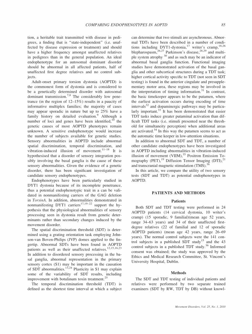
(Z score >2.5) were found in 5 of 24 (21%) AOPTD
patients and in 17 of 34 (50%) first degree relatives
(Fig. 1). Original results are provided (Supporting
Information 2).
Temporal Discrimination Thresholds
All of the 43 control subjects’ Z-scores were less
than 2.5 (range 22.21 to 11.76). Abnormal TDTs (Zscore >2.5) were found in 20 of 24 (83%) AOPTD
patients and 14 of 34 (41%) of first degree relatives
(Fig. 1). Original results are provided (Supporting In-
formation 2).
The frequencies of SDT and TDT abnormalities
were similar in Cervical Dystonia and Writer’s Cramp
patients (Supporting Information 3).
SDT and TDT Testing Compared
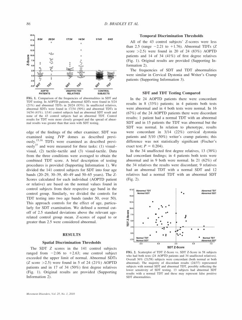
In the 24 AOPTD patients there were concordant
results in 8 (33%) patients; in 4 patients both tests
were abnormal and in 4 both tests were normal. In 16
(67%) of the 24 AOPTD patients there were discordant
results; 1 patient had a normal TDT with an abnormal
SDT and in 15 patients the TDT was abnormal but the
SDT was normal. In relation to phenotype, results
were concordant in 3/14 (22%) cervical dystonia
patients and 5/10 (50%) writer’s cramp patients; this
difference was not statistically significant (Fischer’s
exact test; P 5 0.204).
In the 34 unaffected first degree relatives, 13 (38%)
had concordant findings; in 4 patients both tests were
abnormal and in 9 both were normal. In 21 (62%) of
the 34 relatives the results were discordant; 9 relatives
had an abnormal TDT with a normal SDT and 12
relatives had a normal TDT with an abnormal SDT
(Fig. 2).
FIG. 1. Comparison of the frequencies of abnormalities in SDT andTDT testing. In AOPTD patients, abnormal SDTs were found in 5/24(21%) and abnormal TDTs in 20/24 (83%). In unaffected relatives,abnormal SDTs were found in 17/34 (50%) and abnormal TDTs in14/34 (41%). 1/141 control subjects had an abnormal SDT result andnone of the 43 control subjects had an abnormal TDT. Controlresults for TDT were more closely grouped and the spread of abnor-mal results was greater than that seen with SDT testing.
FIG. 2. Scatterplot of TDT Z-Score vs. SDT Z-Score in 58 subjectswho had both tests (24 AOPTD patients and 34 unaffected relatives).Overall 36% (21/58) subjects were concordant (both normal or bothabnormal). The majority of discordant results (24/37) representedsubjects with normal SDT and abnormal TDT, possibly reflecting thelower sensitivity of SDT testing. 13 subjects had abnormal SDTresults with a normal TDT and these may represent false positiveSDT abnormalities.
86 D. BRADLEY ET AL
Movement Disorders, Vol. 25, No. 1, 2010
DISCUSSION
In our AOPTD patients we found a remarkable level
of discordance (67%) between the SDT and TDT test
results. In the unaffected first degree relatives, although
both tests were abnormal in a significant proportion
(SDT 50%, TDT 41%), there was again a notable dis-
cordance of 62%. Clearly one of these two potential
endophenotypes is more unreliable than the other. The
frequencies of abnormalities in our AOPTD patients
(SDT 21%, TDT 83%) indicate that TDT is a more
sensitive marker of abnormal sensory processing in
AOPTD. Moreover, in control subjects the distribution
of TDT results was narrower (range 22.21 SD to
11.76 SD) than the SDT control range (range 22.06
SD to 12.63 SD) suggesting greater confidence that an
abnormal result is indicative of abnormal central sen-
sory processing. Furthermore, as can be seen from Fig-
ure 1, the range of abnormal Z-scores for the TDT is
much greater than that of the SDT.
The SDT is relatively sensitive to age related
changes in the peripheral nervous system; a number of
discordant results may thus be due to the lower speci-
ficity of SDT testing. There is marked increase in the
sensory threshold with age which reflects the natural
effect of age on the peripheral nervous system. This
age effect renders it impossible to determine with ac-
curacy the upper limit of normal of the SDT over the
age of 65 and probably limits sensitivity of the test
over the age of 50. This variation in the SDT sensitiv-
ity with age might partly (but not completely) explain
why AOPTD patients (who had a mean age of 52
years) had fewer abnormal SDT results than their unaf-
fected first degree relatives (mean age 42 years).
SDT has more potential for error due to the variabil-
ity in stimuli presented to subjects using manually
applied JVP domes in comparison with the electroni-
cally-determined electrical stimuli in the TDT testing
procedure. The basal ganglia,32 and dopaminergic path-
ways in particular,33 play a particular role in timekeep-
ing in the CNS. Thus the TDT may be a more sensi-
tive measure of the postulated dopaminergic dysfunc-
tion in AOPTD patients.38
Validation of TDT as an Endophenotype
There is no gold standard with which to validate any
candidate endophenotype in AOPTD as the genotype is
not known. TDT has been examined in other genetic
forms of dystonia. Fiorio and colleagues found that
TDT was abnormal in DYT1-carriers (both manifesting
and nonmanifesting) compared with noncarrier rela-
tives and healthy controls.17 In addition, in PINK1 Par-
kinsonism they found abnormal TDTs in both homozy-
gous manifesting patients and heterozygous nonmani-
festing relatives compared with controls.29
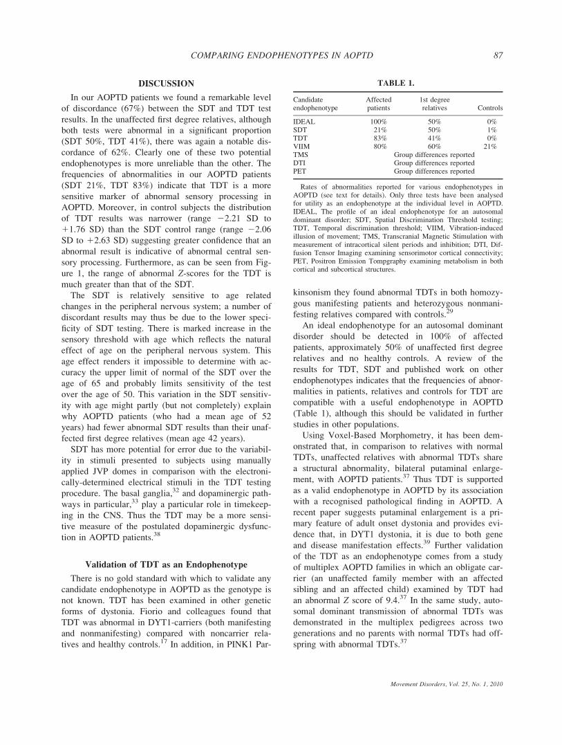
An ideal endophenotype for an autosomal dominant
disorder should be detected in 100% of affected
patients, approximately 50% of unaffected first degree
relatives and no healthy controls. A review of the
results for TDT, SDT and published work on other
endophenotypes indicates that the frequencies of abnor-
malities in patients, relatives and controls for TDT are
compatible with a useful endophenotype in AOPTD
(Table 1), although this should be validated in further
studies in other populations.
Using Voxel-Based Morphometry, it has been dem-
onstrated that, in comparison to relatives with normal
TDTs, unaffected relatives with abnormal TDTs share
a structural abnormality, bilateral putaminal enlarge-
ment, with AOPTD patients.37 Thus TDT is supported
as a valid endophenotype in AOPTD by its association
with a recognised pathological finding in AOPTD. A
recent paper suggests putaminal enlargement is a pri-
mary feature of adult onset dystonia and provides evi-
dence that, in DYT1 dystonia, it is due to both gene
and disease manifestation effects.39 Further validation
of the TDT as an endophenotype comes from a study
of multiplex AOPTD families in which an obligate car-
rier (an unaffected family member with an affected
sibling and an affected child) examined by TDT had
an abnormal Z score of 9.4.37 In the same study, auto-
somal dominant transmission of abnormal TDTs was
demonstrated in the multiplex pedigrees across two
generations and no parents with normal TDTs had off-
spring with abnormal TDTs.37
TABLE 1.
Candidateendophenotype
Affectedpatients
1st degreerelatives Controls
IDEAL 100% 50% 0%SDT 21% 50% 1%TDT 83% 41% 0%VIIM 80% 60% 21%TMS Group differences reportedDTI Group differences reportedPET Group differences reported
Rates of abnormalities reported for various endophenotypes inAOPTD (see text for details). Only three tests have been analysedfor utility as an endophenotype at the individual level in AOPTD.IDEAL, The profile of an ideal endophenotype for an autosomaldominant disorder; SDT, Spatial Discrimination Threshold testing;TDT, Temporal discrimination threshold; VIIM, Vibration-inducedillusion of movement; TMS, Transcranial Magnetic Stimulation withmeasurement of intracortical silent periods and inhibition; DTI, Dif-fusion Tensor Imaging examining sensorimotor cortical connectivity;PET, Positron Emission Tompgraphy examining metabolism in bothcortical and subcortical structures.
87COMPARING ENDOPHENOTYPES IN AOPTD
Movement Disorders, Vol. 25, No. 1, 2010
ALTERNATIVE ENDOPHENOTYPES IN AOPTD
Candidate AOPTD endophenotypes include abnor-
malities in SDT, TDT, VIIM, PET, and TMS. The rel-
ative advantages and disadvantages of these techniques
as potential endophenotypes have been examined in a
number of patient populations.
Vibration-Induced Illusion of Movement (VIIM)
Vibration of a muscle through stimulation of the
muscle spindle 40 can induce an illusion of movement.
This perception is reduced in AOPTD patients.35,41
VIIM abnormalities were examined in a cohort of 30
AOPTD patients, 57 relatives and 19 controls.14 VIIM
abnormalities were found in 80% of AOPTD patients
and approximately 60% of first degree relatives. As an
endophenotype VIIM is not ideal, given that abnormal-
ities were found in 21% of control subjects and thus it
has a sub-optimal specificity and positive predictive
value.14
Transcranial Magnetic Stimulation
Inhibitory mechanisms in the central nervous system
are abnormal in patients with dystonia.42 Transcranial
magnetic stimulation has been used to assess intracorti-
cal activity in DYT1 dystonia. Edwards and colleagues
studied manifesting DYT1 patients, nonmanifesting
DTY1 carriers and controls.36 They reported reduced
intracortical inhibition with reduced cortical silent peri-
ods in DYT1 carriers, regardless of phenotype expres-
sion, which is compatible with the reduced GABAergic
activity postulated in dystonia. Their findings may be
explained by secondary change resulting from a pri-
mary basal ganglia disorder.
Positron Emission Tomography
Eidelberg and colleagues have shown using PET
that metabolism is diffusely altered in DYT1 dystonia.
In both manifesting and nonmanifesting DTY1 carriers
metabolism was shown to be increased in the lentiform
nucleus, cerebellum, and supplementary motor area.19
Additional abnormalities were seen in manifesting sub-
jects only, including hypermetabolism in the midbrain
and thalamus.19 Further PET studies have examined
other dystonia-related mutations, including the less
common DYT6 dystonia linked to 8q21–22. Compared
to DYT1 dystonia, similar metabolism was seen in
DYT6 carriers, both manifesting and nonmanifesting.43
Changes specific to DYT1 included hypermetabolism
in the putamen, anterior cingulate and cebebellar hemi-
spheres, while DYT6 patients had hypometabolism in
the putamen and hypermetabolism in the temporal
lobe.22 PET has also been used to examine motor
learning in nonmanifesting DYT1 carriers and reported
increased metabolic activity during both sequence
learning and motor execution compared with con-
trols.20 PET with [11C] raclopride (RAC) scanning has
been used to examine D2 receptor availability in the
basal ganglia and selected extra-striatal regions in
DYT1 and DYT6 carriers.38 The authors found that
both DYT1 and DYT6 mutation carriers had reduced
receptor availability in caudate, putamen and ventrolat-
eral thalamus compared to controls. The relatively con-
sistent patterns of abnormalities relating to particular
genotypes and phenotypes along with some pene-
trance-related findings are the basis for the proposed
use of functional imaging as an endophenotype in
AOPTD. This modality also provides significant insight
into the pathogenesis of the disorder.44
Diffusion Tensor Imaging
Carbon et al. describe a DTI study of manifesting
and nonmanifesting DTY1 patients. They found that,
compared to controls, the genotype was associated
with microstructural abnormalities in the connectivity
of the primary sensorimotor cortex.21 They further
demonstrate that these changes were more pronounced
amongst manifesting carriers, suggesting a threshold
effect. They postulate that the microstructural abnor-
mality detected in their study could be the structural
basis for the well recognised reduction of GABA-ergic
intracortical inhibition in dystonia. This structural find-
ing may represent an endophenotype in DYT1 dystonia
but has not been examined in AOPTD.
CHOICE OF ENDOPHENOTYPE
In many published endophenotype studies, group
results are presented so that, while significant differen-
ces are demonstrated between affected individuals, rel-
atives and controls, it is not possible to be certain of
the status of any one individual (Table 1). TDT testing
appears capable of assigning status to individuals. TDT
is not without limitations; false negative and false posi-
tive results occur. In this study 4 of the 24 AOPTD
patients had normal TDTs. Furthermore, as part of an
ongoing genetic study in our department, we found
that removing one unaffected relative with an abnormal
TDT (Z-Score 6.6) from a linkage analysis resulted in
a significant increase in the LOD score to greater than
13.0 (unpublished results). A false positive TDT was
found in the control group in a study of TDT in
88 D. BRADLEY ET AL
Movement Disorders, Vol. 25, No. 1, 2010
PINK1; one of the control subjects had a TDT greater
than the chosen cut off for normal of 2 standard devia-
tions above the control mean.29 Overall however, the
number of inappropriate results seems to be low. It is
of critical importance that an endophenotype misclassi-
fies the fewest possible individuals as incorrect assign-
ments in a linkage analysis can negatively affect the
outcome. One example of this relates to a Swiss family
with dopa-responsive dystonia incorrectly assigned to
DYT14.45 In addition, while TDT appears to be rela-
tively sensitive in detecting subclinical basal ganglia
dysfunction, it is not specific to AOPTD because
abnormal TDTs are seen in other basal ganglia disor-
ders. A number of proposed AOPTD endophenotypes
do not reliably dichotomise unaffected relatives to
allow assignment of probable gene carriage. Based on
the available evidence TDT testing satisfies the criteria
for a useful endophenotype.
Acknowledgments: This research was funded by DystoniaIreland, a non-profit patient support charity, (www.dystonia)and by the Health Research Board of Ireland (www.hrb.ie).
Author Roles: David Bradley recruited participants,designed the TDT testing protocol, performed TDT testing,analysed data and wrote the manuscript. Robert Whelandesigned the TDT testing protocol, analysed data, and editedthe manuscript. Richard Walsh recruited participants, per-formed SDT testing and edited the manuscript. JohnO’Dwyer designed the SDT testing protocol, recruited partic-ipants and reviewed the manuscript. Richard Reilly reviewedthe manuscript and provided mentorship. Siobhan Hutchinsonrecruited participants and reviewed the manuscript. FionaMolloy recruited participants and reviewed the manuscript.Michael Hutchinson designed the study, edited the manu-script, and provided mentorship.
Financial Disclosures: R. Reilly and R. Whelan havebeen awarded funding from Enterprise Ireland, a state findingagency. M. Hutchinson sits on the medical advisory board ofBiogen Idec. None of the authors have any further financialdisclosures to make under the headings of stock ownership inmedically-related fields, consultancies, advisory boards, part-nerships, honoraria, grants, intellectual property rights, experttestimony, employment, contracts, or royalties.
REFERENCES
1. Gottesman, II, Shields J. Genetic theorizing and schizophrenia.Br J Psychiatry 1973;122:15–30.
2. Greenberg DA, Delgado-Escueta AV, Widelitz H, et al. Juvenilemyoclonic epilepsy (JME) may be linked to the BF and HLAloci on human chromosome 6. Am J Med Genet 1988;31:185–192.
3. O’Sullivan M, Jarosz JM, Martin RJ, Deasy N, Powell JF, Mar-kus HS. MRI hyperintensities of the temporal lobe and externalcapsule in patients with CADASIL. Neurology 2001;56:628–634.
4. Gershon ES, Goldin LR. Clinical methods in psychiatric genetics.I. Robustness of genetic marker investigative strategies. ActaPsychiatr Scand 1986;74:113–118.
5. Leboyer M, Bellivier F, Nosten-Bertrand M, Jouvent R, Pauls D,Mallet J. Psychiatric genetics: search for phenotypes. TrendsNeurosci 1998;21:102–105.
6. Gottesman, II, Gould TD. The endophenotype concept in psychi-atry: etymology and strategic intentions. Am J Psychiatry 2003;160:636–645.
7. Leube B, Kessler KR, Goecke T, Auburger G, Benecke R. Fre-quency of familial inheritance among 488 index patients with idi-opathic focal dystonia and clinical variability in a large family.Mov Disord 1997;12:1000–1006.
8. Stojanovic M, Cvetkovic D, Kostic VS. A genetic study of idio-pathic focal dystonias. J Neurol 1995;242:508–511.
9. Waddy HM, Fletcher NA, Harding AE, Marsden CD. Agenetic study of idiopathic focal dystonias. Ann Neurol 1991;29:320–324.
10. Muller U. The monogenic primary dystonias. Brain 2009;132:2005–2025.
11. Hallett M. Physiology of dystonia. Adv Neurol 1998;78:11–18.12. Molloy FM, Carr TD, Zeuner KE, Dambrosia JM, Hallett M.
Abnormalities of spatial discrimination in focal and generalizeddystonia. Brain 2003;126:2175–2182.
13. Meunier S, Garnero L, Ducorps A, et al. Human brain mappingin dystonia reveals both endophenotypic traits and adaptive reor-ganization. Ann Neurol 2001;50:521–527.
14. Frima N, Nasir J, Grunewald RA. Abnormal vibration-inducedillusion of movement in idiopathic focal dystonia: an endopheno-typic marker? Mov Disord 2008;23:373–377.
15. O’Dwyer JP, O’Riordan S, Saunders-Pullman R, et al. Sensoryabnormalities in unaffected relatives in familial adult-onset dys-tonia. Neurology 2005;65:938–940.
16. Walsh R, O’Dwyer JP, Sheikh IH, O’Riordan S, Lynch T,Hutchinson M. Sporadic adult onset dystonia: sensory abnormal-ities as an endophenotype in unaffected relatives. J Neurol Neu-rosurg Psychiatry 2007;78:980–983.
17. Fiorio M, Gambarin M, Valente EM, et al. Defective temporalprocessing of sensory stimuli in DYT1 mutation carriers: a newendophenotype of dystonia? Brain 2007;130:134–142.
18. Fiorio M, Tinazzi M, Bertolasi L, Aglioti SM. Temporal process-ing of visuotactile and tactile stimuli in writer’s cramp. AnnNeurol 2003;53:630–635.
19. Eidelberg D, Moeller JR, Antonini A, et al. Functional brain net-works in DYT1 dystonia. Ann Neurol 1998;44:303–312.
20. Ghilardi MF, Carbon M, Silvestri G, et al. Impaired sequencelearning in carriers of the DYT1 dystonia mutation. Ann Neurol2003;54:102–109.
21. Carbon M, Kingsley PB, Su S, et al. Microstructural white matterchanges in carriers of the DYT1 gene mutation. Ann Neurol2004;56:283–286.
22. Carbon M, Su S, Dhawan V, Raymond D, Bressman S, EidelbergD. Regional metabolism in primary torsion dystonia: effects ofpenetrance and genotype. Neurology 2004;62:1384–1390.
23. Sanger TD, Tarsy D, Pascual-Leone A. Abnormalities of spatialand temporal sensory discrimination in writer’s cramp. Mov Dis-ord 2001;16:94–99.
24. Bara-Jimenez W, Catalan MJ, Hallett M, Gerloff C. Abnormalsomatosensory homunculus in dystonia of the hand. Ann Neurol1998;44:828–831.
25. Walsh R, Hutchinson M. Molding the sensory cortex: spatial acu-ity improves after botulinum toxin treatment for cervical dysto-nia. Mov Disord 2007;22:2443–2446.
26. Scontrini A, Conte A, Defazio G, et al. Somatosensory temporaldiscrimination in patients with primary focal dystonia. J NeurolNeurosurg Psychiatry 2009; Jun 18: Epub Ahead of Print.
27. Fiorio M, Tinazzi M, Scontrini A, et al. Tactile temporal dis-crimination in patients with blepharospasm. J Neurol NeurosurgPsychiatry 2008;79:796–798.
28. Lee MS, Kim HS, Lyoo CH. "Off" gait freezing and temporaldiscrimination threshold in patients with Parkinson disease. Neu-rology 2005;64:670–674.
89COMPARING ENDOPHENOTYPES IN AOPTD
Movement Disorders, Vol. 25, No. 1, 2010
29. Fiorio M, Valente EM, Gambarin M, et al. Subclinical sensoryabnormalities in unaffected PINK1 heterozygotes. J Neurol2008;255:1372–1377.
30. Lyoo CH, Lee SY, Song TJ, Lee MS. Abnormal temporal dis-crimination threshold in patients with multiple system atrophy.Mov Disord 2007;22:556–559.
31. Pastor MA, Day BL, Macaluso E, Friston KJ, Frackowiak RS.The functional neuroanatomy of temporal discrimination. J Neu-rosci 2004;24:2585–2591.
32. Rao SM, Mayer AR, Harrington DL. The evolution of brain acti-vation during temporal processing. Nat Neurosci 2001;4:317–323.
33. Malapani C, Rakitin B, Levy R, et al. Coupled temporal memo-ries in Parkinson’s disease: a dopamine-related dysfunction. JCogn Neurosci 1998;10:316–331.
34. Pastor MA, Macaluso E, Day BL, Frackowiak RS. Putaminal ac-tivity is related to perceptual certainty. Neuroimage 2008;41:123–129.
35. Rome S, Grunewald RA. Abnormal perception of vibration-induced illusion of movement in dystonia. Neurology 1999;53:1794–1800.
36. Edwards MJ, Huang YZ, Wood NW, Rothwell JC, Bhatia KP.Different patterns of electrophysiological deficits in manifestingand non-manifesting carriers of the DYT1 gene mutation. Brain2003;126:2074–2080.
37. Bradley D, Whelan R, Walsh R, et al. Temporal discriminationthreshold: VBM evidence for an endophenotype in adult onsetprimary torsion dystonia. Brain 2009;132:2327–2335.
38. Carbon M, Niethammer M, Peng S, et al. Abnormal striatal andthalamic dopamine neurotransmission: Genotype-related featuresof dystonia. Neurology 2009;72:2097–2103.
39. Draganski B, Schneider SA, Fiorio M, et al. Genotype-phenotypeinteractions in primary dystonias revealed by differential changesin brain structure. Neuroimage 2009;47:1141–1147.
40. Proske U, Morgan DL, Gregory JE. Thixotropy in skeletal muscleand in muscle spindles: a review. Prog Neurobiol 1993;41:705–721.
41. Grunewald RA, Yoneda Y, Shipman JM, Sagar HJ. Idiopathicfocal dystonia: a disorder of muscle spindle afferent processing?Brain 1997;120 (Part 12):2179–2185.
42. Berardelli A, Rothwell JC, Hallett M, Thompson PD, ManfrediM, Marsden CD. The pathophysiology of primary dystonia. Brain1998;121 (Part 7):1195–1212.
43. Trost M, Carbon M, Edwards C, et al. Primary dystonia: isabnormal functional brain architecture linked to genotype? AnnNeurol 2002;52:853–856.
44. Carbon M, Eidelberg D. Abnormal structure-function relation-ships in hereditary dystonia. Neuroscience 2009;164:220–229.
45. Wider C, Melquist S, Hauf M, et al. Study of a Swiss dopa-responsive dystonia family with a deletion in GCH1: redefiningDYT14 as DYT5. Neurology 2008;70:1377–1383.
90 D. BRADLEY ET AL
Movement Disorders, Vol. 25, No. 1, 2010