comparing the use of static versus dynamic images to
TRANSCRIPT
CLINICAL RESEARCH
This work waaResearch PrbResearch PcCollaboratordAdjunct ProeProfessor, DfProfessor, EsgAdjunct Pro
THE JOURNA
Comparing the use of static versus dynamicimages to evaluate a smile
Eduardo Mahn, PhD,a Camila S. Sampaio, PhD,b Bruno Pereira da Silva, PhD,c Kyle Stanley, DDS,d
Ana María Valdés, DDS,e Javiera Gutierrez, DDS,f and Christian Coachman, DDSg
ABSTRACTStatement of problem. Smile analysis, as part of the overall facial analysis, is an importantcomponent of diagnosis and treatment planning in the esthetic rehabilitation of a patient. Moststudies that refer to smile analysis are based on static images. A more comprehensive evaluationcan be made with dynamic video images that can be stopped at the most appropriate frame toensure the best static images for analysis.
Purpose. The purpose of this clinical study was to evaluate the posed and dynamic smilesof both sexes, considering the type of smile, prevalence of gingival display, dental displayat rest, dentogingival display at posed and spontaneous smile, and lip mobility, throughdigital image acquisition (photographs and video clips) manipulated by using a softwareprogram.
Material and methods. Three photographs and 1 video clip were made for each of the 380voluntary participants aged between 18 and 32 years by using an iPhone 6 iSight 8 MP camera,Moment lens, and artificial 5500 Kelvin light (IceLight). Digital files were evaluated by using asoftware program (Keynote), determining each point to be evaluated with posed and spontaneoussmiles.
Results. With static images, 90% of women and 74% of men had gingival display, with only35% of women and 21% of men having continuous gingival display. With dynamic analysis,these values increased to 100% of women and 95% of men having gingival display and 62%of men and 81% of women having a continuous gingival display (P<.05). The differencebetween dentogingival display during posed and spontaneous smiles was clear, with 68% ofthe participants having 2.25 mm more gingival display. Women tend to show slightly moredental display at rest, posed and spontaneous dentogingival display, as well as lip mobility,than men.
Conclusions. The type of smile changes significantly when posed and spontaneous smiles arecompared. Women generally show more gingiva and teeth in all the parameters evaluated thanmen. Dental treatments should be individually planned according to each patient’s smilecharacteristics. (J Prosthet Dent 2019;-:---)
When starting esthetic dentaltreatment, the patient’s ex-pectations, the patient’sindividual anatomic charac-teristics, and possible thera-peutic solutions should beconsidered.1 Facial featuresand lip movements should beanalyzed in relation to teethwhen facial, dentolabial, andphonetic parameters are eval-uated1,2 by directly measuringthe lip-tooth relationshipsboth dynamically and at rest.3
A pleasing smile has beenshown to depend not only ontooth position, color, size, andshape but also on the amountof gingival display and theframing of the lips.4 Clinicaldecisions could be affected bysoft tissue display, which isnormally measured fromposed photographs.
The visibility of thegingival tissues depends onthe position of the smile line
s partially supported by the Fondo Nacional de Desarrollo Científico y Tecnológico, Chile (FONDECYT Project 11170920).ofessor and Director of the Esthetic Dentistry Program, Department of Restorative Dentistry, Universidad de los Andes, Santiago, Chile.rofessor, Department of Restorative Dentistry, Universidad de los Andes, Santiago, Chile.Professor, Department of Prosthodontics and Periodontology, University of Sevilla, Sevilla, Spain.fessor, Department of Restorative Sciences, Herman Ostrow School of Dentistry of USC, University of Southern California, Los Angeles, Calif.epartment of Restorative Dentistry, Universidad de los Andes, Santiago, Chile.thetic Dentistry Program, Universidad de los Andes, Santiago, Chile.fessor, Department of Preventive and Restorative Sciences, University of Pennsylvania School of Dental Medicine, Philadelphia, Pa.
L OF PROSTHETIC DENTISTRY 1
Clinical ImplicationsClinicians have based their treatment and researchon a static analysis of patient smiles, whichrepresents a posed smile. However, this could leadto false diagnosis and nonideal treatment becausethe patients’ spontaneous smiles might differsignificantly from the posed smiles by showingmore maxillary and mandibular teeth and moregingival display. Dynamic smile evaluations shouldbe used to determine the full range of a smile inmotion.
2 Volume - Issue -
and the relationship between the upper lip and the sizeand visibility of teeth.5 Smile types can be classified asfollows: a very high smile line, high smile line, averagesmile line, and low smile line.5 For this analysis, bothposed and spontaneous smiles need to be defined andconsidered. The posed or static social smile is a voluntarysmile a person uses in social settings or when beingphotographed, while a spontaneous smile is involuntaryand represents the emotion a person is experiencing atthat moment.6,7 Gingival displays within 0 to 2 mm8 and2 to 4 mm4 have been reported to be estheticallypleasing, while higher or lower smile lines may presentesthetic issues.4,8
Digital imaging has been used to evaluate differentaspects of a smile, initially with static photographs.5,8-10
However, a spontaneous smile is difficult to obtain withstatic photographs,11 and capturing and quantitativelyanalyzing digital images acquired from videos in com-puter software may improve the esthetic assess-ment.3,4,7,11-16
Dentolabial parameters vary according to lip mobilityin both a static posed smile and a smile in motion ascaptured in video.17 The use of dynamic documentationof the smile (DDS) allows esthetic rehabilitative plan-ning from a facial perspective, improving communica-tion with the patient, integration among specialists, andthe predictable quality of the treatment.17 In addition,the improvements in video and photographic quality inmodern smartphones make them useful for clinicians.17
This is why, recently, protocols have been developedusing smartphones, demonstrating the simplicity oftheir application.17 It may no longer be essential fordentists to purchase expensive and bulky photographicequipment.
Digital analysis has revealed that women have ahigher smile line than men.5,14 Moreover, incisor displaychanges with age, and no individuals older than 50 yearspresented a high smile line.13 This change can beexplained by the elasticity of soft tissues, which decreaseswith age-related alterations in the connective tissue
THE JOURNAL OF PROSTHETIC DENTISTRY
metabolism18,19; thus, a high smile line has beendescribed as a sign of youth.8,13
Paradigms regarding smile line and the shape of teethand their difference in men and women still exist inesthetic dentistry.20 The concept that oval teeth werecharacteristic of women and square teeth of men hasbeen disproven recently.20 Moreover, the concept of thesmile line and the normal or average display obtainedfrom static photographs5,10,18 may also require revision.Videos, made with several frames per second, allow theclinician to choose the optimal smile display and providea more accurate and natural assessment than staticphotography.
Therefore, the purpose of this clinical study was toevaluate the smile in static and video images. The nullhypotheses tested were that no difference would befound in posed and spontaneous smiles and that womenand men would not show differences in the differentsmile parameters evaluated.
MATERIAL AND METHODS
This study was performed according to protocolsapproved by the institutional review board of the Uni-versidad de los Andes, Chile. Three hundred and eightydental students (227 women and 153 men) from theUniversity of Los Andes, Chile, were selected; researchdetails were explained, and the participants signed aconsent agreement. Inclusion criteria specified partici-pants should be between 18 and 32 years becausedifferent ages might affect gingival display.13
The operator (J.G.) was calibrated by a specialist(E.M.) to standardize and ensure the quality control ofthe photographs and video clips. Files used for thiscalibration were not used in the results of this study.Photographs and video clips were made by using an8-MP camera (iPhone 6 iSight; Apple Inc) and a60-mm Moment lens (Moment Inc) in artificial andstandardized light calibrated in 5500 Kelvin (Ice Light;Westcott Co).
Digital imaging was made with standardized param-eters: participants were requested to stand in front of ablack screen, and those with long hair were asked to tie itback, with the ears showing. The digital camera waspositioned 40 cm from the tip of the participant’s nose forthe photographs and 70 cm for the video clips. Lightswere positioned on tripods at 45 degrees from the medialsagittal plane and 15 cm from the tip of the participant’snose. The digital files obtained were the following:photographs (posed smile): 1 frontal photograph withthe participant’s mouth closed in maximum inter-cuspation; 1 frontal photograph at rest (after swallowing);and 1 frontal photograph in posed smile. Participantswere requested to keep their eyes focused on the hori-zon. Video clips (spontaneous smile): a 30-second video
Mahn et al
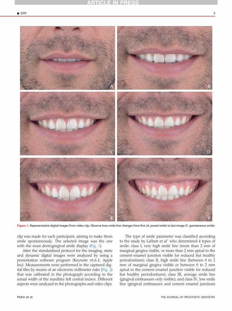
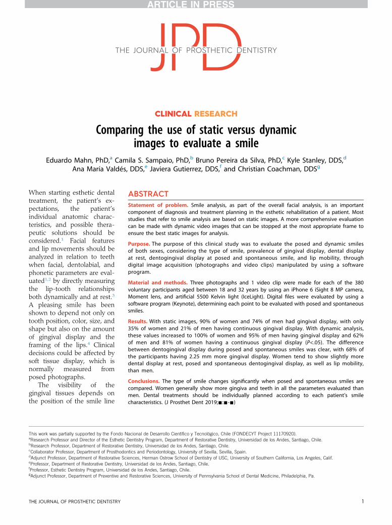
Figure 1. Representative digital images from video clip. Observe how smile line changes from first (A, posed smile) to last image (F, spontaneous smile).
- 2019 3
clip was made for each participant, aiming to make themsmile spontaneously. The selected image was the onewith the most dentogingival smile display (Fig. 1).
After the standardized protocol for the imaging, staticand dynamic digital images were analyzed by using apresentation software program (Keynote v6.6.1; AppleInc). Measurements were performed in the captured dig-ital files by means of an electronic millimeter ruler (Fig. 2)that was calibrated in the photograph according to theactual width of the maxillary left central incisor. Differentaspects were analyzed in the photographs and video clips.
Mahn et al
The type of smile parameter was classified accordingto the study by Liébart et al5 who determined 4 types ofsmile: class I, very high smile line (more than 2 mm ofmarginal gingiva visible, or more than 2 mm apical to thecement-enamel junction visible for reduced but healthyperiodontium); class II, high smile line (between 0 to 2mm of marginal gingiva visible or between 0 to 2 mmapical to the cement-enamel junction visible for reducedbut healthy periodontium); class III, average smile line(gingival embrasures only visible); and class IV, low smileline (gingival embrasures and cement-enamel junctions
THE JOURNAL OF PROSTHETIC DENTISTRY
Figure 2. Different aspects measured in captured digital files by electronic millimeter ruler calibrated according to actual width of maxillary left centralincisor. A, Dental display at rest measured in millimeters in frontal photograph at rest calculated from incisal edge to stomion of upper lip. B,Dentogingival display in social smile expressed by measuring (mm) distance from incisal edge of left maxillary central incisor to lower edge of upper lipfollowing vertical line. C, Digital ruler calibrated with conventional ruler. D, Dentogingival display in spontaneous smile expressed by measuring (mm)distance from incisal edge of left maxillary central incisor to lower edge of upper lip following vertical line.
4 Volume - Issue -
not visible). Figure 3 shows the type of smile consideredfor each of the 4 classifications. The type of smile wascalculated as percentages for women and men at spon-taneous and posed smiles. The prevalence of gingivalsmile display considered all the classes where gingivawas present (classes I, II, and III) and was comparedbetween sexes for posed and spontaneous smiles.
Dental display at rest was measured in millimeters inthe frontal photograph at rest and calculated from theincisal edge to the stomion of the upper lip. The mea-surements for women and men were compared. Den-togingival display in posed and spontaneous smiles wasexpressed by measuring (mm) the distance from theincisal edge of the left maxillary central incisor to thelower edge of the upper lip following a vertical line.Dentogingival display was observed for posed andspontaneous smiles for women and men, and the dif-ference in gingival display was calculated in millimetersby subtracting posed smile dentogingival display fromspontaneous dentogingival display. Lip mobility wascalculated in millimeters by subtracting the dental display
THE JOURNAL OF PROSTHETIC DENTISTRY
at rest from the dentogingival display distance in thespontaneous smile (maximum dentogingival display) forwomen and men. The data of prevalence of gingivalsmile display regarding the sexes at posed and sponta-neous smiles were analyzed by using a statistical softwareprogram (IBM SPSS Statistics, v23.0; IBM Corp) by 1-wayANOVA and the Tukey HSD post hoc test (a=.05).
RESULTS
The type of posed and spontaneous smiles regardingwomen and men is presented in Table 1. In general, theposed smile type most frequently seen was class III(53.9% total) for both sexes, women (54.6%) and men(52.9%). The least frequently seen type was class I (5%total) for both women (7.9%) and men (0.6%). Regardingthe spontaneous smile seen through dynamic videos, theprevious trend changed, and more gingiva was displayed,presenting class II smile as the most frequently seen one(45.3% total) for both sexes (women 44.9%; men 45.7%),followed by class I (women 36.1% and men 16.3%), class
Mahn et al

Figure 3. Types of smile evaluated. A, class I: very high smile line. B, class II: high smile line. C, class III: average smile line. D, class IV: low smile line.
Table 1. Prevalence of type of smile (%) with respect to sexes with posedand spontaneous smiles
Type of smile
Type of Smile
Posed Spontaneous
Women (%) Men (%) Women (%) Men (%)
Class I 7.9 0.6 36.1 16.3
Class II 27.7 20.3 44.9 45.7
Class III 54.6 52.9 18.9 32.7
Class IV 9.7 26.1 0 5.2
- 2019 5
III (women 18.9% and men 32.7%), and finally, class IV(2.1% total; 0% women and 5.2% men). It was observedthat 68.4% of the sample (64.0% from men; 71.4% fromwomen) showed a change (P<.05) in the type of smilefrom posed to spontaneous smile, whereas in 31.6%, thesmile type was maintained (Fig. 4).
The prevalence of gingival smile display consideredclass I, II, and III, which were all classifications that showgingiva with the different types of smile and are shown inTable 2. For the posed smile, a statistically significantdifference was seen (P<.05) when compared with spon-taneous smiles in both men and women. For both posedand spontaneous smiles, women presented a higherpercentage of gingival display than men. For the posedsmile, a total of 83.7% (318 of 380 participants) showedpapillae or more, whereas for spontaneous smiles, thisvalue increased to 97.9% (372 out of 380).
Dental display at rest, posed and spontaneous den-togingival display, lip mobility, and difference in gingivaldisplay between posed and spontaneous smilesregarding the sexes are shown in Table 3. Slightly highervalues were found for all factors in women. Moreover,the minimum and maximum values (lower standarddeviation) were closer to each other for women for allfactors when compared with men.
Mahn et al
DISCUSSION
The first null hypothesis tested was rejected because thepredominant types of smiles changed from posed tospontaneous. In posed smile, the highest prevalence oftype of smile was the average (53.9%), but when evalu-ated in spontaneous smile, a greater part of the teethstarted to show in the smile, and most of the participantspresented a high smile line (45.3%; 2 to 4 mm gingivalexposure). In total, most participants presented a changein type of smile from posed to spontaneous (68.4%).
Different studies have observed the importance oftype of smile and smile lines.1,3-5,7-13,16 For posed smiles,the low smile line was the most frequent, whereas theaverage smile line was the most frequent in spontaneous
THE JOURNAL OF PROSTHETIC DENTISTRY

Figure 4. Observed changes in women and men. Posed smile visualized through digital photographs and spontaneous smile through video clips.Observe that photographs from A to D did not show change in type of smile from posed to spontaneous smile, whereas photographs from E to Hshowed changes. Change in type of smile corresponds to bigger lip movement from posed to spontaneous smile. A-D, “no change in type of smile” and“change in type of smile” images from women. E-H, “no change” and “change” images from men.
6 Volume - Issue -
THE JOURNAL OF PROSTHETIC DENTISTRY Mahn et al

Table 2. Prevalence of gingival smile display with respect to sexes withposed and spontaneous smiles
Gingival Smile Display
Type of Smile
Posed (%) Spontaneous (%)
Men 73.8B 94.8B
Women 90.3A 100A
Means followed by different superscript uppercase letters vertically differ statistically fromeach other (P<.05).
Table 3.Dental display at rest, posed and spontaneous dentogingivaldisplay, lip mobility, and difference in gingival display between posedand spontaneous smiles with respect to sexes (mm) (minimum tomaximum)
Smile Characteristic Men Women
1. Dental display at rest 1 (0-6.5) 1.2 (0-6.5)
2. Posed dentogingival display 7.8 (2-12.5) 8.8 (4-14)
3. Spontaneous dentogingival display 10.1 (3.5-16.5) 11 (6-17)
4. Lip mobility (subtraction 3-1) 9.1 (3.5-18) 9.8 (5-15)
5. Difference in gingival display(spontaneous minus posed)
2.2 (0-9) 2.2 (0-8)
- 2019 7
smiles.9 Another study reported the average smile line asthe most frequent for both posed and spontaneous smilelines.5 All these studies are consistent with the presentstudy if the posed static smile is considered. If any of theclassifications of Tjan et al,10 Liebart et al,5 and Jensenet al18 are used, a total of 81% of women and 62% of menshow a gingival display, making this group the mostcommon and not in need of any kind of correction.Nevertheless, there is a threshold of display that tends tobe less attractive, which the authors believe lies at 4 mmof display. The evaluation of this threshold should be amatter for future investigations. However, when in-dividuals smile spontaneously, this pattern changes, andwhat was previously considered a high smile line ispredominant, corroborating the importance of the dy-namic assessment. It is nearly impossible to capture thehighest smile line of a patient in a single photographicimage,11 which is why video recording is indicated.Studies that used videography also reported changeswhen smile parameters were evaluated between posedand spontaneous smiles.11,13 This is consistent with thisstudy and demonstrates that a video recording is indi-cated when the spontaneous smile requires evaluation.
Such information is relevant for patients with exces-sively short teeth, excessive gingival display, or lack oftooth display frequently associated with esthetic prob-lems.21 The results of the present study show thatgingival display is normal for most individuals, which willhelp the clinician look for other discrepancies such astooth length-width ratio, wear, altered passive eruption,alveolar extrusion, or skeletal vertical maxillary excess. Inthe authors’ clinical experience, when a patient com-plains of excessive gingival display, the problem is oftenbecause of other undetected problems such as unevengingival zeniths or tooth ratio discrepancy. Treatmentoptions might involve crown lengthening alone or inconjunction with restorative treatment.21
The second null hypothesis was also rejected becausefor all parameters evaluated, (dental display at rest,posed dentogingival display, spontaneous dentogingivaldisplay, lip mobility, and the difference between posedand spontaneous dentogingival display), womenshowed higher values for visible teeth. Although thedental gingival display difference at rest between womenand men was only about 0.23 mm, the difference be-tween the sexes regarding posed and spontaneous
Mahn et al
dentogingival display and lip mobility was about 1 mm.Moreover, although more women than men were eval-uated in this study (227 to 153), which could be reflectedin higher differences between the maximum and mini-mum values of the parameters evaluated, this was notfound, indicating that women had more standardizedsmile patterns than men. One millimeter can be thedifference between an esthetically favorable and unfa-vorable smile because the type of smile classificationvaries from 0 to 4 mm, and values within those limits areconsidered extremes. These results are also consistentwith those of previous studies showing that womendisplay more gingiva than men.10,14
When gingival display in women and men wasevaluated, similar mean values were obtained (2.24 mmgingival display for men and 2.25 mm for women). Thismean is consistent with the esthetic smile as defined inprevious studies. These studies reported that a gingivaldisplay of between 0 and 2 mm is acceptable to dentistsand lay people8 and that a smile line height of between 2and 4 mm is perceived most favorably.4 Therefore, withthe average increase in the spontaneous smile of around2.24 mm for men and 2.25 mm women, most of theparticipants evaluated in this study would be consideredas having an esthetically favorable smile.
Although gingival display appears to have a negativesocial connotation, 36.1% of women exposed more than2 mm of gingival tissue in spontaneous smiles. Addi-tional studies are needed to determine the threshold ofgingival display for an esthetically unpleasant smile.
This study used the technology of videographic im-aging, which provides the opportunity to select imagesthat best reflect the specified function among numerousframes that are obtained over time.6,11-13 Videographyappears to be reliable, reproducible, and valid for use inclinical practice.22 After making the videos, analysis andmeasurements can be performed by using a softwareprogram.
This study was limited to a single age range because ithas been shown that maxillary incisor display changeswith age.13 Further studies should focus on type of smiles,changes during spontaneous and posed smiles, and smilecharacteristics such as lip mobility and dental and
THE JOURNAL OF PROSTHETIC DENTISTRY
8 Volume - Issue -
dentogingival display in different age ranges. This studyshowed that the use of digital photographs alone forevaluation and treatment planning is incomplete becausemost of the participants showed a change in the type ofsmile from posed to spontaneous records. Moreover,treatments should be planned individually because a widerange of maximum and minimum values for lip mobility,dental and dentogingival displays at rest, and posed andspontaneous smiles was observed. Women usually pre-sent higher dentogingival display in both posed andspontaneous smiles than men.
CONCLUSIONS
From the findings of this clinical study, the followingconclusions were drawn:
1. Unlike previous reports, a high smile line (class II)was the most frequent type of spontaneous smileseen (45.3%) in this young population (18 to 32years old), while an average smile (class III) was themost frequently seen one when the smile was posed(59.9%).
2. Around two-third (68.4%) of the participantschanged the type of smile when posed and spon-taneous smiles were compared, showing a highersmile line when a spontaneous smile was presented.
3. Women tended to display more teeth than men,reflected in the higher percentages of gingival smiledisplay.
4. Women tended to present slightly higher dentaldisplay at rest, posed and spontaneous dentogin-gival display, lip mobility, and difference in gingivaldisplay between posed and spontaneous smilesthan men.
5. The present study demonstrated that gingival tis-sue is typically shown when people smile naturally,a fact that should lead the clinician to consider itstandard and not in need of treatment. Whenpatients complain about excessive gingival display,the clinician should look for other responsibleelements.
REFERENCES
1. Fradeani M. Evaluation of dentolabial parameters as part of a comprehensiveesthetic analysis. Eur J Esthet Dent 2006;1:62-9.
2. Fradeani M, Barducci G. Esthetic rehabilitation in fixed prosthodontics. Vol 2.Illinois: Quintessence Publishing; 2004. p. 125-7.
THE JOURNAL OF PROSTHETIC DENTISTRY
3. Sarver DM, Ackerman MB. Dynamic smile visualization and quantification:part 1. Evolution of the concept and dynamic records for smile capture. Am JOrthod Dentofacial Orthop 2003;124:4-12.
4. Van der Geld P, Oosterveld P, Van Heck G, Kuijpers-Jagtman AM. Smileattractiveness. Self-perception and influence on personality. Angle Orthod2007;77:759-65.
5. Liébart MF, Fouque-Deruelle C, Santini A, Dillier FL, Monnet-Corti V,Glise JM, et al. Smile line and periodontium visibility. Perio 2004;1:17-25.
6. Ackerman JL, Ackerman MB, Brensinger CM, Landis JR. A morphometricanalysis of the posed smile. Clin Orthod Res 1998;1:2-11.
7. Sarver DM, Ackerman MB. Dynamic smile visualization and quantification:Part 2. Smile analysis and treatment strategies. Am J Orthod DentofacialOrthop 2003;124:116-27.
8. Akhare PJ, Daga A. Effect of the gingival display on posed smile withdifferent facial forms: A comparison of dentists and patients concepts. IndianJ Dent Res 2012;23:568-73.
9. Sepolia S, Sepolia G, Kaur R, Gautam DK, Jindal V, Gupta SC. Visibility ofgingiva-An important determinant for an esthetic smile. J Indian SocPeriodontol 2014;18:488-92.
10. Tjan AH, Miller GD, The JG. Some esthetic factors in a smile. J Prosthet Dent1984;51:24-8.
11. Dindaro�glu F, Do�gan S, Erdinç AM. Smile esthetics: age related changes, andobjective differences between social and spontaneous smiles. J Clin PediatrDent 2011;36:99-106.
12. Ackerman MB, Brensinger C, Landis JR. An evaluation of dynamic lip-toothcharacteristics during speech and smile in adolescents. Angle Orthod2004;74:43-50.
13. Desai S, Upadhyay M, Nanda R. Dynamic smile analysis: changes with age.Am J Orthod Dentofacial Orthop 2009;136:310.e1-10.
14. Maulik C, Nanda R. Dynamic smile analysis in young adults. Am J OrthodDentofacial Orthop 2007;132:307-15.
15. McNamara L, McNamara JA Jr, Ackerman MB, Baccetti T. Hard-and soft-tissue contributions to the esthetics of the posed smile in growing patientsseeking orthodontic treatment. Am J Orthod Dentofacial Orthop 2008;133:491-9.
16. Van der Geld P, Oosterveld P, Schols J, Kuijpers-Jagtman AM. Smile lineassessment comparing quantitative measurement and visual estimation. AmJ Orthod Dentofacial Orthop 2011;139:174-80.
17. Coachman C, Calamita MA, Sesma N. Dynamic documentation of the smileand the 2D/3D digital smile design process. Int J Periodontics RestorativeDent 2017;37:183-93.
18. Jensen J, Joss A, Lang NP. The smile line of different ethnic groups in relationto age and gender. Acta Med Dent Helv 1999;4:38-46.
19. Neumann LM, Christensen C, Cavanaugh C. Dental esthetic satisfaction inadults. J Am Dent Assoc 1989;118:565-70.
20. Mahn E, Walls S, Jorquera G, Valdés AM, Val A, Sampaio CS. Prevalence oftooth forms and their gender correlation. J Esthet Restor Dent 2018;30:45-50.
21. Arias DM, Trushkowsky RD, Brea LM, David SB. Treatment of the patientwith gummy smile in conjunction with digital smile approach. Dent ClinNorth Am 2015;59:703-16.
22. van der Geld PA, Oosterveld P, van Waas MA, Kuijpers-Jagtman AM. Digitalvideographic measurement of tooth display and lip position in smiling andspeech: reliability and clinical application. Am J Orthod Dentofacial Orthop2007;131:301.e1-8.
Corresponding author:Dr Camila S. SampaioDepartment of Restorative DentistryUniversidad de los AndesAvenida Monseñor Alvaro del Portillo12455 SantiagoCHILEEmail: [email protected]
Copyright © 2019 by the Editorial Council for The Journal of Prosthetic Dentistry.https://doi.org/10.1016/j.prosdent.2019.02.023
Mahn et al