comparison of genetic, antigenic and clinical features of extra- osseous ewing sarcoma (eo- ews) and...
TRANSCRIPT

Comparison of genetic, antigenic and clinical features of extra-osseous Ewing Sarcoma (EO-EWS) and osseous Ewing sarcoma (O-EWS)
Amanda Rivera-Begeman; Carrye Cost; Stephen Lessnick; Richard Smith; Charles Timmons; Patrick Leavey
Annual CTOS Meeting; Seattle, Washington, November 2007

Background
Ewing's sarcoma family of tumors (EFT) Osseous EWS (O-EWS), Extraosseous ES
(EO-EWS), Peripheral primitive neuroectodermal tumor (pPNET) and Askin's tumor of chest wall (Carvajal,R. et al; Hematol Oncol Clin North Am 2005)
Cell surface protein MIC-2 (CD99) expressed on both O-EWS and EO-EWS (Ambros, IM; Cancer; 1991)
FLI-1 nuclear immunostain + ve in 70% of EWS and PNET cases (Folpe et al; Am J Surg Pathol; 2000)

Background
EWS/FLI-1 is seen in patients with EFT (O’Sullivan,MJ et al; Hum Pathol; 2001)
Prior reports of treatment for EO-EWS demonstrated no advantage to the addition of Doxorubicin (Raney RB et al.; J Clin Oncol; 1997)
EO-EWS should be treated with strategies used for O-EWS vs. malignant mesenchymal tumors (Castex,M.P.; J Clin Oncol; 2007)

Objective
To describe genetic, antigenic, and clinical features of patients with EO-EWS, primarily those of intra-abdominal origin
To compare genetic and antigenic features of EO-EWS to those of randomly chosen patients with O-EWS

Patients
Eligibility criteriaEFT treated at Children’s
Medical Center Dallas (1995 – 2005; n=52)
Availability of archival diagnostic material and clinical data

Patients
Clinical Characteristic
EO-EWS(n=11)
O-EWS(n=11)
Primary site
Abdominal: n=9 Pelvic: n=5
Brain: n=1 Long Bone: n=5
Nasopharynx: n=1
Rib: n=1
Mean tumor volume 1035 cm3 494 cm3
Metastases at diagnosis
2 5
Median age at diagnosis
14.9 11.4
Radiation 5 7
Surgery 8 3
Died 2 5

MethodsAE1/AE3
CytokeratinCAM5.2
CK8
CK7
CEA Carcino-embryonic antigen
Vimentin Mesenchymal
FLI-1 Nuclear stain
CD99 MIC2
CD56 Neuro-ectodermal
Synaptophysin
NeuronalNSE
Chromogranin
MyogeninMuscle
Desmin
All immunostains were independently reviewed by 2 pathologists (CT, ARB)
Interpretation was subjective +ve vs. –ve No grading was
attempted

Fli-1weak positive
Fli-1strong positive

CD99 (O13)Weak positive
CD99 (O13)strong positive

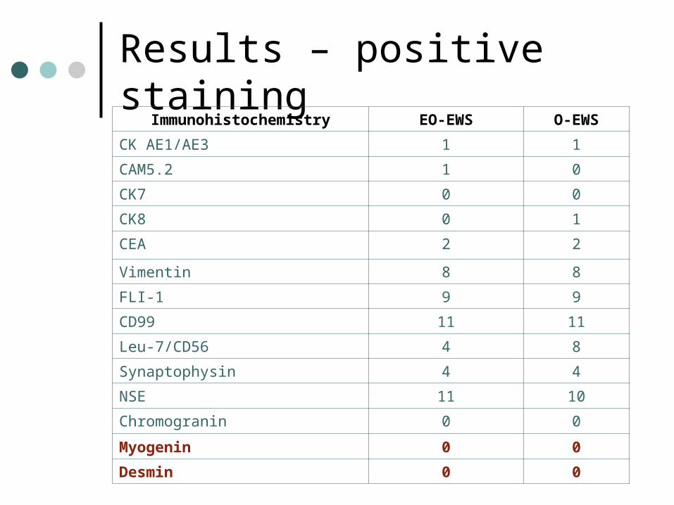
Results – positive staining
Immunohistochemistry EO-EWS O-EWS
CK AE1/AE3 1 1
CAM5.2 1 0
CK7 0 0
CK8 0 1
CEA 2 2
Vimentin 8 8
FLI-1 9 9
CD99 11 11
Leu-7/CD56 4 8
Synaptophysin 4 4
NSE 11 10
Chromogranin 0 0
Myogenin 0 0
Desmin 0 0

Results – positive staining
Immunohistochemistry EO-EWS O-EWS
CK AE1/AE3 1 1
CAM5.2 1 0
CK7 0 0
CK8 0 1
CEA 2 2
Vimentin 8 8
FLI-1 9 9
CD99 11 11
Leu-7/CD56 4 8
Synaptophysin 4 4
NSE 11 10
Chromogranin 0 0
Myogenin 0 0
Desmin 0 0

Cytokeratin Cam 5.2positive
Carcinoembryonic antigen (CEA)positive

Results – positive staining
Immunohistochemistry EO-EWS O-EWS
CK AE1/AE3 1 1
CAM5.2 1 0
CK7 0 0
CK8 0 1
CEA 2 2
Vimentin 8 8
FLI-1 9 9
CD99 11 11
Leu-7/CD56 4 8
Synaptophysin 4 4
NSE 11 10
Chromogranin 0 0
Myogenin 0 0
Desmin 0 0

Results – positive staining
Immunohistochemistry EO-EWS O-EWS
CK AE1/AE3 1 1
CAM5.2 1 0
CK7 0 0
CK8 0 1
CEA 2 2
Vimentin 8 8
FLI-1 9 9
CD99 11 11
Leu-7/CD56 4 8
Synaptophysin 4 4
NSE 11 10
Chromogranin 0 0
Myogenin 0 0
Desmin 0 0
Results – positive staining
Immunohistochemistry EO-EWS O-EWS
CK AE1/AE3 1 1
CAM5.2 1 0
CK7 0 0
CK8 0 1
CEA 2 2
Vimentin 8 8
FLI-1 9 9
CD99 11 11
Leu-7/CD56 4 8
Synaptophysin 4 4
NSE 11 10
Chromogranin 0 0
Myogenin 0 0
Desmin 0 0

Methods
RT-PCR RNA extracted from formalin fixed,
paraffin embedded tumor (Roche high pure RNA paraffin kit)
PCR was performed for EWS/FLI-1• EWS/FLI-1 fusion type identified by melt
curve analysis• EWS-FLI-1 fusion type confirmed by agarose
gel electrophoresis Alternate partners were examined as
necessary (ews/fev, etv1, etv4 and erg)

Results – EWS-FLI-1 type
EO-EWS
O-EWS
EWS-FLI-1
10 10
Type 1 7 10
Type 2 3 0
Non Type 1 or 2
1 1
Not type 1 or 2 EO-EWS: confirmed
t(11:22)(q24;q12) but no translocation products amplified
O-EWS: PCR product melting curve between types 1 and 2 but failed sequencing

Summary
Negative FLI-1 nuclear staining does not exclude EWS-FLI-1 translocation positive EFT
While CEA +ve staining can be seen in EO-EWS it does not differentiate this from O-EWS
More type 2 fusions noted in patients with EO-EWS

Conclusion
Variability may occur in immunostaining and genotype analysis of patients with extra-osseous Ewing sarcoma vs. osseous Ewing sarcoma.