comparison of japanese and indian intestinal microbiota
TRANSCRIPT

ARTICLE OPEN
Comparison of Japanese and Indian intestinal microbiotashows diet-dependent interaction between bacteria and fungiSiddhika Pareek1,2,3,12, Takashi Kurakawa1,2,12, Bhabatosh Das4, Daisuke Motooka5, Shuuichi Nakaya6, Temsunaro Rongsen-Chandola7,Nidhi Goyal7, Hisako Kayama1,2,3, Dylan Dodd8, Ryu Okumura1,2,3, Yuichi Maeda1,2,9, Kosuke Fujimoto1,9, Takuro Nii1,2,9,Takao Ogawa1,2,9, Tetsuya Iida5,10, Nita Bhandari 7, Toshiyuki Kida11, Shota Nakamura5, G. Balakrish Nair4 and Kiyoshi Takeda1,2,3*
The bacterial species living in the gut mediate many aspects of biological processes such as nutrition and activation of adaptiveimmunity. In addition, commensal fungi residing in the intestine also influence host health. Although the interaction of bacteriumand fungus has been shown, its precise mechanism during colonization of the human intestine remains largely unknown. Here, weshow interaction between bacterial and fungal species for utilization of dietary components driving their efficient growth in theintestine. Next generation sequencing of fecal samples from Japanese and Indian adults revealed differential patterns of bacterialand fungal composition. In particular, Indians, who consume more plant polysaccharides than Japanese, harbored increasednumbers of Prevotella and Candida. Candida spp. showed strong growth responses to the plant polysaccharide arabinoxylanin vitro. Furthermore, the culture supernatants of Candida spp. grown with arabinoxylan promoted rapid proliferation of Prevotellacopri. Arabinose was identified as a potential growth-inducing factor in the Candida culture supernatants. Candida spp. exhibited agrowth response to xylose, but not to arabinose, whereas P. copri proliferated in response to both xylose and arabinose. Candidaspp., but not P. copri, colonized the intestine of germ-free mice. However, P. copri successfully colonized mouse intestine alreadyharboring Candida. These findings demonstrate a proof of concept that fungal members of gut microbiota can facilitate acolonization of the intestine by their bacterial counterparts, potentially mediated by a dietary metabolite.
npj Biofilms and Microbiomes (2019) 5:37 ; https://doi.org/10.1038/s41522-019-0110-9
INTRODUCTIONThe human gut bacterial community represents an enormousnumber of 1014 bacteria with more than 1000 different speciesand has diverse roles, such as maintaining immune homeostasis,freeing dietary nutrients for host absorption, and colonizationresistance against pathogens.1 The gut bacterial compositionvaries immensely among individuals in response to intrinsic andextrinsic factors including genetic background, mode of deliveryduring childbirth, age, diet, and diseases.2–4 High throughputsequencing technologies have enabled comprehensive analysesof the human microbiome.5 Several studies investigating thecomposition of human microbiota have shown that environmen-tal factors rather than host genetics play a crucial role in shapingthe intestinal microbial ecosystem.6–8 Among these environmentalfactors, dietary components strongly influence the bacterialcomposition of the gut.9 Generally, plant-based diets are moreprevalent in developing countries, whereas the intake of animal-derived products is higher in developed countries, as shown in thedatabase of Food and Agriculture Organization of the UnitedNations (http://www.fao.org/faostat/en/home). The gut microbiotawas clustered into three enterotypes, characterized by theabundance of Bacteroides, Prevotella, and Ruminococcus10 andlong-term diet were suggested to influence these enterotype
patterns across the populations. Several studies covering differentpopulations worldwide have shown that consumption of animal-based diets and plant-based diets induces differential patterns ofgut bacterial composition.6,9,11–13 However, these studies explor-ing the impact of intestinal microbes and metabolic health haveessentially focused on bacteria in the intestine.14,15 Notably, fungalspecies have been reported to colonize as commensals in the gutof healthy humans and mice.16,17 Although they comprise lessthan 1% of the total gut microbial population,10,18 studies inmurine models have shown its importance during alteration of thegut environment. For instance, Candida albicans is persistentlypresent in a mouse model that develops allergic disorders andautoimmune diseases,19 and was shown to interact with bacteriaduring gastric colonization.20 The commensal bacteria preventfungi from long-term colonization.21 During antibiotic recovery inthe murine cecum, C. albicans was shown to promote therestoration of bacterial diversity.22 In addition, diet has beenshown to modify the abundance of the fungal population as wellas bacterial population in the gut.18 These findings, therefore,underline the necessity to examine the precise mechanism ofinterkingdom interactions to understand microbiome-mediatedeffects on host physiology.
1Department of Microbiology and Immunology, Graduate School of Medicine, Osaka University, Suita, Osaka 565-0871, Japan. 2WPI Immunology Frontier Research Center, OsakaUniversity, Suita, Osaka 565-0871, Japan. 3Core Research for Evolutional Science and Technology, Japan Agency for Medical Research and Development, Tokyo 100-0004, Japan.4Molecular Genetics Laboratory, Center for Human Microbial Ecology, Translational Health Science and Technology Institute, Faridabad 121001, India. 5Department of InfectionMetagenomics, Genome Information Research Center, Research Institute for Microbial Diseases, Osaka University, Osaka 565-0871, Japan. 6Global Applications DevelopmentCenter, Shimadzu Corp, Kyoto 604-8511, Japan. 7Centre for Health Research and Development, Society for Applied Studies, New Delhi 110016, India. 8Department of Pathology,Stanford University School of Medicine, Stanford, CA 94305, USA. 9Department of Respiratory Medicine and Clinical Immunology, Graduate School of Medicine, Osaka University,Osaka 565-0871, Japan. 10Department of Bacterial Infections, Research Institute for Microbial Diseases, Osaka University, Osaka 565-0871, Japan. 11Department of AppliedChemistry, Graduate School of Engineering, Osaka University, Osaka 565-0871, Japan. 12These authors contributed equally: Siddhika Pareek, Takashi Kurakawa.*email: [email protected]
www.nature.com/npjbiofilms
Published in partnership with Nanyang Technological University
1234567890():,;
In this study, we analyzed bacterial and fungal composition ofJapanese and Indian fecal samples. Based on the dietary habitatquestionnaire and their dominant microorganisms, we focused onmetabolism of arabinoxylan, which is one of the major indigestiblepolysaccharides. We then analyzed the potential mechanism forinteraction between gut bacterium and fungus both in vitro andin vivo. The results suggested a dietary metabolite-dependentinteraction between fungi and bacteria, which promotes bacterialgrowth and colonization in the gut.
RESULTSAnalyses of bacterial and fungal composition in Japanese andIndian fecesWe compared the composition of fecal bacteria and fungi fromtwo geographically distinct healthy adult populations living inJapan (n= 47) and India (n= 50) (Table 1). First, bacterialcompositions were compared by 16S rRNA gene sequencing.Firmicutes, Bacteroidetes, Actinobacteria, and Proteobacteria werethe four dominant bacterial phyla in both Japanese and Indiansamples (Supplementary Fig. 1a, b). The ratios of Bacteroidetes toFirmicutes in individuals from India were markedly higher than
those from Japan (Supplementary Fig. 1c, d). Genus level analysisshowed that Bacteroides and Prevotella were the dominantbacterial genera in samples from Japan and India, respectively(Fig. 1a). Principal component analysis (PCA) based on gutbacterial composition showed distinct clustering of Indian andJapanese samples (Fig. 1b). Considering the genus Prevotella,individuals from India harbored Prevotella copri as the dominantbacterial species, followed by Prevotella stercorea and Prevotellasp., while these species were detected only in a small number ofthose from Japan (Fig. 1c). With respect to the genus Bacteroides,Bacteroides sp., Bacteroides uniformis, Bacteroides ovatus, andBacteroides fragilis were prevalent in Japanese samples. Althoughhigher numbers of operational taxonomic units (OTUs) wereobserved in samples from India than Japan, both groups showedsimilar Shannon diversity, which would be due to the highproportion of Prevotella in Indian samples (Fig. 1d, e). Thisobservation is in accordance with previous reports on analysis ofmicrobiota from populations consuming diets rich in plant-derived products.6,7,23
Next, we analyzed fecal fungi from individuals living in Japanand India using an improved sequencing procedure to analyzefungal composition by comparing the internal transcribed spacer1 (ITS1) sequences of rRNA genes.16 Ascomycota and Basidiomy-cota were the main fungal phyla in both Japanese and Indiansamples (Supplementary Fig. 1e, f). The relative abundance ofBasidiomycota in Japanese samples was remarkably higher thanthat in Indians. Saccharomyces and Candida, both of which belongto the Ascomycota, were found to be the major fungal genera inJapanese and Indian samples, respectively (Fig. 2a). The PCAshowed a clear separation between Japanese and Indian samplesin terms of the variation of the fungal composition (Fig. 2b). Forthe genus Candida, the detection ratios and relative abundancesof C. albicans, Candida tropicalis, and Candida glabrata in Indiansamples were markedly higher than those in Japanese (Fig. 2c).Among Saccharomyces, Saccharomyces cerevisiae dominated inJapanese samples. Similar to bacterial case, the Shannon diversityindices for gut fungal species were similar in both Japanese andIndian subjects (Fig. 2d, e). Demographics such as age and sexwere not associated with the bacterial and fungal composition(data not shown). Thus, the composition of both intestinal fungiand bacteria varied between individuals living in these distinctareas.
Growth of Prevotella and Candida on several plantpolysaccharidesGiven that dietary habits predominantly shape gut bacterialecology,9,23 we reasoned that the components of plant-baseddiets might influence the composition of both the bacterial andfungal communities in the intestine. We analyzed the effect ofplant polysaccharides (constituents of cereals), such as starch anddietary fibers, including wheat arabinoxylan (AX),24 and carbox-ymethyl cellulose (CMC), which is the soluble alternative tocellulose, on in vitro growth of Prevotella and Candida, both ofwhich were dominant microorganisms in the intestines of Indianpopulation we recruited. To monitor the growth yields of P. copriand Candida spp. (C. albicans, C. tropicalis, and C. glabrata), thesemicroorganisms were cultured in a medium containing individualpolysaccharides. Previous studies reported that glucose (amonosaccharide) supported the growth of these microorganismswhen used as the sole carbohydrate source.25–27 Therefore, weused glucose as the sole carbohydrate in the growth medium andobserved that P. copri grew rapidly in this condition (Fig. 3a,Supplementary Fig. 2a). In accordance with previous reportsanalyzing Prevotella spp. isolated from ruminant animals,25,28,29 P.copri, which was originally isolated from the human intestine30
and obtained from Japan Collection of Microorganisms (JCM),utilized AX as the sole carbon source and proliferated in the
Table 1. Subjects information in this study.
Issues Japanese Indians Statisticaldifference
District of residence Osaka Delhi
Subject number 47 50
Male (%) 54 53
Age 28.8 ± 6.2 30.6 ± 6.1
Animal exposure (%) 17 38 *
Cattle 0 20 **
Goat/Sheep 0 2
Dog 2 4
Cat 4 2
Others (including noanswer)
11 10
Places of defecation (%)
Private toilet at home 100 88 *
Public toilet 0 18 **
Open field 0 26 ***
Food (frequency of ingestion in 1 week)
Bread 5.6 ± 1.9 14.4 ± 4.3 ****
Ricea 6.5 ± 1.8 4.2 ± 2.7 ****
Maize 1.2 ± 1.9 0.4 ± 0.8 **
Soybeanb 4.6 ± 3.4 0.1 ± 0.4 ****
Yogurt 2.7 ± 2.3 2.2 ± 2.3
Cheese 2.0 ± 1.8 0.1 ± 0.2 ****
Butter 2.9 ± 2.7 1.9 ± 2.6 *
Milk 4.7 ± 3.7 4.6 ± 3.8
Meat 4.2 ± 1.8 0.3 ± 0.8 ***
Fish 2.5 ± 1.5 0.1 ± 0.4 ****
Poultry 2.7 ± 1.7 0.4 ± 0.8 ****
Eggs 4.2 ± 1.9 1.1 ± 2.1 ****
aIncludes rotibIncludes tofu and natto*p < 0.05**p < 0.01***p < 0.001****p < 0.0001
S. Pareek et al.
2
npj Biofilms and Microbiomes (2019) 37 Published in partnership with Nanyang Technological University
1234567890():,;

defined medium though at a slower rate than when using glucose.However, P. copri did not grow in response to other types ofdietary polysaccharides tested in the current study. P. coprishowed a dose-dependent growth response to AX and grewequally in the presence of AX or glucose (Fig. 3b). To confirm thatthe slower growth response of P. copri in the presence of AX is notstrain-specific, we isolated P. copri from the feces of Indiansubjects and analyzed the growth response (Supplementary Fig.2b). The isolated P. copri showed the similar growth response toAX and glucose as the P. copri JCM strain, although it alsoresponded to starch. Next, we analyzed the growth response ofCandida, using publicly available type strains obtained from JCM,to the same sets of dietary polysaccharides, in yeast nitrogen base(YNB) medium. Similar to P. copri, both C. albicans and C. tropicalisgrew in the presence of glucose or AX but not the otherpolysaccharides (Fig. 3c, d, Supplementary Fig. 3a). C. albicans andC. tropicalis achieved higher growth in the presence of AX thanglucose at an equimolar concentration (10 mM) (Fig. 3c–f). Wenext isolated Candida species (C. albicans, C. tropicalis, and C.glabrata) from the fecal samples of Indian subjects. Although C.glabrata did not show a strong growth response to AX, C. albicansand C. tropicalis isolated from Indians showed the similar growthpattern as the JCM strains (Supplementary Fig. 3b–d), suggestingthat the ability to utilize polysaccharides is preserved between thestrains used in the current study. We also analyzed the growthresponses of S. cerevisiae and Bacteroides species, such as B. fragilis,B. ovatus, B. thetaiotaomicron, and B. uniformis, which weredominant in Japanese samples (Supplementary Fig. 4a–e). S.cerevisiae did not show any growth response in the presence of
the above mentioned dietary polysaccharides. Bacteroides speciesgrew in the presence of starch, but not CMC, and some Bacteroidesspecies showed a response to AX. Thus, intestinal microorganismsshowed differential growth responses to various dietarypolysaccharides.
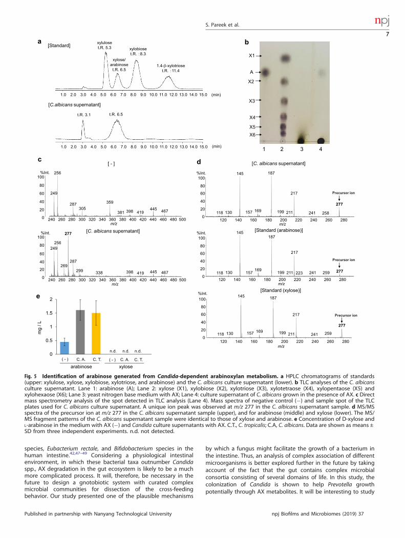
Candida-dependent dietary metabolites that support PrevotellagrowthWe next analyzed the interaction of Candida and Prevotella, bothof which dominated the intestine of Indian subjects and showedgrowth response to AX. Given the effective use of AX by Candidaover Prevotella, we analyzed whether Candida supports Prevotellagrowth in an AX-rich environment (Fig. 4a). Addition of culturesupernatants of C. albicans or C. tropicalis grown in the presence ofAX induced rapid growth of P. copri compared to its growth in thepresence of AX alone. Similarly, Candida strains isolated fromIndian feces promoted P. copri growth (Fig. 4b). These resultssuggest that the fungal supernatants were enriched in metabolicproducts that enabled rapid growth of P. copri.Subsequently, we attempted to identify the molecule in the
culture supernatants of Candida grown in AX-containing mediumthat stimulated P. copri growth. First, the C. albicans culturesupernatant was analyzed by high performance liquid chromato-graphy (HPLC) (Fig. 5a). Because AX is a polymer of β-1,4 linkedD-xylopyranosyl residues that are substituted with monomeric α-L-arabinofuranose units at the second and/or third carbon (C-2 andC-3) positions,31,32 xylo-oligosaccharides or monomeric D-xyloseand L-arabinose were expected to be produced by AX
0.0
0.1
0.2
0.3
0.4
0.5
P. copriP. stercoreaPrevotella sp.P. melaninogenicaP. pallensOther PrevotellaBacteroides sp.B. uniformisB. ovatusB. fragilisB. caccaeB. plebeiusB. eggerthiiB. coprophilusOther BacteroidesB. nordiiB. barnesiae
-0.6
-0.3
0
0.3
0.6
-0.6 -0.3 0 0.3 0.6PC1 (71.1%)
a b c
PC
2 (8
.5%
)
ecnadnuba evitaleR
JapaneseIndians
JapaneseIndians
max0 0.001
Japanese Indians
**** **** * **** **** ****
n.s. ** **** ***
0
500
1000
1500
2000
2500
3000
0 5000 10000 15000 20000 25000 30000
srebmun
UTO devresb
O
Japanese
4.0
4.5
5.0
5.5
6.0
6.5
7.0
7.5
IndiansReads
d eS
hann
on in
dex
******
******
******
****** *** ***
JapaneseIndians
n.s.
Fig. 1 Comparison of fecal bacteria in healthy adults living in Japan and India. a Relative abundances of the major genera. b Principalcomponent analysis of fecal bacteria at the genus level. c Heatmap representing the relative abundances of bacterial species in genusBacteroides and Prevotella. d Rarefaction curves. e Shannon index. n.s. not significant; ****p < 0.0001, ***p < 0.001, **p < 0.01, *p < 0.05.
S. Pareek et al.
3
Published in partnership with Nanyang Technological University npj Biofilms and Microbiomes (2019) 37

degradation. Therefore, they were used as standards. In the C.albicans supernatant, two peaks were observed in HPLC withretention times of 3.1 and 6.5 min; the retention time of 6.5 mincorresponded to that of xylose and arabinose. In an effort todistinguish between xylose and arabinose, supernatants ofC. albicans (the JCM strain and the Indian fecal isolate) wereseparated by thin layer chromatography (TLC) (Fig. 5b, Supple-mentary Fig. 5a). A spot of the C. albicans supernatant wasobserved at a similar position to that of the arabinose standard,while no spots were observed comigrating with the xylosestandard. A similar peak at retention time of 6.5 min in HPLCand spot in TLC were observed in supernatants of C. tropicalis (theJCM strain and the Indian isolate) (Supplementary Fig. 5b–d). Next,the TLC spot of the C. albicans supernatant was isolated andanalyzed by mass spectrometry (MS) (Fig. 5c, d). A specific andstrong signal was observed at m/z 277 in the C. albicanssupernatant, and MS/MS spectra of the m/z 277 ion showed thesame pattern as that of xylose and arabinose. These datacollectively suggest that the C. albicans supernatant containedarabinose. We then measured the concentrations of D-xylose andL-arabinose in the Candida culture supernatants using xylose- orarabinose/galactose-specific enzyme-based colorimetric assays,respectively (Fig. 5e). Xylose was not detected in the supernatantsof C. albicans or C. tropicalis cultures. In contrast, arabinose waselevated when C. albicans or C. tropicalis were cultured in thepresence of AX. These findings indicate that arabinose wasenriched in the Candida spp. supernatant, although AX degrada-tion is expected to produce both xylose and arabinose. Therefore,we speculated that xylose produced by hydrolysis of AX wasrapidly and completely consumed by Candida spp. To assess this,
we analyzed the growth response of Candida (the JCM strain andthe Indian isolate) to the monosaccharides D-xylose andL-arabinose (Fig. 6a–e). D-xylose, but not L-arabinose, inducedprominent growth of both C. albicans, C. tropicalis, and C. glabrataat a level similar to that induced by glucose. These findingsindicate that Candida strains used in the current study metabolizeAX and use xylose for their growth. Then, we analyzed the effectof L-arabinose on the growth of P. copri (the JCM strain and theIndian isolate) (Fig. 6f, g). P. copri showed a marked growthresponse to L-arabinose as well as D-xylose. Consumption ofarabinose by P. copri was also assessed by TLC (Supplementary Fig.6). The spot corresponding to arabinose detected in the super-natant of C. tropicalis culture in the presence of AX was notdetected in the culture supernatant of P. copri grown in the C.tropicalis AX supernatants. Thus, the AX-derived metabolite, whichwas produced by Candida and induced substantial growth of P.copri, could be arabinose. However, we should consider thatseveral other microbes participate in utilizing such diet-derivedmetabolic products in the gut. Indeed, similar to P. copri,Bacteroides species also utilized L-arabinose (Supplementary Fig.7), which indicate the presence of an unknown mechanism thatmight facilitate the preferential selection of P. copri by Candida(see Discussion).
Promotion of Prevotella colonization by Candida in germ-freemiceNext, we analyzed the in vivo interaction of Candida and Prevotellausing a gnotobiotic mouse colonization model. Germ-free (GF)mice were orally administered Candida or Prevotella and then the
0.0
0.2
0.4
0.6
0.8
1.0
-0.8
-0.4
0
0.4
0.8
-0.8 -0.4 0 0.4 0.8
a b c
PC1 (47.3%)P
C2
(21.
6%)
ecnadnuba evitaleR
JapaneseIndians
JapaneseIndians
S. cerevisiaeOther SaccharomycesS. paradoxusC. albicansC. tropicalis
Other CandidaC. orthopsilosisC. parapsilosisC. sakeC. dubliniensis
0 0.001 max
Japanese Indians
C. glabrata
*
**** **** ************ *
******* n.s.
0
20
40
60
80
0 500 1000 1500
d e
srebmun
U TO devresb
O
Japanese IndiansReads
Sha
nnon
inde
x0
12
34
JapaneseIndians
**n.s.n.s.
n.s.n.s.
n.s.
Fig. 2 Comparison of fecal fungi in healthy adults living in Japan and India. a Relative abundances of the major genera. b Principalcomponent analysis of fecal fungi at the genus level. c Heatmap representing the relative abundances of fungal species in genusSaccharomyces and Candida. d Rarefaction curves. e Shannon index. n.s. not significant; ****p < 0.0001; ***p < 0.001; *p < 0.05.
S. Pareek et al.
4
npj Biofilms and Microbiomes (2019) 37 Published in partnership with Nanyang Technological University

microbial load in feces was analyzed (Fig. 7a). C. albicans andC. tropicalis successfully colonized the mouse intestine, reached apeak level within 2 days post administration and maintained peaknumbers for >3 weeks (Fig. 7b, Supplementary Fig. 8). In contrast,P. copri was barely detectable, indicating that Prevotella hadlimited ability to colonize the mouse intestine on its own (Fig. 7c).In the next set of experiments, GF mice were first colonized with C.albicans and then P. copri was orally administered 3 days later. Inthe Candida-enriched intestinal environment, P. copri increasedgradually and outnumbered Candida at day 24 (3 weeks after the
P. copri administration) (Fig. 7d). We also analyzed the localizationof both microorganisms in the colon by fluorescence in situhybridization (FISH) using probes specific for Prevotella andCandida (Fig. 7e). In mice administered C. albicans alone, Candidawas detected in the colonic lumen. In contrast, Prevotella wasbarely detected in the colonic lumen of mice given P. copri alone.However, elevated numbers of Prevotella were observed in thecolonic lumen and feces of mice administered both C. albicansand P. copri (Fig. 7e, Supplementary Fig. 9). These findings indicate
0
0.5
1
1.5
2
2.5
3
0 12 24 36 48 60 72
(-)
Glucose
AX
Starch
CMC
0
0.5
1
1.5
2
2.5
3
0 12 24 36 48 60 72
(-)
Glucose
AX
Starch
CMC
0
0.1
0.2
0.3
0.4
0.5
0.6
0.7
0.8
0 12 24 36 48 60 72
(-)
Glucose
AX
Starch
CMC
0
0.5
1
1.5
2
2.5
3
AX
0
0.5
1
1.5
2
2.5
3
AX
0
0.1
0.2
0.3
0.4
0.5
0.6
0.7
0.8
a
DO(
ecnabrosbA
600)
Time (h)
AX
n.s.n.s.
n.s.n.s.
n.s.********
****
b
c
e f
Time (h) Time (h)
d
*******
********
n.s.n.s.
********
****
**
**** **********
****
Abs
orba
nce
(OD
600)
DO(
ecnabrosbA
600)
Abs
orba
nce
(OD
600)
DO(
ec nabrosbA
600)
Abs
orba
nce
(OD
600)
C. albicans JCM 1542 C. tropicalis JCM 1541
P. copri JCM 13464
n.d.
n.d.
**
n.d.
P. copri JCM 13464
C. albicans JCM 1542 C. tropicalis JCM 1541
Fig. 3 Co-utilization of arabinoxylan by Candida and Prevotella. a, c, d Growth of P. copri (a), C. albicans (c), and C. tropicalis (d) in thepresence or absence of 10mM glucose, arabinoxylan (AX), starch, or carboxymethyl cellulose (CMC). The growth rate in the presence of AXand glucose was statistically compared. b, e, f Growth response of P. copri (b), C. albicans (e), and C. tropicalis (f) in the presence or absence of10mM glucose or the indicated concentrations of AX at 72 h. (−) is base media alone without any carbon source. n.s. not significant, n.d. notdetected. ****p < 0.0001; ***p < 0.001; **p < 0.01.
S. Pareek et al.
5
Published in partnership with Nanyang Technological University npj Biofilms and Microbiomes (2019) 37

that P. copri efficiently colonized and grew in the intestine in thepresence of Candida spp.
DISCUSSIONOur study analyzed the intestinal bacterial and fungal compositionof Japanese and Indian adults and provided evidence for aninterkingdom interaction that is potentially mediated by differ-ences in the host diet. Japanese populations, with high intake ofanimal products, showed an abundance of Bacteroides comparedto Indians, consuming plant-based diets, showing higher levels ofPrevotella. These observations are parallel to the earlier findings inhuman populations with a diet enriched in complex carbohy-drates, such as Hadza hunter-gatherer from Tanzania33 andchildren from rural Africa6 who showed a higher abundance ofPrevotella compared to populations ingesting a western dietharboring higher levels of Bacteroides. In accordance with thosestudies, Prevotella has been shown abundant in individuals withhigh intake of carbohydrates/dietary fiber.23
In addition to bacteria, accumulating evidence indicates thatother domains of life residing in the gut, including viruses,34,35
archaea36 and eukaryotes, such as protozoans37 and fungi,36,38
contribute to the development of gut ecosystem and influencethe host physiology. Indeed, the correlation of dietary habits withthe composition of intestinal archaea and fungi has beenreported.36 However, further studies are required to establishthe mechanisms of their interaction with one another in thecomplex gut environment. A previous study in mice showed thatcommensal bacteria B. thetaiotamicron and Blautia producta canpromote colonization resistance to C. albicans by increasingexpression of antimicrobial peptide LL-37 mediated by hypoxia-inducible factor-1α.39 Another study showed that human gutBacteroides has the glycoside phosphorylase genes that targets β-
1,2-mannnosidic linkages in Candida mannan, and it can utilizeyeast mannan as a food source.40 It was also reported that oraladministration of Saccharomyces in obese mice resulted inalteration of bacterial composition, in which Bacteroides wasdramatically increased and Prevotella was decreased.41 Thesefindings could potentially explain the observation in our studywhere Japanese cohort had a substantial presence of Sacchar-omyces, Bacteroides, and Blautia but lower levels of C. albicans, andIndian cohort had lower levels of Bacteroides. Thus, it will becrucial to carefully study the host-derived influences together withexternal factors including diet in order to completely assess thedifferential colonization of microbial communities acrosspopulations.The information from dietary habitat survey indicated that diets
of Indian subjects were rich in plant-derived carbohydrates (fiber-rich) and therefore representative dietary plant polysaccharideswere included for in vitro growth response assay of Prevotella andCandida. Xylan, second most abundant (after cellulose) plantpolysaccharide, is a known substrate for microbial fermentation inthe gut of ruminants as well as humans.42 Cereal grains, such aswheat, corn, and rye, have higher proportion of xylan.43 P. copri,like ruminant origin P. bryantii,25 grew in response to wheat AX.This is the first study that tested the growth response of Candidaspp. (both JCM and Indian fecal isolates) toward AX. Candidautilized AX from the panel of three polysaccharides and generatedarabinose which potentially enhanced P. copri growth in vitro,suggesting a dietary metabolite mediated interaction betweenfungi and bacteria. It is important to note that while we observedthat arabinose was produced by Candida and consumed by P.copri, the in vitro assays tested for specific metabolites. Thus, acomprehensive characterization of the metabolites is warranted togain deeper insights into the dietary components mediatingmicrobial interactions.Similar to in vitro observations, this interkingdom interaction
was recapitulated in GF mouse system where we observedincreased P. copri numbers in the presence of Candida. However,given the multitude of factors regulating highly dynamic andcomplex intestinal system, other mechanisms might be active tofacilitate this interaction. For instance, some intestinal bacteria,such as Bacteroides, have been known to ferment polysaccharidesof the yeast cell wall such as mannan40 and β-glucans.44 Thus, it isalso possible that in addition to diet-derived sources Prevotellamight benefit from the presence of Candida by other alternatemechanisms. In the present study, we focused on validating theinterkingdom interaction and proposed arabinose as a potentialcandidate. However, additional experiments will be required toestablish its role as a major beneficiary module that facilitatesbacterial growth. For instance, future studies comparing GF micecolonized with respective microbes by administering a customizeddiet rich in AX and AX-free diet group will be able to provide aclearer picture of the impact of AX/arabinose based cross-feedingmechanism in colonization of Prevotella in the intestine.AX itself has a complex chemical structure comprised of linear
D-xylose backbone. In various grain species, the backbone xylosemay be substituted by arabinose and cross-linked with ferulic acid.To acquire energy, microbes need to depolymerize the poly-saccharides by enzymatic cleavage of the chemical linkages. Gutbacteria produce thousands of substrate-specific carbohydrate-active enzymes (CAZymes) that catalyze the breakdown of theunique linkages and have been extensively cataloged.28,45,46 Thus,one of future directions would be identification of CAZymes inCandida spp. and Prevotella for utilizing AX.The present study aims to highlight the importance of analyses
on the lesser-studied microorganisms, particularly fungi, for acomprehensive understanding of the complex interactions in thegut microbial ecosystem. Although our study mainly focused onCandida species, which identified them as AX-degraders, there areseveral AX-degrading microorganisms including Bacteroides
0
0.2
0.4
0.6
0.8
1
0 12 24 36 48 60 72
aAX
C. albicans sup with AX
C. tropicalis sup with AX
(-)
C. albicans sup with (-)C. tropicalis sup with(-)
C. tropicalis sup with glucose
C. albicans sup with glucose
Glucosen.s.
********n.s.
*****
n.s.**** ***
n.s.
Abs
orba
nce
(OD
600)
Time (h)
P. copri JCM 13464
0
0.1
0.2
0.3
0.4
0.5
0.6
0 12 24 36 48 60 72
AX
C. albicans sup with AX
C. tropicalis sup with AX
(-)
C. albicans sup with (-)C. tropicalis sup with(-)
C. tropicalis sup with glucose
C. albicans sup with glucose
Glucose**** ********
n.s. n.s.**
****
****
****
Abs
orba
nce
(OD
600)
****
****
Time (h)
b P. copri
(Isolated from Indian feces)
Fig. 4 Promotion of Prevotella growth by the metabolitesproduced by Candida. Growth of P. copri JCM 13464 (a) and isolatesfrom Indian feces (b) in the presence of glucose, AX, and C. tropicalis-or C. albicans-supernatants from cultures grown in AX. (−) is basemedia alone without any carbon source. Results of statisticalcomparison between C. albicans and C. tropicalis-supernatants fromcultures grown in AX with AX alone are shown. All the graphs showthe mean ± SD of three independent experiments. n.s. notsignificant. ****p < 0.0001, ***p < 0.001, **p < 0.01, *p < 0.05.
S. Pareek et al.
6
npj Biofilms and Microbiomes (2019) 37 Published in partnership with Nanyang Technological University
species, Eubacterium rectale, and Bifidobacterium species in thehuman intestine.42,47–49 Considering a physiological intestinalenvironment, in which these bacterial taxa outnumber Candidaspp., AX degradation in the gut ecosystem is likely to be a muchmore complicated process. It will, therefore, be necessary in thefuture to design a gnotobiotic system with curated complexmicrobial communities for dissection of the cross-feedingbehavior. Our study presented one of the plausible mechanisms
by which a fungus might facilitate the growth of a bacterium inthe intestine. Thus, an analysis of complex association of differentmicroorganisms is better explored further in the future by takingaccount of the fact that the gut contains complex microbialconsortia consisting of several domains of life. In this study, thecolonization of Candida is shown to help Prevotella growthpotentially through AX metabolites. It will be interesting to study
Fig. 5 Identification of arabinose generated from Candida-dependent arabinoxylan metabolism. a HPLC chromatograms of standards(upper: xylulose, xylose, xylobiose, xylotriose, and arabinose) and the C. albicans culture supernatant (lower). b TLC analyses of the C. albicansculture supernatant. Lane 1: arabinose (A); Lane 2: xylose (X1), xylobiose (X2), xylotriose (X3), xylotetraose (X4), xylopentaose (X5) andxylohexaose (X6); Lane 3: yeast nitrogen base medium with AX; Lane 4: culture supernatant of C. albicans grown in the presence of AX. c Directmass spectrometry analysis of the spot detected in TLC analysis (Lane 4). Mass spectra of negative control (−) and sample spot of the TLCplates used for C. albicans culture supernatant. A unique ion peak was observed at m/z 277 in the C. albicans supernatant sample. d MS/MSspectra of the precursor ion at m/z 277 in the C. albicans supernatant sample (upper), and for arabinose (middle) and xylose (lower). The MS/MS fragment patterns of the C. albicans supernatant sample were identical to those of xylose and arabinose. e Concentration of D-xylose andL-arabinose in the medium with AX (−) and Candida culture supernatants with AX. C.T., C. tropicalis; C.A, C. albicans. Data are shown as means ±SD from three independent experiments. n.d. not detected.
S. Pareek et al.
7
Published in partnership with Nanyang Technological University npj Biofilms and Microbiomes (2019) 37

whether the interaction of these microorganisms in the intestine isinvolved in the maintenance of the host health.
MATERIALS AND METHODSFecal collection and processingFecal samples were collected from 47 healthy Japanese adults living in theOsaka area (25 males and 22 females, average age 30.6 ± 6.1 years) and 50healthy Indians living in the Delhi area (27 males and 23 females, averageage 28.8 ± 6.2 years). A spoonful of feces (0.5 g) was collected into a tubecontaining 2ml of RNAlater (Ambion) for nucleic acid extraction.Collections were made immediately after defecation. Each fecal samplefor nucleic acid extraction was weighed and suspended in nine volumes ofRNAlater to make a fecal homogenate (100mg feces/ml). In accordancewith the Declaration of Helsinki, all subjects were adequately informedabout the study. Informed written consent was collected from all the
participants. The ethics committees of Osaka University and theTranslational Health Science and Technology Institute (Faridabad)approved this study. The protocol numbers are 12237, and SAS/THSTI/001/2013-2014, respectively. The samples were transported betweenJapan and India in accordance with the Nagoya protocol.
Extraction of DNA for bacterial analysisFor DNA extraction, 1 ml of phosphate-buffered saline (PBS) was added to200 μl of fecal homogenate. The fecal homogenate was centrifuged at13,000 × g for 10 min and 1ml of the supernatant was discarded. Afteranother wash with 1ml of PBS, the pellets were stored at −30 °C until usefor DNA extraction. Glass beads (0.3 g; diameter, 0.1 mm) (BioSpecProducts), 300 μl Tris-SDS solution and 500 μl Tris-EDTA (TE)-saturatedphenol were added to 200 μl of the fecal homogenate, and the mixturewas vortexed vigorously for 30 s using a FastPrep-24 (M.P. Biomedicals) at5.0 power level for 30 s. After centrifugation at 20,000 × g for 5 min at 4 °C,
0
0.1
0.2
0.3
0.4
0.5
0.6
0.7
0.8
0 12 24 36 48 60 72
0
0.5
1
1.5
2
2.5
0 12 24 36 48 60 720
0.5
1
1.5
2
2.5
0 12 24 36 48 60 72
a b
Time (h) Time (h)
****
****
n.s.n.s.
******
n.s. n.s.
n.s.
n.s. n.s.
DO(
ecnabrosbA
600)
Abs
orba
nce
(OD
600)
C. albicans JCM 1542 C. tropicalis JCM 1541
**
f
Time (h)
** ** * *
n.s.
Abs
orba
nce
(OD
600)
P. copri JCM 13464
*0
0.5
1
1.5
2
2.5
0 12 24 36 48 60 72
0
0.5
1
1.5
2
2.5
0 12 24 36 48 60 72
Abs
orba
nce
(OD
600)
c C. albicans(Isolated from Indian feces)
Time (h)
*****
****
n.s. n.s. n.s.
0
0.4
0.8
1.2
1.6
0 12 24 36 48 60 72
DO(
ecnabrosbA
600)
C. tropicalis(Isolated from Indian feces)
d
Time (h)
n.s.
****n.s.
*** ****
****
C. glabrata(Isolated from Indian feces)
e
Time (h)
********
****
**** ********
Abs
orba
nce
(OD
600)
0
0.2
0.4
0.6
0.8
0 12 24 36 48 60 72
(-)
Glucose
D-Xylose
L-Arabinose
DO(
ecnabrosbA
600)
P. copri(Isolated from Indian feces)
g
Time (h)
n.s.n.s.n.s. **** **
Fig. 6 In vitro growth of Candida and Prevotella in response to monosaccharides. a–g Growth of C. albicans JCM 1542 (a), C. tropicalis JCM1541 (b), C. albicans isolated from Indian feces (c), C. tropicalis isolated from Indian feces (d), C. glabrata isolated from Indian feces (e), P. copriJCM 13464 (f), and P. copri isolated from Indian feces (g) in the presence of 10 mM glucose, D-xylose or L-arabinose. Data from threeindependent experiments are shown as means ± SD. Statistical comparison between glucose and D-xylose (a–e) and between glucose andL-arabinose (f, g) is shown. n.s. not significant. ****p < 0.0001, **p < 0.01, *p < 0.05.
S. Pareek et al.
8
npj Biofilms and Microbiomes (2019) 37 Published in partnership with Nanyang Technological University

Fig. 7 In vivo interaction of Prevotella and Candida in colonization of the mouse intestine. a Schematic diagram of fungal and bacterialadministration in the mouse intestine: germ-free BALB/c mice were administered with C. albicans (n= 4), P. copri (n= 5) or C. albicans+ P. copri(n= 5). C. albicans was orally administered on day 0, and P. copri was orally administered on days 3–9. b–d Copy numbers of C. albicans (b, d)and P. copri (c, d) per gram of feces at the indicated time points (days) in mono- and co-administered groups. The number of mice, in whichthe copy numbers of the microorganisms were above the detection limit, is indicated on the graph. Data are representative of twoindependent experiments and are shown as means ± SD. The mean values are calculated based on copy numbers that were above thedetection limit. e FISH using Candida-specific probe Dual 1249 (green), Prevotella-specific probe PRV392 (red), and 4′, 6-diamidino-2-phenylindole (DAPI; blue) on Carnoy’s fixed colon sections harvested from mice 26 days after the initial colonization. Scale bars, 10 µm.
S. Pareek et al.
9
Published in partnership with Nanyang Technological University npj Biofilms and Microbiomes (2019) 37

400 μl of the supernatant was collected and an equal volume of phenol-chloroform-isoamyl alcohol (25:24:1) was added to the supernatant. Aftercentrifugation at 20,000 × g for 5 min at 4 °C, 250 μl of the supernatant wascollected and subjected to isopropanol precipitation. Finally, the DNA wassuspended in 200 μl of TE buffer and stored at −30 °C.
Determination of bacterial composition by MiSeq ampliconsequencingEach DNA library was prepared according to the “Illumina 16SMetagenomic Sequencing Library Preparation Guide” with primer set27Fmod: 5ʹAGRGTTTGATCMTGGCTCAG-3ʹ and 338R: 5ʹ-TGCTGCCTCCCGTAGGAGT-3ʹ targeting the V1–V2 region of 16S rRNA genes; 251-bp pairedend sequencing of the amplicons was performed on a MiSeq system(Illumina) using a MiSeq Reagent v2 500 cycle kit. The paired endsequences obtained were merged using PEAR (http://sco.h-its.org/exelixis/web/software/pear/). Subsequently, 30,000 reads per sample wererandomly sampled according to the minimum read in a sample usingseqtk (https://github.com/lh3/seqtk) for taxonomic assignment. Thesesampled sequences were then clustered into OTUs defined at 97%similarity cutoff using UCLUST version 1.2.22q. Representative sequencesfor each OTU were classified taxonomically using RDP Classifier version2.250 with the Greengenes database (gg_13_8). The Mann–Whitney U testwas conducted for statistical analyses by using R 3.2.2. Although therarefaction curves did not reach saturation (Fig. 1d) due to the limitedreads in a sample, we could observe the tendency that the observed OTUnumbers in Indians were higher than those in Japanese at all the points.
Extraction of DNA for fungal analysisFive-hundred microliters of fecal homogenate (50 mg feces) were washedtwice with 1ml of PBS and fungal DNA was extracted by using thePowerSoil DNA isolation kit (MO BIO Laboratories) according to themanufacturer’s protocol. The fungal DNA was stored at −20 °C until use.Polymerase chain reaction (PCR) was performed with primers ITS1-F (5′-CTTGGTCATTTAGAGGAAGTAA-3′) and ITS2 (5′-GCTGCGTTCTTCATCGATGC-3′), which are specific to the fungal ITS1 region.16 Each reaction mixture(50 μl) was composed of 1× PCR buffer, each deoxynucleoside tripho-sphate at 200 μM, each primer at 0.4 μM, 2.5 units of rTaq (Takara), and 1 μlof fungal DNA as the template. The amplification program consisted of onecycle at 95 °C for 2 min, 40 cycles at 95 °C for 20 s, 56 °C for 30 s, and 72 °Cfor 30 s, followed by 1 cycle at 72 °C for 10min. The PCR productscontaining the fungal ITS1 region, whose length was widely distributedfrom approximately 250–700 bps, were purified and subjected to SingleMolecule Real-Time (SMRT) sequencing using a PacBio RSII instrument(Pacific Biosciences).
Determination of fungal composition by PacBio technologyA DNA library was prepared using the DNA Template Prep kit 2.0 (PacificBiosciences) according to the manufacturer's instructions. Sequencing wasperformed with the PacBio RS II system using the DNA Sequencing Kit C2(Pacific Biosciences) with P4 polymerase. Circular Consensus Sequence(CCS) constructed from more than eight full-pass subreads were producedusing PacBio SMRT Analysis, and then primer sequences were removedusing the FASTX-Toolkit (http://bbmap.sourceforge.net/). For fungalanalyses, 2202 reads in average in 1 sample were generated. Sequenceswere clustered into OTUs, defined at 95% similarity using UCLUST version1.2.22q (http://sco.h-its.org/exelixis/web/software/pear/). Representativesequences for each OTU were classified taxonomically using RDP Classifierversion 2.2 with the ntF-ITS1 database.16 The Mann–Whitney U test and theFisher’s probability test were used to compare the relative abundance anddetection ratio for statistical analyses, respectively.
MicroorganismsBacteroidales strains used in this study, P. copri JCM 13464T, B. fragilis JCM11019T, B. ovatus JCM 5824T, B. thetaiotaomicron JCM 5827T, and B.uniformis JCM 5828T were obtained from the Japan Collection ofMicroorganisms (JCM). The fungal strains C. tropicalis JCM 1541T, C.albicans JCM 1542T, and S. cerevisiae JCM 7255T were also obtained fromJCM. For isolation of P. copri strains from Indian feces, fecal dilutions withPBS were spread on sheep blood agar (BD) and cultured in an anaerobiccondition at 37 °C for 48 h. The colonies were picked up and subjected tocolony PCR using g-Prevo-F (5′-CACRGTAAACGATGGATGCCCACRGTAAACGATGGATGCC-3′) and g-Prevo-R (5′-GGTCGGGTTGCAGACC-3′).51 The
positive colonies were again subjected to colony PCR using 8F (5′-AGAGTTTGATCMTGGCTCAG-3′) and 15R (5′-AAGGAGGTGATCCARCCGCA-3′)52
targeting full length of 16S rRNA gene. After purification of the ampliconby using GEL/PCR Purification Mini Kit (FAVORGEN), a full length of 16SrRNA gene were analyzed using BigDye Terminator (Applied Biosystems)on ABI 3730 sequencer (Applied Biosystems) by the following primer: 520R(5′-ACCGCGGCTGCTGGC-3′), 520F (5′-CAGGAGTGCCAGCAGCCGCGG-3′),800R (5′-CAGGACTACCAGGGTATCTAAT-3′), 930F (5′-GCACAAGCGGTGGAGCATGTGG-3′), or 1100R (5′-AGGGTTGCGCTCGTTG-3′).52 For isolation ofCandida strains, fecal dilutions with PBS were spread on potato dextroseagar (Merck) with 0.05% (w/v) chloramphenicol and cultured in an aerobiccondition at 37 °C for 48 h. The colonies were picked up and subjected tocolony PCR using the following primer: UNI1 (5′-ATGAAGAACGCAGCGAAATGCGATA-3′) and UNI2 (5′-GTTGGTTTCTTTTCCTCC-3′).53 The PCRproduct was purified by GEL/PCR Purification Mini Kit (FAVORGEN)according to the manufacture’s protocol. The ITS2 region was sequencedby using the BigDye Terminator (Applied Biosystems) with the FSeq (5′-ATGCCTGTTTGAGCGTC-3′) or RSeq (5′-CCTACCTGATTTGAGGTC-3′)53 onABI 3730 sequencer (Applied Biosystems). Three strains of P. copri, and 2, 5,and 10 strains of C. albicans, C. tropicalis, and C. glabrata were successfullyisolated from Indian fecal samples, respectively. One representative strainfrom Prevotella and Candida isolates was used for experiments.
ReagentsWheat AX (medium viscosity, 31 centistokes) was obtained fromMegazyme, CMC from Nacalai Tesque and soluble starch from Sigma-Aldrich. Monosaccharides D-xylose and D-glucose were purchased fromNacalai Tesque. Xylulose and L-arabinose were purchased from Sigma-Aldrich. Xylo-oligosaccharides (xylobiose, xylotriose, xylotetraose, xylopen-taose, and xylohexaose; X1–X6) as standards for TLC were purchased fromMegazyme and Silica Gel 60 F254 TLC plates (5 cm × 10 cm) were obtainedfrom Merck. Colorimetric detection reagent, orcinol monohydrate, waspurchased from Sigma-Aldrich. α-Cyano-4-hydroxycinnamic acid (CHCA)and 2,5-dihydroxybenzoic acid (DHB) were purchased from Shimadzu GLC.Angiotensin II, 3-aminoquinoline (3-AQ), and N-acetyl-renin substrate werepurchased from Sigma-Aldrich. Ammonium dihydrogen phosphate waspurchased from Merck Millipore. Trifluoroacetic acid (TFA) was purchasedfrom Wako Pure Chemical Industries.
In vitro growth of bacteria and fungiFor experiments analyzing growth response toward polysaccharides andmonosaccharides, bacterial strains were grown in a modified chemicallydefined medium as described previously25 with the addition of tryptone(0.2%; BD Biosciences). First, P. copri, B. fragilis, B. ovatus, B. thetaiotaomi-cron, and B. uniformis were cultured on blood agar plates from theirglycerol stocks. Single colonies of the bacteria were picked individually andcultured in a Ruskinn Bugbox Plus anaerobic chamber (The BakerCompany; 10% H2, 10% CO2, 80% N2) at 37 °C overnight in the 4ml GifuAnaerobic Medium (GAM) (OD600 1.0–1.5). The bacterial cultures were thenpelleted by centrifugation at 1300 × g for 5 min. Culture pellets werewashed and re-suspended in 1× modified chemically defined medium.Twenty microliters of the culture were then added to 2 ml modifiedchemically defined medium supplemented with either AX or starch or CMCor D-glucose or D-xylose or L-arabinose as the sole carbohydrate source asindicated. The final concentration of all the carbohydrates usedcorresponded to 10mM, as reported in the growth studies of ruminantPrevotella bryantii.25 For dose dependent growth analysis towards AX,0.07–16.7 mM monosaccharide equivalents were used as the solecarbohydrate source. To assess the growth response of fungi in thepresence of dietary polysaccharides and monosaccharides, YNB medium(BD Difco) was used. Initially, C. tropicalis, C. albicans, C. glabrata, or S.cerevisiae were cultured in yeast extract, peptone and dextrose (YPD) broth(BD Biosciences) in an aerobic environment at 30 °C. Subsequently, thecultures were pelleted and washed using 1× YNB medium. Similar tobacteria, 20 μl of fungal cultures grown overnight were inoculated into2ml of YNB supplemented with the above mentioned carbohydrate. Forexperiments assessing the interaction between Candida and Prevotella, the72 h culture supernatants of C. tropicalis or C. albicans grown in thepresence of glucose or AX were filter sterilized (0.22 μm) and added to themodified chemically defined medium in the ratio of 3:1 (v/v). Growth rateswere assessed by measuring optical density at 600 nm wavelength using abiophotometer (Eppendorf) over a period of 72 h with readings taken atmultiple time points. All the experiments were performed in triplicate. For
S. Pareek et al.
10
npj Biofilms and Microbiomes (2019) 37 Published in partnership with Nanyang Technological University

statistical analysis, two-way analysis of variance with Dunnett’s post hoctest was performed using GraphPad Prism (version 7.01 for Windows,GraphPad Software, La Jolla, CA, USA).
Measurement of arabinose and xylose concentrationThe 72 h culture supernatants of C. albicans or C. tropicalis (5 ml) wereconcentrated by evaporating to dryness using an EZ-2 Plus Genevaccentrifugal evaporator (SP Scientific) and the dried contents were dissolvedin 250 µl of distilled water (20-fold concentrated). The concentrations ofliberated arabinose and xylose were quantified using the L-arabinose/D-galactose assay kit (K-ARGA, Megazyme) and the D-xylose assay kit (K-XYLOSE, Megazyme), respectively.
High performance liquid chromatographyC. albicans and C. tropicalis were grown in the presence of AX for 72 h. Theculture supernatants (5 ml) were then evaporated using an EZ-2 PlusGenevac centrifugal evaporator. The dried samples were dispersed inwater and then filtered to remove insoluble solids before HPLC analysis.HPLC was performed using a Shimadzu Prominence HPLC systemequipped with a Softa 400 ELSD detector and a COSMOSIL Sugar-Dcolumn (Φ4.6 mm× 250mm; mobile phase: CH3CN/H2O(3/1), flow rate:1.0 ml/min, temperature: 30 °C).
Thin layer chromatographyThe capacity of C. albicans, C. tropicalis, and P. copri to hydrolyze AX or AX-derived metabolites was assessed by resolving and detecting thehydrolysis products using TLC. C. albicans and C. tropicalis were grown inthe presence of AX for 72 h. P. copri was grown in the culture supernatantof C. tropicalis in the presence of AX. The culture supernatants (5 ml) wereevaporated using an EZ-2 Plus Genevac centrifugal evaporator. The drymatter was then resuspended in 100 µl of distilled water and 2 µl wasspotted onto a DC-Kieselgel Silica Gel 60 F254 TLC plate to resolve theproducts. Monomeric xylose (X1), and xylo-oligosaccharides (X2–X6)(0.5 mg/ml each) and arabinose (1.5 mg/ml) were used as standards. TLCplates were developed using an n-butanol: acetic acid: distilled water(10:5:1 v/v/v) as an eluent.54,55 The products were then visualized byspraying the plates with a 1:1 (v/v) mixture of methanolic orcinol (0.2% w/v)and sulfuric acid (20% v/v) followed by heating the plates at 100 °C for5min.28,56
Mass spectrometryMass spectrometry analysis was performed with a matrix-assisted laser/desorption ionization quadrupole ion trap time-of-flight (MALDI-QIT-TOF)mass spectrometer (AXIMA Resonance; Shimadzu/Kratos) in the positive-ion mode. Ionization was performed with a 337 nm pulsed N2 laser. Heliumand argon gases were used for ion cooling and collision-induceddissociation, respectively. A matrix solution was prepared by dissolving10mg DHB in 1ml of 50% acetonitrile containing 0.1% TFA aqueoussolution. As calibrants for the instrument, angiotensin II and N-acetyl-reninsubstrate were dissolved in 30% acetonitrile containing 0.1% TFA aqueoussolution to 10 pmol/μl. The two peptide solutions were mixed, and 0.5 μl ofthe mixed solution and 0.5 μl of DHB matrix solution were deposited ontoa MALDI target plate sequentially. A liquid matrix 3-AQ/CHCA wasprepared by mixing 3-AQ and CHCA based on a procedure as describedpreviously.57 Briefly, CHCA solution was prepared by dissolving 10mg ofCHCA in 600 μl of 50% acetonitrile containing 10mM ammoniumdihydrogen phosphate solution, then 20mg of 3-AQ was dissolved in150 μl of CHCA solution, and diluted 10-fold using 50% acetonitrileaqueous solution. C. albicans culture supernatant (15ml, after culture for72 h in AX-containing medium) was evaporated and resuspended in 500 µlof distilled water; 1 µl was spotted onto a TLC plate to resolve the products.The surfaces of the sample spot areas (indicated as C. albicans in Fig. 5c) onfive TLC plates were scraped off and collected into a polypropylenemicrotube, and the surface of a TLC plate without sample loading(indicated as (−) in Fig. 5c) was collected into another microtube for use asthe negative control. The collected silica was suspended in 1 ml of water,shaken at room temperature for 20min, and centrifuged at 20,000 × g for10min. The supernatant was collected into a new microtube, concentratedto dryness using a centrifugal concentrator (SPD-2010; Thermo Scientific),and reconstituted in 10 μl of water. An aliquot of 0.5 μl of the analytesolution was mixed with an equal volume of 3-AQ/CHCA matrix solution.The mixed solution was deposited onto the MALDI target plate and
incubated at 60 °C for 1 h on a heating block (ALB-121; Scinics). By meansof this preparation step, saccharides were labeled with 3-AQ. After thetarget plate was cooled to room temperature, it was introduced into theinstrument and the analyte was measured. The instrument was calibratedusing H adducted ions of angiotensin II ([M+ H]+, m/z= 1046.54) and N-acetyl-renin substrate ([M+ H]+, m/z= 1800.94) before sample analysis.
MiceGF (IQI/Jic[Gf] ICR as well as BALB/c) mice were purchased from CLEA,Japan. All mice were maintained in GF conditions at the ExperimentalAnimal Facility, Graduate School of Medicine, Osaka University. All animalexperiments were performed in accordance with the guidelines of theAnimal Research Committee at Osaka University. The protocol number isDOUI28-026-007.
Colonization of P. copri and C. albicans in GF miceTo assess the interaction between P. copri and C. albicans in mice, weprepared three separate groups of GF mice which was based on theadministration of P. copri JCM 13464T and C. albicans JCM 1542T asindicated in the schematics of Fig. 4a. In the first group, GF mice weregavaged with C. albicans suspension (109 colony forming units [CFU]/mouse) alone. This day of C. albicans gavaging was labeled day 0 toindicate the starting point of the experiment. In the second group, themice were orally administered with P. copri suspension (about 1010 CFU ofbacteria/mouse) alone. P. copri administration started on day 3 andcontinued for 7 consecutive days (i.e., until day 9).58 Finally, the mice in thethird group were gavaged with C. albicans on day 0 using the samesuspension as the first group. Similar to the second group, P. copri wasadministered from day 3 until day 9, using the same P. copri suspensionthat was used for the second group. All the three experimental groupswere kept separate and provided with CRF-1 diet, which contains 3.1 g/100 g of fiber derived from wheat and alfalfa (Oriental Yeast Co., Ltd.). Theexperiment was performed twice independently using Jcl-ICR GF malemice (10–13 weeks old, 3 mice per group) or BALB/c GF male mice(13–17 weeks old, 4 or 5 mice per group). For preparing an oral suspension,C. albicans was cultured in 7.5 ml YPD medium for 16 h and centrifuged at1870 × g for 5 min. The pellet was resuspended in 10ml PBS andtransferred to the mouse facility in 1.5 ml screw cap tubes. The C. albicanssuspension of 200 μl was orally administered to mice in both the first andthird groups on day 0. Next, P. copri suspension was prepared by culturingsingle colony of P. copri (obtained on a blood agar plate from the glycerolstock) for 12 h in 7.5ml GAM broth. The culture was centrifuged at 1870 × gfor 5 min and then resuspended in 2.5 ml prereduced 1× PBS. Tightlysealed 1.5-ml tubes containing the P. copri suspension were transported tothe mouse facility in an AnaeroPack Rectangular Jar (Mitsubishi GasChemical Company, Inc.) to ensure anaerobic conditions during transpor-tation. For both the second and third groups 200 μl of P. copri suspensionwas orally administered per mouse, from day 3 until day 9, using the sameculture procedure. Fecal samples were collected at the indicated timepoints (Fig. 7a) and DNA was extracted as described in bacterial DNAextraction section above, except bead size used for fungal DNA extractionwas 1.0 mm.
Quantitative PCRFor enumeration of P. copri, C. albicans, and C. tropicalis in mouse samplesby quantitative PCR (qPCR), the following primers sets were used: PCFw(5′-CCGGACTCCTGCCCCTGCAA-3′) and PCRv (5′-GTTGCGCCAGGCACTGCGAT-3′) for P. copri.59 Candida_albicans_138Fw (5′-GCCGCCAGAGGTCTAAACTT-3′) and Candida_albicans_234Rv (5′-GAACCAAGAGATCCGTTGTTGA-3′) for C. albicans16; and Ctro (5′-TATTGAACAAATTTCTTTGGTGGC-3′)and UNI2 (5′-GTTGGTTTCTTTTCCTCC-3′) for C. tropicalis.53 qPCR assayswere performed in 96-well optical plates (Watson Biolab). Each reactionconsisted of 5 μl of 10-fold diluted DNA as the template and 15 μl ofmaster mix solution (4.6 μl PCR-grade water, 0.2 μl forward primer from10 μM stock, 0.2 μl reverse primer from 10 μM stock and 10 μl probe GoTaqqPCR master mix [Promega] for a final reaction volume of 20 μl). Plateswere sealed with Titer Stick HC Film (Biolabs). Reactions were performedusing an AB Biosystems StepOnePlus™ System using the followingprogram: 1 cycle of 94 °C for 5 min; 40 cycles of 94 °C for 20 s, 55 °C for20 s, and 72 °C for 30 s, followed by 1 cycle of 40 °C for 30 s. Absolute copynumbers per gram of feces were calculated based on standard curvevalues obtained for respective bacterial and fungal analyses (ranging from10 to 1 × 10⁵ copies/reaction). The Ct value could not be estimated with
S. Pareek et al.
11
Published in partnership with Nanyang Technological University npj Biofilms and Microbiomes (2019) 37

<10 copies from bacteria or fungi, and, therefore, the detection limit wasset to 10 copies/reaction, which corresponded to 106 copies/g feces ofboth types of microorganism. A melting curve analysis was performed afteramplification to distinguish the targeted PCR products from nontargetedones. The melting curve was obtained by slow heating from 60 to 95 °Cwith continuous fluorescence collection. To confirm the specificity of theprimers used in this study, DNA was extracted from 26 fungal species(Supplementary Table 1), and 5 ng of DNA of each species was subjectedto qPCR. Although Candida_albicans_138Fw/234Rv was found to cross-react to C. dubliniensis as well as C. albicans, Ctro/UNI2 was found to bespecific to the target species. PCFw/PCRv reacted to type strain of P. copriand did not cross-react to non-targeted bacterial species, however, it alsodid not react to several P. copri strains which were isolated from fecalsamples in this study (Supplementary Table 1).
Fluorescence in situ hybridizationThe colons were isolated from mice at day 26 after colonization of C.albicans and fixed in methanol-Carnoy’s fixative (60% methanol, 30%chloroform, and 10% acetic acid). Paraffin-embedded sections (5 μm) werethen dewaxed and hydrated. Probe Cy3-conjugated Dual 1249 (5′-GCCAAGGCTTATACTCGCT-3′)60 and Cy5-conjugated PRV392 (5′-GCACGCTACTTGGCTGG-3′)61 were used for detection of Candida and Prevotella,respectively. To evaluate the number of Candida and Prevotella in feces,PFA-fixed fecal suspensions were spread on 10mm square compartmentsof a slide glass and dried up at 40 °C for 1 h. The sections were incubatedwith 1 µg of the respective probes in 200 µl hybridization buffer (750mMNaCl, 100 mM Tris-HCl [pH 7.4], 5 mM EDTA, 0.01% bovine serum albumin,10% dextran sulfate) at 40 °C for 16 h. The sections were thoroughly rinsedusing washing buffer (50 mM NaCl, 4 mM Tris-HCl [pH 7.4], 0.02 mM EDTA),at 45 °C for 20min and counterstained with 4′,6-diamidino-2-phenylindole(DAPI) (Vector Laboratories). Then, the sections were examined using aconfocal microscope (FV1000-D; Olympus). C. albicans and P. copricolonization were recorded at three points along the length of the colon(proximal, middle, and distal), in each mouse from each group. Thenumbers of Candida and Prevotella in square regions (200 µm × 200 µmand 40 µm × 40 µm, respectively) were counted and the total number ofeach microbe in 1 g of feces was calculated.
DATA AVAILABILITYAll the sequences obtained by bacterial and fungal analyses have been deposited inDRA at DDBJ (https://www.ddbj.nig.ac.jp/index-e.html) with accession numberDRA007592.
Received: 25 July 2019; Accepted: 22 November 2019;
REFERENCES1. Guarner, F. & Malagelada, J. R. Gut flora in health and disease. Lancet 361,
512–519 (2003).2. Claesson, M. J. et al. Gut microbiota composition correlates with diet and health
in the elderly. Nature 488, 178–184 (2012).3. Khachatryan, Z. A. et al. Predominant role of host genetics in controlling the
composition of gut microbiota. PloS ONE 3, e3064 (2008).4. Mariat, D. et al. The Firmicutes/Bacteroidetes ratio of the human microbiota
changes with age. BMC Microbiol. 9, 123 (2009).5. Lloyd-Price, J. et al. Multi-omics of the gut microbial ecosystem in inflammatory
bowel diseases. Nature 569, 655–662 (2019).6. De Filippo, C. et al. Impact of diet in shaping gut microbiota revealed by a
comparative study in children from Europe and rural Africa. Proc. Natl Acad. Sci.USA 107, 14691–14696 (2010).
7. Yatsunenko, T. et al. Human gut microbiome viewed across age and geography.Nature 486, 222–227 (2012).
8. Rothschild, D. et al. Environment dominates over host genetics in shaping humangut microbiota. Nature 555, 210–215 (2018).
9. David, L. A. et al. Diet rapidly and reproducibly alters the human gut microbiome.Nature 505, 559–563 (2014).
10. Arumugam, M. et al. Enterotypes of the human gut microbiome. Nature 473,174–180 (2011).
11. Tyakht, A. V. et al. Human gut microbiota community structures in urban andrural populations in Russia. Nat. Commun. 4, 2469 (2013).
12. Nakayama, J. et al. Diversity in gut bacterial community of school-age children inAsia. Sci. Rep. 5, 8397 (2015).
13. Daniel, H. et al. High-fat diet alters gut microbiota physiology in mice. ISME J. 8,295–308 (2014).
14. Structure, function and diversity of the healthy human microbiome. Nature 486,207–214, https://doi.org/10.1038/nature11234 (2012).
15. Turnbaugh, P. J. et al. An obesity-associated gut microbiome with increasedcapacity for energy harvest. Nature 444, 1027–1031 (2006).
16. Motooka, D. et al. Fungal ITS1 deep-sequencing strategies to reconstruct thecomposition of a 26-Species community and evaluation of the gut mycobiota ofhealthy Japanese individuals. Front. Microbiol. 8, 238 (2017).
17. Dollive, S. et al. Fungi of the murine gut: episodic variation and proliferationduring antibiotic treatment. PloS ONE 8, e71806 (2013).
18. Heisel, T. et al. High-fat diet changes fungal microbiomes and interkingdomrelationships in the murine gut. mSphere 2, https://doi.org/10.1128/mSphere.00351-17 (2017).
19. Sonoyama, K. et al. Gut colonization by Candida albicans aggravates inflamma-tion in the gut and extra-gut tissues in mice. Med. Mycol. 49, 237–247 (2011).
20. Mason, K. L. et al. Interplay between the gastric bacterial microbiota and Candidaalbicans during postantibiotic recolonization and gastritis. Infect. Immun. 80,150–158 (2012).
21. Hummel, R. P., Oestreicher, E. J., Maley, M. P. & Macmillan, B. G. Inhibition ofCandida albicans by Escherichia coli in vitro and in the germfree mouse. J. Surg.Res. 15, 53–58 (1973).
22. Mason, K. L. et al. Candida albicans and bacterial microbiota interactions in thececum during recolonization following broad-spectrum antibiotic therapy. Infect.Immun. 80, 3371–3380 (2012).
23. Wu, G. D. et al. Linking long-term dietary patterns with gut microbial enterotypes.Science 334, 105–108 (2011).
24. Shewry, P. R. & Hey, S. J. The contribution of wheat to human diet and health.Food Energy Secur. 4, 178–202 (2015).
25. Dodd, D., Kiyonari, S., Mackie, R. I. & Cann, I. K. Functional diversity of fourglycoside hydrolase family 3 enzymes from the rumen bacterium Prevotellabryantii B14. J. Bacteriol. 192, 2335–2345 (2010).
26. Ramirez, M. A. & Lorenz, M. C. Mutations in alternative carbon utilization path-ways in Candida albicans attenuate virulence and confer pleiotropic phenotypes.Eukaryot. Cell 6, 280–290 (2007).
27. Pemmaraju, S. C., Pruthi, P. A., Prasad, R. & Pruthi, V. Modulation of Candidaalbicans biofilm by different carbon sources. Mycopathologia 181, 341–352(2016).
28. Dodd, D. et al. Biochemical analysis of a beta-D-xylosidase and a bifunctionalxylanase-ferulic acid esterase from a xylanolytic gene cluster in Prevotella rumi-nicola 23. J. Bacteriol. 191, 3328–3338 (2009).
29. Flint, H. J., Whitehead, T. R., Martin, J. C. & Gasparic, A. Interrupted catalyticdomain structures in xylanases from two distantly related strains of Prevotellaruminicola. Biochim. Biophys. Acta 1337, 161–165 (1997).
30. Hayashi, H., Shibata, K., Sakamoto, M., Tomita, S. & Benno, Y. Prevotella copri sp.nov. and Prevotella stercorea sp. nov., isolated from human faeces. Int. J. Syst.Evolut. Microbiol. 57, 941–946 (2007).
31. Courtin, C. M. & Delcour, J. A. Arabinoxylans and endoxylanases in wheat flourbread-making. J. Cereal Sci. 35, 225–243 (2002).
32. Scheller, H. V. & Ulvskov, P. Hemicelluloses. Annu. Rev. plant Biol. 61, 263–289(2010).
33. Schnorr, S. L. et al. Gut microbiome of the Hadza hunter-gatherers. Nat. Commun.5, 3654 (2014).
34. Virgin, H. W. The virome in mammalian physiology and disease. Cell 157, 142–150(2014).
35. Cadwell, K. The virome in host health and disease. Immunity 42, 805–813 (2015).36. Hoffmann, C. et al. Archaea and fungi of the human gut microbiome: correlations
with diet and bacterial residents. PloS ONE 8, e66019 (2013).37. Berrilli, F., Di Cave, D., Cavallero, S. & D’Amelio, S. Interactions between parasites
and microbial communities in the human gut. Front. Cell. Infect. Microbiol. 2, 141(2012).
38. Iliev, I. D. et al. Interactions between commensal fungi and the C-type lectinreceptor Dectin-1 influence colitis. Science 336, 1314–1317 (2012).
39. Fan, D. et al. Activation of HIF-1alpha and LL-37 by commensal bacteria inhibitsCandida albicans colonization. Nat. Med. 21, 808–814 (2015).
40. Cuskin, F. et al. Human gut Bacteroidetes can utilize yeast mannan through aselfish mechanism. Nature 517, 165–169 (2015).
41. Everard, A., Matamoros, S., Geurts, L., Delzenne, N. M. & Cani, P. D. Saccharomycesboulardii administration changes gut microbiota and reduces hepatic steatosis,low-grade inflammation, and fat mass in obese and type 2 diabetic db/db mice.mBio 5, e01011–e01014 (2014).
42. Dodd, D., Mackie, R. I. & Cann, I. K. Xylan degradation, a metabolic property sharedby rumen and human colonic Bacteroidetes. Mol. Microbiol. 79, 292–304 (2011).
S. Pareek et al.
12
npj Biofilms and Microbiomes (2019) 37 Published in partnership with Nanyang Technological University

43. Selvendran, R. R. The plant cell wall as a source of dietary fiber: chemistry andstructure. Am. J. Clin. Nutr. 39, 320–337 (1984).
44. Manners, D. J., Masson, A. J., Patterson, J. C., Bjorndal, H. & Lindberg, B. The structureof a beta-(1-6)-D-glucan from yeast cell walls. Biochem. J. 135, 31–36 (1973).
45. Ndeh, D. et al. Complex pectin metabolism by gut bacteria reveals novel catalyticfunctions. Nature 544, 65–70 (2017).
46. Sonnenburg, E. D. et al. Specificity of polysaccharide use in intestinal bacteroidesspecies determines diet-induced microbiota alterations. Cell 141, 1241–1252(2010).
47. Reichardt, N. et al. Specific substrate-driven changes in human faecal microbiotacomposition contrast with functional redundancy in short-chain fatty acid pro-duction. ISME J. 12, 610–622 (2018).
48. Wu, M. et al. Genetic determinants of in vivo fitness and diet responsiveness inmultiple human gut Bacteroides. Science 350, aac5992 (2015).
49. Riviere, A., Gagnon, M., Weckx, S., Roy, D. & De Vuyst, L. Mutual cross-feedinginteractions between bifidobacterium longum subsp. Longum NCC2705 andEubacterium rectale ATCC 33656 explain the bifidogenic and butyrogenic effects ofarabinoxylan oligosaccharides. Appl. Environ. Microbiol. 81, 7767–7781 (2015).
50. Cole, J. R. et al. The ribosomal database project (RDP-II): introducing myRDP spaceand quality controlled public data. Nucleic Acids Res. 35, D169–D172 (2007).
51. Matsuki, T. et al. Development of 16S rRNA-gene-targeted group-specific primersfor the detection and identification of predominant bacteria in human feces.Appl. Environ. Microbiol. 68, 5445–5451 (2002).
52. Turner, S., Pryer, K. M., Miao, V. P. & Palmer, J. D. Investigating deep phylogeneticrelationships among cyanobacteria and plastids by small subunit rRNA sequenceanalysis. J. Eukaryot. Microbiol. 46, 327–338 (1999).
53. Heisel, T. et al. Complementary amplicon-based genomic approaches for thestudy of fungal communities in humans. PloS ONE 10, e0116705 (2015).
54. Kurokawa, J. et al. Clostridium thermocellum cellulase CelT, a family 9 endoglu-canase without an Ig-like domain or family 3c carbohydrate-binding module.Appl. Microbiol. Biotechnol. 59, 455–461 (2002).
55. Han, Y. et al. Comparative analyses of two thermophilic enzymes exhibiting bothbeta-1,4 mannosidic and beta-1,4 glucosidic cleavage activities from Caldanaer-obius polysaccharolyticus. J. Bacteriol. 192, 4111–4121 (2010).
56. Han, S. O., Yukawa, H., Inui, M. & Doi, R. H. Isolation and expression of the xynBgene and its product, XynB, a consistent component of the Clostridium cellulo-vorans cellulosome. J. Bacteriol. 186, 8347–8355 (2004).
57. Kaneshiro, K., Fukuyama, Y., Iwamoto, S., Sekiya, S. & Tanaka, K. Highly sensitiveMALDI analyses of glycans by a new aminoquinoline-labeling method using 3-aminoquinoline/alpha-cyano-4-hydroxycinnamic acid liquid matrix. Anal. Chem.83, 3663–3667 (2011).
58. Kovatcheva-Datchary, P. et al. Dietary fiber-induced improvement in glucosemetabolism is associated with increased abundance of prevotella. Cell Metab. 22,971–982 (2015).
59. Scher, J. U. et al. Expansion of intestinal Prevotella copri correlates with enhancedsusceptibility to arthritis. eLife 2, e01202 (2013).
60. Lakner, A., Essig, A., Frickmann, H. & Poppert, S. Evaluation of fluorescence in situhybridisation (FISH) for the identification of Candida albicans in comparison withthree phenotypic methods. Mycoses 55, e114–e123 (2012).
61. Diaz, P. I. et al. Molecular characterization of subject-specific oral microfloraduring initial colonization of enamel. Appl. Environ. Microbiol. 72, 2837–2848(2006).
ACKNOWLEDGEMENTSWe thank T. Kondo and Y. Magota for technical assistance, and C. Hidaka forsecretarial assistance. K.T. was supported by Grant-in-Aid for Scientific Research of theMinistry of Education, Culture, Sports, Science, and Technology of Japan(JP15H02511) and Japan Agency for Medical Research and Development(JP18gm1010004). S.P. was supported by Grant-in-Aid for Japan Society for thePromotion of Science fellow (JP15J06509).
AUTHOR CONTRIBUTIONSS.P. and Ta.K. equally contributed to this study. S.P. and Ta.K. planned and performedthe experiments and wrote the paper. B.D., D.M., T.R.C., N.G., K.F., T.I., N.B., S.N. andG.B.N. analyzed the bacterial and fungal compositions. S.N. and To.K. performed theHPLC and MS analyses. D.D. analyzed the bacterial growth response. H.K., R.O., Y.M.,T.N. and T.O. performed the mouse experiments. K.T. planned and directed theresearch and wrote the paper.
COMPETING INTERESTSThe authors declare no competing interests.
ADDITIONAL INFORMATIONSupplementary information is available for this paper at https://doi.org/10.1038/s41522-019-0110-9.
Correspondence and requests for materials should be addressed to K.T.
Reprints and permission information is available at http://www.nature.com/reprints
Publisher’s note Springer Nature remains neutral with regard to jurisdictional claimsin published maps and institutional affiliations.
Open Access This article is licensed under a Creative CommonsAttribution 4.0 International License, which permits use, sharing,
adaptation, distribution and reproduction in anymedium or format, as long as you giveappropriate credit to the original author(s) and the source, provide a link to the CreativeCommons license, and indicate if changes were made. The images or other third partymaterial in this article are included in the article’s Creative Commons license, unlessindicated otherwise in a credit line to the material. If material is not included in thearticle’s Creative Commons license and your intended use is not permitted by statutoryregulation or exceeds the permitted use, you will need to obtain permission directlyfrom the copyright holder. To view a copy of this license, visit http://creativecommons.org/licenses/by/4.0/.
© The Author(s) 2019
S. Pareek et al.
13
Published in partnership with Nanyang Technological University npj Biofilms and Microbiomes (2019) 37