comparison of the effects of moderate and severe hypercapnic
TRANSCRIPT

Yang et al. BMC Anesthesiology (2015) 15:67 DOI 10.1186/s12871-015-0050-8
RESEARCH ARTICLE Open Access
Comparison of the effects of moderate and severehypercapnic acidosis on ventilation-induced lunginjuryWanchao Yang1,2†, Ziyong Yue1,2†, Xiaoguang Cui1,2, Yueping Guo1,2, Lili Zhang1,2, Huacheng Zhou1,2
and Wenzhi Li1*
Abstract
Background: We have proved that hypercapnic acidosis (a PaCO2 of 80-100 mmHg) protects against ventilator-inducedlung injury in rats. However, there remains uncertainty regarding the appropriate target PaCO2 or if greater CO2 “doses”(PaCO2 > 100 mmHg) demonstrate this effect. We wished to determine whether severe acute hypercapnic acidosis canreduce stretch-induced injury, as well as the role of nuclear factor-κB (NF-κB) in the effects of acute hypercapnic acidosis.
Methods: Fifty-four rats were ventilated for 4 hours with a pressure-controlled ventilation mode set at a peak inspiratorypressure (PIP) of 30 cmH2O. A gas mixture of carbon dioxide with oxygen (FiCO2 = 4-5%, FiCO2 = 11-12% or FiCO2 =16-17%; FiO2 = 0.7; balance N2) was immediately administered to maintain the target PaCO2 in the NC (a PaCO2 of35-45 mmHg), MHA (a PaCO2 of 80-100 mmHg) and SHA (a PaCO2 of 130-150 mmHg) groups. Nine normalor non-ventilated rats served as controls. The hemodynamics, gas exchange and inflammatory parameters weremeasured. The role of NF-κB pathway in hypercapnic acidosis-mediated protection from high-pressure stretch injurywas then determined.
Results: In the NC group, high-pressure ventilation resulted in a decrease in PaO2/FiO2 from 415.6 (37.1) mmHg to179.1 (23.5) mmHg (p < 0.001), but improved by MHA (379.9 ± 34.5 mmHg) and SHA (298.6 ± 35.3 mmHg). The lunginjury score in the SHA group (7.8 ± 1.6) was lower than the NC group (11.8 ± 2.3, P < 0.05) but was higher than theMHA group (4.4 ± 1.3, P < 0.05). Compared with the NC group, after 4 h of high pressure ventilation, the MHA andSHA groups had decreases in MPO activity of 67% and 33%, respectively, and also declined the levels of TNF-α(58% versus 72%) and MIP-2 (76% versus 60%) in the BALF. Additionally, both hypercapnic acidosis groups reducedstretch–induced NF-κB activation (p < 0.05) and significantly decreased lung ICAM-1 expression (p < 0.05).
Conclusions: Moderate hypercapnic acidosis (PaCO2 maintained at 80-100 mmHg) has a greater protective effect onhigh-pressure ventilation-induced inflammatory injury. The potential mechanisms may involve alterations in NF-κB activity.
Keywords: Hypercapnic acidosis, Ventilation-induced lung injury, Nuclear factor kappa B, Cytokines
BackgroundMechanical ventilation with high pressure or high volumehas been reported that can induce lung injury as the typicalof hyaline membrane formation, pulmonary edema, anddeterioration in oxygenation [1,2]. Dreyfuss et al. reportedthat ventilation with 45 cm H2O peak inspiratory pressure
* Correspondence: [email protected]†Equal contributors1Department of Anesthesiology, Second Affiliated Hospital of HarbinMedical University; Anesthesiology Key Laboratory, Harbin MedicalUniversity, Harbin 150086, ChinaFull list of author information is available at the end of the article
© 2015 Yang et al.; licensee BioMed Central. TCommons Attribution License (http://creativecreproduction in any medium, provided the orDedication waiver (http://creativecommons.orunless otherwise stated.
(PIP) resulted in pathophysiological changes in the lungs,such as the destruction of epithelial lining and basementmembrane [1]. Recent researches have focused on trying todecrease mortality by reducing ventilator-induced lunginjury (VILI) [3,4]. These attempts impose restrictions onthe tidal volume (VT) and inflation pressure and may leadto hypercapnic acidosis (HA). Studies have also found thathypercapnic acidosis can directly attenuate experimentalacute lung injuries induced by ischemia-reperfusion [5],free radicals [6], endotoxin [7,8], systemic sepsis [9,10], andVILI both ex vivo [11,12] and in vivo [12-14]. These studies
his is an Open Access article distributed under the terms of the Creativeommons.org/licenses/by/4.0), which permits unrestricted use, distribution, andiginal work is properly credited. The Creative Commons Public Domaing/publicdomain/zero/1.0/) applies to the data made available in this article,

Yang et al. BMC Anesthesiology (2015) 15:67 Page 2 of 11
indicated that hypercapnic acidosis may reduce lung injury,with the mechanisms hypothesized to function throughanti-inflammatory and lung surfactant effects [15] as wellas a reduction in NO [16] and attenuation of the nuclearfactor kappa B (NF-κB) pathway [12]. NF-κB is a keytranscriptional factor that modulates the gene expres-sion of various pro-inflammatory cytokines and adhe-sion molecules [17-19]. More recent studies havedemonstrated that the effects of HA—both beneficial anddeleterious—may be mediated at least in part via theinhibition of NF-κB activity [12,20].In our previous study, the protective effects of hypercap-
nic acidosis associated with a particular level of PaCO2
have been demonstrated in moderate-range hypercapnicacidosis (PaCO2 maintained at 80-100 mmHg) [21].Although the dose-response characteristics of hypercapnicacidosis have previously been demonstrated in the settingof ischemia-reperfusion injury [22], there remains uncer-tainty regarding the appropriate target PaCO2 in the settingof VILI in vivo. Our previous results from an in vivo modelof cerebral ischemia showed that a PaCO2 of 100 mmHgmay be the upper limit of the neuroprotective range ofhypercapnia [23]. This evidence indicates that higherdoses of PaCO2 likely had adverse effects on neurologicoutcomes in a rat cerebral ischemia model. Whether thisphenomenon is similar to that in high-pressure ventilation-induced lung injury is less clear.We hypothesized that greater CO2 “doses” (PaCO2 >
100 mmHg) in rats receiving high-pressure ventilation(HPV) were associated with decreased protective effectsof HA in reducing pulmonary inflammatory injury. Wesought: (1) to compare the effects of moderate hypercapnicacidosis (PaCO2 of 80-100 mmHg) and severe hypercapnicacidosis (PaCO2 of 130-150 mmHg) on HPV-inducedinflammatory injury and (2) to elucidate the role of theNF-κB pathway in this process.
MethodsExperimental protocolThe experimental protocols were approved by theInstitutional Animal Care and Use Committee ofHarbin Medical University, and conducted in compliancewith the animal-use guidelines (SYXK (Hei) 2006-033).Seventy-two adult Wistar rats (weight 250-300 g) wereanesthetized with an intraperitoneal injection of 30 mg/kgof pentobarbital sodium. The internal carotid artery wascannulated with a 20-gauge catheter to aspirate blood forblood gas analysis and arterial pressure monitoring. Therectal temperature was maintained at 37.0-38.5°C.Mechanical ventilation delivered via tracheostomy wasinitiated in the pressure-controlled mode (Kent ScientificVentilator-Dual Mode, USA) with 15 cmH2O PIP, apositive end-expiratory pressure (PEEP) of 2 cmH2O, afrequency of 30 breaths/min to maintain the PaCO2 at
35-45 mmHg, an inspiration-to-expiration (I:E) ratio of 1:2,and an inhaled oxygen fraction (FiO2) of 0.7 for 15 min,after which baseline data were collected.Prior to randomization, the following values needed to
be stable: PaO2/FiO2 > 300 mmHg, PaCO2 30-45 mmHg,and HCO3
- > 20 mmol · L-1. If any parameter was notfulfilled, the animals were excluded from the protocoland further data analysis.
Experimental groupsSeventy two rats were randomly assigned to 8 blocks of9 animals each, with random numbers generated bySPSS (version 13.01S; Beijing Stats Data Mining Co. Ltd,Beijing, China). Among them, two blocks were randomlyassigned to the sham group (anaesthetized and non-ventilated rats) and NV group (ventilated with PIP = 15cmH2O and inhaled FiO2 of 0.7 for 4 h) served as con-trols for assessing the expression of NF-κB p65 proteinand the inflammatory mediators in the lung. The left 6blocks were assigned to three groups through mergingtwo blocks of rats randomly, and including the Normo-capnia (NC) group (PaCO2 = 35-45 mmHg, n = 18),PaCO2 was maintained in the normal range throughinhaling the gas mixture (FiO2 0.7, FiCO2 4-5%, balanceN2); the Moderate Hypercapnic Acidosis (MHA) group(PaCO2 = 80-100 mmHg, n = 18), PaCO2 was maintainedthrough inhaling the gas mixture (FiO2 0.7, FiCO2 11-12%, balance N2); and the Severe Hypercapnic Acidosis(SHA) group (PaCO2 = 130-150 mmHg, n = 18), PaCO2
was maintained through inhaling the gas mixture (FiO2
0.7, FiCO2 16-17%, balance N2). The rats in the threegroups were ventilated for 4 h in the supine position witha PIP of 30 cmH2O via the pressure-controlled mode(inspiratory time = 0.7 s; PEEP = 2 cmH2O and respiratoryrate = 30 breaths/min). For all rats, anesthesia wasmaintained with sodium pentobarbital (2-4 mg · kg-1 · hr-1)and pancuronium bromide (0.03-0.07 mg · kg-1 · hr-1).Throughout the experiment, frequent checks were madeto ensure that the animals were adequately anesthetized.This was performed by applying a painful stimulus to apaw and observing blood pressure responses. LactatedRinger’s solution was infused i.v. at 10 ml · kg-1 · hr-1 tocompensate for blood sampling.
Measurement of physiologic indicesIn all experimental series, the systemic mean blood pres-sure (MAP) and heart rate (HR) were recorded at baseline,initiation of test conditions, and at 1-hour intervals there-after. These were measured using an MP150 Workstationand analyzed using the AcqKnowledge software (BIOPACSystems, Inc., Santa Barbara, CA) according to themanufacturer’s specifications. Tidal volumes (VT) weredetermined every 60 min with a VT Plus HF Gas FlowAnalyzer (Fluke Corporation, USA). Inhaled and exhaled

Yang et al. BMC Anesthesiology (2015) 15:67 Page 3 of 11
CO2 and O2 were tested using a gas monitor (DATEXInstrumentarium, Helsinki, Finland). Arterial bloodsamples were taken at baseline and every 60 min afterrandomization in each series, and blood gas analysiswas performed (Rapidlab 248, Bayer Company, USA).
Assay of inflammatory mediators in BALF andmyeloperoxidase activity in lungsAt the end of the experiment, animals were exsanguinated,and the heart and lungs were dissected from the thorax.The right lobe bronchus was lavaged using sterile salinewith 5 ml of saline (0.9%, 4°C) by three separate washes,and 4 ml of bronchoalveolar lavage fluid (BALF) wascollected. A 1.0 ml aliquot was used for cell counts. Theremaining fluid was centrifuged (300 × g at 4°C for10 min), and the cell-free supernatant was divided into two1-ml aliquots. One aliquot was snap-frozen in liquidnitrogen and stored at -80°C for subsequent analysisof tumor necrosis factor (TNF-a), interleukin (IL)-1β,and macrophage inflammatory protein-2 (MIP-2) using acommercial enzyme-linked immunosorbent assay kits(R&D Systems, Minneapolis, MN, USA). The remainingaliquot was frozen at -20°C for a measurement of the totalprotein concentration (BCA; Pierce, Rockford, IL). Theright lobe of the lung was stored at -80°C and was laterground into homogenate to measure myeloperoxidase(MPO) activity using a kit (Jiancheng Bio-Technology,Nanjing, China) and a spectrophotometer. One unit ofMPO was defined as the quantity that degraded 1.0 mmolof peroxide per minute at 37°C. The results were expressedas units per gram of wet lung tissue (U/g).
Histology and immunohistochemistryA 1 cm3 core sample was extracted from the visuallyestimated center of the left upper lobe of the lung,fixed in 4% buffered formalin and embedded in paraffin.The samples were then sectioned, stained with hematoxylinand eosin, and examined by a pathologist who was blindedto the protocol. The evaluation was based on the followingcriteria as described previously [24]: (1) neutrophilinfiltration; (2) interstitial edema; (3) alveolar edema;(4) hyaline membrane formation. Each criterion was scoredon a semiquantitative scale of 0-4, where 0 = normal,1 = minimal change, 2 = mild change, 3 = moderatechange and 4 = severe change. An overall histologicalscore was calculated by totaling the scores for criteria1 through 4. The left lower lobe of the lung was used todetermine the wet: dry weight ratio (WW: DW) of the lung.The expression of intercellular adhesion molecule-1
(ICAM-1) and NF-κB in lung sections was measured byimmunohistochemical staining with an ICAM-1 detec-tion kit (Zhongshan Golden Bridge Biotechnology,Beijing, China) and an NF-κB p65 antibody (Santa CruzBiotechnology, Inc., Santa Cruz, CA). Specific labeling
was detected with an Elite ABC peroxidase kit anddiaminobenzidine (DAB) (Zhongshan Golden BridgeBiotechnology). Briefly, slides were systematically scannedat a lower magnification to define the lung injury byevaluating H&E-stained slides along with consecutiveimmunohistochemistry-stained slides. Eight to tenrepresentative digital images were acquired from each slideusing a 40× objective. Brown granules were quantified aspositively stained cells or nuclei in each high-powered field(400 x magnification). The results were expressed as thepercentage of positively stained cells or ratio of nuclei tototal cells from 8–10 digital images per animal and fouranimals per group.
Determination of NF-κB and IκB-α concentrationTissues were homogenized in RIPA buffer and lysed for30 min on ice. Samples were then sonicated, vortexedand centrifuged at 12,000 × g for 20 min at 4°C. Nuclearand cytoplasmic fractionation was performed using an EZnuclei isolation kit (Applygen Technologies Inc.; China).Nuclear P65 concentrations and cytoplasmic IκB-α con-centrations were determined using western blot analysis.Briefly, the supernatants were collected and separatedusing SDS-polyacrylamide gels, blotted onto mem-branes and incubated with the primary rabbit polyclonalanti-NF-κB p65 antibody, rabbit polyclonal anti-I-kappaBKinase (IκB)-α antibody or ICAM-1 (M-19) antibody(Santa Cruz Biotechnology Inc., USA). Signals on themembranes were detected with an Odyssey InfraredImaging System (LI-COR Bioscience, USA). The level ofmeasured materials was normalized to the level of β-actin.Total lung NF-κB activity was determined using anactivated NF-κB ELISA assay kit (TransAM NF-κB; ActiveMotif, Carlsbad, CA). In this kit, an oligonucleotide con-taining an NF-κB consensus site (5′-GGGACTTTCC-3′)was absorbed onto polystyrene microwells.
Statistical analysisAll data are presented as mean (SD). Statistical analyseswere performed using SPSS (version 13.01S; Beijing StatsData Mining Co. Ltd, Beijing, China). Power calculationswere performed prior to the commencement of thestudy. A sample size of 8 in each block will be sufficientto detect a difference of 0.1 U/g in MPO between thetreatment and the control groups assuming a standarddeviation of 0.1 U/g as reported in this population, at80% power and 5% level of significance. This numberhas been increased to 9 per block (total of 72) to allowfor a predicted drop-out from treatment of around10%. Group comparisons were evaluated with a one-wayANOVA followed by the Student–Newman–Keuls (SNK)test for multiple comparisons. MAP, HR, VT, pH, PaCO2
and PaO2/FiO2 levels were evaluated by repeated measuresANOVA with time (five levels: baseline, at 1 hour, 2 hours,

Yang et al. BMC Anesthesiology (2015) 15:67 Page 4 of 11
3 hours and 4 hours after ventilation) as a within-subjectfactor and group (three levels: NC, MHA and SHA) as abetween-subject factor. Overall significant differences intime, group and interaction between time and group weredetermined by a two-tailed p < 0.05.
ResultsSeven rats (two from each HPV group and one from NVgroup) that died before the 3 h mark were excluded fromthe study because of progressive hypotension. Forty-eightrats in HPV groups and eight rats in NV group survivedthe 4 h ventilation protocol and were included in the sub-sequent data analysis. The physiological characteristics ofthe NC, MHA and SHA groups were similar at baseline.
Hemodynamic variables and arterial blood gas analysisThe hemodynamics induced by the VILI process andmanagement are shown in Table 1. The VT decreased
Table 1 Hemodynamic parameters and gas exchange at diffe
Baseline 1 h
MAP(mmHg)
NC 126 ± 8 124 ± 10
MHA 122 ± 13 130 ± 12
SHA 127 ± 15 137 ± 7*†
HR(1/min)
NC 347 ± 26 324 ± 31
MHA 362 ± 15 323 ± 17
SHA 347 ± 24 284 ± 29*†#
VT(ml)
NC 2.4 ± 0.3 9.5 ± 0.9
MHA 2.4 ± 0.2 10.1 ± 0.9
SHA 2.4 ± 0.2 9.9 ± 0.7
PaO2/FiO2(mmHg)
NC 361.4 ± 33.3 415.6 ± 37.1
MHA 355.3 ± 39.5 428.6 ± 36.1*
SHA 350.1 ± 34.8 456.8 ± 40.6*†
pH
NC 7. 42 ± 0.10 7. 39 ± 0.03
MHA 7. 39 ± 0.08 7. 10 ± 0.02*†
SHA 7. 37 ± 0.06 6. 94 ± 0.04*†#
PaCO2 (mmHg)
NC 41. 3 ± 10.1 36. 5 ± 4.0
MHA 38. 7 ± 9.3 88. 7 ± 8.8*†
SHA 41. 7 ± 7.3 140.1 ± 12.6*†#
Values are means ± SD; n = 16.NC = high-pressure ventilation with a peak inspiratory pressure (PIP) of 30 cmH2O withplus moderate hypercapnic acidosis (inhaled 11-12% CO2 to maintain Paco2 = 80-100 mmaintain Paco2 = 130-150 mmHg); MAP = systemic mean blood pressures; HR = heart roxygen; PaCO2 = arterial carbon dioxide partial pressure; VT = tidal volume.*p < 0.05 versus baseline except VT (versus 1 h); †p < 0.05 versus NC group; #p < 0.05 ve
gradually during the last 2 h period compared with thefirst 2 h period in the NC and SHA groups, but thiseffect was not observed in the MHA group (Table 1).The results of the blood gas analysis are shown in
Table 1, including the PaO2/FiO2, PaCO2 and pH values.In the NC group, high-pressure ventilation resulted in adecrease in PaO2/FiO2 from 415.6 (37.1) mmHg to 179.1(23.5) mmHg (p < 0.001). This gradually increased andwas significantly improved in both hypercapnic acidosisgroups (p < 0.05), but the values of PaO2/FiO2 in theMHA group were significantly higher than those in theSHA group at the end of the protocol (p < 0.05).
Pulmonary permeability changes and neutrophil countsAs shown in Figure 1, BALF protein concentrationsand the WW: DW in both hypercapnic acidosisgroups were significantly reduced compared with theNC group (p < 0.05). Inhalation of CO2 during VILI
rent timepoints for each group
2 h 3 h 4 h
119 ± 12 113 ± 12* 90 ± 24*
134 ± 9*† 135 ± 12*† 127 ± 12†
134 ± 7† 123 ± 12†# 113 ± 17*†#
295 ± 38* 283 ± 39* 258 ± 28*
294 ± 23* 283 ± 20* 270 ± 19*
258 ± 40*†# 243 ± 38*†# 224 ± 35*†#
9.2 ± 0.7 8.4 ± 1.0 5.9 ± 1.3*
9.9 ± 1.1 9.4 ± 0.9 8.8 ± 1.3†
9.7 ± 0.9 9.2 ± 0.9 7.8 ± 1.3*
426.9 ± 32.7 270.1 ± 48.8* 179.1 ± 23.5*
448.4 ± 35.9* 432.2 ± 44.2*† 379.9 ± 34.5†
439.6 ± 25.2* 388.8 ± 49.8†# 298.6 ± 35.3*†#
7. 37 ± 0.04 7. 32 ± 0.03* 7. 29 ± 0.08*
7. 07 ± 0.04*† 7. 07 ± 0.04*† 7. 06 ± 0.04*†
6. 94 ± 0.03*†# 6. 93 ± 0.04*†# 6. 93 ± 0.03*†#
40. 6 ± 4.8 43.0 ± 5.2 47.8 ± 9.6*
95. 1 ± 9.9*† 97.0 ± 9.5*† 93.6 ± 12.1*†
141.6 ± 11.1*†# 144.2 ± 14.7*†# 142.8 ± 16.7*†#
normocapnia (inhaled 4-5% CO2 to maintain Paco2 = 35-45 mmHg); MHA = NCmHg); SHA = NC plus severe hypercapnic acidosis (inhaled 16-17% CO2 to
ates; PaO2/FiO2 = ratio of arterial oxygen tension to fraction of the inspired
rsus MHA group.

Figure 1 Effect of hypercapnic acidosis on major markers in BALF in rats’ lungs induced by high-pressure ventilation (HPV) for 4 h.(A) Bronchoalveolar lavage fluid (BALF) total protein levels (microgram/ml) were augmented by high-pressure ventilation but diminished by bothhypercapnic groups. (B, C) The total and neutrophil cell count in the lavage group increased following HPV but were attenuated in the hypercapnicgroups. (D) Pulmonary edema formation was quantified by measuring the wet and desiccated dry weights of the lung tissue. n = 8 for each group.ap < 0.05 versus the NV group; bp < 0.05 versus the NC group; cp < 0.05 in the SHA group versus the MHA group.
Table 2 Comparison of the level of TNF-a, IL-1β, and MIP-2 in BALF; MPO activity in lungs among groups
NV group NC group MHA group SHA group
TNF-a (pg/ml) 64 ± 18 335 ± 107* 141 ± 49*† 93 ± 33*#
IL-1β (pg/ml) 223 ± 76 1571 ± 421* 709 ± 205*† 973 ± 224*†
MIP-2 (pg/ml) 74 ± 19 479 ± 114* 113 ± 31*† 190 ± 51*†#
MPO (U/g) 0.3 ± 0.1 1.2 ± 0.4* 0.4 ± 0.1† 0.8 ± 0.2*†#
Values are means ± SD; n = 8.NV = normal-pressure ventilation with a peak inspiratory pressure (PIP) of 15cmH2O; NC = high-pressure ventilation with a peak inspiratory pressure (PIP) of 30cmH2O; MHA = NC plus moderate hypercapnic acidosis (inhaled 11-12% CO2 tomaintain Paco2 = 80-100 mmHg); SHA = NC plus severe hypercapnic acidosis(inhaled 16-17% CO2 to maintain Paco2 = 130-150 mmHg); BALF, bronchoalveolarlavage fluid; *p < 0.05 versus NV group; †p < 0.05 versus NC group; #p < 0.05versus MHA group.
Yang et al. BMC Anesthesiology (2015) 15:67 Page 5 of 11
significantly decreased the total cell counts and neutrophilcounts at the end of the experiment, and moderatehypercapnic acidosis had a greater effect than severehypercapnic acidosis.
Cytokines in BALF and myeloperoxidase activity in lungtissuesThe levels of TNF-α and MIP-2 in the BALF were muchlower in the NV group, but both significantly increasedin the NC group (Table 2, p < 0.05). Hypercapnic acidosisattenuated TNF-α and MIP-2 levels in both HA groups(p < 0.05). After 4 h of high pressure ventilation, theMHA and SHA groups had decreases in MPO activityof 67% and 33%, respectively, compared with the NCgroup (Table 2).
HistologyCompared with the NV group (Figure 2A), microscopicfindings in the lungs from NC rats (Figure 2B) showedmoderate to severe edema in the alveolar septum andspaces, hyaline membrane formation. Much less severechanges were present in lungs from both hypercapnicacidosis groups (Figure 2C and D). The lung injury scorein the NC group (11.8 ± 2.3) was higher than that in theNV group (2.9 ± 0.9, P < 0.001). SHA group (7.8 ± 1.6)
was lower than the NC group (P < 0.05) but was higherthan the MHA group (4.4 ± 1.3, P < 0.05) (Figure 2E).
Lung ICAM-1 expressionThe presence of ICAM-1 as assessed by immunostainingsignificantly increased in the NC group compared with thenormal ventilation group (Figure 3A and B). Hypercapnicacidosis apparently inhibited ICAM-1 expression (Figure 3Aand B), but significant differences were not observedbetween the MHA and SHA groups (Figure 3A and B).

Figure 2 Histologic analysis of lungs. (A and a) NV group; (B and b) NC group; (C and c) MHA group; (D and d) SHA group and (E) lung injuryscores in the four groups. NV = normal-pressure ventilation with normocapnia; NC = high-pressure ventilation with normocapnia; MHA =moderatehypercapnic acidosis; SHA = severe hypercapnic acidosis. The A, B, C and D panels represent 100x magnification, and the a, b, c and d panels represent400x magnification. Severe edema in the alveolar septum and spaces with hyaline membrane formation (red arrow) were seen in the NC group, andrare neutrophil infiltration, moderate interstitial edema and less hyaline membrane formation were observed in both hypercapnic acidosis groups.Horizontal bars represent the median. n = 12 for each group. ap <0.05 versus the NV group; bp <0.05 versus the NC group; cp < 0.001 in the SHA groupversus the MHA group.
Yang et al. BMC Anesthesiology (2015) 15:67 Page 6 of 11
Western blot analysis of ICAM-1 levels also revealed anincrease in expression in the NC group compared to shamanimals, and a reduction in ICAM-1 was observed in bothHA groups (Figure 4A and C).
Lung NF-κB expression and IκB-α degradationMost NF-κB signals were located in the alveoli and smallairway epithelial cells and were mainly expressed in thenucleus (Figure 5A and B). The expression of NF-κB inlung tissues was significantly decreased in the HA groupscompared with the normal ventilation group (Figure 5C,P < 0.05). Furthermore, HA significantly reduced totallung tissue NF-κB activity compared with the normal-ventilated group as evidenced by the ELISA assay(Figure 5D, P < 0.05). Western blot analysis for nuclear p65also revealed an increase in expression in the NC groupand a reduction in both HA groups (Figure 4A and B,P < 0.05). With 4 h of normocapnic HPV, IκB-α protein
expression significantly decreased, but levels were relativelyhigher in both the moderate and severe hypercapnicgroups (Figure 4A and D, P < 0.05).
DiscussionOur study demonstrates that compared with high PaCO2
(130-150 mmHg) ventilation, rats receiving ventilation witha PaCO2 of 80-100 mmHg achieved better oxygenationwith fewer histopathologic changes and less inflammatoryinjury. Furthermore, the hypercapnic acidosis induced byinhalational application of CO2 led to downregulationof NF-κB activity accompanied by a reduction in lungICAM-1 expression.This study was performed using a normal rat lung
model, which does not reflect the same pathophysiologyobserved in humans or in acute respiratory distresssyndrome (ARDS) [25]. As we know that the studyby Sinclair [13] demonstrated that high tidal volume
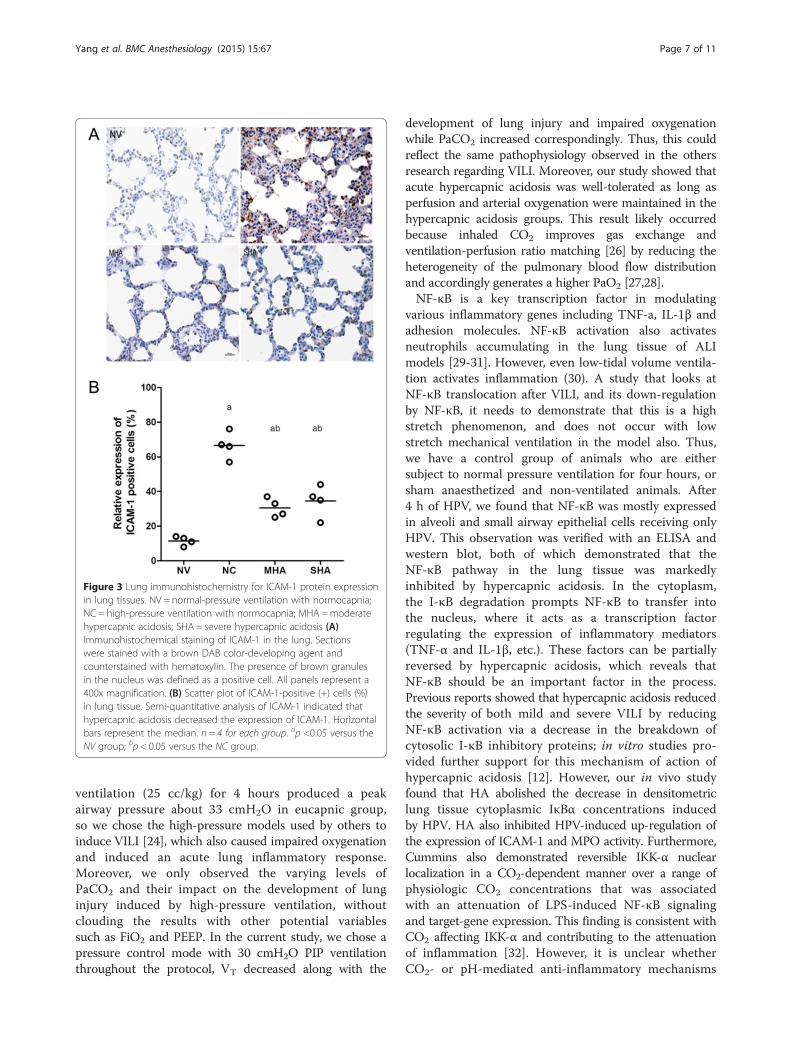
Figure 3 Lung immunohistochemistry for ICAM-1 protein expressionin lung tissues. NV = normal-pressure ventilation with normocapnia;NC = high-pressure ventilation with normocapnia; MHA =moderatehypercapnic acidosis; SHA = severe hypercapnic acidosis (A)Immunohistochemical staining of ICAM-1 in the lung. Sectionswere stained with a brown DAB color-developing agent andcounterstained with hematoxylin. The presence of brown granulesin the nucleus was defined as a positive cell. All panels represent a400x magnification. (B) Scatter plot of ICAM-1-positive (+) cells (%)in lung tissue. Semi-quantitative analysis of ICAM-1 indicated thathypercapnic acidosis decreased the expression of ICAM-1. Horizontalbars represent the median. n = 4 for each group. ap <0.05 versus theNV group; bp < 0.05 versus the NC group.
Yang et al. BMC Anesthesiology (2015) 15:67 Page 7 of 11
ventilation (25 cc/kg) for 4 hours produced a peakairway pressure about 33 cmH2O in eucapnic group,so we chose the high-pressure models used by others toinduce VILI [24], which also caused impaired oxygenationand induced an acute lung inflammatory response.Moreover, we only observed the varying levels ofPaCO2 and their impact on the development of lunginjury induced by high-pressure ventilation, withoutclouding the results with other potential variablessuch as FiO2 and PEEP. In the current study, we chose apressure control mode with 30 cmH2O PIP ventilationthroughout the protocol, VT decreased along with the
development of lung injury and impaired oxygenationwhile PaCO2 increased correspondingly. Thus, this couldreflect the same pathophysiology observed in the othersresearch regarding VILI. Moreover, our study showed thatacute hypercapnic acidosis was well-tolerated as long asperfusion and arterial oxygenation were maintained in thehypercapnic acidosis groups. This result likely occurredbecause inhaled CO2 improves gas exchange andventilation-perfusion ratio matching [26] by reducing theheterogeneity of the pulmonary blood flow distributionand accordingly generates a higher PaO2 [27,28].NF-κB is a key transcription factor in modulating
various inflammatory genes including TNF-a, IL-1β andadhesion molecules. NF-κB activation also activatesneutrophils accumulating in the lung tissue of ALImodels [29-31]. However, even low-tidal volume ventila-tion activates inflammation (30). A study that looks atNF-κB translocation after VILI, and its down-regulationby NF-κB, it needs to demonstrate that this is a highstretch phenomenon, and does not occur with lowstretch mechanical ventilation in the model also. Thus,we have a control group of animals who are eithersubject to normal pressure ventilation for four hours, orsham anaesthetized and non-ventilated animals. After4 h of HPV, we found that NF-κB was mostly expressedin alveoli and small airway epithelial cells receiving onlyHPV. This observation was verified with an ELISA andwestern blot, both of which demonstrated that theNF-κB pathway in the lung tissue was markedlyinhibited by hypercapnic acidosis. In the cytoplasm,the I-κB degradation prompts NF-κB to transfer intothe nucleus, where it acts as a transcription factorregulating the expression of inflammatory mediators(TNF-α and IL-1β, etc.). These factors can be partiallyreversed by hypercapnic acidosis, which reveals thatNF-κB should be an important factor in the process.Previous reports showed that hypercapnic acidosis reducedthe severity of both mild and severe VILI by reducingNF-κB activation via a decrease in the breakdown ofcytosolic I-κB inhibitory proteins; in vitro studies pro-vided further support for this mechanism of action ofhypercapnic acidosis [12]. However, our in vivo studyfound that HA abolished the decrease in densitometriclung tissue cytoplasmic IκBα concentrations inducedby HPV. HA also inhibited HPV-induced up-regulation ofthe expression of ICAM-1 and MPO activity. Furthermore,Cummins also demonstrated reversible IKK-α nuclearlocalization in a CO2-dependent manner over a range ofphysiologic CO2 concentrations that was associatedwith an attenuation of LPS-induced NF-κB signalingand target-gene expression. This finding is consistent withCO2 affecting IKK-α and contributing to the attenuationof inflammation [32]. However, it is unclear whetherCO2- or pH-mediated anti-inflammatory mechanisms
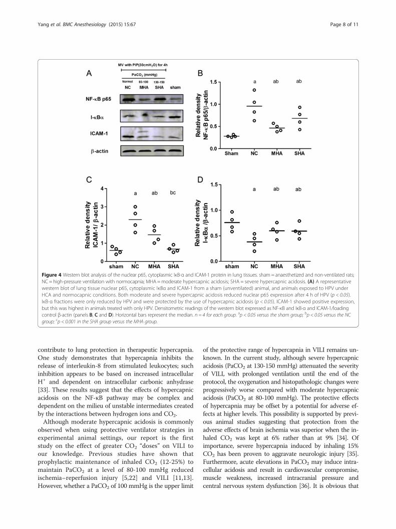
Figure 4 Western blot analysis of the nuclear p65, cytoplasmic IκB-α and ICAM-1 protein in lung tissues. sham = anaesthetized and non-ventilated rats;NC = high-pressure ventilation with normocapnia; MHA=moderate hypercapnic acidosis; SHA = severe hypercapnic acidosis. (A) A representativewestern blot of lung tissue nuclear p65, cytoplasmic IκBα and ICAM-1 from a sham (unventilated) animal, and animals exposed to HPV underHCA and normocapnic conditions. Both moderate and severe hypercapnic acidosis reduced nuclear p65 expression after 4 h of HPV (p < 0.05).IκB-α fractions were only reduced by HPV and were protected by the use of hypercapnic acidosis (p < 0.05). ICAM-1 showed positive expression,but this was highest in animals treated with only HPV. Densitometric readings of the western blot expressed as NF-κB and IκB-α and ICAM-1/loadingcontrol β-actin (panels B, C and D). Horizontal bars represent the median. n = 4 for each group. ap < 0.05 versus the sham group; bp < 0.05 versus the NCgroup; cp < 0.001 in the SHA group versus the MHA group.
Yang et al. BMC Anesthesiology (2015) 15:67 Page 8 of 11
contribute to lung protection in therapeutic hypercapnia.One study demonstrates that hypercapnia inhibits therelease of interleukin-8 from stimulated leukocytes; suchinhibition appears to be based on increased intracellularH+ and dependent on intracellular carbonic anhydrase[33]. These results suggest that the effects of hypercapnicacidosis on the NF-κB pathway may be complex anddependent on the milieu of unstable intermediates createdby the interactions between hydrogen ions and CO2.Although moderate hypercapnic acidosis is commonly
observed when using protective ventilator strategies inexperimental animal settings, our report is the firststudy on the effect of greater CO2 “doses” on VILI toour knowledge. Previous studies have shown thatprophylactic maintenance of inhaled CO2 (12-25%) tomaintain PaCO2 at a level of 80-100 mmHg reducedischemia–reperfusion injury [5,22] and VILI [11,13].However, whether a PaCO2 of 100 mmHg is the upper limit
of the protective range of hypercapnia in VILI remains un-known. In the current study, although severe hypercapnicacidosis (PaCO2 at 130-150 mmHg) attenuated the severityof VILI, with prolonged ventilation until the end of theprotocol, the oxygenation and histopathologic changes wereprogressively worse compared with moderate hypercapnicacidosis (PaCO2 at 80-100 mmHg). The protective effectsof hypercapnia may be offset by a potential for adverse ef-fects at higher levels. This possibility is supported by previ-ous animal studies suggesting that protection from theadverse effects of brain ischemia was superior when the in-haled CO2 was kept at 6% rather than at 9% [34]. Ofimportance, severe hypercapnia induced by inhaling 15%CO2 has been proven to aggravate neurologic injury [35].Furthermore, acute elevations in PaCO2 may induce intra-cellular acidosis and result in cardiovascular compromise,muscle weakness, increased intracranial pressure andcentral nervous system dysfunction [36]. It is obvious that

Figure 5 Lung Immunohistochemistry for NF-κB p65 protein expression in lung tissues. NV = normal-pressure ventilation with normocapnia;NC = high-pressure ventilation with normocapnia; MHA =moderate hypercapnic acidosis; SHA = severe hypercapnic acidosis. (A and B) arrowsindicate the position of NF-κB p65 expression, which was only observed in the nuclei of airway epithelial cells and alveoli of HPV-treated animals.(C) Scatter plot of NF-κB p65 -positive (+) cells (%) in lung tissue. NF-κB expression also increased after 4 h of HPV compared to the normalventilation group (p < 0.05), but this was attenuated by both moderate and severe hypercapnia. (D) ELISA analysis of total lung tissue NF-κBindicated that the NF-κB activity also increased after 4 h of HPV compared to the normal ventilation group but was attenuated by both moderateand severe hypercapnia. All panels represent a 400x magnification. Horizontal bars represent the median. n = 4 for each group. ap < 0.05 versus theNV group; bp < 0.001 versus the NC group.
Yang et al. BMC Anesthesiology (2015) 15:67 Page 9 of 11
acidosis is a double-edged sword that may be difficult toapply in critically ill patients. Nevertheless, the results ofour study show that induction of HA by the addition of
carbon dioxide to the inspired gas may necessitate attentionto the potential for adverse effects at higher levels of PaCO2
in patients with ARDS during mechanical ventilation. On

Yang et al. BMC Anesthesiology (2015) 15:67 Page 10 of 11
the other hand, our studies must be viewed as hypothesis-generating and should be tested by intensive studiesin preclinical models.Despite its interesting results, this exploratory study
was limited in several ways. First, the animals wereanesthetized with an intraperitoneal injection ofpentobarbital sodium alone, which may not havereached the depth of anesthesia corresponding toclinical practice. Further studies should avoid thisinappropriate anesthesia. Second, the pulmonary vasocon-striction and lung mechanics (i.e., plateau pressures,compliance, etc.) were not measured, which limited theinterpretation of the specific effects of hypercapnic acid-osis on pulmonary and systemic hemodynamic parame-ters. Third, the duration of the ALI models was limited to4 h of mechanical ventilation and was too short toextrapolate to clinical practice. The results indicate that itis reasonable to believe that the acidosis generated byacute hypercapnia may be an important factor in acutemodels of VILI. Further study should be performed toevaluate the effects of hypercapnia in ALI models ofconsiderably longer duration.
ConclusionThis study demonstrates that an increased level ofcarbon dioxide has a protective effect against VILI in rats.Animals exposed to moderate hypercapnia (a PaCO2 of80-100 mmHg) remained in a more favorable conditionand had less histopathologic changes and inflammatoryinjury than animals with severe hypercapnia (a PaCO2
of 130-150 mmHg). The protective mechanism is likelyassociated with the inhibition of NF-κB expression duringhigh pressure stretch.
Competing interestsThe authors declare that they have no competing interests.
Authors’ contributionsWY and ZY participated in the design of the study and the experiments,were involved in the data extraction and the statistical analysis, and draftedthe manuscript. XC and YG participated in the design of the study and inexperiments and helped draft the manuscript. LZ and HZ participated in thedesign of the study and in the experiments. WL participated in the design ofthe study, was involved in the interpretation of the results, and helped draftthe manuscript. All authors read and approved the final manuscript forpublication.
AcknowledgmentsThis study was supported by the graduate (2012RFXXS064) and(2014RFQGJ113) from the Harbin Science and Technology CommissionFoundation.
Author details1Department of Anesthesiology, Second Affiliated Hospital of HarbinMedical University; Anesthesiology Key Laboratory, Harbin MedicalUniversity, Harbin 150086, China. 2Education Department of HeilongjiangProvince, Anesthesiology Key Laboratory, Harbin Medical University,Harbin, Heilongjiang Province, China.
Received: 6 August 2014 Accepted: 22 April 2015
References1. Dreyfuss D, Basset G, Soler P, Saumon G. Intermittent positive-pressure
hyperventilation with high inflation pressures produces pulmonarymicrovascular injury in rats. Am Rev Respir Dis. 1985;132(4):880–4.
2. Dreyfuss D, Saumon G. Ventilator-induced lung injury: lessons fromexperimental studies. Am J Respir Crit Care Med. 1998;157(1):294–323.
3. Amato MB, Barbas CS, Medeiros DM, Magaldi RB, Schettino GP,Lorenzi-Filho G, et al. Effect of a protective-ventilation strategy on mortality inthe acute respiratory distress syndrome. N Engl J Med. 1998;338(6):347–54.
4. Maeda YFY, Uchiyama A, Matsuura N, Mashimo T, Nishimura M. Effects ofpeak inspiratory flow on development of ventilator-induced lung injury inrabbits. Anesthesiology. 2004;101(3):722–8.
5. Laffey JGTM, Engelberts D, Luo X, Yuan S, Keith Tanswell A, Post M, et al.Therapeutic hypercapnia reduces pulmonary and systemic injuryfollowing in vivo lung reperfusion. Am J Respir Crit Care Med.2000;162(6):2287–94.
6. Shibata KCN, Engelberts D, Takeuchi A, Fedorko L, Kavanagh BP.Hypercapnic acidosis may attenuate acute lung injury by inhibitionof endogenous xanthine oxidase. Am J Respir Crit Care Med.1998;158(5 Pt 1):1578–84.
7. Laffey JGHD, Hopkins N, Hyvelin JM, Boylan JF, McLoughlin P. Hypercapnicacidosis attenuates endotoxin-induced acute lung injury. Am J Respir CritCare Med. 2004;169(1):46–56.
8. Nichol AD, O’Cronin DF, Naughton F, Hopkins N, Boylan J, McLoughlin P.Hypercapnic acidosis reduces oxidative reactions in endotoxin-induced lunginjury. Anesthesiol. 2010;113(1):116–25.
9. Costello J, Higgins B, Contreras M, Chonghaile MN, Hassett P, O’Toole D,et al. Hypercapnic acidosis attenuates shock and lung injury in early andprolonged systemic sepsis. Crit Care Med. 2009;37(8):2412–20.
10. Higgins BD, Costello J, Contreras M, Hassett P, O’Toole D, Laffey JG.Differential effects of buffered hypercapnia versus hypercapnic acidosis onshock and lung injury induced by systemic sepsis. Anesthesiol.2009;111(6):1317–26.
11. Broccard AFHJ, Vannay C, Markert M, Sauty A, Feihl F, Schaller MD.Protective effects of hypercapnic acidosis on ventilator-induced lung injury.Am J Respir Crit Care Med. 2001;164(5):802–6.
12. Contreras M, Ansari B, Curley G, Higgins BD, Hassett P, O’Toole D, et al.Hypercapnic acidosis attenuates ventilation-induced lung injury by a nuclearfactor-kappaB-dependent mechanism. Crit Care Med. 2012;40(9):2622–30.
13. Sinclair SEKD, Lamm WJ, Starr IR, Chi EY, Hlastala MP. Hypercapnic acidosis isprotective in an in vivo model of ventilator-induced lung injury. Am J RespirCrit Care Med. 2002;166(3):403–8.
14. Peltekova V, Engelberts D, Otulakowski G, Uematsu S, Post M, Kavanagh BP.Hypercapnic acidosis in ventilator-induced lung injury. Intensive Care Med.2010;36(5):869–78.
15. Laffey JGED, Duggan M, Veldhuizen R, Lewis JF, Kavanagh BP. Carbondioxide attenuates pulmonary impairment resulting from hyperventilation.Crit Care Med. 2003;31(11):2634–40.
16. Kregenow DA, Swenson ER. The lung and carbon dioxide: implications forpermissive and therapeutic hypercapnia. Eur Respir J. 2002;20(1):6–11.
17. Blackwell TS, Christman JW. The role of nuclear factor-kappa B in cytokinegene regulation. Am J Respir Cell Mol Biol. 1997;17(1):3–9.
18. Christman JW, Lancaster LH, Blackwell TS. Nuclear factor kappa B: a pivotalrole in the systemic inflammatory response syndrome and new target fortherapy. Intensive Care Med. 1998;24(11):1131–8.
19. Chen LW, Egan L, Li ZW, Greten FR, Kagnoff MF, Karin M. The two faces ofIKK and NF-kappaB inhibition: prevention of systemic inflammation butincreased local injury following intestinal ischemia-reperfusion. Nat Med.2003;9(5):575–81.
20. O’Toole D, Hassett P, Contreras M, Higgins BD, McKeown ST, McAuley DF, et al.Hypercapnic acidosis attenuates pulmonary epithelial wound repair by anNF-kappaB dependent mechanism. Thorax. 2009;64(11):976–82.
21. Yang WC, Song CY, Wang N, Zhang LL, Yue ZY, Cui XG, et al. Hypercapnicacidosis confers antioxidant and anti-apoptosis effects against ventilator-inducedlung injury. Lab Invest. 2013;93(12):1339–49.
22. Laffey JGJR, Engelberts D, Tanswell AK, Post M, Lindsay T, Mullen JB, et al.Effects of therapeutic hypercapnia on mesenteric ischemia-reperfusioninjury. Am J Respir Crit Care Med. 2003;168(11):1383–90.
23. Zhou Q, Cao B, Niu L, Cui X, Yu H, Liu J, et al. Effects of permissivehypercapnia on transient global cerebral ischemia-reperfusion injury in rats.Anesthesiol. 2010;112(2):288–97.

Yang et al. BMC Anesthesiology (2015) 15:67 Page 11 of 11
24. Imanaka H, Shimaoka M, Matsuura N, Nishimura M, Ohta N, Kiyono H.Ventilator-induced lung injury is associated with neutrophil infiltration,macrophage activation, and TGF-beta 1 mRNA upregulation in rat lungs.Anesth Analg. 2001;92(2):428–36.
25. Gattinoni L, Pesenti A, Avalli L, Rossi F, Bombino M. Pressure-volume curveof total respiratory system in acute respiratory failure. Computed tomographicscan study. Am Rev Respir Dis. 1987;136(3):730–6.
26. Sinclair SE, Kregenow DA, Starr I, Schimmel C, Lamm WJ, Hlastala MP, et al.Therapeutic hypercapnia and ventilation-perfusion matching in acute lunginjury: low minute ventilation vs inspired CO2. Chest. 2006;130(1):85–92.
27. Brogan TV, Hedges RG, McKinney S, Robertson HT, Hlastala MP, Swenson ER.Pulmonary NO synthase inhibition and inspired CO2: effects on V’/Q’ andpulmonary blood flow distribution. Eur Respir J. 2000;16(2):288–95.
28. Akca O, Doufas AG, Morioka N, Iscoe S, Fisher J, Sessler DI. Hypercapniaimproves tissue oxygenation. Anesthesiol. 2002;97(4):801–6.
29. Liu SF, Ye X, Malik AB. Inhibition of NF-kappaB activation by pyrrolidinedithiocarbamate prevents In vivo expression of proinflammatory genes.Circ. 1999;100(12):1330–7.
30. Schwartz MD, Moore EE, Moore FA, Shenkar R, Moine P, Haenel JB, et al.Nuclear factor-kappa B is activated in alveolar macrophages from patientswith acute respiratory distress syndrome. Crit Care Med. 1996;24(8):1285–92.
31. Blackwell TS, Blackwell TR, Holden EP, Christman BW, Christman JW. In vivoantioxidant treatment suppresses nuclear factor-kappa B activation andneutrophilic lung inflammation. J Immunol. 1996;157(4):1630–7.
32. Cummins EP, Oliver KM, Lenihan CR, Fitzpatrick SF, Bruning U, Scholz CC, et al.NF-kappaB links CO2 sensing to innate immunity and inflammation inmammalian cells. J Immunol. 2010;185(7):4439–45.
33. Coakley RJ, Taggart C, Greene C, McElvaney NG, O’Neill SJ. Ambient pCO2modulates intracellular pH, intracellular oxidant generation, and interleukin-8secretion in human neutrophils. J Leukoc Biol. 2002;71(4):603–10.
34. Vannucci RC, Towfighi J, Heitjan DF, Brucklacher RM. Carbon dioxideprotects the perinatal brain from hypoxic-ischemic damage: an experimentalstudy in the immature rat. Pediatr. 1995;95(6):868–74.
35. Vannucci RC, Towfighi J, Brucklacher RM, Vannucci SJ. Effect of extremehypercapnia on hypoxic-ischemic brain damage in the immature rat.Pediatr Res. 2001;49(6):799–803.
36. Marini JJ. Pressure-targeted, lung-protective ventilatory support in acutelung injury. Chest. 1994;105(3 Suppl):109S–15.
Submit your next manuscript to BioMed Centraland take full advantage of:
• Convenient online submission
• Thorough peer review
• No space constraints or color figure charges
• Immediate publication on acceptance
• Inclusion in PubMed, CAS, Scopus and Google Scholar
• Research which is freely available for redistribution
Submit your manuscript at www.biomedcentral.com/submit