comparison of the endovac system and conventional needle
TRANSCRIPT

1168 © 2017 Nigerian Journal of Clinical Practice | Published by Wolters Kluwer ‑ Medknow
Objective: This study aimed to compare the EndoVac system and conventional needle irrigation in removing smear layer (SR) from primary molar root canals. Materials and Methods: Fifty extracted human primary second molar roots were instrumentedup toanapical sizeof0.04/35and randomlydivided into twomaingroups; Group 1: EndoVac system (n = 25) and Group 2: Conventional needleirrigation (n = 25) and three subgroups (a) NaOCl + ethylenediaminetetraaceticacid (EDTA) (n = 20) (b) ozonated water (OW) + EDTA (n = 20) and(c) saline (control, n = 10). After a standardized final irrigation protocolperformedforallteeth,scanningelectronmicroscopeimagesweretakenat×1000magnification for each thirds of each root canal. Data were analyzed by theweighted kappa, Kruskal–Wallis, and Wilcoxon signed rank tests. Results: EndoVac was more effective than conventional needle in the removal of SR from the apical third of the root canal system (P < 0.05). The OW + EDTA regimen providedsimilar SR removal comparedwith NaOCl + EDTA.Conclusions: EndoVac has better performance than conventional needle irrigation in the removal of the SR in theapicalthirdsoftheprimarymolarrootcanals.Asafinalirrigationregimen,theOW+EDTAregimenisaseffectiveastheNaOCl+EDTAregimen.
Keywords: Apical negative pressure, irrigation, ozonated water, primary teeth, smear layer
Comparison of the EndoVac System and Conventional Needle Irrigation on Removal of the Smear Layer in Primary Molar Root CanalsB Buldur, A Kapdan
Address for correspondence: Dr. B Buldur, Department of Pediatric Dentistry, Faculty of Dentistry, Cumhuriyet University, Kampüs, 58140, Sivas, Turkey.
E-mail: [email protected]
Smear layer (SR) is an amorphous, irregular surface of organic and inorganic debris retained on the dentin and other surfaces after instrumentation.[8] This irregular layer needs to be removed since it limits the penetration of irrigation solutions, and it acts as a substrate for bacteria and a barrier between fillings and root canalwall.[9]Thehermeticalsealingofresorbablefillingstothedentinal tubules and canal walls of PT is very important for clinical success.[3] Barcelos et al.[4] found that SR removal improved root canal treatment successfully in PTina24-monthperiodinan in vivo study.
Mechanical instrumentation cannot clean the canal entirely. Therefore, chemical debridement with an irrigation solution is necessary to eliminate bacteria,
Original Article
IntroductIon
Early loss of primary teeth (PT) causes functional, esthetical, and developmental problems. Root canal
treatment is indicated for PT having irreversible pulpitis symptoms or necrosis.[1] Pulpectomy procedures of PT involve mechanical instrumentation with hand or rotary files, irrigationwith various irrigants, and obturation ofrootcanalwitharesorbablefilling.[2] Clinical success of PT pulpectomy has been shown in many studies.[3-5]
PT root canals are rarely straight and almost have lateral canals, apical deltas, fins, and anastomoses intheir morphology. Severely divergent, curved primary molar roots and anatomical variations due to radicular resorption, and dentin apposition on the root canal limit the chemomechanical debridement efficacy ofinstrumentation and irrigation.[6,7]
Department of Pediatric Dentistry, Faculty of Dentistry, Cumhuriyet University, Sivas, Turkey
Ab
str
Ac
t
This is an open access article distributed under the terms of the Creative Commons Attribution-NonCommercial-ShareAlike 3.0 License, which allows others to remix, tweak, and build upon the work non-commercially, as long as the author is credited and the new creations are licensed under the identical terms.
For reprints contact: [email protected]
How to cite this article: Buldur B, Kapdan A. Comparison of the EndoVac system and conventional needle irrigation on removal of the smear layer in primary molar root canals. Niger J Clin Pract 2017;20:1168-74.
Date of Acceptance: 29-Mar-2016
Access this article onlineQuick Response Code:
Website: www.njcponline.com
DOI: 10.4103/1119-3077.181351
PMID: *******
[Downloaded free from http://www.njcponline.com on Thursday, October 26, 2017, IP: 165.255.142.217]

Buldur and Kapdan: Effect of irrigation on smear layer in primary teeth
1169Nigerian Journal of Clinical Practice ¦ Volume 20 ¦ Issue 9 ¦ September 2017
flush debris, and remove the SR from root canalsystem.[10] Many types of irrigation solutions were used such as NaOCl, chlorhexidine, hydrogen peroxide, and saline, yet there is no consensus on which irrigant and concentration should be used for primary root canal treatment. NaOCl is the most commonly used irrigant in endodontics since its great antimicrobial efficacy, theproperty to dissolve vital and necrotic tissues, low cost, and availability.[11]Exactly, 2.5%NaOCl ismostwidelyused for PT root canals.[2] Ethylenediaminetetraacetic acid(EDTA)isachelatingagentthatisneededasafinalrinse for the removal of a SR, and 17% concentrationis widely used.[12] A NaOCl following EDTA regimen is the most commonly used final irrigation regimen inendodontic treatment.[13] However, alternative irrigant regimens have been investigated, since the extrusion of NaOCl on vital tissues and periapical areas causes several complications and also special care is needed for the upcoming permanent successor in pediatric endodontic treatment. In addition, Niu et al.[14] reported that more debris is removed by irrigation with EDTA followed by NaOCl than with EDTA alone, but also, final irrigation with NaOCl following EDTA causesmore dentinal erosion.
Ozone (O3) is a powerful oxidizing agent that has great antimicrobial effects and higher biocompatibility.[15] O3 can be used in dentistry either in gaseous, aqueous, oroiled forms. Ozonated water (OW) is an alternative to NaOCl for eliminating cytotoxic effects on vital tissues.[16] Many studies[17-19] have investigated the antimicrobial efficacy ofOW, but to our knowledge, nostudy has examined the SR removal effect of an OW and EDTAcombinationasafinalirrigationregimen.
Although conventional needle irrigation is the most used technique in endodontics, the replenishmentand exchange of the irrigant is limited at the apical part, lateral canal, and isthmus.[20] Furthermore, positive pressure with the risk of extruding irrigants to periradicular tissues can lead to postoperative pain as well as tissue and permanent teeth bud damage.[21,22] A conventional needle is not successful for delivering safely and effectively high volumes of irrigation solution to the entire root canal and untouchable parts.[23] Therefore, new irrigation systems and devices have been introduced to increase the effectiveness of root canal debridement.
EndoVac (Discus Dental, Culver City, CA, USA) is an apical negative pressure system that delivers irrigants safely to apical areas and unreachable parts in the root canal system.[24] The superiority of EndoVac system to conventional needle irrigation regarding debridement efficacy, SR removal, and antimicrobial efficacy on
permanent teeth has been reported.[25,26] In addition, EndoVac system extrudes less irrigant to the periapical area and decreases the risk of NaOCl accidents.[21]
The rationale of the study was that the dental literature shows a lack of importance of irrigation in primary root canal treatment. The effect of different irrigation solutions and delivery systems in primary root canal treatment has not been well-elucidated. To our knowledge, no study was conducted on the SR removal of both OW and the EndoVac system on PT.
The aim of this in vitro study was to compare the SR removal of two final irrigation regimens using theEndoVac system and conventional needle irrigation method with different irrigation solutions in primary molar root canals. The null hypotheses of the present study were (a) there is no difference between EndoVac and conventional needle irrigation systems regarding removing the SR from primary molar root canals (b) there is no difference between OW + EDTA andNaOCl + EDTA final irrigation regimens regardingremoving the SR from primary molar root canals.
MAterIAls And MethodsTeeth selectionEthical approval was obtained from the Health Ethics Committee of the University of Cumhuriyet University, Sivas, Turkey (ID: 2012-04/04). The study wasconducted on the largest palatinal or distal roots of, respectively, human primary maxillary or mandibular second molars. Freshly extracted primary molar teeth were collected, and each tooth was radiographed digitally todeterminecurvature<30°andtherootresorptionscaledegree as “resi or res1/4,” described by Fanning.[27] Teeth withfractured,calcified,orpreviousrootcanaltreatmentwere excluded. The sample consisted of 50 primarymolars, and the sample size was calculated as α=0.01,β=0.10,1–β = 0.90, and P =0.91234.[28] Only one root of each tooth was used; nonused roots were removed by diamond blazer.All teeth were stored at +4°C in aphysiological saline supplemented with 0.02% sodiumazide.
Specimen preparationEach tooth was decoronated and the root length standardized to 11 mm. The working length (WL) wasdetermined by inserting a size 10 K-file (DentsplyMaillefer, Ballaigues, Switzerland) into each root canal until visible apically under a magnifying loupe ×20 andby subtracting 1mm from this point.[29] Thereafter, each root apical foramina was closed with soft modeling wax (Cera Reus, SA, Reus, Spain) to create a closed system.[29] Horizontal grooves were placed for mechanical
[Downloaded free from http://www.njcponline.com on Thursday, October 26, 2017, IP: 165.255.142.217]

Buldur and Kapdan: Effect of irrigation on smear layer in primary teeth
1170 Nigerian Journal of Clinical Practice ¦ Volume 20 ¦ Issue 9 ¦ September 2017
retention in the experimental setup. Root cementum was coated with two layers of nail varnish to prevent bacterial retention.[29] Each root was inserted into polyvinylsiloxane impression material and adapted to the previously prepared experimental setup.[25] Each root canal was instrumented crown down with a nickel–titanium rotary Profile. 04 ISO (Dentsply Tulsa, Tulsa, OK) up to anapical 0.04/35 file by using 1 mL 2.5% NaOCl at eachfilechangewitha27-gaugeneedleinaccordancewiththemanufacturer’s recommendations.[30]
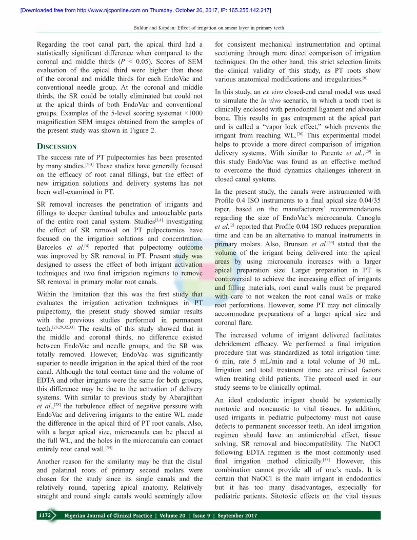
Experimental materialsAnexperimentalsetupwaspreparedbasedonthefixturepreviously designed[29] to facilitate consistent irrigation protocols performed by one single operator. A separate fluid collection trap was not used to measure irrigantvolume suctioned by the EndoVac system [Figure1].
2.5%NaOCl, 17%EDTA (Werax, Izmir,Turkey), 0.9%sterile saline and 4 parts per million (ppm) OW were used as irrigation solutions in the study. The ozonation of water was performed by bubbling O3 through sterile distilled water at 4 mg/L using the O3 generator digitally (TeknO3zone, Izmir, Turkey).
Experimental groups and final irrigationFifty extracted human primary second molar roots were randomly divided into 2 groups; Group 1. EndoVac(n = 25), Group 2 conventional needle (n = 25)and three subgroups (two experimental subgroups; (a)NaOCl+EDTA(n=20),(b)OW+EDTA(n=20),and control group (c) saline (n=10).
Each tooth had the same total final irrigation time of6 min with average rate was 5 mL/min, and the totalirrigant volume delivered was 30 mL for each canal.Final irrigation procedure was carried out as:
Group 1 (EndoVac group)• Subgroup 1a (NaOCl + EDTA) (n = 20):
Experimental group consisted of a 30 s period ofirrigation with 2.5% NaOCl using the macrocanula,followedbyleavingthecanalfullofirrigantfor30s.Three irrigation cycles were performed by using the microcanula placed, respectively, at WL for 6 s, 2mmshorterWL for6 s, andWL for6 s.Thefirstcyclewas30sof2.5%NaOCl, followedby30sofsoaking; the second cycle was 1 min %17 EDTA,followed by 1 min of soaking; and the third cyclewas 1 min of 2.5% NaOCl, followed by 1 min ofsoaking
• Subgroup 1b (OW + EDTA) (n = 20): Sameprocedure as group NaOCl + EDTA. Differently,4ppmOWwasusedinsteadof2.5%NaOCl
• Subgroup 1c (saline) (n = 10): 0.9% sterile salinewas used as the only irrigant.
Group 2 (conventional needle irrigation group)• Subgroup 2a (NaOCl + EDTA) (n = 20): 27 gauge
needlewasinsertedintothecanal2mmshorterWL,and 2.5% NaOCl was delivered into the canal for1minactiveandfollowedby1minofsoaking.17%EDTAwasdeliveredfor1minactiveandsoakedfor1 min. Finally, 2.5% NaOCl was delivered into thecanalfor1minactive,followedby1minofsoaking
• Subgroup 2b (OW + EDTA) (n = 20): Sameprocedure as group NaOCl + EDTA. Differently,4ppmOWwasusedinsteadof2.5%NaOCl
• Subgroup 2C (saline) (n = 10): 0.9% sterile salinewas used as the only irrigant.
After the final irrigation procedure, all experimentalcanals were rinsed with sterile saline and dried with sterile paper points, and a sterile cotton pellet was placed into the access cavity and sealed with Cavit G (3MESPE,Seefeld,Germany).Eachtoothwasremovedfrom polyvinylsiloxane and stored in bottles containing physiological saline supplemented with 0.02% sodiumazide.
Scanning electron microscopeTwo opposing longitudinal grooves were prepared on both the buccal and lingual external root surfaces using a diamond disc without penetrating into the canal. Root surfaces were rinsed with compressed water and air to avoid contamination with external debris. Roots were then split open by inserting a chisel into the grooves and twisting. The most visible and intact half part of each root was used for the study. Each specimen was dehydrated in graded ethanol series 25%, 50%, 75%, 90% for 25min and finally in100% ethanol for 1 h. The specimens were criticallypoint-dried, mounted on aluminum stubs, sputter-coated with gold/palladium and examined with a scanning electron microscope (SEM) (Leo 440 CCD, Leica, Bensheim, Germany). Coronal (8–10 mm from apex),middle (5–7 mm from apex) and apical (1–3 mmfrom apex) thirds of each specimen were examined, and photographs were taken at ×1000 magnificationand labeled by an independent SEM technician. Two independent examiners who were unaware of which specimens belonged to which groups blindly analyzed and scored for the degree of SR removal. Each examiner scoredallphotographstwiceata2-weekinterval.
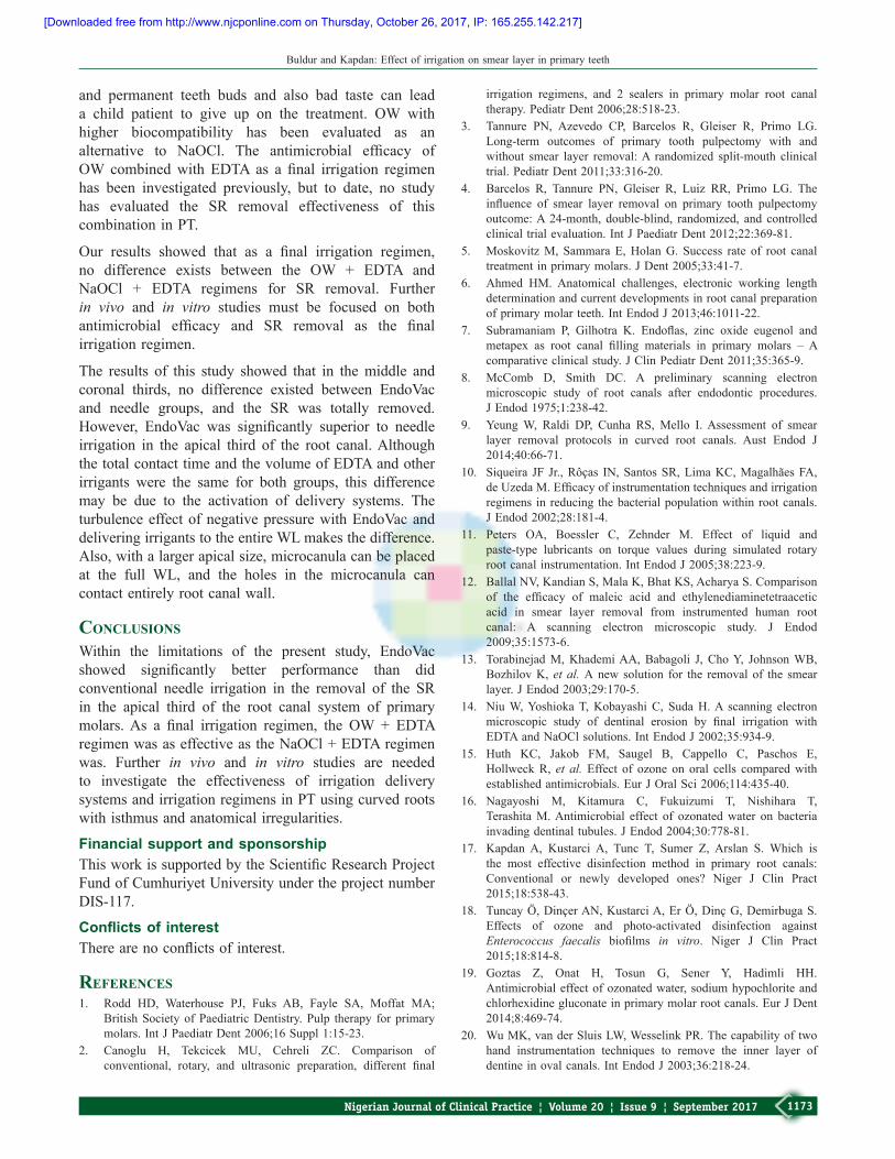
A 5-level scoring system described by Hülsmannet al.[31]was used for the degree ofSR removal: 1= noSR, dentinal tubules open [Figure 2a]; 2 = smallamount of SR, some dentinal tubules open [Figure 2b];3 = homogenous SR covering the root canal wall, onlya few dentinal tubules open [Figure 2c]; 4 = completeroot canal wall covered by a homogenous SR, no open
[Downloaded free from http://www.njcponline.com on Thursday, October 26, 2017, IP: 165.255.142.217]

Buldur and Kapdan: Effect of irrigation on smear layer in primary teeth
1171Nigerian Journal of Clinical Practice ¦ Volume 20 ¦ Issue 9 ¦ September 2017
dentinal tubules [Figure2d];5=heavy,nonhomogeneousSR, no open dentinal tubules [Figure2e].
Statistical analysisAll data were processed by SPSS 15.0 software (SPSSInc., Chicago, IL, USA). Intra- and inter-examiner reliability for SEM assessment was verified by aweightedcoefficientkappa(Kw)test.ThescoresofSEMevaluation were analyzed by the Kruskal-Wallis and Wilcoxonsignedranktestsat0.05significancelevel.
results
The kappa test showed good inter- and intra-examiner agreement, with values at 0.90 or above. Table 1 shows
the results of the SEM evaluation of remaining SR for EndoVac and conventional needle groups regarding final irrigation regimen and root canal part. EndoVacshowed better results than did the conventional needle at each root canal part, but only statistical significancewas found at the apical third (P < 0.05). Regarding thefinal irrigation regimen, saline was the least effectivegroup (P < 0.05). There was no statistically significantdifference between other irrigation regimens (P > 0.05).
Table 1: Scores of scanning electron microscope evaluation of remaining smear layer for EndoVac and conventional needle groups regarding final irrigation regimens and root canal parts
Groups Total scores Score 1 (%) Score 2 (%) Score 3 (%) Score 4 (%) Score 5 (%)EndoVacNaOCl+EDTA 30 23(30.67) 7(9.33) 0 (0.00) 0 (0.00) 0 (0.00)OW+EDTA 30 24(32.00) 6(8.00) 0 (0.00) 0 (0.00) 0 (0.00)Saline 15 0 (0.00) 0 (0.00) 0 (0.00) 5(6.67) 10(13.33)
Conventional needleNaOCl+EDTA 30 16(21.33) 4(5.33) 6(8.00) 3(4.00) 1(1.33)OW+EDTA 30 17(22.67) 3(4.00) 5(6.67) 4(5.33) 1(1.33)Saline 15 0 (0.00) 0 (0.00) 0 (0.00) 3(4.00) 12(16.00)
EndoVacCoronal 25 20(26.67) 0 (0.00) 0 (0.00) 3(4.00) 2(2.67)Middle 25 19(25.33) 1(1.33) 0 (0.00) 2(2.67) 3(4.00)Apical 25 8(10.67) 12(16.00) 11(14.67) 7(9.33) 12(16.00)
Conventional needleCoronal 25 17(22.67) 3(4.00) 0 (0.00) 2(2.67) 3(4.00)Middle 25 16(21.33) 4(5.33) 0 (0.00) 1(1.33) 4(5.33)Apical 25 0 (0.00) 0 (0.00) 11(14.67) 7(9.33) 7(9.33)EDTA=Ethylenediaminetetraacetic acid; NaOCl=Sodium hypochlorite; OW=Ozonated water
Figure 1: An experimental setup to perform irrigation by single operator.(a)20mLsyringe,(b)connectorbetween20mLsyringeandthe master suction tip, (c) connector between the master suction tip and high vacuum line, (d) the master suction tip, (e) connector between the high vacuum line and EndoVac hand piece, (f) high vacuum line, (g) connector to dental unit
Figure 2:Examplesofscanningelectronmicroscopeimagesofa5-levelscoringsystemat×1000magnification.(a)Score1=nosmearlayer,dentinaltubulesopen,(b)score2=smallamountofsmearlayer,somedentinaltubulesopen;(c)score3=homogenoussmearlayercoveringtheroot canal wall, only a few dentinal tubules open; (d) score 4 = complete root canal wall covered by a homogenous smear layer, no open dentinal tubules; (e) score 5= heavy, nonhomogeneous smear layer, no opendentinal tubules
d
cba
e
[Downloaded free from http://www.njcponline.com on Thursday, October 26, 2017, IP: 165.255.142.217]
Buldur and Kapdan: Effect of irrigation on smear layer in primary teeth
1172 Nigerian Journal of Clinical Practice ¦ Volume 20 ¦ Issue 9 ¦ September 2017
Regarding the root canal part, the apical third had a statistically significant difference when compared to thecoronal and middle thirds (P < 0.05). Scores of SEMevaluation of the apical third were higher than those of the coronal and middle thirds for each EndoVac and conventional needle group. At the coronal and middle thirds, the SR could be totally eliminated but could not at the apical thirds of both EndoVac and conventional groups.Examples of the5-level scoring systemat×1000magnificationSEMimagesobtainedfromthesamplesofthe present study was shown in Figure2.
dIscussIon
The success rate of PT pulpectomies has been presented by many studies.[3-5] These studies have generally focused on the efficacy of root canal fillings, but the effect ofnew irrigation solutions and delivery systems has not been well-examined in PT.
SR removal increases the penetration of irrigants and fillings to deeper dentinal tubules and untouchable partsof the entire root canal system. Studies[3,4] investigating the effect of SR removal on PT pulpectomies have focused on the irrigation solutions and concentration. Barcelos et al.[4] reported that pulpectomy outcome was improved by SR removal in PT. Present study was designed to assess the effect of both irrigant activation techniques and two final irrigation regimens to removeSR removal in primary molar root canals.
Within the limitation that this was the first study thatevaluates the irrigation activation techniques in PTpulpectomy, the present study showed similar results with the previous studies performed in permanent teeth.[28,29,32,33] The results of this study showed that in the middle and coronal thirds, no difference existed between EndoVac and needle groups, and the SR was totally removed. However, EndoVac was significantlysuperior to needle irrigation in the apical third of the root canal. Although the total contact time and the volume of EDTA and other irrigants were the same for both groups, this difference may be due to the activation of delivery systems. With similar to previous study by Abarajithan et al.,[28] the turbulence effect of negative pressure with EndoVac and delivering irrigants to the entire WL made the difference in the apical third of PT root canals. Also, with a larger apical size, microcanula can be placed at the full WL, and the holes in the microcanula can contact entirely root canal wall.[34]
Another reason for the similarity may be that the distal and palatinal roots of primary second molars were chosen for the study since its single canals and the relatively round, tapering apical anatomy. Relatively straight and round single canals would seemingly allow
for consistent mechanical instrumentation and optimal sectioning through more direct comparison of irrigation techniques.On theotherhand, this strict selection limitsthe clinical validity of this study, as PT roots show variousanatomicalmodificationsandirregularities.[6]
In this study, an ex vivo closed-end canal model was used to simulate the in vivo scenario, in which a tooth root is clinically enclosed with periodontal ligament and alveolar bone. This results in gas entrapment at the apical part and is called a “vapor lock effect,” which prevents the irrigant from reaching WL.[30] This experimental model helps to provide a more direct comparison of irrigation delivery systems. With similar to Parente et al.,[29] in this study EndoVac was found as an effective method to overcome the fluid dynamics challenges inherent inclosed canal systems.
In the present study, the canals were instrumented with Profile0.4 ISO instruments toafinalapical size0.04/35taper, based on the manufacturers’ recommendations regarding the size of EndoVac’s microcanula. Canoglu et al.[2]reportedthatProfile0.04ISOreducespreparationtime and can be an alternative to manual instruments in primary molars. Also, Brunson et al.[34] stated that the volume of the irrigant being delivered into the apical areas by using microcanula increases with a larger apical preparation size. Larger preparation in PT is controversial to achieve the increasing effect of irrigants and filling materials, root canal walls must be preparedwith care to not weaken the root canal walls or make root perforations. However, some PT may not clinically accommodate preparations of a larger apical size and coronalflare.
The increased volume of irrigant delivered facilitates debridement efficacy. We performed a final irrigationprocedure that was standardized as total irrigation time: 6 min, rate 5 mL/min and a total volume of 30 mL.Irrigation and total treatment time are critical factors when treating child patients. The protocol used in our study seems to be clinically optimal.
An ideal endodontic irrigant should be systemically nontoxic and noncaustic to vital tissues. In addition, used irrigants in pediatric pulpectomy must not cause defects to permanent successor teeth. An ideal irrigation regimen should have an antimicrobial effect, tissue solving, SR removal and biocompatibility. The NaOCl following EDTA regimen is the most commonly used final irrigation method clinically.[35] However, this combination cannot provide all of one’s needs. It is certain that NaOCl is the main irrigant in endodontics but it has too many disadvantages, especially for pediatric patients. Sitotoxic effects on the vital tissues
[Downloaded free from http://www.njcponline.com on Thursday, October 26, 2017, IP: 165.255.142.217]
Buldur and Kapdan: Effect of irrigation on smear layer in primary teeth
1173Nigerian Journal of Clinical Practice ¦ Volume 20 ¦ Issue 9 ¦ September 2017
and permanent teeth buds and also bad taste can lead a child patient to give up on the treatment. OW with higher biocompatibility has been evaluated as an alternative to NaOCl. The antimicrobial efficacy ofOW combinedwith EDTA as a final irrigation regimenhas been investigated previously, but to date, no study has evaluated the SR removal effectiveness of this combination in PT.
Our results showed that as a final irrigation regimen,no difference exists between the OW + EDTA andNaOCl + EDTA regimens for SR removal. Further in vivo and in vitro studies must be focused on both antimicrobial efficacy and SR removal as the finalirrigation regimen.
The results of this study showed that in the middle and coronal thirds, no difference existed between EndoVac and needle groups, and the SR was totally removed. However, EndoVac was significantly superior to needleirrigation in the apical third of the root canal. Although the total contact time and the volume of EDTA and other irrigants were the same for both groups, this difference may be due to the activation of delivery systems. The turbulence effect of negative pressure with EndoVac and delivering irrigants to the entire WL makes the difference. Also, with a larger apical size, microcanula can be placed at the full WL, and the holes in the microcanula can contact entirely root canal wall.
conclusIons
Within the limitations of the present study, EndoVac showed significantly better performance than didconventional needle irrigation in the removal of the SR in the apical third of the root canal system of primary molars.As a final irrigation regimen, the OW + EDTAregimenwasaseffectiveastheNaOCl+EDTAregimenwas. Further in vivo and in vitro studies are needed to investigate the effectiveness of irrigation delivery systems and irrigation regimens in PT using curved roots with isthmus and anatomical irregularities.
Financial support and sponsorshipThisworkissupportedbytheScientificResearchProjectFund of Cumhuriyet University under the project number DIS-117.
Conflicts of interestTherearenoconflictsofinterest.
references1. Rodd HD, Waterhouse PJ, Fuks AB, Fayle SA, Moffat MA;
British Society of Paediatric Dentistry. Pulp therapy for primary molars.IntJPaediatrDent2006;16Suppl1:15-23.
2. Canoglu H, Tekcicek MU, Cehreli ZC. Comparison ofconventional, rotary, and ultrasonic preparation, different final
irrigation regimens, and 2 sealers in primary molar root canaltherapy.PediatrDent2006;28:518-23.
3. Tannure PN, Azevedo CP, Barcelos R, Gleiser R, Primo LG.Long-term outcomes of primary tooth pulpectomy with and without smear layer removal: A randomized split-mouth clinical trial.PediatrDent2011;33:316-20.
4. Barcelos R, Tannure PN, Gleiser R, Luiz RR, Primo LG. The influence of smear layer removal on primary tooth pulpectomyoutcome:A 24-month, double-blind, randomized, and controlledclinicaltrialevaluation.IntJPaediatrDent2012;22:369-81.
5. MoskovitzM, SammaraE,HolanG. Success rate of root canaltreatmentinprimarymolars.JDent2005;33:41-7.
6. Ahmed HM. Anatomical challenges, electronic working length determination and current developments in root canal preparation ofprimarymolarteeth.IntEndodJ2013;46:1011-22.
7. Subramaniam P, Gilhotra K. Endoflas, zinc oxide eugenol andmetapex as root canal filling materials in primary molars – Acomparativeclinicalstudy.JClinPediatrDent2011;35:365-9.
8. McComb D, Smith DC. A preliminary scanning electronmicroscopic study of root canals after endodontic procedures. JEndod1975;1:238-42.
9. Yeung W, Raldi DP, Cunha RS, Mello I. Assessment of smear layer removal protocols in curved root canals. Aust Endod J 2014;40:66-71.
10. Siqueira JF Jr.,Rôças IN,SantosSR,LimaKC,MagalhãesFA,deUzedaM.Efficacyofinstrumentationtechniquesandirrigationregimens in reducing the bacterial population within root canals. JEndod2002;28:181-4.
11. Peters OA, Boessler C, Zehnder M. Effect of liquid andpaste-type lubricants on torque values during simulated rotaryrootcanalinstrumentation.IntEndodJ2005;38:223-9.
12. BallalNV,KandianS,MalaK,BhatKS,AcharyaS.Comparisonof the efficacy of maleic acid and ethylenediaminetetraaceticacid in smear layer removal from instrumented human root canal: A scanning electron microscopic study. J Endod 2009;35:1573-6.
13. TorabinejadM,KhademiAA,Babagoli J, ChoY, JohnsonWB,Bozhilov K, et al. A new solution for the removal of the smear layer.JEndod2003;29:170-5.
14. NiuW,YoshiokaT,Kobayashi C, SudaH.A scanning electronmicroscopic study of dentinal erosion by final irrigation withEDTAandNaOClsolutions.IntEndodJ2002;35:934-9.
15. Huth KC, Jakob FM, Saugel B, Cappello C, Paschos E,Hollweck R, et al. Effect of ozone on oral cells compared with establishedantimicrobials.EurJOralSci2006;114:435-40.
16. Nagayoshi M, Kitamura C, Fukuizumi T, Nishihara T,Terashita M. Antimicrobial effect of ozonated water on bacteria invadingdentinaltubules.JEndod2004;30:778-81.
17. Kapdan A, Kustarci A, Tunc T, Sumer Z, Arslan S. Which isthe most effective disinfection method in primary root canals: Conventional or newly developed ones? Niger J Clin Pract 2015;18:538-43.
18. TuncayÖ,DinçerAN,KustarciA,ErÖ,DinçG,DemirbugaS.Effects of ozone and photo-activated disinfection against Enterococcus faecalis biofilms in vitro. Niger J Clin Pract 2015;18:814-8.
19. Goztas Z, Onat H, Tosun G, Sener Y, Hadimli HH.Antimicrobial effect of ozonated water, sodium hypochlorite and chlorhexidine gluconate in primary molar root canals. Eur J Dent 2014;8:469-74.
20. WuMK,vanderSluisLW,WesselinkPR.Thecapabilityoftwohand instrumentation techniques to remove the inner layer ofdentineinovalcanals.IntEndodJ2003;36:218-24.
[Downloaded free from http://www.njcponline.com on Thursday, October 26, 2017, IP: 165.255.142.217]

Buldur and Kapdan: Effect of irrigation on smear layer in primary teeth
1174 Nigerian Journal of Clinical Practice ¦ Volume 20 ¦ Issue 9 ¦ September 2017
21. Desai P, Himel V. Comparative safety of various intracanalirrigationsystems.JEndod2009;35:545-9.
22. KuvvetliSS,SandalliN,TopcuogluN,KulekciG.AntibacterialefficacyofdiodeandEr:YAGlaser irradiation inexperimentallycontaminated primary molar root canals. J Clin Pediatr Dent 2009;34:43-8.
23. HaapasaloM,ShenY,WangZ,GaoY.Irrigationinendodontics.BrDentJ2014;216:299-303.
24. Schoeffel GJ. The EndoVac method of endodontic irrigation:Safetyfirst.DentToday2007;26:92,94,96.
25. Miller TA, Baumgartner JC. Comparison of the antimicrobialefficacy of irrigation using the EndoVac to endodontic needledelivery.JEndod2010;36:509-11.
26. Chen JE, Nurbakhsh B, Layton G, Bussmann M, Kishen A.Irrigation dynamics associated with positive pressure, apical negative pressure and passive ultrasonic irrigations: A computational fluid dynamics analysis. Aust Endod J2014;40:54-60.
27. Fanning EA. Most cited: Number 1. A longitudinalstudy of tooth formation and root resorption. N Z Dent J 2008;104:60-1.
28. AbarajithanM,DhamS,VelmuruganN,Valerian-AlbuquerqueD,Ballal S, Senthilkumar H. Comparison of Endovac irrigation system with conventional irrigation for removal of intracanal smear layer: An in vitro study. Oral Surg Oral Med Oral Pathol
OralRadiolEndod2011;112:407-11.29. Parente JM, Loushine RJ, Susin L, Gu L, Looney SW,
Weller RN, et al. Root canal debridement using manual dynamic agitation or the EndoVac for final irrigation in a closed systemandanopensystem.IntEndodJ2010;43:1001-12.
30. Tay FR, Gu LS, Schoeffel GJ,Wimmer C, Susin L, Zhang K,et al. Effect of vapor lock on root canal debridement by using a side-vented needle for positive-pressure irrigant delivery. JEndod2010;36:745-50.
31. Hülsmann M, Rümmelin C, Schäfers F. Root canal cleanlinessafter preparation with different endodontic handpieces and hand instruments: A comparative SEM investigation. J Endod 1997;23:301-6.
32. Saber Sel-D, HashemAA. Efficacy of different final irrigationactivation techniques on smear layer removal. J Endod2011;37:1272-5.
33. Ribeiro EM, Silva-Sousa YT, Souza-Gabriel AE,Sousa-Neto MD, Lorencetti KT, Silva SR. Debris and smear removal in flattened root canals after use of different irrigantagitationprotocols.MicroscResTech2012;75:781-90.
34. Brunson M, Heilborn C, Johnson DJ, Cohenca N. Effect ofapical preparation size and preparation taper on irrigant volume delivered by using negative pressure irrigation system. J Endod 2010;36:721-4.
35. ZehnderM.Rootcanalirrigants.JEndod2006;32:389-98.
[Downloaded free from http://www.njcponline.com on Thursday, October 26, 2017, IP: 165.255.142.217]