comparison unipolar and bipolar electrograms cardiac...
TRANSCRIPT
VOL 56, No 5, NOVEMBER 1977
5. Crampton RS, Hunter -FP Jr: Low-energy ventricular defibrillation andminiature defibrillators. JAMA 235: 2284, 1976
6. Schuder JC, Stoeckle H, Dolan AM: Transthoracic ventricular defibrilla-tion with square-wave stimuli. Circ Res 15: 258, 1964
7. Schuder JC, Rahmoeller GA, Nellis SH, Stoeckle H, Mackenzie JW:Transthoracic ventricular defibrillation with very high amplitude rec-tangular pulses. J Appl Physiol 22: 1110, 1967
8. Stoeckle H, Nellis SH, Schuder JC: Incidence of arrhythmias in the dogfollowing transthoracic ventricular defibrillation with unidirectional rec-tangular stimuli. Circ Res 23: 343, 1968
9. Schuder JC, Gold JH: Design of an ultrahigh-energy hydrogenthyratron/SCR research defibrillator. Med Instrum 10: 146, 1976
10. Garner HE, Mather EC, Hoover TR, Brown RE, Halliwell WC:Anesthesia of bulls undergoing surgical manipulation of the vas deferen-tia. Can J Comp Med 39: 250, 1975
11. Weingarten M, Lowe HJ: A new circuit injection technic for syringe-
measured administration of methoxyflurane: a new dimension inanesthesia. Anesth Analg (Cleve) 52: 634, 1973
12. Schuder JC, Stoeckle H, Gold JH: Effectiveness of transthoracic ventric-ular defibrillation with square and trapezoidal waveforms. In Proceedingsof Cardiac Defibrillation Conference, Purdue University, WestLafayette, Indiana, 1975, p 109
13. Peleska B: Cardiac arrhythmias following condenser discharges and theirdependence upon strength of current and phase of cardiac cycle. Circ Res13: 21, 1963
14. Peleska B: Cardiac arrhythmias following condenser discharges ledthrough an inductance: Comparison with effects of pure condenser dis-charges. Circ Res 16: 11, 1965
15. Ten Eick RE, Wyte SR, Ross SM, Hoffman BF: Postcountershockarrhythmias in untreated and digitalized dogs. Circ Res 21: 375, 1967
16. Pansegrau DG. Abboud FM: Hemodynamic effects of ventriculardefibrillation. J Clin Invest 49: 282, 1970
A Comparison of Unipolar and Bipolar Electrogramsfor Cardiac Pacemaker Sensing
VINCENT DECAPRIO, PH.D., PHILIP HURZELER, PH.D., AND SEYMOUR FURMAN, M.D.
SUMMARY Simultaneous unipolar and bipolar electrograms wererecorded and compared from 49 pacemaker patients with bipolarendocardial electrodes. Average bipolar depolarization signal voltageequalled that of unipolar but showed greater variation. Bipolar andunipolar slew rates were equal in both mean and variance. The prox-imal pole voltage had little effect on the bipolar result in 8% of thecases, tended to cancel the tip voltage in 49% of the cases and aug-mented the tip voltage in 43% of the electrograms.
FROM THE FIRST DAYS of cardiac pacing, two vari-eties of stimulating electrodes have been used: unipolar andbipolar. The unipolar electrode has one pole (cathode ornegative stimulating pole) in contact with cardiac tissue, andthe other (anode or positive pole) outside of the heart, eitherin subcutaneous tissue or on the surface of the body. Thebipolar electrode has both the cathode (sometimes called itsdistal or tip pole) and the anode (proximal or ring pole) atthe cardiac tissue being stimulated.The same electrodes are used to sense cardiac activity as
well as to stimulate the heart. Bipolar and unipolar elec-trodes are not equivalent in transmitting the cardiac electro-gram to the pacemaker. Only the electrical events at the tippole describe the unipolar electrogram; the remote anodecontributes negligible voltage, since its location is extracar-diac. ' 2 The bipolar electrode exhibits a large anodal voltage(ring signal), similar in magnitude to the tip signal, but theresulting electrogram is also dependent upon the orien-tation of the electrode within the heart. It has long been
From the Department of Surgery, Division of Cardiothoracic Surgery,Montefiore Hospital and Medical Center, Bronx, and the Polytechnic Insti-tute of New York, Brooklyn, New York.
Supported in part by USPHS Grant HL 04666-17.Taken from a dissertation submitted to the Faculty of the Polytechnic
Institute of New York in partial fulfillment of the requirements for the Doc-tor of Philosophy degree (Bioengineering), 1977.
Address for reprints: Dr. Philip Hurzeler, Cardiac Pacemaker Center,Montefiore Hospital and Medical Center, 111 East 210th Street, Bronx, NewYork 10467.
The average bipolar R wave duration was 28% less, the T waveamplitude 34% less, and the ST-segment elevation 37% less than theunipolar values.By consistently attenuating the undesirable T waves and ST eleva-
tions, while leaving the depolarization signal unaffected, the bipolarelectrode offered the advantage of a superior signal-to-noise ratio forsensing depolarization. In one case, however, the bipolar signal wasso small as to cause a clinical sensing failure.
demonstrated that unfavorable electrode orientation canproduce low voltage bipolar signals, even in the presence ofhigh voltage tip and ring signals.3The unipolar system is not orientation sensitive, because
of its virtually indifferent anode, and has been considered toyield larger and morphologically consistent electrograms.2The preference for unipolar sensing is such that, in the eventof unsatisfactory bipolar sensing, conversion to unipolar isfrequently attempted.2 3
Several clinical instances in which bipolar sensing wassuperior to unipolar, or in which conversion from bipolar tounipolar produced no beneficial effect, caused a re-evalua-tion of the previously held beliefs. The difference betweenthe two electrograms was evaluated by measuring andrecording signals returning via the pacemaker electrode dur-ing implant (acute) and during pulse generator replacement(chronic) on satisfactorily functioning bipolar electrodes.The signals compared were, in each case, the unipolar sig-nal from the tip pole, and the bipolar signal developedbetween tip and ring poles.
Methods
Right ventricular, high fidelity (0.1Hz - 2kHz) endocar-dial electrograms from bipolar pacemaker electrodes weremeasured in 49 patients. Twenty-one electrodes were acuteand 28 were chronic (in service 2-83 months). A three-chan-nel lead selector and high impedance isolated preamplifier of
750 CIRCULATION
by guest on June 25, 2018http://circ.ahajournals.org/
Dow
nloaded from

PACEMAKER SENSING/DeCaprio, Hurzeler, Furman
limb lead
I
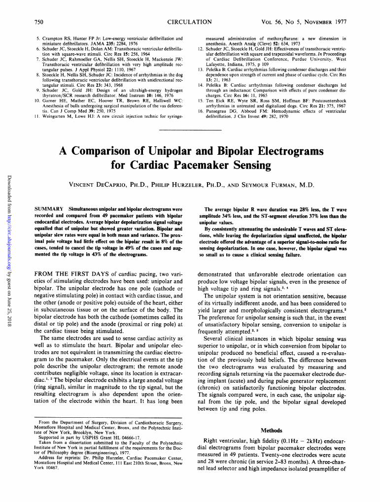
FIGURE 1. The recording system routes simultaneous unipolar and bipolar signals from the catheter electrode and astandard limb lead to an isolated preamplifier and onto magnetic tape. The tape is later played into a photographic re-corder to obtain a hard copy. The illustrated configuration records the following: a lead IH ECG on channel 1, a bipolarelectrogram on channel 2, a unipolar electrogram from the tip electrode on channel 3, and added voice comment on chan-nel 4. To record a unipolar ring signal, the tip and ring connections to the lead selector are interchanged.
custom design* allowed simultaneous bipolar and unipolartracings to be recorded with a peripheral lead II ECG (fig.1). A Hewlett-Packard Model 3960A instrumentation gradetape recorder stored the data for later playback to ModelsDR-8 and DR-12 Electronics for Medicine multichannel,high speed photographic recorders. Readings from the trac-ings were then statistically analyzed by a General Electriccomputer time sharing network.
Those parameters of the ventricular electrogram thatmost affect pacer sensing have been described elsewhere5 andin this study were similarly measured for both unipolar andbipolar signals, viz., peak-to-peak voltage and maximumslew rate (slope, or dv/dt) of the QRS complex, the ST-seg-ment elevation and the peak-to-peak voltage and maximumslew rate of the T wave. The duration of the intrinsic deflec-tion was taken as the width of the intracardiac R wave mea-sured between crossings of the isoelectric line; however, thedurations of some irregularly shaped curves could not bedetermined without extrapolation (e.g. fig. 2, middle) andare, therefore, estimates. Parameter values were averagedover one or two respiratory cycles before tabulation.The electrograms were recorded from electrodes with a
low, stable, clinically useful stimulation threshold and im-pedance, and satisfactory radiographic visualization. Theelectrode surface areas and bipole separations are shown intable 1. Only complexes from the most prominent focus,either conducted or idioventricular, were analyzed. Allrecordings were free from pacemaker stimuli and artifacts.Our recording arrangement causes a positive voltage at
the tip electrode to register as an upward deflection. Uni-polar signals were recorded with the negative input of a highimpedance bioelectric amplifier connected to a large surface
area metallic plate, temporarily inserted in the subcutane-ous pulse generator site. Bipolar signals were measured withthe tip electrode connected to the positive input and the ringelectrode connected to the negative input.
Early results showed that the interpretation of some
,p - 1-Pk-to-PNsk Volta*SRO". - 5i SIs. Rate
D - DurationS-S - S-S S555nt Diop1coen't
T-
Slwolt
5t. - S 55 S1* 5.t.
*Courtesy of Medtronic, Inc.
< S ~~~~SRX ISOLCDtRIC LIPC
FIGURE 2. Three diferent representative and diagrammatic acuteelectrograms, both unipolar and bipolar. Intermediate configura-tions exist but the parameters can be similarly defined.
751
by guest on June 25, 2018http://circ.ahajournals.org/
Dow
nloaded from

VOL 56, No 5, NOVEMBER 1977
TABLE 1. Bipolar Electrode SpecificationsCathodal Bipole
Quantity Manufacturer, model surface area separation
33 Medtronic 6901 11 sq mm 28 mm8 Medtronic 5816 87 sq mm 15 mm6 G. E. bipolar 12 sq mm 20 mm2 Pacesetter systems 12.2 sq mm 29 mm
bipolar signals was facilitated by simultaneously observingthe unipolar ring signal. Therefore, in the last 30 instancesunipolar ring signals were also recorded by moving the posi-tive input terminal from the tip to the ring electrode, andcontinuing the simultaneous recording. At the conclusion ofthe recording, simultaneous tip and bipolar electrograms,and simultaneous proximal and bipolar electrograms were
available for analysis.Signals which vary with time and may be displayed as a
plot of amplitude as a function of time (e.g., electrograms)are said to be in the "time domain" in the mathematic ter-minology of waveform analysis. Such signals, by means ofthe "Fourier Transform" can be recast into the mathe-matically equivalent "frequency domain" and be visualizedas a spectrum or plot of amplitude versus sinusoidal frequen-cy.6 Both representations are equally accurate and precisealthough each offers specific conveniences. The frequencydomain form is especially useful to designers of electroniccircuitry, but as the time domain form preserves themorphologic features of the waveform, it permits correla-tion of physiologic events with pacemaker sensing circuitresponse. Since the frequency domain offers no further in-sights into the electrophysiologic process, it was not used inthis study.
Results
One-third (16 of 49) of the bipolar depolarization signalsresembled the bottom curve of figure 2 with a narrow tri-angular deflection crossing the isoelectric line more thanonce. Of these, the peak was positive, indicating propaga-
tion from ring to tip in 11 cases, and negative, indicatingpropagation from tip to ring, in five cases. The remaining 33bipolar waveforms could not be classified into uniquemorphological subgroups.The bipolar depolarization signal voltage (measured
within 0.1 mV) was greater than its unipolar mate in 21cases (43%), equal in four cases (8%) and less in 24 cases
(49%). In one of the latter 24 cases the bipolar voltage was
too small to be sensed by the pacemaker; the matching uni-polar tip electrogram was larger and sensed. The maximumslew rate always occurred during an intrinsic deflection,which was usually a downward slope, virtually a straight linesegment. The maximum bipolar slew rates, measured within0.2 V/sec, were greater than their unipolar mates in 15 cases
(31%), equal in 22 cases (45%) and less in 12 cases (24%).The unipolar tip signals always showed a larger slew ratethan the corresponding unipolar ring signal.
Statistical evaluation of the unipolar and bipolar electro-grams as independent groups revealed that average uni-polar and bipolar voltages and slew rates are equal. Theaverage bipolar R wave duration is 28% less, the T wave
voltage 34%, and the ST-segment elevation 37% less than theaverage unipolar parameter (table 2). In the bar graphs ofthe bipolar and unipolar voltage distributions (fig. 3) thebipolar signals appear to show a larger variance, which isnot significant according to an F test with 90% confidence.By comparing the amplitudes of the unipolar tip and ring
signals for each electrode, a ring-to-tip voltage ratio was
computed. If the ring electrogram is small in comparison tothe tip electrogram a large voltage difference exists and thebipolar signal then approximates the unipolar tip signal; it isa quasi-unipolar signal (fig. 4-1). Twenty-two cases with a
ratio greater than 0.5 (a ring signal amplitude at least halfthe tip) were considered true bipolar signals, the result of tipand ring contributions of similar magnitude. The truebipolar signals exhibited a standard deviation of ± 6.8 mVwith a mean amplitude of 11.3 mV and a greater variancethan the unipolar group at the 90% level of the F test.
Data from each bipolar electrogram were then subtractedfrom the values of its unipolar mate, and the individualchanges in voltage, slew rate, and duration were computedfor all 49 cases and averaged. As six of the 21 acute bipolarelectrograms had no discernible T wave, and the ST seg-
ment of chronic electrograms is isoelectric, in neither in-stance could a change be calculated. Therefore, for the Twave voltage and slew rate, and ST displacement, the meanvalues of the independent groups (table 2) were used to com-pute the percentage change (table 3). The probability (Pr)that the unipolar value may be equal to the bipolar value,based on Student's t-test,7 is included in the righthandcolumn. Any Pr value less than 0.05 indicates a significantchange (table 3).
Bipolar R wave amplitudes were an average 3.03% lessthan unipolar ones. This difference, however, is not sup-
ported at any reasonable level of significance. The R wave
duration, T wave amplitude, T wave slew rate, and ST-seg-ment elevation are all significantly reduced with bipolarsensing (table 3).
Finally, the unipolar-bipolar pairs were further dividedinto acute and chronic subgroupings. A comparison of theunpaired (not from the same electrode) acute and chronicvalues revealed similar changes for both unipolar andbipolar signals; amplitude is maintained, though only withmarginal confidence; and slew rate is decreased by about40% (table 4).5 Here, Pr is the probability that no acute-to-chronic difference exists. Values less than .05 indicatesignificance.
TABLE 2. Unipolar and Bipolar Parameters
Unipolar BipolarMean SD Range Mean SD Range
Amplitude (mV) 12.2 5.2 4.0-29.2 11.8 6.0 3.3-31.3Slew rate (V/sec) 2.8 1.7 0.6-7.0 2.8 1.8 0.6-7.3Duration (msec) 88.3 30.3 25.0-150.0 61.9 29.3 10.0-145.0T wave voltage (mV) 2.4 2.2 0-8.4 1.6 1.3 0-5.2T wave slew rate (V/sec) .02 .03 0-.11 .02 .02 0-.08Acute ST displacement (mV) 2.6 1.6 0-5.7 1.6 1.5 0-4.0
752 CIRCULATION
by guest on June 25, 2018http://circ.ahajournals.org/
Dow
nloaded from

PACEMAKER SENSING/DeCaprio, Hurzeler, Furman
PEAK-TO-PEAK AMPLITUDE
VOLTAGElAFvrh2.0
4.0
3.0
8.0
*0.0
12.0
14.0
19.0
20.0
22.0
15 14 13 12 11 10 9 8 7 6 S 4 3 2 1
NUMBER OF CASES
24.0
320.0
32.0 m
1 2 3 4 5 6 7 8 9 10 11 12 13 14 15
NUMBER OF CASES
FIGURE 3. Comparison of the bargraphs of the 49 unipolar and bipolar electrograms suggest greater bipolar variance,
significant marginally only when those with a ring signal less than half the tip signal were excluded.
-B
3A
B
c
J
lOi,w
don
1, mnvlm
4
FIGURE 4. Four sets ofelectrograms indicate several clinicalfind-ings. Curve A is a lead HI ECG, B is the bipolar electrogram, C isthe unipolar tip electrogram and D is the unipolar ring signal. OnlyA, B and C are simultaneous recordings. Calibrations accompanyeach intracardiac electrogram. Time lines are 200 msec apart. 1) Aquasi-unipolar signal. With a small ring signal, the bipolar signal,though shorter in duration, resembles the unipolar tip signal. 2)Bipolar signal enhancement. A difference in activation time at eachpole causes a bipolar signal larger than either tip or ring signal. 3)Bipolar signal attenuation. Synchronous tip and ring depolariza-tions, though both large, produce a bipolar signal much smallerthan either. 4) The superior bipolar signal-to-noise ratio provides an
exceptionally clean signal of activation, with greatly reduced Twaves and ST-segment elevations.
Discussion
Ventricular electrograms may be resolved into four bio-electric events: the intrinsic deflection (the intracardiac Rwave), repolarization (the T wave), far-field phenomena (dis-tant electrical activity) and the injury current (ST-segmentelevation).5 The most important event for pacer sensing isthe intrinsic deflection, the rapid, nearly straightline down-ward deflection of the unipolar electrogram, which occurs
when the muscle adjacent to the electrode becomes electro-negative as the depolarization wave passes. With bipolarelectrodes, the wavefront appears first at one pole, thentravels to the other. An exception is the quasi-unipolar sig-nal where the wavefront does not produce similar intrinsicdeflections at each pole. The bipolar voltage, i.e., the poten-tial difference between the two poles at any time, is a func-tion of three variables, tip voltage, ring voltage and traveltime between poles, against only one (tip voltage) for theunipolar electrogram. Because of the increased number ofvariables, the greater variance in bipolar amplitude is notsurprising. Further evidence for inconsistency in bipolaramplitude is found in the large signal variation associatedwith normal respiration.8The interelectrode distance (bipole separation), the vel-
ocity of the spreading depolarization wave and especiallythe direction of the depolarization pathway through the ven-
tricle determine the timing of the tip and ring intrinsicdeflections. In an idealized model of myocardial tissue ac-
tivation (fig. 5) identical signals appear on both poles of a
bipolar electrode. In the two extreme cases the bipole maybe oriented either normal (N) (at a 90° angle) or parallel (P)to the path of the depolarization wave, here considered a
TABLE 3. Percent Change from UnipolarMean SD Pr
Voltage -3.03 30.30 .32Slew rate .32 26.54 .98Duration -27.64 30.13 <10-6
Mean T voltage -33.60 .007Mean T slew rate -29.17 .045Mean ST displacement -37.40 <10-6
753
I
0 P---.,---\4 by guest on June 25, 2018http://circ.ahajournals.org/
Dow
nloaded from

VOL 56, No 5, NOVEMBER 1977
TABLE 4. Acute-to-Chronic R Wave ChangeAcute Chronic
Mean SD Mean SD Pr
UnipolarVoltage (mV) 12.4 5.2 12.1 a.3 .825Slew rate (V/sec) 3.5 1.7 2.3 1.6 .012
BipolarVoltage (mV) 13.4 6.1 10.5 5.8 .094Slew rate (V/sec) 3.6 1.7 2.2 1.7 .008
sheet of charge extending the full width of a strip of cardiacmuscle. For the N orientation, the wavefront strikes bothpoles simultaneously and no bipolar potential results(fig. 4-3).
Alternatively, the P orientation causes a difference in ar-
rival times at the two poles resulting in an additive bipolarsignal (fig. 4-2). The amplitude of the bipolar signal is alsoinfluenced by the distances from each pole to the activetissue and the width of the depolarization wavefront.9A parallel (P) orientation, with a bipolar signal larger
than either ring or tip, occurred in 10 of the 30 cases in whichthe ring signal was explicitly recorded. In two of the 10, thebipolar was larger than the tip signal because of the ring con-
tribution. In the other eight, the cause was a timing differ-ence between the two smaller unipolar signals.
In the 21 cases in which the bipolar signal amplitude was
greater than the associated tip signal, the tip signal alonewas always large enough for clinically satisfactory sensing.No bipolar signal of sufficient amplitude and slew rate totrigger a pacer has been formed from two insufficient uni-polar signals. The probability of such an occurrence is ex-
tremely small since the small, wide intrinsic deflections
p N
N
T p
FIGURE 5. An idealized model depicting the activation of aof cardiac muscle adjacent to two bipolar electrodes represei
uniform depolarization process as a line ofdipoles spreadinggroup of very long muscle fibers (rectangle) (after Wilson e,The tip (T) and ring (R) electrodes of bipolar catheters posinormal (N) andparallel (P) to thefibers areprojected onto theof depolarization (dashed line). As the wave passes the poleselectrode, the ring, tip and bipolar curves are generated. Withmal orientation, the coincident arrival ofequal potentials on ti
poles results in complete subtraction. The parallel oriendelays the signal at one of the poles to produce a bipolarwhich may be larger than either unipolar signal.
associated with poor electrode position or an infarcted myo-cardium'0 arrive nearly simultaneously at both poles andtend to be attenuated rather than enhanced with bipolar sensing.(However, since only clinically satisfactory signals wererecorded, and a unipolar tip signal greater than 2 mV was acriterion, such bipolar signals were not demonstrated in thisstudy.) Thus, the clinical practice of unipolarizing a bipolarelectrode with an N orientation to improve its sensing per-formance,* does not have a useful converse: bipolarizing apair of poorly performing unipolar electrodes will not help.The purpose of a pacemaker's sensing amplifier is to re-
spond selectively to cardiac electrical activity. An adequatesignal, returning via the electrode, triggers the sensing cir-cuit into modifying the pacemaker's output. Adequacydepends on the signal's amplitude and frequency content,which can be characterized by its voltage, rate of change(slew rate),2' and duration (the time between isoelectriccrossings of the depolarization signal). Signals of very shortduration tend to be rejected by the sensing circuit as spurioushigh frequency noise, and will trigger the circuit only ifamplitude is high. The precise effect of signal duration onsensing is determined by the circuit's high frequency filtercharacteristics, and varies among designs. Pacemakers are
tested with a haversine test signal to simulate an intracar-diac R wave.t A typical sensing amplifier is most sensitive toa 25 msec duration haversine pulse and requires only a 2 mVamplitude to trigger at that duration. As the duration isdecreased to 6 msec, a 4 mV haversine is required (personalcommunication, J. Hartlaub, Medtronic, Inc.).
Because tip and ring voltage is nearly equal throughoutmost of the R wave, a substantial voltage difference existsonly when the depolarization wavefront is between poles.Usually, a bipolar signal of shorter duration and dissimilarmorphology from either tip or ring is produced (fig. 4-4).Even if the amplitude of the ring signal is significantly lessthan the tip, a bipolar signal morphologically similar to thetip is generated, but again, with a shorter duration (fig. 4-1).Forty-four of the 49 cases had bipolar durations shorter thanunipolar tip (mean decrease 28%), but as signal durationsnever fell below 10 msec (mean 75 msec) it is unlikely that a
DIPOLAR signal from a bipolar electrode would be rejected as noise.
The remaining three contributors to the electrogram (Twave, ST elevation and far-field activity) are physiologicnoise to a sensing system and should be rejected. The myo-
cardial repolarization wavefront is wide" and appears nearlysimultaneously on both poles of the bipolar system. It is,therefore, significantly attenuated. The bipolar T waves ofthis study were reduced an average of 34% compared tothose of the simultaneous bipolar (figs. 4-3 and 4-4). Theacute ST-segment elevation is believed due to a local cur-
rent of injury caused by pressure of the electrode tip againstPts thee the endocardium. The electrogram from the ring, which is
over a distant from the injury, also displays an ST elevation
,t al.7).tioned'planeof thea nor-
he two'tationsignal
*In this instance good signals on tip and ring cancel each other to form anattenuated bipolar signal. Elimination of one leaves a good signal fully avail-able.
tA haversine pulse is one half cycle of a sine wave, squared. It may be elec-tronically generated and its amplitude and duration independently varied totest pacemaker sensitivity. See Pacemaker Standard (proposed), August,1975, Para. 4.1.4.1., Association for the Advancement of Medical Instru-mentation, 1901 Fort Meyers Drive, Arlington, Va. 22209.
CIRCULATION754
-e .'-BIPOLAR
by guest on June 25, 2018http://circ.ahajournals.org/
Dow
nloaded from

PACEMAKER SENSING/DeCaprio, Hurzeler, Furman
(though smaller than the tip electrogram) and the bipolarsignals therefore display lower ST elevations than the uni-polar signals. Far-field potentials from distant electrical ac-
tivity, (e.g., activation of the opposite ventricle, skeletalmuscle potentials, atrial activity, nonphysiologic signals,etc.), if large enough, can falsely trigger the pacemaker.Unipolar signal amplitude is inversely proportional to thedistance between the electrode and the signal source. Bipolarsignal amplitude is inversely proportional to the square ofthe distance from a point midway between the two poles andthe signal source.*12 The smaller lead-field of the bipolarelectrode renders it superior in rejecting electromagnetic in-terference and distant physiologic activity.18By the attenuation or enhancement of select events the
bipolar electrode can provide a signal-to-noise ratio superiorto that of a unipolar electrode. It attenuates not only elec-trical noise"3 but also the physiologic signals which should betreated as noise. T waves, ST-segment elevations and poten-tials from areas of the heart far from the electrode are
reduced before they enter the pacer's sensing amplifier(where they may be further attenuated by filtering). Attimes, however, the price of low noise can be R wave
attenuation unable to trigger a pacemaker in 2% of our
cases. Often a small but artifact-free signal may be an ad-vantage over one larger but noisier (better signal-to-noiseratio).'4 If the T waves are considered noise, the signal (Rwave)-to-noise ratio was greater for bipolar electrodes in 33of our 49 cases (67%).
Conclusions
Bipolar and tip unipolar signals, simultaneously derivedfrom the same catheter electrode, are usually unequal inamplitude, yet the averages of the two groups are equal. Theprobability of obtaining a signal much larger or muchsmaller than average is greater with bipolar sensing. Theorientation of a bipolar electrode within the heart deter-
*A unipolar electrode is equivalent to a monopole and the bipolar elec-trode to a dipole in electric field theory.
mines the amplitude and slew rate of the electrogram. Theorientation can cause either a subtraction or an addition ofthe two unipolar amplitudes at each pole. The addition oftwo smaller unipolar signals, and not a large ring signal, ac-counted for the majority of the cases having a bipolar signallarger than the unipolar tip signal.The depolarization signal of bipolar electrograms is
shorter in duration than unipolar, yet not so short as to bemistaken for electrical noise by the sensing amplifier.
Bipolar sensing reduces both the physiologic (T waves, STelevations and far-field) and nonphysiologic (electromag-netic interference) noise in sensing systems providing a sig-nal-to-noise ratio superior to unipolar.
References
1. Lewis T: The Mechanism and Graphic Registration of the Heart Beat.London, Shaw and Sons, Ltd, 1925
2. Barold SS, Keller JE: Sensing problems with demand pacemakers. InCardiac Pacing, edited by Samet P. New York, Grune and Stratton, 1973
3. Barold SS, Gaidula JJ: Failure of demand pacemakers from low-voltagebipolar ventricular electrograms. JAMA 215: 923, 1971
4. Greatbatch W: In Technical and clinical aspects of pacing electrodes.Ann NY Acad Sci 167: 865, 1969
5. Hurzeler P, DeCaprio V, Furman S: Endocardial electrograms and pace-maker sensing. Med Instrum 10: 178, 1976
6. Geddes LA, Baker LE: Principles of Applied Biomedical Instrumenta-tion, chap 14. New York, Wiley, 1968, pp 446A467
7. Crow EL, Davis FA, Maxfield MW: Statistics Manual. New York,Dover Publications, 1960
8. DeCaprio V, Gaspar H, Escher DJW, Furman S: Respiratory cyclevariations in pacer sensing signals. Med Instru 10: 55, 1976
9. Wilson FN, MacLeod AG, Barker PS: The distribution of the currents ofaction and injury displayed by heart muscle and other excitable tissue. InSelected Papers of Dr. Frank N. Wilson, edited by Johnston FD,Lepeschkin E. Ann Arbor, JW Edwards, 1954, pp 307
10. Chatterjee K, Davies G, Harris A: Fall of endocardial potentials afteracute myocardial infarction. Lancet 1: 1308, 1970
11. MacLeod AG: The electrogram of cardiac muscle: An analysis which ex-plains the regression or T deflection. Am Heart J 15: 165, 1938
12. Brody DA, Bradshaw JC, Evans JW: A theoretical basis for determiningheart-lead relationships of the equivalent cardiac multipole. IEEE TransBiomed Eng BME8: 139, 1961
13. Kahn AR, Schlentz RJ: Design and construction methods for protectingimplanted cardiac pacemakers from electromagnetic interference. InCardiac Pacing, edited by Thalen H. Netherlands, Van Gorcum and Co,1973
14. Chatterjee K, Swan HJC, Ganz W, Gray R, Loebel H, Forrester J,Chonette D: Use of a balloon-tipped flotation electrode catheter for car-diac monitoring. Am J Cardiol 36: 55, 1975
755
by guest on June 25, 2018http://circ.ahajournals.org/
Dow
nloaded from

V DeCaprio, P Hurzeler and S FurmanA comparison of unipolar and bipolar electrograms for cardiac pacemaker sensing.
Print ISSN: 0009-7322. Online ISSN: 1524-4539 Copyright © 1977 American Heart Association, Inc. All rights reserved.
is published by the American Heart Association, 7272 Greenville Avenue, Dallas, TX 75231Circulation doi: 10.1161/01.CIR.56.5.750
1977;56:750-755Circulation.
http://circ.ahajournals.org/content/56/5/750the World Wide Web at:
The online version of this article, along with updated information and services, is located on
http://circ.ahajournals.org//subscriptions/
is online at: Circulation Information about subscribing to Subscriptions:
http://www.lww.com/reprints Information about reprints can be found online at: Reprints:
document. Permissions and Rights Question and Answer information about this process is available in the
located, click Request Permissions in the middle column of the Web page under Services. FurtherEditorial Office. Once the online version of the published article for which permission is being requested is
can be obtained via RightsLink, a service of the Copyright Clearance Center, not theCirculationpublished in Requests for permissions to reproduce figures, tables, or portions of articles originallyPermissions:
by guest on June 25, 2018http://circ.ahajournals.org/
Dow
nloaded from