compendium of dermatoscopy - j.a.k marketing · if no pigment network, streaks, irregular globules...
TRANSCRIPT

COMPENDIUM OF DERMATOSCOPY
| 02 |
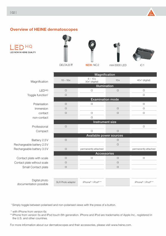
Overview of HEINE dermatoscopes
1 Simply toggle between polarised and non-polarised views with the press of a button.
* with iPhone from version 6s** iPhone from version 5s and iPod touch 6th generation. iPhone and iPod are trademarks of Apple Inc., registered in
the U.S. and other countries.
For more information about our dermatoscopes and their accessories, please visit www.heine.com.
Magnification
Magnification 10 – 16 x6 – 10 x
30x* (digital)10 x 40 x* (digital)
IlluminationLEDHQ ¨ ¨ ¨ ¨
Toggle function1 ¨ ¨
Examination modePolarisation ¨ ¨ ¨
Immersion ¨ ¨ ¨
contact ¨ ¨ ¨ ¨
non-contact ¨
Instrument sizeProfessional ¨ ¨
Compact ¨ ¨
Available power sourcesBattery 2.5 V ¨ ¨
Rechargeable battery 2.5 V ¨
Rechargeable battery 3.5 V ¨ permanently attached permanently attached
AccessoriesContact plate with scale ¨ ¨ ¨ ¨
Contact plate without scale ¨ ¨
Small Contact plate ¨ ¨
Digital photo documentation possible
SLR Photo adaptor iPhone® / iPod® ** iPhone® / iPod® **
mini 3000 LED iC 1NEW: NC 2DELTA 20 T

| 03 |
Dermatoscopy of pigmented and non-pigmented skin lesions – a guide
Andreas Blum, Herwig Swoboda and Rainer Hofmann-Wellenhof
This guide summarises the key principles of dermatoscopy which have been updated in line with the literature. It is based on the first guide, written by Professor Stolz. All images are new. The DELTA 20 Plus Dermatoscope or DELTA 20 T Dermatoscope together with the HEINE SLR Photo adaptor were used for the photography.
Important information for the userKnowledge in medicine is continually changing as a result of research and clinical experience. The authors of this guide have taken great care to ensure the diagnostic information given in this guide is in line with the current state of knowledge.However, this does not release the user of the obligation to use additional, more current written sources of information to check whether the more current information differs from the information given in this guide. The classification and diagnosis of skin tumours and skin lesions is the user’s own responsibility.
Konstanz / Germany and Graz / Austria, July 2013
Original text in German, Translation: thebigword.
TABLE OF CONTENTS
1. Introduction ........................................................................................................................................ 04
2. Devices for dermatoscopy ................................................................................................................. 05
3. Methodology and dermoscopic criteria for the examination .............................................................. 08
3.1. Algorithm for differentiating between melanocytic and non-melanocytic skin tumours ................................................................................................... 08
First Step ................................................................................................................................... 09
Second Step ..............................................................................................................................13
Third Step .................................................................................................................................. 21
3.2. Colours ....................................................................................................................................... 23
3.3. Further diagnostic criteria ........................................................................................................... 24
4. Differentiating between benign and malignant skin tumours .............................................................. 25
4.1. The AC rule ................................................................................................................................ 25
4.2. The AC rule and other clues ....................................................................................................... 26
4.3. Our personal rule ........................................................................................................................ 26
5. Further literature ................................................................................................................................. 34

| 04 |
1. Introduction
The annual incidence of melanomas, basal-cell carcinomas and squamous-cell carcinomas continues to rise significantly, which means that malignant skin tumours in people with a light skin type (Fitzpatrick type I – III) are now among the most common malignant tumours in men and women. Early detection of a melanoma decreases mortality in particular, early detection of basal-cell carcinoma decreases morbidity, and early detection of squamous-cell carcinoma decreases morbidity more than mortality. The development, introduction, further expansion and research of dermatoscopy has played a significant part in the early detection, and therefore in the better prognosis, of malignant skin tumours. In addition, the rate of unnecessary excisions of benign skin tumours has significantly decreased. The documentation of dermatoscopic images is not only helpful for the communication with patients and histopathologists, but is also an essential part of check-ups, research and teaching.
The purpose of this guide is to provide some concrete basics and a structured procedure for diagnosing pigmented and non-pigmented skin lesions. Only through continual use in patients, correlating dermatoscopic images with the histologies of excised skin tumours and by participating in further training will the user acquire the necessary routine and increasing certainty in using the dermatoscope.
1989 20021991 2007 2013 2016
19961990 2003 2008 2015 2018
| 05 |
2. Devices for dermatoscopy
The various dermatoscopes available from HEINE Optotechnik are easy to use, robust and reliable reflected-light microscopes with approx. 10 x – 16 x magnification, with or without polarisation technology, which are ideal for the routine examination of pigmented and non-pigmented skin lesions and other indication determinations. Nowadays, the dermatoscope plays an essential and very useful part in the diagnostics of most dermatologists working in the clinical environment.
By wetting the skin with immersion fluid the skin lesion can be assessed with the illuminated dermatoscope through the contact plate. Suitable immersion fluids include skin disinfectants, isopropyl alcohol, ultrasound gel or dermatoscopy oil. Alcoholic media with a microbiocidic effect have the benefit of also having a disinfecting effect. In principle, immersion fluid is not required for the DELTA 20 Plus with polarisation filter; however it is useful in the case of melanocytic and / or vascular skin tumours.
Some dermatoscopes like the DELTA 20 T can toggle between polarised and non-polarised illumination by the press of a button. This allows the recognition of the “Blink Sign” when switching between illumination modes while observing crystalline structures and the presence of milia cysts.In principle, immersion fluid is not required when using the DELTA 20 T. However it is useful when extensively utilising the non-polarised illumination mode.
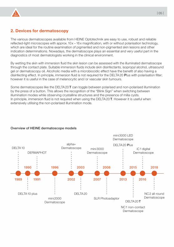
Overview of HEINE dermatoscope models
DELTA 10alpha+
Dermatoscope
DERMAPHOTmini 3000
Dermatoscope
mini 3000 LEDDermatoscope
DELTA 20 Plus
mini 2000Dermatoscope
DELTA 10 plus DELTA 20
SLR PhotoadaptorDELTA 20 T
NC 1 non-contact Dermatoscope
iC 1 digitalDermatoscope
NC 2 all-round Dermatoscope

| 06 |
Fig. 1: With polarisation filter, without immersion fluid and with full illumination (4 LEDs).
Fig. 3: With polarisation filter, with immersion fluid (HEINE Dermatoscopy Oil) and with full illumination (4 LEDs).
Fig. 2: With polarisation filter, without immersion fluid and with semi-illumination (2 LEDs).
Fig. 4: With polarisation filter, with immersion fluid (HEINE Dermatoscopy Oil) and with semi-illumination (2 LEDs).
Below you will find a series of dermatoscopic images of an early invasive melanoma (< 0.2 mm tumour thickness, predominantly melanoma in-situ) with details of the various filters, applications of immersion fluid and the intensity of illumination (Fig. 1 – 8):

| 07 |
Fig. 5: Without polarisation filter, without immersion fluid and with full illumination (4 LEDs).
Fig. 7: Without polarisation filter, with immersion fluid (HEINE Dermatoscopy Oil) and with full illumination (4 LEDs).
Fig. 6: Without polarisation filter, without immersion fluid and with semi-illumination (2 LEDs).
Fig. 8: Without polarisation filter, with immersion fluid (HEINE Dermatoscopy Oil) and with semi-illumination (2 LEDs).
In most cases, in particular in case of vascular lesions, we apply the dermatoscope with polarisation filter, immersion fluid (e.g. HEINE Dermatoscopy Oil on the skin) and full illumination (4 LEDs). With very light skin or light lesions it has been proven appropriate to use semi-illumination of the dermatoscope, i.e. two instead of four LEDs. Specific structures (e.g. pseudohorn cysts, comedo-like openings, cerebriform pattern) are easier to identify without a polarisation filter.

| 08 |
3. Methodology and dermatoscopic criteria for the examination
Regardless of the number of skin lesions examined, the diagnosis of pigmented and non-pigmented skin lesions is based on knowledge of the various differential structures and colours in the dermatoscopic image and their correct classification.
3.1. Algorithm for differentiating between melanocytic and non-melanocytic skin tumours
The first diagnostic differentiation should classify the skin lesions into a melanocytic or non-melanocytic skin tumour (Fig. 9). The algorithm modified according to Kreusch and Stolz can be used to classify nearly all pigmented and non-pigmented skin lesions into one of the listed diagnoses. Rarely occurring skin tumours that cannot be differentiated with this algorithm fall into the “Third Step” group due to the absence of characteristic differential structures and are therefore classified as “suspect”. This increases the certainty for the patient and the examiner.
Fig. 9: Multistep algorithm for differentiating between melanocytic and non-melanocytic skin tumours (modified according to Kreusch and Stolz).
First step
Second step
Third step
Seborrheic keratosis
CAVE
AngiomaAngiokeratoma
Basal-cell carcinoma
Actinic keratosis
Bowen’s Disease
Keratoacanthoma
Squamous-cellcarcinoma
Melanocytic lesion
Pigment network, streaks, globules, structureless brown / blue lesionException: dermatofibroma, solar lentigo, accessory nipple
Pseudohorn cysts, comedo-like openings, fingerprint-like structures, erebriform structures, opaque colours
None of the named structures / vascular polymorphismsolitary blue, black or reddish nodular lesion
Red to blue-black lacunas, white veil possibleHyperkeratoses
Arborizing vessels, blue-grey oval globules,ulceration, spoke-wheel areas, leaf-like structures
White halo around keratotic follicles, pseudo-network with erythema, Follicles filled with keratin, possible vessels around follicles
Reddish / brownish plaque, hyperkeratoses possible,dotted or glomerular vessels in a line-like arrangement
Coronary vessels around a central plug of keratin
Hyperkeratoses, haemorrhages, possible white halo around follicles

*
| 09 |
First step
The presented algorithm is divided into three steps. The first step analyses if a network (Fig. 10), streaks (Fig.11), globules (Fig.11) or homogeneous blue pigmentation are visible dermatoscopically. If so, a melanocytic skin tumour is present. Exceptions include dermatofibromas with post-inflammatory hyperpigmentation at the rim, actinic lentigines and accessory nipple with a distinct network.
Fig. 10: Pigment network (arrow) and streaks (arrow with *) in an early invasive melanoma < 0,2 mm tumour thickness, predominantly melanoma in-situ). Image taken with polarisation filter, with immersion fluid (HEINE Dermatoscopy Oil) and with full illumination (4 LEDs).

| 10 |
Fig. 12: Homogeneous blue pigmentation in a blue nevus (image taken with polarisation filter, with immersion fluid (HEINE Dermatoscopy Oil) and with full illumination (4 LEDs).
Fig. 11: Globules in a melanocytic nevus. Image taken with polarisation filter, with immersion fluid (HEINE Dermatoscopy Oil) and with full illumination (4 LEDs).
| 11 |
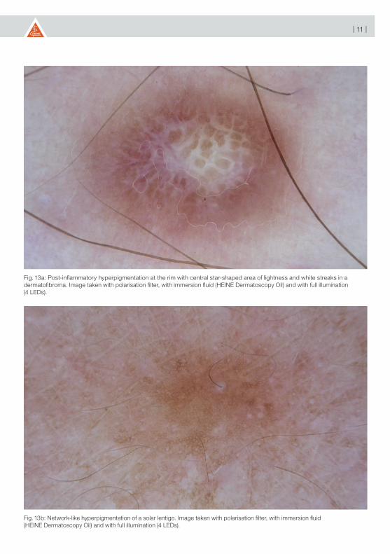
Fig. 13b: Network-like hyperpigmentation of a solar lentigo. Image taken with polarisation filter, with immersion fluid (HEINE Dermatoscopy Oil) and with full illumination (4 LEDs).
Fig. 13a: Post-inflammatory hyperpigmentation at the rim with central star-shaped area of lightness and white streaks in a dermatofibroma. Image taken with polarisation filter, with immersion fluid (HEINE Dermatoscopy Oil) and with full illumination (4 LEDs).

| 12 |
Fig. 13c: Network-like hyperpigmentation of an accessory nipple. Image taken with polarisation filter, with immersion fluid (HEINE Dermatoscopy Oil) and with full illumination (4 LEDs).

| 13 |
Second step
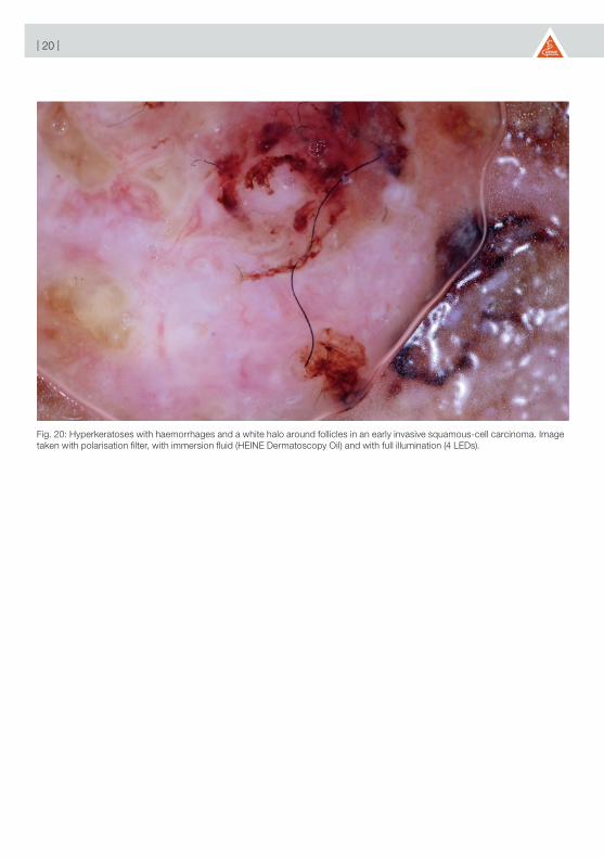
If no pigment network, streaks, irregular globules or homogeneous blue pigmentation are visible, the second step is to look for the differential structures of seborrheic keratosis (Fig. 14a – d), angioma (Fig. 15a and b) or angiokeratoma, basal-cell carcinoma (Fig. 16a and b), actinic keratosis (Fig. 17a – c), Bowen’s disease (Fig. 18), keratoacanthoma (Fig. 19) and squamous-cell carcinoma (Fig. 20). Corresponding examples are provided in the image section of these instructions.
Fig. 14a: Fingerprint-like lines in an early seborrheic keratosis. Image taken with polarisation filter, with immersion fluid (HEINE Dermatoscopy Oil) and with full illumination (4 LEDs).

| 14 |
Fig. 14c: Fingerprint-like lines in the periphery, central cerebriform structures with pseudohorn cysts in a seborrheic keratosis. Image taken with polarisation filter, with immersion fluid (HEINE Dermatoscopy Oil) and with full illumination (4 LEDs).
Fig. 14b: Fingerprint-like lines with cerebriform structures in a seborrheic keratosis. Image taken with polarisation filter, with immersion fluid (HEINE Dermatoscopy Oil) and with full illumination (4 LEDs).

| 15 |
Fig. 14d: Pseudohorn cysts, comedo-like openings, fingerprint-like lines in the periphery, in a seborrheic keratosis with opaque colouring. Image taken without polarisation filter, with immersion fluid (HEINE Dermatoscopy Oil) and with semi-illumination (2 LEDs).
Fig. 15a: Reddish lacunas with whitish, thin streaks in a senile angioma. Image taken with polarisation filter, with immersion fluid (HEINE Dermatoscopy Oil) and with full illumination (4 LEDs).
| 16 |
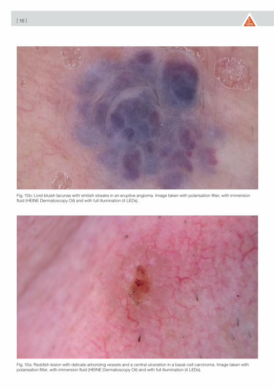
Fig. 15b: Livid-bluish lacunas with whitish streaks in an eruptive angioma. Image taken with polarisation filter, with immersion fluid (HEINE Dermatoscopy Oil) and with full illumination (4 LEDs).
Fig. 16a: Reddish lesion with delicate arborizing vessels and a central ulceration in a basal-cell carcinoma. Image taken with polarisation filter, with immersion fluid (HEINE Dermatoscopy Oil) and with full illumination (4 LEDs).

| 17 |
Fig. 16b: Reddish lesion with delicate arborizing vessels, two large blue-grey ovoid nests of a basal-cell carcinoma and a laterally positioned angioma with reddish lacunas (collision tumour). Image taken with polarisation filter, with immersion fluid (HEINE Dermatoscopy Oil) and with semi-illumination (2 LEDs).
Fig. 17a: Pseudo-network with erythema and discrete pigmentation, with keratin-filled follicles and discrete vessels of an early actinic keratosis (grade 1). Image taken with polarisation filter, with immersion fluid (HEINE Dermatoscopy Oil) and with full illumination (4 LEDs).

| 18 |
Fig. 17b: Keratin-filled follicle with progressive central hyperkeratoses of an advanced actinic keratosis (grade II). Image taken with polarisation filter, with immersion fluid (HEINE Dermatoscopy Oil) and with full illumination (4 LEDs).
Fig. 17c: Progressive hyperkeratoses with residual erythema in an older actinic keratosis (grade III). Image taken with polarisation filter, with immersion fluid (HEINE Dermatoscopy Oil) and with full illumination (4 LEDs).

| 19 |
Fig. 18: Reddish plaque with dotted vessels in a line-like arrangement in a case of relapsed Brown‘s disease. Image taken with polarisation filter, with immersion fluid (HEINE Dermatoscopy Oil) and with full illumination (4 LEDs).
Fig. 19: Coronary vessels around a central keratin plug with haemorrhages in a keratoacanthoma. Image taken with polarisation filter, with immersion fluid (HEINE Dermatoscopy Oil) and with full illumination (4 LEDs).
| 20 |
Fig. 20: Hyperkeratoses with haemorrhages and a white halo around follicles in an early invasive squamous-cell carcinoma. Image taken with polarisation filter, with immersion fluid (HEINE Dermatoscopy Oil) and with full illumination (4 LEDs).
| 21 |
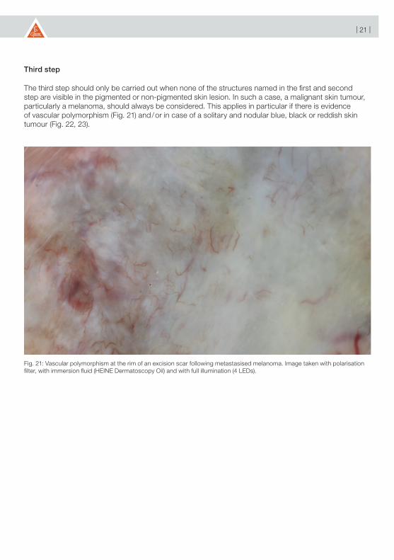
Third step
The third step should only be carried out when none of the structures named in the first and second step are visible in the pigmented or non-pigmented skin lesion. In such a case, a malignant skin tumour, particularly a melanoma, should always be considered. This applies in particular if there is evidence of vascular polymorphism (Fig. 21) and / or in case of a solitary and nodular blue, black or reddish skin tumour (Fig. 22, 23).
Fig. 21: Vascular polymorphism at the rim of an excision scar following metastasised melanoma. Image taken with polarisation filter, with immersion fluid (HEINE Dermatoscopy Oil) and with full illumination (4 LEDs).
| 22 |
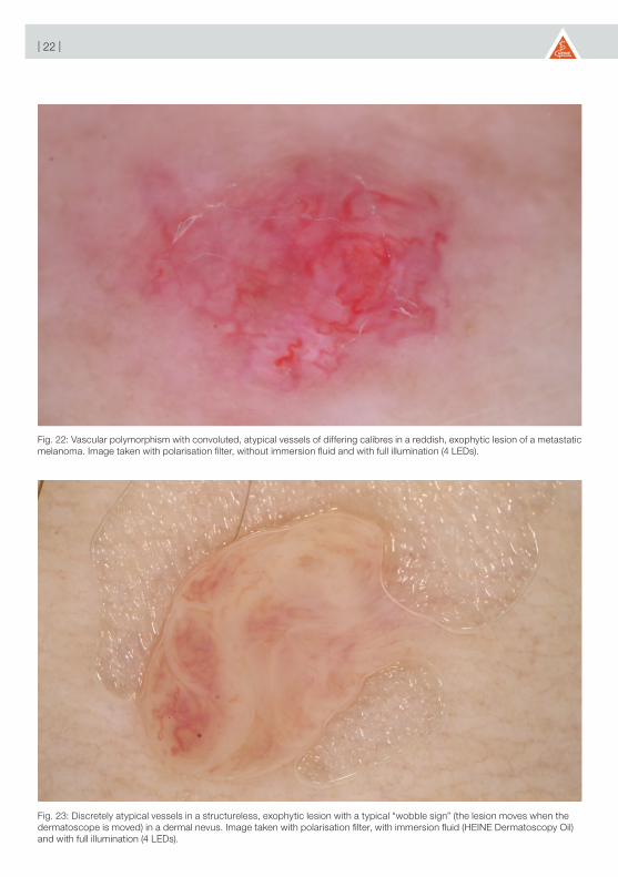
Fig. 23: Discretely atypical vessels in a structureless, exophytic lesion with a typical “wobble sign” (the lesion moves when the dermatoscope is moved) in a dermal nevus. Image taken with polarisation filter, with immersion fluid (HEINE Dermatoscopy Oil) and with full illumination (4 LEDs).
Fig. 22: Vascular polymorphism with convoluted, atypical vessels of differing calibres in a reddish, exophytic lesion of a metastatic melanoma. Image taken with polarisation filter, without immersion fluid and with full illumination (4 LEDs).

| 23 |
3.2. Colours
The normal epidermis can appear from whitish, yellowish to brown in the colour spectrum (Fig. 24). When the horny layer (acanthosis) becomes thicker, the colour shade can become yellow-brown to grey-brown. Melanin, the major pigment in the skin, is found in the basal membrane between the epidermis and dermis. The more melanin rises to the stratum corneum of the epidermis and therefore to the surface of the skin, the more a black colour is clearly visible. The deeper the melanin is in the skin, the more brown (basal membrane), grey (upper dermis) or steel-blue (middle dermis) colour shades are visible. The deeper the melanin is in the skin, the less distinct the colours are. This also applies to the position of vessels. Vessels in different positions and forms can appear accordingly as colours ranging from light to dark red, sometimes blue or dark blue, and in rare cases even black. The colour white can only be defined when compared with the surrounding healthy skin and is a sign of regression of the benign or malignant skin tumour.
Fig. 24: Colours visible through the dermatoscope according to the position of the melanin (left) and the blood vessels (right).
Epidermis
Dermis
Melanin Vessels
| 24 |
3.3. Further diagnostic criteria
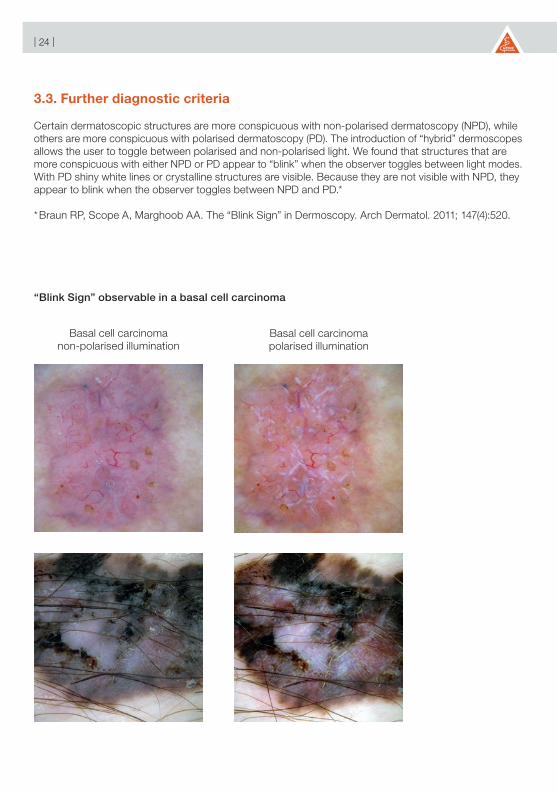
Certain dermatoscopic structures are more conspicuous with non-polarised dermatoscopy (NPD), while others are more conspicuous with polarised dermatoscopy (PD). The introduction of “hybrid” dermoscopes allows the user to toggle between polarised and non-polarised light. We found that structures that are more conspicuous with either NPD or PD appear to “blink” when the observer toggles between light modes. With PD shiny white lines or crystalline structures are visible. Because they are not visible with NPD, they appear to blink when the observer toggles between NPD and PD.*
* Braun RP, Scope A, Marghoob AA. The “Blink Sign” in Dermoscopy. Arch Dermatol. 2011; 147(4):520.
“Blink Sign” observable in a basal cell carcinoma
Basal cell carcinomanon-polarised illumination
Basal cell carcinomapolarised illumination

| 25 |
4. Differentiating between benign and malignant skin tumours
Even if the diagnosis as to whether the tumour is benign or malignant has been clinically confirmed, it is very useful to start by looking at the tumour through the dermatoscope. Look at the tumour’s overall structures, colours and patterns. Look at the rim and how the tumour appears to be growing, irrespective of its grade. Familiarize yourself with these structures, colours and patterns to learn how to identify very small, early malignant tumours of the skin with a favourable prognosis.
Which pattern does the pigmented or non-pigmented skin lesion show you? There are many international studies relating to pattern analysis. This pattern analysis has been and continues to be the basis for creating a variety of scores that are used to differentiate between melanocytic and non-melanocytic, benign and malignant skin tumours. So as not to complicate matters, we will not address all of these scores and algorithms here, but will instead show you a straightforward method and rule to help you to accurately detect melanomas, in addition to basal-cell and squamous-cell carcinomas. During this process, you will find that you will also diagnose a very small number of benign skin tumours as malignant, and thus remove these “unnecessarily” – however, this happens to even the “best” dermatoscopy specialists and serves to ensure that absolutely no melanoma is missed.
4.1. The AC rule
The clinical and dermatoscopic rule is the AC rule, which stands for asymmetry and colour variations (Table 1). A malignant skin tumour, particularly a melanoma, grows chaotically. This means asymmetry occurs in both the external form and in the internal structures (i.e. the tumour is no longer mirrored in the main axes layered vertically on top of one another), and the variety of colour increases over time as the tumour grows. Visible jet-black or blue-grey colours are a clear sign of a melanoma. Not all melanomas follow this rule. For example, there can be clinically and dermoscopically symmetrical melanomas which are purely red in colour and amelanotic. In addition, purely nodular and sometimes predominantly black or purely red melanomas can occur. In general the Ugly Duckling rule helps here: an individual tumour which is completely different to the patient’s other tumours, should undergo a highly critical clinical and, above all, dermatoscopic examination.
Asymmetry-Colour-Variation-Rule (The AC rule)
Asymmetry Form and internal structures
Colour variations Jet-black, blue, grey, brown, red, white
Table 1
When analysing symmetry or asymmetry, be aware that there is actually very rarely a symmetrical skin tumour. Nature is not perfect in this sense, and therefore symmetry can be evaluated with a bit of generosity.With this AC rule described above, medical laypersons achieved an accuracy rate of 91 % in the detection of melanomas in a clinical assessment, and 94 % if a dermatoscope was used.

| 26 |
4.2. The AC rule and other clues
The following clues can also be used alongside the AC rule to help analyse unclear skin tumours and rule out malignant skin tumours (Table 2): peripheral black dots, radial lines or pseudopods (= thickened radial lines) in only a segmental and partial arrangement, polymorphic vessels, thickened atypical network or branched lines, unknown nodular structure. Just one of these patterns is needed in addition to the AC rule to suspect a melanoma or basal-cell carcinoma.
The AC rule and clues
AC rule Asymmetry and colour variations
Clues radial lines or pseudopods (= thickened radial lines) in only a segmental and partial arrangement (Fig. 29, 30)
polymorphic vessels (Fig. 35) thickened atypical network or branched lines (Fig. 29, 30) unknown nodular blue, black or red tumour (Fig. 32, 33, 34)
Table 2
Dermatoscopy can also be used on the palms of the hands, soles of the feet and on (non-varnished) nails. However, on these surfaces the structures and colours are not quite as visible as on normal skin. Nevertheless, both the AC rule and AC rule and clues can still be applied.
4.3. Our personal rule
Finally, we have our additional, personal rule on detecting melanomas or other malignant skin tumours:
Have I seen something like this before?
Do I know of a benign skin tumour that could look like this?
Would I want to leave this skin tumour on my own skin and live with it?
I am curious what that is (according to Jürgen Kreusch).
In general the following rule applies: in case of doubt, an excision or at least a biopsy is always the best option if you cannot guarantee, to the patient and yourself, that the skin tumour you have examined is not a melanoma or other malignant skin tumour.
For many years, our best continued dermatoscopic training has been the clinical and dermatoscopic photography of skin tumours prior to surgery and their subsequent comparison with the histology results. This is a method that we would highly recommend to everyone. We have learnt much from doing this and we know that every week we will discover something new, as melanomas are precisely extremely varied in their appearance. This knowledge and respect for this forms the basis for our daily work.

| 27 |
Fig. 25: Normal streaks and network arranged almost symmetrically and central hyperpigmentation of a lentiginous melanocytic nevus with brown and black colouring. Image taken with polarisation filter, with immersion fluid (HEINE Dermatoscopy Oil) and with full illumination (4 LEDs).
Fig. 26: Slightly asymmetrical lesion with normal globules of a compound nevus brown in colour. Image taken with polarisation filter, with immersion fluid (HEINE Dermatoscopy Oil) and with reduced light intensity (2 LEDs).

| 28 |
Fig. 27: Slightly asymmetrical lesion with normal network and globules of a lentiginous melanocytic nevus brown in colour. Image taken with polarisation filter, with immersion fluid (HEINE Dermatoscopy Oil) and with full illumination (4 LEDs).
Fig. 28: Slightly asymmetrical lesion with prominent network and peripheral globules (starburst pattern) of a Reed nevus brown in colour. Image taken with polarisation filter, with immersion fluid (HEINE Dermatoscopy Oil) and with full illumination (4 LEDs).

| 29 |
Fig. 29: Asymmetrical lesion with atypical network, streaks and dots of an atypical compound nevus brown, grey and blue in colour. Image taken without polarisation filter, with immersion fluid (HEINE Dermatoscopy Oil) and with full illumination (4 LEDs).
Fig. 30: Asymmetrical lesion with atypical network, streaks, pseudopods and dots of a melanoma in-situ brown and black in colour. Image taken with polarisation filter, with immersion fluid (HEINE Dermatoscopy Oil) and with full illumination (4 LEDs).
| 30 |
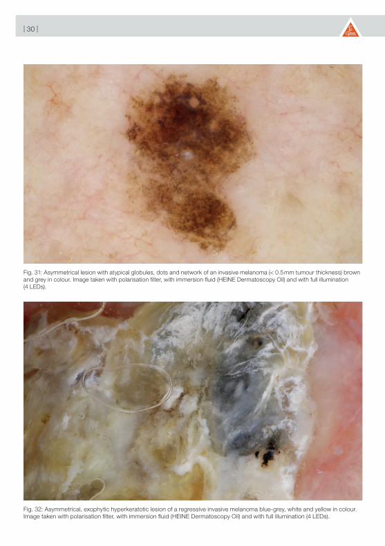
Fig. 31: Asymmetrical lesion with atypical globules, dots and network of an invasive melanoma (< 0.5 mm tumour thickness) brown and grey in colour. Image taken with polarisation filter, with immersion fluid (HEINE Dermatoscopy Oil) and with full illumination (4 LEDs).
Fig. 32: Asymmetrical, exophytic hyperkeratotic lesion of a regressive invasive melanoma blue-grey, white and yellow in colour. Image taken with polarisation filter, with immersion fluid (HEINE Dermatoscopy Oil) and with full illumination (4 LEDs).
| 31 |
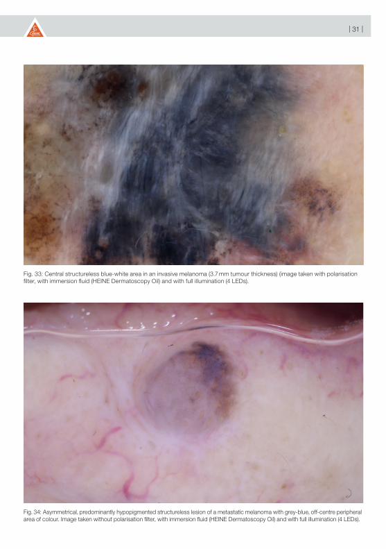
Fig. 33: Central structureless blue-white area in an invasive melanoma (3.7 mm tumour thickness) (image taken with polarisation filter, with immersion fluid (HEINE Dermatoscopy Oil) and with full illumination (4 LEDs).
Fig. 34: Asymmetrical, predominantly hypopigmented structureless lesion of a metastatic melanoma with grey-blue, off-centre peripheral area of colour. Image taken without polarisation filter, with immersion fluid (HEINE Dermatoscopy Oil) and with full illumination (4 LEDs).

| 32 |
Fig. 35: Asymmetrical, non-pigmented, structureless lesion with arborizing horizontal vessels of superficial basal-cell carcinoma red in colour. Image taken without polarisation filter, with immersion fluid (HEINE Dermatoscopy Oil) and with full illumination (4 LEDs).
Fig. 36: Asymmetrical lesion with pigmentation outside the scar and “circles in the circles” of a recurrent lentigo maligna on the face with various brown shades. Image taken with polarisation filter, with immersion fluid (HEINE Dermatoscopy Oil) and with full illumination (4 LEDs).

| 33 |
Fig. 37: Slightly asymmetrical lesion with parallel pigmentation in the sulci of the hairless skin of a melanocytic nevus on the heel with brown shades. Image taken with polarisation filter, with immersion fluid (HEINE Dermatoscopy Oil) and with full illumination (4 LEDs).

| 34 |
5. Further literature
1. Argenziano G, Fabbrocini G, Carli P, De Giorgi V, Sammarco E, Delfino M (1998) Epiluminescence microscopy for the diagnosis of doubtful melanocytic skin lesions – Comparison of the ABCD rule of dermatoscopy and a new 7-Point checklist based on pattern analysis. Arch Dermatol 134: 1563-1570.
2. Argenziano G, Soyer HP, Chimenti S, Talamini R, Corona R, Sera F, et al. (2003) Dermoscopy of pigmented skin lesions: Results of a consensus meeting via the Internet. J Am Acad Dermatol 48: 679-693.
3. Argenziano G, Longo C, Cameron A, Cavicchini S, Gourhant JY, Lallas A, et al. (2011) Blue-black rule: a simple dermoscopic clue to recognize pigmented nodular melanoma. Br J Dermatol 165: 1251-1255.
4. Argenziano G, Zalaudek I, Hofmann-Wellenhof R, Bakos RM, Bergman W, Blum A, et al. (2011) Total body skin examination for skin cancer screening in patients with focused symptoms. J Am Acad Dermatol. 2011 Jul 12. [Epub ahead of print]
5. Bauer J, Leinweber B, Metzler G, Blum A, Hofmann-Wellenhof R, Leitz N, et al. (2006) Correlation with digital dermoscopic images can help dermatopathologists to diagnose equivocal skin tumours. Br J Dermatol 155: 546-551
6. Blum A, Clemens J, Argenziano G (2006) Modified dermoscopic algorithm for the differentiation between melanocytic and nonmelanocytic skin tumors. J Cut Med Surg 10: 73-78
7. Blum A. Hofmann-Wellenhof R, Luedtke H, Ellwanger U, Steins A, Roehm S, et al. (2004) Value of the clinical history for different users of dermoscopy compared with results of digital analysis. J Eur Acad Dermatol Venereol 18: 665-669
8. Blum A, Simionescu O, Argenziano G, Braun R, Cabo H, Eichhorn A, et al. (2011) Dermoscopy of Pigmented Lesions of the Mucosa and the Mucocutaneous Junction. Results of a Multicenter Study by the International Dermoscopy Society (IDS). Arch Dermatol 147: 1181-1187.
9. Bowling J, Argenziano G, Azenha A, Bandic J, Bergman R, Blum A, et al. (2007) Dermoscopy key points: recommendations from the international dermoscopy society. Dermatology 214: 3-5
10. Gewirtzman AJ, Saurat JH, Braun RP (2003) An evaluation of dermoscopy fluids and application techniques. Br J Dermatol 149: 59-63.
11. Haenssle HA, Korpas B, Hansen-Hagge C, Buhl T, Kaune KM, Johnsen S, Rosenberger A, et al. (2010) Selection of patients for long-term surveillance with digital dermoscopy by assessment of melanoma risk factors. Arch Dermatol 146: 257-264.
12. http://ado-homepage.de/leitlinien/
13. Kittler H, Pehamberger H, Wolff K, Binder M (2002) Diagnostic accuracy of dermoscopy. Lancet Oncol 3: 159-165.
14. Kraus SL, Haenssle HA (2013) Early detection of cutaneous melanoma by sequential digital dermatoscopy (SDD). J Dtsch Dermatol Ges 4. doi: 10.1111/ddg.12072.
15. Kreusch J, Rassner G. Auflichtmikroskopie pigmentierter Hauttumoren. Thieme Verlag, Stuttgart, New York, 1991.
16. Luttrell MJ, Hofmann-Wellenhof R, Fink-Puches R, Soyer HP (2011) The AC Rule for melanoma: a simpler tool for the wider community. J Am Acad Dermatol 65: 1233-1234.
17. Luttrell MJ, McClenahan P, Hofmann-Wellenhof R, Fink-Puches R, Soyer HP (2012) Laypersons’ sensitivity for melanoma identification is higher with dermoscopy images than clinical photographs. Br J Dermatol 167: 1037-1041.
18. Menzies SW, Ingvar C, McCarthy WH (1996) A sensitivity and specificity analysis of the surface microscopy features of invasive melanoma. Melanoma Res 6: 55-62.

| 35 |
19. Menzies SW, Kreusch J, Byth K, Pizzichetta MA, Marghoob AA, Braun R, et al. (2008) Dermoscopic Evaluation of Amelanotic and Hypomelanotic Melanoma. Arch Dermatol 144: 1120-1127.
20. Menzies SW, Moloney FJ, Byth K, Avramidis M, Argenziano G, Zalaudek I, et al. (2013) Dermoscopic Evaluation of Nodular Melanoma. JAMA Dermatol 3:1-11. doi: 10.1001/jamadermatol.2013.2466.
21. Pehamberger H, Steiner A, Wolff K (1987) In vivo epiluminescence microscopy of pigmented skin lesions. I. Pattern analysis of pigmented skin lesions. J Am Acad Dermatol 17: 571-583.
22. Ronger S, Touzet S, Ligeron C, Balme B, Viallard AM, Barrut D, et al. (2002) Dermoscopic examination of nail pigmentation. Arch Dermatol 138: 1327-1333.
23. Rosendahl C, Cameron A, Argenziano G, Zalaudek I, Tschandl P, Kittler H (2012) Dermoscopy of squamous cell carcinoma and keratoacanthoma. Arch Dermatol 1;148: 1386-1392.
24. Rosendahl C, Tschandl P, Cameron A, Kittler H (2011) Diagnostic accuracy of dermatoscopy for melanocytic and nonmelanocytic pigmented lesions. J Am Acad Dermatol 64: 1068-1073.
25. Saida T, Miyazaki A, Oguchi S, Ishihara Y, Yamazaki Y, Murase S, et al. (2004) Significance of dermoscopic patterns in detecting malignant melanoma on acral volar skin: results of a multicenter study in Japan. Arch Dermatol 140: 1233-1238.
26. Soyer HP, Kenet RO, Wolf IH, Kenet BJ, Cerroni L (2000) Clinicopathological correleation of pigmented skin lesions using dermoscopy. Eur J Dermatol 10: 22-28.
27. Stanganelli, I, Argenziano G, Sera F, Blum A, Özdemir F, Karaarslan IK, et al. (2012) Dermoscopy of scalp tumours: a multi-centre study conducted by the international dermoscopy society. J Eur Acad Dermatol Venereol 26: 953-963.
28. Steiner A, Pehamberger H, Wolff K (1987) In vivo epiluminescence microscopy of pigmented skin lesions. II. Diagnosis of small pigmented skin lesions and early detection of malignant melanoma. J Am Acad Dermatol 17: 584-591.
29. Stolz W, Braun-Falco O, Bilek P, Burgdorf HC, Landthaler M. Farbatlas der Dermatoskopie. Georg Thieme Verlag, Stuttgart, 2004.
30. Stolz W, Riemann A, Cognetta Ab, Pillet L, Abmayr W, Hölzel D, et al. (1994) ABCD rule of dermatoscopy: a new practical method for early recognition of malignant melanoma. Eur J Dermatol 4: 521-527.
31. Vestergaard ME, Macaskill P, Holt PE, Menzies SW (2008) Dermoscopy compared with naked eye examination for the diagnosis of primary melanoma – a meta-analysis of studies performed in a clinical setting. Br J Dermatol 159: 669-676.
32. Zalaudek I, Argenziano G, Soyer HP, Corona R, Sera F, Blum A, et al. (2006) Three-point checklist of dermoscopy: an open internet study. Br J Dermatol 154: 431-437.
33. Zalaudek I, Giacomel J, Schmid K, Bondino S, Rosendahl C, Cavicchini S, et al. (2012) Dermatoscopy of facial actinic keratosis, intraepidermal carcinoma, and invasive squamous cell carcinoma: a progression model. J Am Acad Dermatol 66: 589-597.

| 36 |
NOTES

| 37 |
NOTES

| 38 |
NOTES


GERMANY
HEINE Optotechnik GmbH & Co. KGKientalstr. 782211 Herrsching Tel. +49 (0) 81 52-38 0 Fax +49 (0) 81 52-3 82 02 E-Mail: [email protected]
NORTH AMERICA
HEINE USA LTD.10 Innovation WayDover, NH 03820Tel. (603) 7 42-71 03 Fax (603) 7 42-72 17Toll Free (800) 367-4872E-Mail: [email protected]
AUSTRALIA
HEINE AUSTRALIA PTY. LTD.Unit 9, 98 Old Pittwater Road PO Box 7218 Warringah Mall NSW 2100 Tel. +61 (0) 2-99 38 95 00 Fax +61 (0) 2-99 39 23 05 E-Mail: [email protected] SWITZERLAND
HEINE (Schweiz) AGTobeläckerstr. 9 CH-8212 Neuhausen am RheinfallTel. +41 (0) 52-6 72 22 66 Fax +41 (0) 52-6 72 63 77E-Mail: [email protected]
We reserve the right to change specification without notice. 06/18. med 112339 e