complementary structure of dna proc roy soc london a 223,80 1953 watson and crick
TRANSCRIPT

8/3/2019 Complementary Structure of DNA Proc Roy Soc London a 223,80 1953 Watson and Crick
http://slidepdf.com/reader/full/complementary-structure-of-dna-proc-roy-soc-london-a-22380-1953-watson-and 1/18
The complementary structure of deoxyribonucleic acid
BY F. H. C. CRICKANDJ. D. WATSON*t
Medical Research Council Unit for the Study of the Molecular Structure of
Biological Systems, Cavendish Laboratory, University of Cambridge
(Communicated by Sir Lawrence Bragg, F.R.S.-Received 24 August 1953)
[Plate 2]
This paper describes a possible structure for the paracrystalline form of the sodium salt of
deoxyribonucleic acid. The structure consists of two DNA chains wound helically rounda common axis, and held together by hydrogen bonds between specific pairs of bases. The
assumptions made in deriving the structure are described, and co-ordinates are given forthe principal atoms. The structure of the crystalline form is discussed briefly.
INTRODUCTION
The basic chemical formula of DNA is now fairly well established. It is a very longchain molecule formed by,the joining together of complex monomeric units called
nucleotides. Four main types of nucleotides are found in DNA, and it is probablethat their sequence along a given chain is irregular. The relative amounts of the
four nucleotides vary from species to species. The linkage between successive
nucleotides is regular and involves 3'-5'-phospho-di-ester bonds.
Information about the three-dimensional shape is much less complete than that
about its chemical formula. Physical-chemical studies, involving sedimentation,diffusion and light-scattering measurements, have suggested that the DNA chains
exist in the form of thin rather rigid fibres approximately 20 A in diameter andmany thousand of angstr6ms in length (Jordan I95I; Sadron I953). Very recentlythese indirect inferences have been directly confirmed by the electron micrographsof Williams (i952) and of Kahler & Lloyd (I953). Both sets of investigators have
presented very good evidence for the presence in preparations of DNA of very longthin fibres with a diameter of 15 to 20 A, and so there now appears little doubt about
the general asymmetrical shape of DNA.
The only source of detailed information about the configuration of the atoms
within the fibres is X-ray analysis (Astbury 1947; Wilkins, Stokes & Wilson i953;Franklin & Gosling 1953a). DNA's from various sources can be extracted, purifiedand drawn into fibres which are highly birefringent and give remarkably good X-ray
diagrams. The same type of X-ray pattern is obtained from all soures of DNA, and
the unit cell found is many times larger than that of the fundamental chemical unit,the nucleotide.
It seems improbable that the structure can be solved solely by modern crystallo-
graphic methods such as inequalities or vector superposition. These methods have
so far been successfully used with relatively simple compounds. The DNA unit cell,
however, is very large, and in fact contains a larger number of atoms than in any* Aided by a Fellowship from the National Foundation for Infantile Paralysis (U.S.A.).
f Present address, Biology Division, California Institute of Technology, Pasadena 4,California.
[ 80]
8/3/2019 Complementary Structure of DNA Proc Roy Soc London a 223,80 1953 Watson and Crick
http://slidepdf.com/reader/full/complementary-structure-of-dna-proc-roy-soc-london-a-22380-1953-watson-and 2/18
The complementary tructureof deoxyribonucleicacid
structure, crystalline or fibrous, so far determined. Moreover, the number of X-rayreflexions is small, as there are few reflexions at spacings less than 3A, and so the
classical method of trial and error seems the most promising approach.It has therefore seemed worth while for us to build models of idealized poly-
nucleotide chains to see if stereochemical considerations might tell us somethingabout their arrangement in space. In doing so we have utilized interatomic distances
and bond angles obtained from the simpler constituents of DNA and have only
attempted to formulate structures in which configurational parameters assume
accepted dimensions. We have only considered such structures as would fit the
preliminary X-ray data of Wilkins, Franklin and their co-workers. Our search has
so far yielded only one suitable structure. This structure, of which a preliminaryaccount has already appeared (Watson & Crick 1953a), consists of two intertwined
polynucleotide chains helically arranged about a common axis. The two chains are
joined together by hydrogenbonds between a
purinebase on one chain and a
pyrimidine base on the other. This structure appears to us most promising, and in
fact we believe that its broad features are correct. In this paper we shall present the
assumptions used in formulating this structure and give precise co-ordinates for
the principal atoms. We shall make no attempt to test the structure with the
experimental X-ray evidence as this is being done by others.
CHEMICAL BACKGROUND
The DNA molecule can be formally divided into two parts, the backbone and the
side groups. The backbone, as shown in figure 1, is very regular and is made up of
alternate sugar (2-deoxy-D-ribose) and phosphate groups joined together in
regular, 3', 5'-phosphate-di-ester linkages (Brown & Todd 1952; Dekker, Michelson
& Todd 1953). The side groups consist of either a purine or a pyrimidine base, onlyone of which is attached to any given sugar. Two purines, adenine and guanine, and
two pyrimidines, cytosine and thymine, are commonly present. In addition, a
third pyrimidine 5-methyl-cytosine (Wyatt 1952) occurs in small amounts in certain
organisms, while in the T-even phages cytosine is absent and is replaced by a fourth
pyrimidine, 5-hydroxy-methyl-cytosine (Wyatt & Cohen 1952).The glycosidic combination of the base and the sugar is known as a nucleoside,
while the phosphate ester of a nucleoside is called a nucleotide. The deoxyriboseresidue in each of the nucleotides is in the furanose form (Brown & Lythgoe 1950)and is glycosidically bound to N3 in the pyrimidine nucleosides and to N9 in the
purine nucleosides (for a review, see Tipson 1945). The configuration at the glyco-sidic linkage has been shown to be fi in deoxyadenosine and deoxycytidine (Toddet al. unpublished) and is considered by analogy to be the same in the other natural
deoxyribonucleosides.A DNA chain may contain thousands of nucleotides and is thought in view of the
regular internucleotide linkage to be unbranched. Very little is known about the
precise sequenceof the different
nucleotides,but as far as can be
now ascertainedthe order is irregular and any sequence of nucleotides is possible.At pH values > 2, the primary phosphoryl groups are ionized, and so most in-
vestigations have utilized the sodium salt. The crystallographic analysis has so far
81
Vol. 223. A. 6

8/3/2019 Complementary Structure of DNA Proc Roy Soc London a 223,80 1953 Watson and Crick
http://slidepdf.com/reader/full/complementary-structure-of-dna-proc-roy-soc-london-a-22380-1953-watson-and 3/18
F. H. C. Crick and J. D. Watson
dealt exclusively with this salt, and our structural suggestions are correspondinglylimited to this form.
H
H
C4, C1,
C3L.- C2,
/\
p/o
LOG HI COI
0 I0
c u
\ H
X-ray photographs of DNA fibres were obtained in 1938 by Astbury & Bell
(1938) and more recently by Wilkins & Franklin and their collaborators at King's
College, London (Wilkins etal. 1953; Franklin & Gosling I953 a, c). The photographs
were taken of purified samples which had been drawn into birefringentfibres in
which the DNA molecules are orientated approximately parallel to the fibre axis.
The photographs of Wilkins & Franklin and their collaborators are appreciably
sharper than those of Astbury & Bell, and we shall restrict our discussion to
their work.
It is observed' that DNA can exist in two different formst, a crystalline form
structure A, and a paracrystalline form structure B. The crystalline form occurs at
* The information reported in this section was very kindly reported to us prior to its
publication by Drs Wilkins and Franklin. We are most heavily indebted in this respect to
the King's College Group, and we wish to point out that without this data the formulation
of ourstructure would have been most
unlikely,if not impossible. We should at the same time
mention that the details of their X-ray photographs were not known to us, and that the
formulation of the structure wasargely theesultof extensive model building in which the
main effort was to find any structure whichwasstereochemically feasible.
t The existencef the two forms was first suggestedy powder hotogra phs of DNAe lls
(Riley &ster 951).Riley &3Oster I951)?
82

8/3/2019 Complementary Structure of DNA Proc Roy Soc London a 223,80 1953 Watson and Crick
http://slidepdf.com/reader/full/complementary-structure-of-dna-proc-roy-soc-london-a-22380-1953-watson-and 4/18
The complementary structure of deoxyribonucleic acid 83
75 % relative humidity and contains about 30 % water by weight. Its repeat dis-
tance along the fibre axis is 28 A. At higher humidities this form takes up more
water, increases in length by about 30 % and assumes the alternative paracrystallineform. In contrast to the
crystallineform which lacks
any strongmeridional reflexion
the paracrystalline form gives a very strong meridional reflexion at 3-4A. In con-
junction with the increase in fibre length, the repeat along the fibre axis increases
to 34 A. Both forms give equatorial reflexions corresponding to sideways repeats of
22 to 25 A, and it appears that their diameters are approximately the same. The
transition between the two forms is freely reversible, and it seems likely that theyare related in a simple manner.
They have further shown (Wilkins et al. i953) that the X-ray pattern of both the
crystalline and paracrystalline forms is the same for all sources of DNA rangingfrom viruses to mammals. At first sight this seems surprising, as the ratios of the
various nucleotides vary from one source to another and it might have been expectedthat the size and shape of the structural unit would vary correspondingly. On the
other hand, we should recall that the sequence of nucleotides within a given DNA
chain is irregular, and so the fact that DNA forms a repetitive structure (much less
a crystalline structure!) is itself unusual.
It seemed to us that the most likely explanation of these observations was that
the structure was based upon features common to all nucleotides. This suggestedthat in the first instance one should consider mainly the configuration of the
phosphate-sugar chain, with an 'average' base attached to each sugar. In other
words, an idealized polynucleotide with all the monomers the same.For such a model it is stereochemically plausible to assume that all the sugar and
phosphate groups are in equivalent positions and have identical environments
irrespective of which nucleotide is being considered. This implies that one nucleotide
is related to another by a symmetry operation, and in the case of a single optically
active chain, this operation is necessarily a rotation about an axis accompanied by
a translation along the axis. This corresponds to a screw axis, and the operation if
repeated leads in general to a helix, as pointed out before by Pauling, Corey &
Branson (I951) and by Crane (1950).The idea that the DNA structure is helical* is supported by two general features
of the experimental data. First, it provides a simple explanation of the fact that the
fibre axis repeat ( --30 A) is many times longer than the probable axial spacing
between nucleotides (- 3 A), since a helical structure composed of identical mono-
mers will give a spacing related to the pitch of the helix (Cochran, Crick & Vand
I952). Secondly, the unit-cell dimensions of the crystalline form (Franklin &
Gosling 1953c) are pseudo-hexagonal in cross-section, as one might expect if the
structure was based on helical bundles approximately cylindrical in shape.We have therefore attempted to build helical structures in which the repeat
distance along the fibre axis is that reported by Wilkins, Franklin and co-workers.
Before doing so, however, it was necessary to decide whether to build models of the* We should mention that on several occasions Dr Wilkins in personal conversation in-
dicated that the paracrystalline X-ray pattern had helical features. Our postulation of ahelical structure was, however, the consequence of the above reasons, and we feel independentof Dr Wilkins's suggestion.
6-2

8/3/2019 Complementary Structure of DNA Proc Roy Soc London a 223,80 1953 Watson and Crick
http://slidepdf.com/reader/full/complementary-structure-of-dna-proc-roy-soc-london-a-22380-1953-watson-and 5/18

8/3/2019 Complementary Structure of DNA Proc Roy Soc London a 223,80 1953 Watson and Crick
http://slidepdf.com/reader/full/complementary-structure-of-dna-proc-roy-soc-london-a-22380-1953-watson-and 6/18
The complementary structure of deoxyribonucleic acid
translation from structure B to structure A is accompanied by a visual shorteningof the fibre by roughly 30 % (Franklin & Gosling I953 a). The longitudinal com-
ponent is thus no longer 3.4A but 3-4A x 0 70 = 2-4A. The unit cell of structure A,
therefore, contains two polynucleotide chains each of which contains about 12nucleotides per fibre axis repeat, since 2-4 x 12 28. As the transformation from
A to B is readily reversible, it seems most improbable that the chains would be
grouped in threes in structure B, and we believe that in this form also the funda-
mental structural unit contains two helically arranged polynucleotide chains.
It is necessary to decide what part of the nucleotide to place in the centre of the
helix. Initially, it seemed reasonable to believe that the basic structural arrange-ment would be dictated by packing consideration at the centre and that the core
would contain atomic groups common to all the nucleotides. Our first attempts,
therefore, involvedpossible
models with thephosphate groups
in the centre, the
sugar groups further out and with the bases on the outside (the alternative
arrangement of placing the sugar in the centre, is very improbable due to the
irregular shape of the deoxyribofuranose group.)Now the phosphate group carries a negative charge which is neutralized by the
presence of a Na+ ion. We thought it possible that this electrostatic attraction
might dominate the structure and that the correct solution to DNA structure mightfall out if we found a satisfactory way of packing the charged groups. We decided
momentarily to ignore the sugar and base constituents and to build up regular
patterns of co-ordination for the Na+ and phosphate groups. In particular, we
tried arrangements in which both of these ions were at the same distance from thefibre axis. No difficulty was found in obtaining repeat distances of 3-4A in the
fibre direction as long as we considered only the charged groups. When, however,
we attempted the next step of joining up the phosphate groups with the sugar
groups we ran into difficulty. The phosphate groups tended to be either too far
apart for the sugars to reach between them, or to be so close together that the
sugars would fit in only by grossly violating van der Waals contacts. At first this
seemed surprising, as the sugar-phosphate backbone contains, per residue, five
single bonds, about all of which free rotation is possible. It might be thought that
such a backbone would be very flexible and compliant. On the contrary, we cameto realize that because of the awkward shape of the sugar, there are relatively few
configurations which the backbone can assume. It therefore seemed that our
initial approach would lead nowhere and that we should give up our attempt to
place the phosphate groups in the centre. Instead, we believe it most likely that
the bases form the central core and that the regular sugar-phosphate backbone
forms the circumference.
Before building models of this type, it is necessary to know the approximateradius at which to place the backbone. As mentioned before, both the crystallineand paracrystalline forms give equatorial reflexions corresponding to sideways
spacings of 22 to 24A (Wilkins et al. 1953; Franklin & Gosling 1953a), and so it
seems very likely that both have effective radii of approximately 10A. This im-
poses a severe restriction on the types of models, for the polynucleotide chain has
a maximum length. The distance between successive phosphorus atoms in a fully
85

8/3/2019 Complementary Structure of DNA Proc Roy Soc London a 223,80 1953 Watson and Crick
http://slidepdf.com/reader/full/complementary-structure-of-dna-proc-roy-soc-london-a-22380-1953-watson-and 7/18
F. H. C. Crick and J. D. Watson
extended chain is only about 7 A, and so the maximum length of the ten nucleotide
repetitive unit is but 70 A. This is almost exactly the length of one repeat of a
helical chain of radius 10A and pitch 34 A, and so we can immediately conclude
that the polynucleotide chain can have at most one revolution per fibre axisrepeat. If the DNA molecule contained only one chain we could be more definite
and conclude that the X-ray evidence demands one turn in 34 A. As the molecule,
however, contains two chains, the possibility remains that they are related by a
diad parallel to the fibre axis and that each chain makes only half a revolution
in 34A.
These possibilities can be differentiated by building models. We find that we
can build models of one chain with a rotation of approximately 40? per residue
but that it is difficult, if not impossible, with a rotation of only 20?. The van der
Waals contacts in this latter case are much tooclose,
and itappears probable
that
no structure of this type can exist. It, therefore, seems probable that each chain
is in a nearly fully extended condition and makes one revolution every 34 A. It
should be noted that this argument rules out the possibility that the two inter-
twined chains are related by a diad parallel to the fibre axis, for if true, the fibre
axis repeat would be halved to 17A.
It seems most likely that the two chains will be held together by hydrogenbonds between the bases. Both the purine and pyrimidine bases can form
hydrogen bonds at several places on their periphery, and such instability would
result from their absence that we may be confident of their presence. These bonds
are strongly directional in character and can form only in the plane of the bases.
They cannot be formed, however, between bases belonging to the same chain,
since successive bases are located approximately on top of each other, and if we
would draw a vector joining their centres, it would lie almost perpendicular to
the plane in which they can form hydrogen bonds. Instead, we may expect the
hydrogen bonds to be formed between bases belonging to the opposing chains and
in doing so to unite the bases in pairs. This can be done in a regular manner onlyif we always join a purine with a pyrimidine. This is accomplished more suitably
by forming two hydrogen bonds per pair; one from purine position 1 to pyrimidine
position 1, the other from purine position 6 to pyrimidine position 6.We should note the reason why the two chains cannot be linked together by two
purines or by two pyrimidines. It arises from our postulate that each of the sugar-
phosphate backbone chains is in the form of a regular helix. This implies that the
glycosidic bonds (the link between the sugar and the base) always occur in identical
orientation with regard to the helical axis. The two glycosidic bonds of a pair will
therefore be fixed in space and have a constant distance between them. This
distance, however, is different for each of the three possible types of pairs, purinewith purine, pyrimidine with pyrimidine and purine with pyrimidine. The only
way, therefore, to keep this distance fixed and to insert both types of bases into
the structure is to restrict the pairing to the mixed variety.We believe that the bases will most likely be present in the tautomeric forms
shown in figure 2, and so in general only specific pairs of bases will bond together.These pairs are adenine with thymine (figure 3), and guanine with cytosine (figure 4).
86

8/3/2019 Complementary Structure of DNA Proc Roy Soc London a 223,80 1953 Watson and Crick
http://slidepdf.com/reader/full/complementary-structure-of-dna-proc-roy-soc-london-a-22380-1953-watson-and 8/18
The complementary tructureof deoxyribonucleicacid
When 5-methyl cytosine is present it should also pair with guanine as the methyl
group is located on the side opposite to that involved in the pairing process. For
similar reasons, 5-hydroxy-methyl-cytosine should likewise pair with guanine.
It is easy to see why the other types of pairs will not occur. If, for instance,adenine is paired with cytosine, there are two hydrogen atoms between the amino
nitrogens and none between the two ring nitrogens. For similar reasons guaninecannot be paired with thymine.
0 NH2
thymine adenine
NH2 0
IC
N /6\ Nc \ NC
8CHC2 4CH HC2
N N
thymiose adenine
FIGURE2. The formulae of the four common bases of DNA,showing the tautomeric forms assumed.
Whenmodelsemployinghis pairingarrangementrebuilt, severaladditional
structuraleaturesbecomeapparent.In the firstplace,we findby trialthat themodelcanonlybe built in the right-handed*ense. Left-handed elicescanbeconstructed nly by violating he permissiblean derWaalscontacts. Secondly,in orderto maintainthe equivalenceof the sugarand phosphategroups t is
necessaryo have the two chains(butnot the bases)relatedby a diadperpen-dicular o the fibreaxis. This is possiblebecause he two glycosidicbondsof a
purine-pyrimidineairarenot onlythe samedistanceapart n bothof ourchosen
pairs,but are foundto be related o eachotherby a diad,andcanthus be fittedinto the structure itherwayround(seefigures3 and4). It is this featurewhichallowsall four bases to occuron both chains.The insertionof the perpendiculardiadrequireshe chains o run n oppositedirectionsachainhasa direction eter-minedbythesequence ftheatoms nit) andplaces hesugar-phosphateackbone
6 C
N NH2\ /N
cytosine guanine
FIGURRE 2. The formulae of the four common bases of DNA,
showing the tautomeric forms assumed.
When models employing this pairing arrangement are built, several additional
structural features become apparent. In the first place, we find by trial that themodel can only be built in the right-handed* sense. Left-handed helices can be
constructed only by violating the permissible van der Waals contacts. Secondly,in order to maintain the equivalence of the sugar and phosphate groups it is
necessary to have the two chains (but not the bases) related by a diad perpen-dicular to the fibre axis. This is possible because the two glycosidic bonds of a
purine-pyrimidine pair are not only the same distance apart in both of our chosen
pairs, but are found to be related to each other by a diad, and can thus be fitted
into the structure either way round (see figures 3 and 4). It is this feature which
allows all four bases to occur on both chains. The insertion of the perpendiculardiad requires the chains to run in opposite directions (a chain has a direction deter-
mined by the sequence of the atoms in it) and places the sugar-phosphate backbone
* The Fischer convention has recently been shown to be correct (Bijvoet, Peerd.eman &
van Bommel 1951).
87
8/3/2019 Complementary Structure of DNA Proc Roy Soc London a 223,80 1953 Watson and Crick
http://slidepdf.com/reader/full/complementary-structure-of-dna-proc-roy-soc-london-a-22380-1953-watson-and 9/18
F. H. C. Crick and J. D. Watson
of each chain in identical orientations with regard to the purine and pyrimidineside groups.
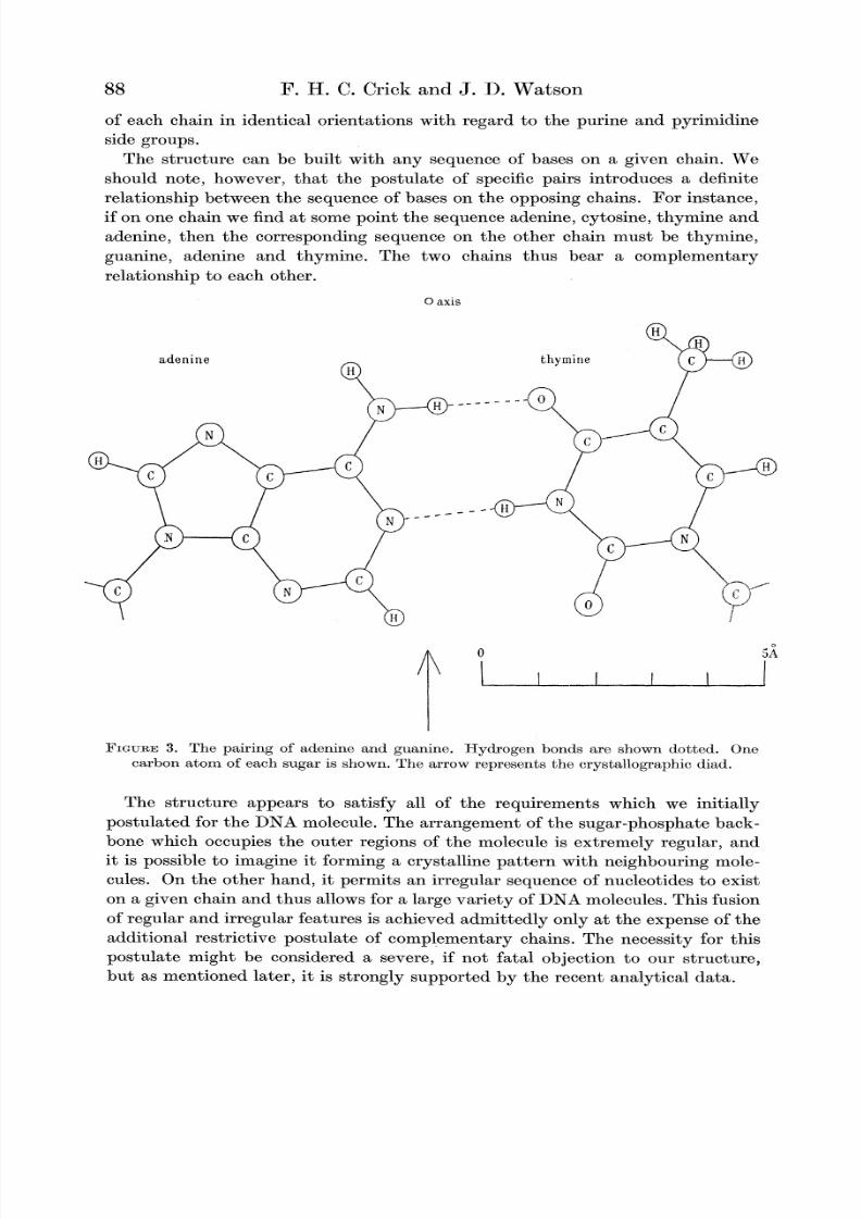
The structure can be built with any sequence of bases on a given chain. We
should note, however, that the postulate of specific pairs introduces a definiterelationship between the sequence of bases on the opposing chains. For instance,
if on one chain we find at some point the sequence adenine, cytosine, thymine and
adenine, then the corresponding sequence on the other chain must be thynmine,
guanine, adenine and thymine. The two chains thus bear a complementary
relationship to each other.
0 axis
adenine thymine
C N
\ I l I IA
FIGURE 3. The pairing of adenine and guanine. Hydrogen bonds are shown dotted. One
carbon atom of each sugar is shown. The arrow represents the crystallographic diad.
The structure appears to satisfy all of the requirements which we initially
postulated for the DNA molecule. The arrangement of the sugar-phosphate back-
bone which occupies the outer regions of the molecule is extremely regular, and
it is possible to imagine it forming a crystalline pattern with neighbouring mole-
cules. On the other hand, it permits an irregular sequence of nucleotides to exist
on agiven
chain and thus allows for alarge variety
of DNA molecules. This fusion
of regular and irregular features is achieved admittedly only at the expense of the
additional restrictive postulate of complementary chains. The necessity for this
postulate might be considered a severe, if not fatal objection to our structure,but as mentioned later, it is strongly supported by the recent analytical data.
88

8/3/2019 Complementary Structure of DNA Proc Roy Soc London a 223,80 1953 Watson and Crick
http://slidepdf.com/reader/full/complementary-structure-of-dna-proc-roy-soc-london-a-22380-1953-watson-and 10/18
The complementary structure of deoxyribonucleic acid
DETAILED CONFIGURATION OF THE DOUBLE HELIX
We shall refer first to the specific pairs of bases. Adenine and thymine are shown
pairedin
figure 3,while
guanineand
cytosineare shown
pairedin
figure4. These
drawings are to scale and have been constructed as far as possible by utilizing bond
angles and bond lengths which have been reported to occur in these compounds.
O axis
guanine cytosine
5A
l I I I I
FIGURE 4. The pairing of guanine and cytosine. Hydrogen bonds are shown dotted. One
carbon atom of each sugar is shown. The arrow represents the crystallographic diad.
The crystal structures of both adenine and guanine have been studied by Broom-
head (1948, 195i), while the structure of cytosine is known through Furberg's
(I950) analysis of the crystal structure of cytidine. More recently Broomhead's
data on adenine have been refined by Cochran (I95I) and the atomic parametersof this compound are now accurate to within 0.02A.
As yet, no determination has been made of the structure of thymine, but it seems
unlikely that its ring configuration will differ markedly from cytosine. Any devia-
tions which might occur would have only a negligible effect on the pairing con-
figuration,and we have utilized the idealized
thymine configurationof
figure3.
We also lack information about the exact angles at the ,f-glycosidic bond. There is
no reason, however, to believe that they should differ significantly from those in
cytidine or in the cyclic adenosine nucleoside studied by Zussman (I953), and theylikewise have been assigned symmetrically.
89

8/3/2019 Complementary Structure of DNA Proc Roy Soc London a 223,80 1953 Watson and Crick
http://slidepdf.com/reader/full/complementary-structure-of-dna-proc-roy-soc-london-a-22380-1953-watson-and 11/18
F. H. C. Crick and J. D. Watson
The configuration of the adenine-thymine pair is stereochemically most satis-
factory. The direction of the vector from the amino nitrogen to the keto oxygenlies exactly in the NH direction, as does the vector from the purine nitrogen atom
1 to the pyrimidine nitrogen atom 1. Both of the hydrogen bonds should thereforebe of maximum stability (Donohue I952). In addition, the two glycosidic bonds
of the pair are related by a diad to within 1?, which is less than the accuracy to
which the configuration of the bases is known. The distance apart of the Crcarbon atoms of the two sugars is close to 11 A.
There is more ambiguity about the guanine-cytosine pair. This arises largelyfrom doubt about the exact structure of guanine (Broomhead 1951). In particular,we are doubtful about the exact position of the keto oxygen atom. In figure 4 we
have used the published position, and this makes the relative positions of the
glycosidicbonds different from the
adenine-thymine pair byabout 2?. This differ-
ence would be negligible if the guanine keto oxygen was symmetrically placed.It is also uncertain as to whether this pair might form a third hydrogen bond
between the amino group of guanine and the keto oxygen of cytosine. This pointis unlikely to be settled until the configurations of both these bases are known to
a greater accuracy. It seems clear, nevertheless, that these uncertainties are
only of second-order importance, and that for all practical considerations the two
pairs should be considered structurally equivalent.The phosphate-sugar backbones were constructed utilizing a sugar configuration
reported for ribose by Furberg (1950). A similar configuration for a pentose ring
has also been reported by Beevers & Cochran (1947) in the fructofuranoside ringof sucrose. It seems probable that the furanose ring is puckered, and we have
tentatively placed the C3, atom out of the ring in such a direction that its oxygenatom 03 is brought closer to the common plane. A tetrahedral arrangement has
been assumed for the bond angles around the phosphorus atom. The bond lengthsabout the phosphorus have been assigned unsymmetrically following the sugges-tion of Pauling & Corey (1953), the two P-0 bonds in the backbone have lengthsof 1-65A while the remaining non ester P-0 bonds are thought to have the
shorter length of 1-45A. As a result of Furberg's analysis of cytidine (I950) there
seems little doubt that the glycosidic bond is a single bond. We can thus be surethat the sugar group instead of being coplanar with the nitrogen base, as postu-lated by Astbury (I947), is more nearly perpendicular to it.
The paired bases are arranged so as to be approximately perpendicular to the
fibre axis. This places the glycosidic bonds in a similar arrangement, while the
puckered plane of the sugar ring assumes a position nearly parallel to the fibre
axis. Each backbone chain completes one revolution after 10 residues in 34 A,and so the rotation per residue is 36?. The phosphorus atoms are at radii of 10A,and the backbone has a configuration roughly similar to that described by Furberg
(I952) in his paper dealing with suitable configurations for single helically arranged
polynucleotide chains.
General views of the structure are shown in the photographs of figures 5 and 6,
plate 2, which illustrate the salient features of a scale model. The drawings in
figures 7 and 8 are given to demonstrate more accurately the exact configuration
90

8/3/2019 Complementary Structure of DNA Proc Roy Soc London a 223,80 1953 Watson and Crick
http://slidepdf.com/reader/full/complementary-structure-of-dna-proc-roy-soc-london-a-22380-1953-watson-and 12/18
Crick & Watson
FIGGURE 5.
Proc. Roy. Soc. A, volume223, plate 2
FIGURE 6.
FIGURE 5. Photograph of a rough scale model of the structure. The chemical bonds in the
phosphate sugar backbone are represented by wire. (All the hydrogen atoms and the
two oxygen atoms of the phosphate group not in ester linkage have been omitted.)The pairs of bases are represented by metal plates. The fibre axis is represented by a
Perspex rod.
FIGURE 6. Another view of the model shown in figure 5. The white plates represent the area
between the bases in which hydrogen bonding takes place.
(F'actng p. 90)

8/3/2019 Complementary Structure of DNA Proc Roy Soc London a 223,80 1953 Watson and Crick
http://slidepdf.com/reader/full/complementary-structure-of-dna-proc-roy-soc-london-a-22380-1953-watson-and 13/18
The complementary tuctureof deoxyribonucleicacid
of the backbone. Figure 7 shows two successive residues on the same chain pro-
jected on to a plane perpendicular to the fibre axis, while in figure 8 is shown a
projection of a sugar-phosphate residue on to a plane whose normal is perpen-dicular to the fibre axis. It can be
seen that the atoms forming the sequenceOaxis
A
-diad
O 5A
J , i I
IFIGUREE 7. A projection of two successive residues of one chain of the structure. The direction
of projection is parallel to the fibre axis. The figures show the height of each atom (in
angstr6ms) above the level of the lower base.
C4,--C5,--O,--P-O3 all lie in such a plane; co-ordinatesof the principalbackboneatoms are given to 0-05 A in table 1. No attempt has been made to place the sodium
ion or the water molecules, though it is possible that some of these groups are
located in relatively constant positions.Because the two backbones are related by a diad, the distance between their
effective 'centres of gravity' is much greater than might be imagined from the
location of the glycosidic bonds. Instead of being separated by only . of the fibre
axis repeat (the angle of the pair of glycosidic bonds is close to 90?), they are
91

8/3/2019 Complementary Structure of DNA Proc Roy Soc London a 223,80 1953 Watson and Crick
http://slidepdf.com/reader/full/complementary-structure-of-dna-proc-roy-soc-london-a-22380-1953-watson-and 14/18
92 F. H. C. Crick and J. D. Watson
separated by approximately 3 of the 34A repeat. In contrast to the outside of
the molecule, the centre tends to give the impression of a one-stranded helix. This
is a consequence of the intimate pairing of the bases.
0 5A
I I' I I I
FIGURE 8. A projection of one residue in a direction perpendicular to both the fibre axis
and to the plane containing the atoms C4,-C,--5--P--O3,.
TABLE 1. CO-ORDINATES FOR THE ATOMS OF THE BACKBONE,
FOR A SINGLE RESIDUE
atom
P
0,IOilOIII
OIvC5,
C4,
Ca,C2,Cl,
01'Ol,N
diad
p (A)
10-08-95
11-25
9-65
10-359.69.65
9-28-65
8-2
8-8
6-7
oe:
0.00
- 3-6?
+ 0-7?
+ 8.9?- 5-3?
-22-2?
- 13-2?
- 7.3?+ 0-4?- 3-5?- 11-8?
- 4-2?
+ 39.0?
Z (A)
0-0
+0-8
+0-8
-0-5-1-3
-2-8
-3-2
-2-05-2.8
-4-15
-4-35-4-15
-4-15
Each of the van der Waals contacts appears to be acceptable. They are five
relatively short contacts between the phosphate oxygen atoms and hydrogenatoms. None, however, is less than 25 A, a quite acceptable length for side-by-side contacts. The position of the plane of the bases with respect to the sugar does
not appear to be the optimum, but it is nevertheless within the range stated by
Furberg as possible. Another short contact is found between the hydrogen atoms
attached to the C3, and C5,atoms of the sugar. This contact, however, is also side
by side, and so the postulated length (2-1A) appears permissible. The stagger of

8/3/2019 Complementary Structure of DNA Proc Roy Soc London a 223,80 1953 Watson and Crick
http://slidepdf.com/reader/full/complementary-structure-of-dna-proc-roy-soc-london-a-22380-1953-watson-and 15/18
The complementary structure of deoxyribonucleic acid 93
hydrogen atoms between the C4-C5, bond is not optimal, but the deviation is
only 25? and so allowable.
We can therefore conclude that the model is stereochemically feasible. Never-
theless, it is certainly not ideal, and it is possible that it could be improved byslightly altering the assumptions made about the configuration of the phosphorus
atoms, especially its bond lengths, and by altering the configuration of the sugar.We have assumed that the puckering of the sugar ring is achieved by throwing the
C3, atom out of the plane of the ring; a better model might result by choosing a
different shape. Alternatively, it may be that an attraction between the ringsof the bases is pulling the backbone out of its potential minimum.
THE CRYSTALLINE FORM
The transition to the crystalline form is accompanied by a decrease in watercontent (Franklin & Gosling 1953a), and it seems very probable that this form
exists in a more tightly packed condition than the paracrystalline form. It is thus
. o . oa
0
I i0
a co v O -
0- 0
b Ib w l
FIGURE 9. To show that if the bases are staggered, tilting will reduce the translation in the
axis direction (represented by a dotted arrow). The solid arrow represents the per-
pendicular distance between the bases, which remains constant. a and b, not staggered;
c and d, staggered; a and c, before tilting; b and d, after tilting.
not surprising to observe that the change to the crystalline state is characterized
by a visual shortening of the fibre length of about 30% (Franklin & Gosling
I953a). There is little if any change in the diameter of the fibre, and so it seems
likely that the fibre axis translation per nucleotide is reduced from 3-4 to approxi-
mately 2-5A. This conclusion might appear difficult to believe, as the van der
Waals separation of the rings of the bases must remain the same and thus might
appear to oppose a fibre shortening, but in fact the vertical translation can be
reduced if thepaired
bases are tilted anti-clockwise(when
viewed firom the
fibre axis).The manner in which this might occur is shown in figure 9. It can be seen that
shortening will only take place if successive pairs of bases are not stacked directlyon top of one another, but are displaced to one side. In fact, if the bases are not

8/3/2019 Complementary Structure of DNA Proc Roy Soc London a 223,80 1953 Watson and Crick
http://slidepdf.com/reader/full/complementary-structure-of-dna-proc-roy-soc-london-a-22380-1953-watson-and 16/18
F. H. C. Crick and J. D. Watson
displaced, tilting will result in an increase of the fibre-axis translation. Of
course, in our structure the successive pairs are displaced helically, not simply
sideways as in figure 9, but this in no way destroys the general argument.
We should note that the hydrogen bonding arrangement remains unchanged bythe tilting, as both members of a pair are similarly rotated about the perpen-dicular diad between the bases. This would not be so if the bases were instead
related by a diad parallel to the fibre axis. In this latter case, the configurationof the backbone could be made equivalent only by tilting the two members of a
pair in opposite directions and thus by effectively destroying the hydrogen bonds.
Thus, if tilting is shown to occur in the crystalline state, we should have strongreasons for believing that the backbones are related by perpendicular diads.
We have not attempted to construct a detailed model with tilted bases, as we
feel that this could be done more suitably in conjunction with the detailed X-rayevidence. Nevertheless, for the reasons outlined above, we believe that such a
model can be built and that it will involve the same basic structural features
proposed here for the paracrystalline form.
DISCUSSION
Our structure bears only superficial resemblances to the majority of structures
previously suggested. Most of these earlier formations (Astbury I947; Furberg
I952) have involved single stranded structures and must be rejected on the basis
of the density considerations outlined in the beginning of this paper. The only
multi-stranded structure which previously has been seriously proposed is that of
Pauling & Corey, who very kindly sent their manuscript to us prior to its publica-tion. Their structure involved three intertwined helical chains in which the core
of the molecule was formed by phosphate groups. Their proposal was submitted
without knowledge of the work at King's College, London, by Wilkins and
Franklin and their co-workers, and appears in the light of their experimentalresults to be untenable. The main objection to their proposal involves the number
of chains. As indicated earlier the density of the crystalline form (Franklin &
Gosling I953c) strongly suggests the presence of two chains, and we find it
difficult to imagine that any three-chained proposal can be made which will fitthe experimental evidence.
The structure accounts in a nice way for the analytical data on the compositionof DNA. By requiring specific pairing of purine and pyrimidine groups, it providesfor the first time a suitable explanation for the recent chemical data (Chargaff
1951; Wyatt 1952; Chargaff, Crampton & Lipschitz 1953), which indicated not
only a molar equivalence of the purines and pyrimidines, but also the molar
equivalence of adenine and thymine, and of guanine and cytosine. The ratio of
adenine to guanine varies greatly in DNA's from different sources, and it is difficult
to imagine a structural explanation for the equivalence of adenine with thymineand of guanine with cytosine which does not involve specific pairing.
As far as we can tell our structure is compatible with the X-ray evidence of
Wilkins and Franklin and their co-workers (Wilkins et al. I953; Franklin &
Gosling 1953 a). In a preliminary report on their work, they have independently
94

8/3/2019 Complementary Structure of DNA Proc Roy Soc London a 223,80 1953 Watson and Crick
http://slidepdf.com/reader/full/complementary-structure-of-dna-proc-roy-soc-london-a-22380-1953-watson-and 17/18
The complementarystructureof deoxyribonucleicacid
suggested that the basic structure of the paracrystalline form is helical and con-
tains two intertwined chains. They also suggest that the sugar-phosphate back-
bone forms the outside of the helix and that each chain repeats itself after one
revolution in 34 A.* Nevertheless, these crystallographic conclusions are tentative,and the structure can in no sense be considered proved until a satisfactory solution
to the structure of the crystalline form is obtained.
In conclusion, we may mention that the complementary relationship between
the two chains is very likely related to the biological role of DNA. It is generallyassumed that DNA is a genetic substance and in some way possesses the capacityfor self-duplication. It seems to us that the presence of a complementary structure
strongly suggests that the self-duplicating process will be found to involve the
alternative formation of complementary chains, and that each chain will be found
capableof
servingas a
templatefor the formation of its
complement.A fuller
exposition of these latter ideas is given elsewhere (Watson & Crick I953 b,c).
We are most indebted to Dr M.H. F. Wilkins both for informing us of unpublished
experimental observations and for the benefit of numerous discussions. We are also
grateful to Dr J. Donohue for constant advice on the problems of tautomerism
and van der Waals contacts, and to Professor A. R. Todd, F.R.S., for advice on
chemical matters, and for allowing us access to unpublished work.
One of us (J. D.W.) wishes in addition to acknowledge the very kind hospitality
provided during his stay at the Cavendish Laboratory by Sir Lawrence Bragg,
F.R.S., and by the members of the Medical Research Council Unit located there.
He is especially grateful to the encouragement provided by Dr J. C. Kendrew and
Dr M. F. Perutz. In conclusion he would like to mention Professor S. E. Luria
of the University of Illinois to whom he is indebted for both the opportunity to
come to and to remain in Cambridge.
REFERENCES
Astbury, W. T. 1947 Symp. Soc. Exp. Biol. 1, Nucleic Acid, p. 66. Cambridge UniversityPress.
Astbury, W. T. & Bell, F. 0. 1938 Nature, London, 141, 747.Beevers, C. A. & Cochran, W. I947 Proc. Roy. Soc. A, 190, 257.
Bijvoet, J. M., Peerdeman, A. F. & van Bommel, A. J. 195I Nature, Lond., 168, 271.
Broomhead, J. M. 1948 Acta Cryst. 1, 324.
Broomhead, J. M. I95I Acta Cryst. 4, 92.
Brown, D. M. & Lythgoe, B. 1950 J. Chem. Soc. p. 1990.
Brown, D. M. & Todd, A. R. 1952 J. Chem. Soc. p. 52.
Chargaff, E. 1951 J. Cell. Comp. Physiol. 38, 41.
Chargaff, E., Crampton, C. F. & Lipschitz, R. 1953 Nature, Lond., 172, 289.
Cochran, W. 1951 Acta Cryst. 4, 81.
Cochran, W., Crick, F. H. C. & Vand, V. 1952 Acta Cryst. 5, 581.
Crane, H. R. I950 Sci. Mon. 70, 376.
* More recently, Franklin & Gosling (I953b) have suggested that the X-ray data for the
crystalline form also supports a structure of this general type. They also mention that the
equatorial reflexions for the paracrystalline form suggest that the diameter of our model isa little too large. Note added in proof: Wilkins, Seeds, Stokes & Wilson (1953) have also
presented X-ray evidence for the crystalline form being a pair of helices.
95

8/3/2019 Complementary Structure of DNA Proc Roy Soc London a 223,80 1953 Watson and Crick
http://slidepdf.com/reader/full/complementary-structure-of-dna-proc-roy-soc-london-a-22380-1953-watson-and 18/18
966 F. H. C. Crickand J. D. Watson. H. C. Crickand J. D. Watson
Dekker, C. R., Michelson, A. M. & Todd, A. R. 1953 J. Chem. Soc. p. 947.
Donohue, J. 1952 J. Phys. Chem. 56, 502.
Franklin, R. E. & Gosling, R. G. i953 a Nature, Lond., 171, 740.
Franklin, R. E. & Gosling, R. G. I953b Nature, Lond., 172, 156.
Franklin, R. E. & Gosling, R. G. 1953c Acta Cryst. 6, 673, 678.Furberg, S. 195o Acta Cryst. 3, 325.
Furberg, S. I952 Acta chem. scand. 6, 634.
Jordan, D. 0. 1951 Progr. biophys. 2, 51.
Kahler, H. & Lloyd, B. J. 1953 Biochim. Biophys. Acta, 10, 355.
Michelson, A. M. & Todd, A. R. I953 J. Chem. Soc. p. 951.
Pauling, L., Corey, R. B. & Branson, H. R. 1951 Proc. Nat. Acad. Sci., Wash., 37, 205.
Pauling, L. & Corey, R. B. 1953 Proc. Nat. Acad. Sci., Wash., 39, 84.
Riley, D. P. & Oster, G. 1951 Biochim. Biophys. Acta, 7, 526.
Sadron, C. 1953 Progr. biophys. 3,
Tipson, R. S. 1945 Advanc. Carbohyd. Chem. 1, 238. New York: Academic Press Inc.
Watson, J. D. & Crick, F. H. C. 1953a Nature, Lond., 171, 737.
Watson, J. D. & Crick, F. H. C. I953b Nature, Lond., 171, 964.Watson, J. D. & Crick, F. H. C. 1953 Cold Spr. Harb. Symp. Quant. Biol. (in the Press).
Wilkins, M. H. F., Gosling, R. G. & Seeds, W. E. 1951 Nature, Lond., 167, 759.
Wilkins, M. H. F., Seeds, W. E., Stokes, A. R. & Wilson, H. R. I953 Nature, Lond., 172,759.
Wilkins, M. H. F., Stokes, A. R. & Wilson, H. R. I953 Nature, Lond., 171, 738.
Williams, R. C. 1952 Biochim. Biophys. Acta, 9, 237.
Wyatt, G. R. 1952 The chemistry and physiology of the nucleus. New York: Academic Press.
Wyatt, G. R. & Cohen, S. S. 1952 Nature, Lond., 170, 846.
Zussman, J. 1953 Acta Cryst. 6, 504.
Structure of microseismic waves: estimation of direction
of approach by comparison of vertical and
horizontal components
BY J. DARBYSHIRE
National Institute of Oceanography
(Communicated by G. E. R. Deacon, F.R.S.-Received 25 September 1953-Read 11 March 1954)
Analysis of microseisms recorded at Kew Observatory on 8 to 10 October 1951 affords
further confirmation of the wave-interference theory of microseism generation, and allowsthose of 8 to 10 October to be attributed to a fast-moving depression between the Azoresand Iceland.
Although the bearing of the microseism-generating area changes by more than 90? duringthe period investigated, there is no appreciable difference in the ratio of the mean ampli-tudes of the north-south and east-west horizontal components as would be expected if themicroseisms consisted entirely of Rayleigh waves. An investigation of the phase differences
between the three components, using Lee's method, suggests that the microseisms consist of
Rayleigh and Love waves in comparable proportions. Making use of this assumption, the
vertical component, which is not affected by the Love waves, is correlated with the twohorizontal components with an electronic correlating device, and the bearing of the micro-
seism area can be deduced from the correlation coefficients. The calculated bearings agreereasonably well with those obtained from the meteorological charts.
The bearing of a storm on 12 to 15 November 1945, studied in a previous paper, was also
calculated satisfactorily.