complexformationofcadmiumwithsugarresidues,nucleobases
TRANSCRIPT

Zurich Open Repository andArchiveUniversity of ZurichMain LibraryStrickhofstrasse 39CH-8057 Zurichwww.zora.uzh.ch
Year: 2013
Complex formation of cadmium with sugar residues, nucleobases,phosphates, nucleotides, and nucleic acids
Sigel, Roland K O ; Skilandat, Miriam ; Sigel, Astrid ; Operschall, Bert P ; Sigel, Helmut
Abstract: Cadmium(II), commonly classified as a relatively soft metal ion, prefers indeed aromatic-nitrogen sites (e.g., N7 of purines) over oxygen sites (like sugar-hydroxyl groups). However, mattersare not that simple, though it is true that the affinity of Cd(2+) towards ribose-hydroxyl groups isvery small; yet, a correct orientation brought about by a suitable primary binding site and a reducedsolvent polarity, as it is expected to occur in a folded nucleic acid, may facilitate metal ion-hydroxylgroup binding very effectively. Cd(2+) prefers the guanine(N7) over the adenine(N7), mainly because ofthe steric hindrance of the (C6)NH(2) group in the adenine residue. This Cd(2+)-(N7) interaction in aguanine moiety leads to a significant acidification of the (N1)H meaning that the deprotonation reactionoccurs now in the physiological pH range. N3 of the cytosine residue, together with the neighboring(C2)O, is also a remarkable Cd(2+) binding site, though replacement of (C2)O by (C2)S enhances theaffinity towards Cd(2+) dramatically, giving in addition rise to the deprotonation of the (C4)NH(2)group. The phosphodiester bridge is only a weak binding site but the affinity increases further from themono- to the di- and the triphosphate. The same also holds for the corresponding nucleotides. Complexstability of the pyrimidine-nucleotides is solely determined by the coordination tendency of the phosphategroup(s), whereas in the case of purine-nucleotides macrochelate formation takes place by the interactionof the phosphate-coordinated Cd(2+) with N7. The extents of the formation degrees of these chelatesare summarized and the effect of a non-bridging sulfur atom in a thiophosphate group (versus a normalphosphate group) is considered. Mixed ligand complexes containing a nucleotide and a further mono- orbidentate ligand are covered and it is concluded that in these species N7 is released from the coordinationsphere of Cd(2+). In the case that the other ligand contains an aromatic residue (e.g., 2,2’-bipyridineor the indole ring of tryptophanate) intramolecular stack formation takes place. With buffers like Trisor Bistris mixed ligand complexes are formed. Cd(2+) coordination to dinucleotides and to dinucleosidemonophosphates provides some insights regarding the interaction between Cd(2+) and nucleic acids.Cd(2+) binding to oligonucleotides follows the principles of coordination to its units. The availablecrystal studies reveal that N7 of purines is the prominent binding site followed by phosphate oxygens andother heteroatoms in nucleic acids. Due to its high thiophilicity, Cd(2+) is regularly used in so-calledthiorescue experiments, which lead to the identification of a direct involvement of divalent metal ions inribozyme catalysis.
DOI: https://doi.org/10.1007/978-94-007-5179-8_8
Posted at the Zurich Open Repository and Archive, University of ZurichZORA URL: https://doi.org/10.5167/uzh-79639Book SectionAccepted Version

Originally published at:Sigel, Roland K O; Skilandat, Miriam; Sigel, Astrid; Operschall, Bert P; Sigel, Helmut (2013). Complexformation of cadmium with sugar residues, nucleobases, phosphates, nucleotides, and nucleic acids. In:Sigel, Astrid; Sigel, Helmut; Sigel, Roland K O. Cadmium: From Toxicity to Essentiality. Dordrecht:Springer, 191-274.DOI: https://doi.org/10.1007/978-94-007-5179-8_8
2

1
Chapter 8
Complex Formation of Cadmium with Sugar Residues, Nucleobases,
Phosphates, Nucleotides, and Nucleic Acids
Roland K. O. Sigel, Miriam Skilandat, Astrid Sigel, Bert P. Operschall, and Helmut Sigel
Contents
ABSTRACT
1 INTRODUCTION
2 COMPARISONS OF THE PROPERTIES OF CADMIUM(II) WITH THOSE OF ZINC(II),
CALCIUM(II), MAGNESIUM(II), AND OTHER RELATED METAL IONS
3 CADMIUM(II)-SUGAR INTERACTIONS
3.1 Hydroxyl Coordination in Carboxyhydrates Is Rare
3.2 The Metal Ion Affinity of Ribose-Hydroxyl Groups Is Small
3.3 A Favorable Steric Setting and a Reduced Solvent Polarity May Promote Metal Ion-
Hydroxyl (or Carbonyl) Group Binding
4 INTERACTIONS OF CADMIUM(II) WITH NUCLEOBASE RESIDUES
4.1 Cadmium(II) Complexes of Purine Derivatives
4.2 Cadmium(II) Complexes of Pyrimidine Derivatives
4.3 Cadmium(II) Complexes of Some Less Common Nucleobase Residues
4.3.1 Tubercidin _______________________________________________ R. K. O. Sigel ()) ∙ M. Skilandat Institute of Inorganic Chemistry, University of Zürich, Winterthurerstrasse 190, CH-8057 Zürich, Switzerland e-mail: [email protected]
A. Sigel · B. P. Operschall · H. Sigel ()) Department of Chemistry, Inorganic Chemistry, University of Basel, Spitalstrasse 51, CH-4056 Basel, Switzerland e-mail: [email protected]; [email protected]; [email protected] ___________________________________________________ A. Sigel, H. Sigel, and R. K. O. Sigel, Cadmium: From Toxicity to Essentiality
Metal Ions in Life Sciences 11 (2013) xxx–yyy DOI:
Springer Science & Business Media BV, 2013

2
4.3.2 Orotidine
4.3.3 Xanthosine
4.3.4 Thiouridines
4.3.5 2-Thiocytidine
5 COMPLEXES OF CADMIUM(II) WITH PHOSPHATES
6 CADMIUM(II) COMPLEXES OF NUCLEOTIDES
6.1 Some General Considerations
6.2 Complexes of Nucleoside 5'-Monophosphates
6.2.1 Equilibrium Constants to Be Considered
6.2.2 Properties of Pyrimidine-Nucleoside 5'-Monophosphate Complexes
6.2.3 Properties of Purine-Nucleoside 5'-Monophosphate Complexes
6.3 Complexes of 5'-Di- and Triphosphates
6.4 Complexes of Less Common Nucleotides
6.4.1 Tubercidin 5'-Monophosphate
6.4.2 Nucleoside 2'- and 3'-Monophosphates
6.4.3 Orotidinate 5'-Monophosphate
6.4.4 Xanthosinate 5'-Monophosphate
6.4.5 Thiouracil Nucleotides
6.4.6 Flavin Mononucleotide
7 CADMIUM(II) COMPLEXES OF NUCLEOTIDE ANALOGUES
7.1 Properties of 1,N6-Ethenoadenosine and of Its Phosphates
7.2 Complexes of Nucleoside 5'-O-Thiomonophosphates
7.3 Complexes of Acyclic Nucleotide Analogues
7.4 Cadmium(II) Binding to Nucleotides Containing a Platinum(II)-Coordinated
Nucleobase Residue
8 A SHORT APPRAISAL OF MIXED LIGAND COMPLEXES CONTAINING A
NUCLEOTIDE
8.1 Definitions and Some General Comments
8.2 Ternary Cadmium(II) Complexes Containing ATP4– and a Buffer Molecule

3
8.3 Mixed Ligand Complexes Containing a Nucleotide and a Further Monodentate or
Bidentate Ligand. Release of Purine-N7 and Formation of Stacks
9 CADMIUM(II) BINDING IN DINUCLEOTIDES AND DINUCLEOSIDE
MONOPHOSPHATES
9.1 The Phosphodiester Link
9.2 The Guanine Residue in a Dinucleotide
9.3 The Non-bridging Sulfur of the Thiophosphodiester Link
9.4 Dinucleoside Monophosphates
10 CADMIUM(II) BINDING TO NUCLEIC ACIDS
10.1 Cadmium(II)-Rescue Experiments
10.2 Crystal Structures of RNA or DNA-Protein Complexes Containing Cd2+
10.3 Cadmium(II)-Induced EPR Silencing
11 CONCLUDING REMARKS
ABBREVIATIONS AND DEFINITIONS
ACKNOWLEDGMENTS
REFERENCES
Abstract Cadmium(II), commonly classified as a relatively soft metal ion, prefers indeed
aromatic-nitrogen sites (e.g., N7 of purines) over oxygen sites (like sugar-hydroxyl groups).
However, matters are not that simple, though it is true that the affinity of Cd2+ towards ribose-
hydroxyl groups is very small; yet, a correct orientation brought about by a suitable primary
binding site and a reduced solvent polarity, as it is expected to occur in a folded nucleic acid,
may facilitate metal ion-hydroxyl group binding very effectively. Cd2+ prefers the guanine(N7)
over the adenine(N7), mainly because of the steric hindrance of the (C6)NH2 group in the
adenine residue. This Cd2+-(N7) interaction in a guanine moiety leads to a significant
acidification of the (N1)H meaning that the deprotonation reaction occurs now in the
physiological pH range. N3 of the cytosine residue, together with the neighboring (C2)O, is also
a remarkable Cd2+ binding site, though replacement of (C2)O by (C2)S enhances the affinity
towards Cd2+ dramatically, giving in addition rise to the deprotonation of the (C4)NH2 group.

4
The phosphodiester bridge is only a weak binding site but the affinity increases further from the
mono- to the di- and the triphosphate. The same also holds for the corresponding nucleotides.
Complex stability of the pyrimidine-nucleotides is solely determined by the coordination
tendency of the phosphate group(s), whereas in the case of purine-nucleotides macrochelate
formation takes place by the interaction of the phosphate-coordinated Cd2+ with N7. The extent
of the formation degrees of these chelates are summarized and the effect of a non-bridging sulfur
atom in a thiophosphate group (versus a normal phosphate group) is considered. Mixed ligand
complexes containing a nucleotide and a further mono- or bidentate ligand are covered and it is
concluded that in these species N7 is released from the coordination sphere of Cd2+. In the case
that the other ligand contains an aromatic residue (e.g., 2,2'-bipyridine or the indole ring of
tryptophanate) intramolecular stack formation takes place. With buffers like Tris or Bistris mixed
ligand complexes are formed. Cd2+ coordination to dinucleotides and to dinucleoside
monophosphates provides some insights regarding the interaction between Cd2+ and nucleic
acids. Cd2+ binding to oligonucleotides follows the principles of coordination to its units. The
available crystal studies reveal that N7 of purines is the prominent binding site followed by
phosphate oxygens and other heteroatoms in nucleic acids. Due to its high thiophilicity, Cd2+ is
regularly used in so-called thiorescue experiments, which lead to the identification of a direct
involvement of divalent metal ions in ribozyme catalysis.
Keywords cadmium · calcium · equilibrium constants · magnesium · metal ions · methods ·
ribozymes · RNA · zinc
1 Introduction
Cadmium is widely distributed in the environment at relatively low concentrations, except where
it accumulated due to anthropogenic activities [1] (see also Chapters 2 and 3 of this book).
Cadmium chemically resembles zinc and any differences are attributable to the larger size of
Cd2+ compared with that of Zn2+ [2]. Indeed, cadmium occurs in the earth's crust and the upper

5
lithosphere mainly together with zinc (zinc being present to ca. 0.02% [3]). The Cd/Zn ratio has
been estimated to be about 1:250 [4], and cadmium is thus gained as by-product from zinc ores
(Chapter 3).
Cadmium is a toxic element (see Chapters 1, 14, 15) that accumulates especially in kidney
and liver [4] being bound preferably to metallothionein (Chapters 6, 11). On the other hand, the
chemical similarity of Cd2+ to Zn2+ is confirmed by the fact that carbonic anhydrase of marine
phytoplankton contains Cd2+ (Chapter 16), whereas the corresponding zinc enzymes are found in
organisms from all kingdoms [5] catalyzing the reversible hydration of carbon dioxide. In marine
diatoms cadmium, cobalt, and zinc can functionally substitute for one another to maintain
optimal growth [6]. Cadmium-carbonic anhydrase is involved in the acquisition of inorganic
carbon for photosynthesis [6].
Interestingly, already more than 50 years ago Wacker and Vallee detected cadmium as well
as other metal ions in RNA from horse kidney cortex [7], but there is no indication for a
"positive" role of any cadmium-nucleic acid interaction. However, nowadays Cd2+ is often
applied as a probe to study the effects of metal ions on ribozymes [8,9] as well as the metal ion-
binding properties of nucleic acids in general [10]. With this general analytical use in mind, this
chapter has been written, concentrating on Cd2+ and several related metal ions (see Section 2) as
well as on the nucleobases (NB) and their derivatives which are important for RNA and DNA
(Figure 1) [11–14]. The four main nucleobases of RNA are adenine (Ade), guanine (Gua),
insert Figure 1 close to here (width: 8 cm)
cytosine (Cyt), and uracil (Ura); in DNA uracil is replaced by thymine (Thy). Hypoxanthine
(Hyp) is included for comparisons with guanine. Below we will first consider some
physicochemical properties of Cd2+ and related metal ions. Next, the interaction of these metal
ions with sugar, phosphate, and nucleobase residues will be addressed, followed by nucleotides
and nucleic acid complexes.
2 Comparisons of the Properties of Cadmium(II) with Those of Zinc(II),
Calcium(II), Magnesium(II), and Other Related Metal Ions

6
Despite its essentiality for marine diatoms [5,6], cadmium is best known for its toxicity to
mammals [1,4,15,16] and in this context it is interesting to consider the interdependencies
between Cd2+ and other elements. In Figure 2, which may not be complete, the most obvious
interdependencies with Cd2+ are shown [17]. An arrow from element A to B, A→B, indicates
that administration of element A may reduce toxicity due to element B. Hence, the toxicity of
Cd2+ may be reduced by the ions of Ca, Zn, Cu, Fe, Co, and the metalloid Se. However, low
levels of element A, e.g., of Ca2+, Zn2+, and Cu2+, may increase the toxicity of element B (Cd2+),
or high levels of element B (Cd2+) may inhibit salutary effects of element A (Zn2+). Such
interrelations are common, though not easy to reveal and to understand.
insert Figure 2 close to here (width: 6 cm)
Ignoring strontium and lead from the nine metals shown in Figure 2, we are left with the
essential divalent metal ions Mg2+, Ca2+, Fe2+, Co2+, Zn2+, and Cu2+, and the toxic Cd2+. Cd2+,
larger than Zn2+, has nearly the size of Ca2+ (see below) and this has led to its use as Ca2+ probe
[15,18] (see also Chapter 6). However, in its binding strength to ligands Cd2+ is more like Zn2+
(see below) and thus, it is employed as a Zn2+ probe as well [15] (Chapter 6). Although Cd2+ has
a larger ionic radius than Mg2+, it has recently been widely applied to mimic Mg2+ in ribozymes
[8,19]. Like Mg2+, Cd2+ preferentially forms non-distorted octahedral complexes, and it
selectively replaces Mg2+ bound to purine-N7 sites via an innersphere coordination mode [19].
Based on the mentioned observations, we focus now on the properties of Cd2+ in
comparison to those of Zn2+, Ca2+, and Mg2+ as well as their interactions with the bio-ligands
relevant for nucleic acids as considered in this chapter. The properties and complexes of Fe2+,
Co2+, and Cu2+ are to some extent taken into account as well, to allow, where needed, more
systematic-type comparisons. With this in mind the content of Table 1 has been assembled [20–
27].
insert Table 1 close to here (landscape)
Columns 2 to 4 of Table 1 list the coordination numbers of the divalent metal ions
considered together with their corresponding ionic radii. Evidently, the radii of Zn2+ and Mg2+

7
are very similar, as are those of Cd2+ and Ca2+. The alkaline earth ions like to bind to oxygen
donors (column 6) whereas the 3d metal ions as well as Zn2+ and Cd2+ have a preference for N
sites, especially heteroaromatic amines, as we will see later. Among all these metal ions Cd2+ has
the highest affinity for thiolate sites [15,17], which is of relevance for so-called rescue
experiments [8,28] (see Sections 9.3 and 10.1).
Column 6 of Table 1 provides the acidity constants (pKa/aq) for the hydrolysis reaction (1),
charges being omitted:
M(H2O)n M(OH)(H2O)n–1 + H+ (1a)
K
M(H2O)nH = = Ka/aq (1b)
This hydrolysis reaction leads to hydroxo complexes and the coordinated OH– species can act as
nucleophiles [24,29] or participate in general base catalysis [24], important for metal ion-
containing catalytic cores of ribozymes, where also large pKa shifts can occur [30]. The pKa/aq
values of Table 1 show that in a simple aqueous solution at neutral pH all metal ions listed are
present as M2+ ions, except Cu2+, which forms a CuO precipitate [15]; of course, such a solid
does not occur in plasma where Cu2+ is complexed by proteins [15].
Columns 7 and 8 of Table 1 provide the stability constants as defined by equation (2), of
simple acetate, CH3COO–, and ammonia, NH3, complexes (charges omitted):
M2+ + L M(L) (2a)
KM(L)
M = [M(L)]/([M2+][L]) (2b)
At first sight the M(NH3)2+ complexes of the transition elements seem to be much more stable
than those of the corresponding M(CH3COO)+ complexes. However, in the physiological pH
range around 7.5 one needs to take into account the competition between M2+ and H+ for binding
at the ligand, that is, the size of the acidity constant of the protonated ligands is important. The
acidity constants are defined by equation (3) (charges omitted):
H(L) L + H+ (3a)
KH(L)
H = [L][H+]/[H(L)] = Ka (3b)
Considering that the pKa of acetic acid, CH3COOH, is close to 4.6 and the one of the ammonium

8
ion, NH , close to 9.4, it is evident that at pH ca. 7.5 the acetate ion is freely accessible for M2+
binding, whereas NH3 exists overwhelmingly in the form of the NH ion. The competition
between M2+ and H+ for binding at the ligand can be accounted for by defining so-called
conditional or apparent (app) stability constants, which then hold only for a given pH. This
constant, KM(L)app
M , depends on the acidity constant of the ligand (eq. 3) and the stability constant
of the complex (eq. 2) and is defined by equation (4) [15,22]:
KM(L)app
M = KM(L)
M
1
1 + [H+ ]/KH(L)H
(4a)
= KM(L)
M
KH(L)H
[H+ ] + KH(L)H
(4b)
log KM(L)app
M = log KM(L)
M + log [ KH(L)
H /([H+] + KH(L)
H )] (4c)
For pH >> pKH(L)
H the unbound ligand is predominantly in its basic form and log KM(L)app
M = log
KM(L)
M . Examples are neutral solutions of carboxylic acids with pKa ca. 4 to 5. For pH << pKH(L)
H
the unbound ligand is predominantly protonated and equation (4c) reduces to log KM(L)app
M = log
KM(L)
M – pKH(L)
H + pH. An example for neutral solutions is the ammonium side chain of aliphatic
amino acids with pKa ca. 9.5 to 10. When the pH is within ±2 log units of pKa, the complete
equation (4) should be employed.
With the above reasonings in mind, columns 7 to 9 of Table 1 should be compared: For
acetate pH = 7.5 >> pKH(Ac)
H = 4.56 [25], that is, virtually all ligand is in its basic CH3COO–
form and the apparent and conventional stability constants are equal. For ammonia this is
different; at pH 7.5 only about 1.3% of the ligand is present in its free NH3 form but 98.7% exist
as NH . Consequently, log K
M(NH3 )appM < log
K
M(NH3 )M ; in fact, only for Cu2+ it holds log
KM(Ac)
M < log K
M(NH3 )appM ; for all other metal ions the M(Ac)+ complexes are more stable than
the M(NH3)2+ species.
The above lesson is of relevance for nucleic acids and their constituents. Considering that
the monoprotonated phosphate groups of nucleotides have pKa values of about 6.2 to 6.5 [31,32],
the competition of the proton is not very pronounced at the physiological pH of about 7.5 and for
RO–P(O) –OR' bridges of nucleic acids (pKa ca. 1 [33,34]) no proton competition exists at all.
This is also true for the (N3)H+ sites of cytidine residues (pKa ca. 4.3 [31,35]), the (N1)H+ sides

9
of adenosines (pKa ca. 3.8 [31,36]) as well as the (N7)H+ of guanosines (pKa ca. 2.5 [31,37,38]),
but not for the (N1)H units of guanosines with pKa values of about 9.4 [31,37,38]. In these latter
cases a strong competition for binding at (N1)– between the proton and metal ion exists. These
general considerations will be reflected in the discussions to follow.
3 Cadmium(II)-Sugar Interactions
3.1 Hydroxyl Coordination in Carboxylates Is Rare
Knowledge on binding of metal ions to carbohydrates is scarce [32,39,40] and little information
exists on Cd2+. Ca2+ binding to neutral monosaccharides is very weak unless they form a
favorable tridentate disposition of three hydroxyl groups [18]. The same may be surmised for
Cd2+, though it has been concluded based on crystal structure studies that sugar-hydroxyl groups
are good ligands for alkaline earth ions but not for transition and heavy metal ions [41]. The
reason for this conclusion is most likely that it is based on solid-state studies of nucleosides (and
derivatives) and there the N-sites become important for the latter type of metal ions (vide infra),
but still there is a polymeric Cd2+/5'-IMP2– complex with a M–O2'/3' chelation [11]: This
polymer consists of [Cd2(IMP·H)2(IMP)(H2O)6]·6H2O units with two independent Cd2+ ions,
one of which binds two ribose oxygen atoms, a purine-N7, and three water molecules [42]. There
are also indications that in M(D-fructose)X2·4H2O (X = Cl– or Br–) species, where M2+ = Mg2+,
Zn2+ or Cd2+, M2+ binds O2 and O3 of D-fructose [43].
Metal ion binding to sugars is strengthened when a suitable primary binding site is
provided, e.g., a carboxylate group [18]. Similarly, potentiometric and spectroscopic studies in
aqueous solution (25°C; I = 0.15 M, KNO3) indicate that with 2-amino-2-deoxy-D-mannose
metal ions bind not only to the primary amino site but also to the hydroxyl group (O3)H [44]:
Cu2+, Ni2+, and Co2+ form 5-membered chelates and the same may be surmised for Cd2+. With 2-
amino-2-deoxy-D-glucose the complexes are less stable, which is in accord with the less
favorable arrangements of the hydroxyl groups [44]. Similarly, metal ions coordinate initially via

10
a 6-membered chelate to 1,3-diamino-2-propyl α-D-mannopyranoside and these coordinated
metal ions should then, at least in theory, be able to interact with a hydroxyl group forming an 8-
membered chelate, which, however, is not observed for the complexes of Cu2+, Ni2+, and Zn2+
[45].
3.2 The Metal Ion Affinity of Ribose-Hydroxyl Groups Is Small
How is the situation with the ribose and 2-deoxyribose residues, which are of significance for the
nucleoside derivatives considered herein? The cis arrangement of the 2- and 3-hydroxyl groups
as present in a ribose moiety favors deprotonation of one of the two OH groups because in the
resulting anion intramolecular hydrogen bond formation occurs [34]. Yet, this favored
deprotonation with pKa = 12.5 is far above the physiological pH range, meaning that such a
deprotonation can occur in a biological system only in a very special environment [30].
However, e.g., it can be facilitated by metal ions like Cu2+ which is apparently able to bind to the
cis-glycol unit of a ribose moiety in aqueous solution at high pH values [43,46,47], as proven in
experiments with adenosine. In contrast, 2'-deoxyadenosine shows no deprotonation of the 3'-
hydroxyl group under the corresponding conditions. Since the acidifying power of Cd2+ is much
smaller than the one of Cu2+ (cf. the pKa/aq values in Table 1, column 6), Cd2+ is not expected to
achieve this type of binding.
In this context also a stability constant study in aqueous solution (25°C; I = 0.1 M, NaNO3)
of complexes formed with 2'AMP2– and 3'AMP2– is of relevance [48]. The complex stability of
Cu(2'AMP) is enhanced by 0.25 log unit compared to the stability expected for a sole phosphate
coordination; the stability of the Cu(3'AMP) complex is only very slightly enhanced, if at all.
The different stability enhancements point to different structural properties of the two ligands. In
case 7-membered chelates were formed by coordination of the phosphate-bound Cu2+ with the
neighboring hydroxyl group, the situation in 2'AMP2– and 3'AMP2– would be equivalent and the
same stability enhancement would be expected. Hence, a significant hydroxyl group interaction
needs to be ruled out and this leaves as the only explanation of the observed results an interaction
of Cu2+ in Cu(2'AMP) with N3 of the purine residue giving rise to an 8-membered macrochelate.

11
Indeed, space-filling molecular models indicate that 2'AMP2– in its preferred anti
conformation is perfectly suited for this type of macrochelate formation [48]. Furthermore, a
crystal structure study [49] shows that Mg2+ is coordinated in Mg(2'AMP) to the phosphate
group and that it interacts in addition in an outersphere manner, i.e., via a water molecule, with
N3 of the adenine residue. Finally, among the 10 metal ions studied in solution, only Ni2+, Zn2+,
and Cd2+ are likely to form small amounts of base-backbound species [50]; the stability
enhancements for their M(2'AMP) complexes are just at the edge of significance [48]. No
stability enhancement is observed for Cd(3'AMP) [48].
3.3 A Favorable Steric Setting and a Reduced Solvent Polarity May Promote
Metal Ion-Hydroxyl (or -Carbonyl) Group Binding
More insight into Cd2+ binding to hydroxyl (and carbonyl) groups in solution encompassing the
neutral pH range and having a phosphate group as a primary binding site, can be gained by
considering the metal ion-binding properties of the keto-triose derivative dihydroxacetone
phosphate (DHAP2–) and the other three related compounds shown in Figure 3. The combination
insert Figure 3 close to here (width: 8 cm)
of coordinating groups seen at C1 and C2 for DHAP2– and glycerol 1-phosphate (G1P2–) is
representative for many sugar moieties. From a steric point of view, an interaction of a
phosphate-coordinated metal ion with the neighboring keto or hydroxyl group is very well
possible in both instances. The questions are: Does such an interaction occur in aqueous
solution? Are 7-membered chelates formed as expressed in a simplified manner (with charge
neglection) in equilibrium (5)?
(5)
Any kind of chelate formation has to enhance complex stability [51]. Hence, a possibly
increased stability, defined as log ∆M/L, of Cd(DHAP) or Cd(G1P), if compared with a pure

12
phosphate coordination, could therefore be attributed to the participation of the oxygen at C2 in
Cd2+ binding, i.e., equilibrium (5) would then truly exist and at least in part be on its right side.
However, the results log ∆Cd/DHAP = 0.02 ± 0.05 and log ∆Cd/G1P = –0.02 ± 0.06 (25°C; I = 0.1 M,
NaNO3) [52,53] are both zero within their error limits, and thus, no increased complex stability
is observed and it must therefore be concluded that in aqueous solution equilibrium (5) is on its
left side and that the (C2)=O and (C2)–OH groups do not participate in Cd2+ binding.
Corresponding results have been obtained for the complexes of Co2+, Cu2+, and Zn2+ [52,53].
However, it needs to be added that a decreased solvent polarity is expected to increase the
affinity in general, as has been shown to occur also in larger RNAs [54], and to favor weakly
coordinating oxygen sites [30]. Indeed, for Cu(DHAP) and Cu(G1P) it has been shown that in
water containing 50% 1,4-dioxane (v/v) the chelated species in equilibrium (5) reach a formation
degree of about 45% [52,53]. A similar chelate formation must be anticipated for the
corresponding Cd2+ complexes in solutions with a reduced dielectric constant or permittivity (ε).
It is worthwhile to note that in the discussed examples the hydroxyl and carbonyl groups behave
within the error limits quite alike.
A change in size of the potential 7-membered chelate ring (eq. 5) to a 6-membered one,
demonstrates the importantce of the steric orientation for weak interactions. Acetyl phosphate
(AcP2–) and acetonylphosphonate (AnP2–) may form with metal ions 6-membered chelates as is
show in equilibrium (6), where X = O (AcP2–) or CH2 (AnP2–):
(6)
In fact, for Cd(AcP) and Cd(AnP) small, but significant, stability enhancements are observed,
i.e., log ∆Cd(AcP = 0.19 ± 0.06 and log ∆Cd/AnP = 0.18 ± 0.06, respectively [52,55]. These stability
enhancements correspond to a formation degree of about 35% for the chelate in the
intramolecular equilibrium (6); the interrelation between log ∆M/L and % M(L)cl will be presented
in Section 4.2 [51]. For now it is enough to add that the formation degree of the chelated species
is hardly affected in mixed ligand complexes as is evidenced from examples with Cu(Arm)2+,
where Arm = 2,2'-biypridine or 1,10-phenanthroline [52,56].

13
From the information presented above it follows that sugar hydroxyl (or carbonyl) groups
are weak binding sites which will interact with Cd2+ (or other M2+) in aqueous solution only
under rather specific conditions. Indeed, from comparison of the results to be discussed in
Section 6.2.3 for M(NMP) complexes, where NMP2– = nucleoside 5'-monophosphate, with the
above data it follows that N7 of purine-nucleobase residues has a more pronounced affinity for
Cd2+ than a sugar hydroxyl group. On the other hand, the ligands shown in Figure 4, which
contain different primary binding sites next to a hydroxyl group, allowing formation of 5-
membered chelates, provide some further insights into hydroxyl group coordination as relevant
for nucleic acids.
insert Figure 4 close to here (width: 8 cm)
The experimentally measured stability constants (eq. 2), the stability enhancements, log
∆M/L, and the percentages of the closed isomers, % M(L)cl (in analogy to equilibria 5, 6) are
listed in Table 2 [57] for the 1:1 complexes of several metal ions with the three ligands seen in
Figure 4.
insert Table 2 close to here (landscape)
Unfortunately, not all desired data, especially for the Cd2+ complexes are available, but enough
to draw a number of conclusions. Also, part of the data available for the complexes of
hydroxyacetate refer to the rather high ionic strength of 2 M. Fortunately, the change in I from
0.1 to 2 M affects the overall stability constant (eq. 2), especially in the case of Zn(HOAc)+, but
has no remarkable influence on log ∆M/HOAc and % M(HOAc) , as is proven by the results for
Cu2+ and Zn2+. Consequently, all the listed values in Table 2 for the stability enhancements and
the percentages of the chelated isomers can directly be compared with each other. Many
comparisons are possible, a few follow below:
(i) The possibility to form 5-membered chelates is evidently a favorable setting; metal ion-
hydroxyl group interactions occur in all instances. By taking also the preceding results into
account, it follows that the stability of the chelates decreases with increasing ring size in the
order 5-membered > 6-membered > 7-membered ring.
(ii) There is no correlation between the global stability of a complex (eq. 2) and the formation

14
degree of the chelated isomer, which is the result of an intramolecular and thus concentration-
independent equilibrium (see eqs. 5, 6). For example, Ca(HOAc)+ is less stable than Cd(HOAc)+,
yet the formation degree of the closed isomer is significantly larger in Ca(HOAc)+.
(iii) However, there is a correlation between the charge of the primary binding site and the
extent of chelate formation; the percentages of the closed forms increase in the order M(HMP)cl
< M(HOAc) < M(HOMPy) . This is a reflection of charge neutralization at M2+ leading to a
reduced affinity of the metal ion towards hydroxyl groups [57].
(iv) Point (iii) has an interesting bearing for nucleic acids: It allows the conclusion that a metal
ion coordinated to the singly negatively charged phosphodiester bridge is better suited for a
hydroxyl group interaction than a metal ion bound to a twofold negatively charged terminal
phosphate group. Regarding ribozymes this result is revealing. It may be added that the acetate
ion, CH3COO–, may be considered as a mimic of the phosphodiester unit, RO–P(O) –OR' in the
nucleic acid backbone as far as metal ion coordination is concerned [58] (see also Section 5).
(v) Similarly, the fact that pyridine-type nitrogens are ideal primary binding sites is in
agreement with the suggested chelate formation in Cu(2'AMP) involving N3 of the adenine
residue and the (C2)OH group (see Section 3.2). Note, the basicity, and thus the metal ion
affinity, of N3 should not be underestimated: The micro acidity constant for the (N3)H+ site in
otherwise neutral adenosine was estimated as pka/(N3)H = 1.5 ± 0.3 [36]; such a value is ideal for
outersphere interactions [59–61], e.g., with Mg2+ (see the discussed solid-state structure of
Mg(2'AMP) in Section 3.2).
(vi) The stability enhancements observed for the Cd(L) species are about half the size of those
found for the corresponding Zn(L) complexes; consequently, it holds % Cd(L)cl < % Zn(L)cl.
Yet, here a caveat is needed: If ligands are synthesized with a pocket that fits well the size of
Cd2+ (but not of Zn2+), then the situation towards hydroxyl group interactions may change
dramatically [57]. Clearly, a nucleic acid cavity fitting the size of Ca2+ is expected to be also
ideal for Cd2+. Some ribozymes show a distinct specificity for Ca2+, e.g., the antigenomic HDV
ribozyme [62], group I introns [63–65], and group II introns, the latter being severely hampered
in catalysis [66] and folding [67,68].
(vii) The results given in the lower part of Table 2 confirm the earlier conclusions (see the

15
discussed Cu(DHAP) and Cu(G1P) complexes [52,53]) that a reduced solvent polarity favors
M2+-hydroxyl interactions.
(viii) A final point, which follows by taking the results of the whole section as well as further
data [57] into account, is that, assuming a suitable primary binding site is present, the metal ion
affinity to hydroxyl and carbonyl groups is quite alike, but the one towards ether oxygen atoms is
much smaller, e.g., the stability enhancement for the Cu2+ complex of methoxyacetate, CH3OAc–
= CH3OCH2COO–, amounts only to log ∆Cu/CH3OAc = 0.36 ± 0.11 [57] compared to log ∆Cu/HOAc =
0.79 ± 0.08 (Table 2).
4 Interactions of Cadmium(II) with Nucleobase Residues
The common purine and pyrimidine nucleobases are shown in Figure 1. We will concentrate in
this section on (N9)-substituted purines and (N1)-substituted pyrimidines, the substituent being
an alkyl group, and on the nucleosides which carry a (2'-deoxy)ribose residue at the
corresponding position. Metal ion binding of the free nucleobases is not of relevance in the
present context. It is evident from Figure 1 that these nucleobase residues possess quite a number
of potential metal ion-binding sites [30,58], yet from a narrow point of view one may say that N7
is the crucial site for purines and N3/(N3)– for the pyrimidines. The details will be discussed
below.
4.1 Cadmium(II) Complexes of Purine Derivatives
The dominance of the N7 site for the coordination chemistry of the purine nucleosides and
related systems is even true under rather exceptional circumstances, that is, even protonation at
N1 does not necessarily prevent metal ion binding at N7 though it certainly diminishes it [69].
Considering the importance of N7, it seemed appropriate for us to evaluate its metal ion-
binding properties somewhat more in detail. We are doing this by mimicking the N7 site of the
purine nucleobases by the imidazole derivatives shown in the upper part of Figure 5. The N1 site
16
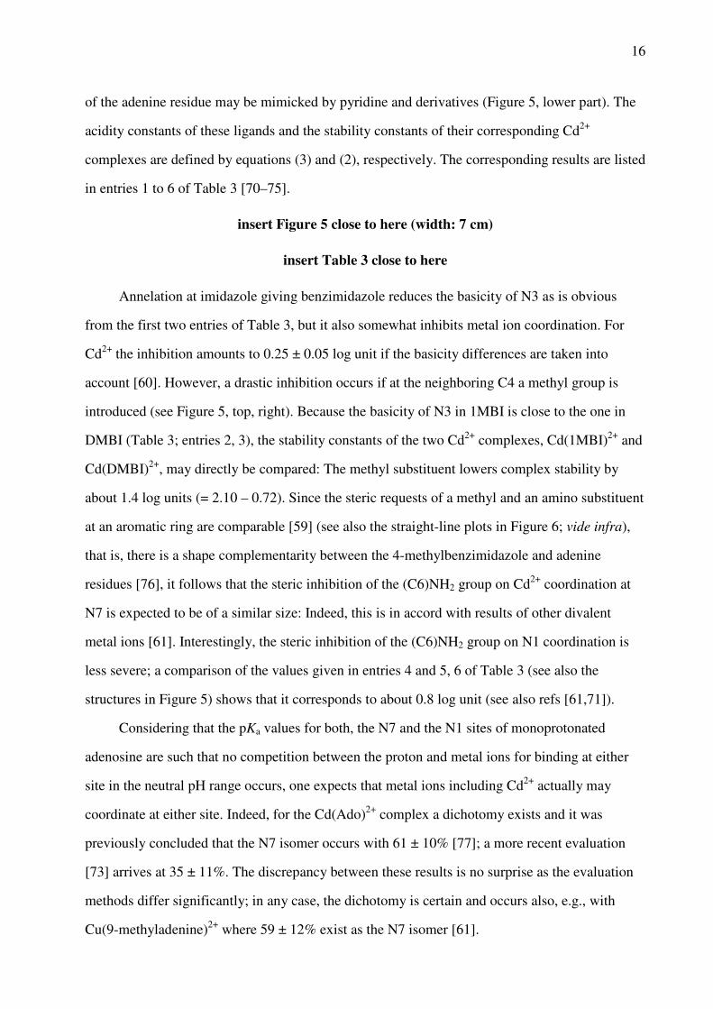
of the adenine residue may be mimicked by pyridine and derivatives (Figure 5, lower part). The
acidity constants of these ligands and the stability constants of their corresponding Cd2+
complexes are defined by equations (3) and (2), respectively. The corresponding results are listed
in entries 1 to 6 of Table 3 [70–75].
insert Figure 5 close to here (width: 7 cm)
insert Table 3 close to here
Annelation at imidazole giving benzimidazole reduces the basicity of N3 as is obvious
from the first two entries of Table 3, but it also somewhat inhibits metal ion coordination. For
Cd2+ the inhibition amounts to 0.25 ± 0.05 log unit if the basicity differences are taken into
account [60]. However, a drastic inhibition occurs if at the neighboring C4 a methyl group is
introduced (see Figure 5, top, right). Because the basicity of N3 in 1MBI is close to the one in
DMBI (Table 3; entries 2, 3), the stability constants of the two Cd2+ complexes, Cd(1MBI)2+ and
Cd(DMBI)2+, may directly be compared: The methyl substituent lowers complex stability by
about 1.4 log units (= 2.10 – 0.72). Since the steric requests of a methyl and an amino substituent
at an aromatic ring are comparable [59] (see also the straight-line plots in Figure 6; vide infra),
that is, there is a shape complementarity between the 4-methylbenzimidazole and adenine
residues [76], it follows that the steric inhibition of the (C6)NH2 group on Cd2+ coordination at
N7 is expected to be of a similar size: Indeed, this is in accord with results of other divalent
metal ions [61]. Interestingly, the steric inhibition of the (C6)NH2 group on N1 coordination is
less severe; a comparison of the values given in entries 4 and 5, 6 of Table 3 (see also the
structures in Figure 5) shows that it corresponds to about 0.8 log unit (see also refs [61,71]).
Considering that the pKa values for both, the N7 and the N1 sites of monoprotonated
adenosine are such that no competition between the proton and metal ions for binding at either
site in the neutral pH range occurs, one expects that metal ions including Cd2+ actually may
coordinate at either site. Indeed, for the Cd(Ado)2+ complex a dichotomy exists and it was
previously concluded that the N7 isomer occurs with 61 ± 10% [77]; a more recent evaluation
[73] arrives at 35 ± 11%. The discrepancy between these results is no surprise as the evaluation
methods differ significantly; in any case, the dichotomy is certain and occurs also, e.g., with
Cu(9-methyladenine)2+ where 59 ± 12% exist as the N7 isomer [61].

17
The N7 sites of inosine or (2'-deoxy)guanosine exist also deprotonated in the neutral pH
range (Table 3, entries 8–10) and thus are easily accessible for metal ions. Such (N7)-bound
metal ions form commonly a hydrogen bond from a ligated water molecule to (C6)O (cf. Figure
1) [11,78]; in contrast to an amino group a carbonyl or keto group at C6 does not exercize any
steric hindrance (see also Section 4.2). Of course, at higher pH values the neutral (N1)H sites
may be deprotonated. This reaction is expressed in eq. (7), where L represents a nucleobase
residue. Naturally, the (L – H)– species formed in this way may also form complexes according
to eq. (8) , and the M(L)2+ complex may be deprotonated at (N1)H as well (eq. 9):
L (L – H)– + H+ (7a)
KLH = [(L – H)–][H+]/[L] (7b)
M2+ + (L – H)– M(L – H)+ (8a)
KM(L − H)
M = [M(L – H)+]/([M2+][(L – H)–]) (8b)
M(L)2+ M(L – H)+ + H+ (9a)
KM(L)
H = [M(L – H)+][H+]/[M(L)2+] (9b)
Evidently, equilibria (2a) and (7a) to (9a) are connected with each other via the equilibrium
scheme (10) [79]:
(10)
This scheme involves four equilibrium constants and because it is of a cyclic nature, only three
constants are independent of each other; the fourth constant is automatically determined by the
other three as follows from eq. (11):
log KM(L)
M – pKM(L)
H = log KM(L − H)
M – pKLH (11a)
pKM(L)
H = pKLH + log
KM(L)
M – log KM(L − H)
M (11b)
The acidification of the (N1)H sites in M(L)2+ complexes, as caused by the (N7)-
18
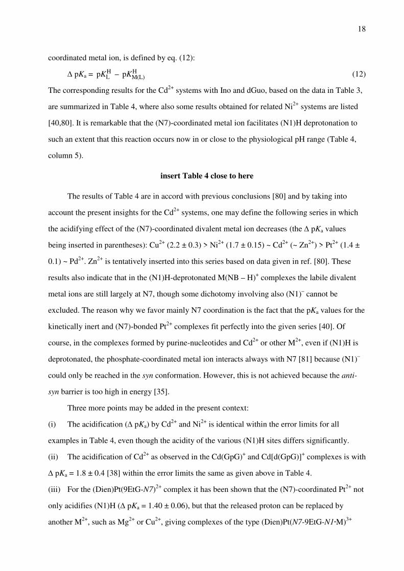
coordinated metal ion, is defined by eq. (12):
∆ pKa = pKLH –
pKM(L)
H (12)
The corresponding results for the Cd2+ systems with Ino and dGuo, based on the data in Table 3,
are summarized in Table 4, where also some results obtained for related Ni2+ systems are listed
[40,80]. It is remarkable that the (N7)-coordinated metal ion facilitates (N1)H deprotonation to
such an extent that this reaction occurs now in or close to the physiological pH range (Table 4,
column 5).
insert Table 4 close to here
The results of Table 4 are in accord with previous conclusions [80] and by taking into
account the present insights for the Cd2+ systems, one may define the following series in which
the acidifying effect of the (N7)-coordinated divalent metal ion decreases (the ∆ pKa values
being inserted in parentheses): Cu2+ (2.2 ± 0.3) > Ni2+ (1.7 ± 0.15) ~ Cd2+ (~ Zn2+) > Pt2+ (1.4 ±
0.1) ~ Pd2+. Zn2+ is tentatively inserted into this series based on data given in ref. [80]. These
results also indicate that in the (N1)H-deprotonated M(NB – H)+ complexes the labile divalent
metal ions are still largely at N7, though some dichotomy involving also (N1)– cannot be
excluded. The reason why we favor mainly N7 coordination is the fact that the pKa values for the
kinetically inert and (N7)-bonded Pt2+ complexes fit perfectly into the given series [40]. Of
course, in the complexes formed by purine-nucleotides and Cd2+ or other M2+, even if (N1)H is
deprotonated, the phosphate-coordinated metal ion interacts always with N7 [81] because (N1)–
could only be reached in the syn conformation. However, this is not achieved because the anti-
syn barrier is too high in energy [35].
Three more points may be added in the present context:
(i) The acidification (∆ pKa) by Cd2+ and Ni2+ is identical within the error limits for all
examples in Table 4, even though the acidity of the various (N1)H sites differs significantly.
(ii) The acidification of Cd2+ as observed in the Cd(GpG)+ and Cd[d(GpG)]+ complexes is with
∆ pKa = 1.8 ± 0.4 [38] within the error limits the same as given above in Table 4.
(iii) For the (Dien)Pt(9EtG-N7)2+ complex it has been shown that the (N7)-coordinated Pt2+ not
only acidifies (N1)H (∆ pKa = 1.40 ± 0.06), but that the released proton can be replaced by
another M2+, such as Mg2+ or Cu2+, giving complexes of the type (Dien)Pt(N7-9EtG-N1·M)3+

19
[82]. The same may be surmised for Cd2+. This shows that "clustering" of metal ions at a guanine
residue is possible; an observation relevant for ribozymes [30].
4.2 Cadmium(II) Complexes of Pyrimidine Derivatives
Among the three pyrimidine-nucleobase residues shown in Figure 1 only the cytosine moiety is
not protonated at N3 in the physiological pH range and hence, freely available for metal ion
coordination. Therefore, this residue will be discussed first. The cytosine residue is an
ambivalent ligating site as follows from crystal structure studies; e.g., Pt2+ coordinates to N3,
Ba2+ to (C2)O, and Cu2+ binds to both sites [35,40]. Thus, in the latter instance chelate formation
occurs and for aqueous solution then the intramolecular equilibrium (13) needs to be considered:
M(Cyd) M(Cyd) (13)
Of course, a M2+ interaction may be innersphere or outersphere, but in any case a 'closed' (cl)
species results. The 'open' (op) species may be N3- or (C2)O-bound, depending on the metal ion
involved. Overall, one may imagine that 4-membered chelates exist, or if a water molecule
participates, that a 6-membered so-called semichelate forms. In addition, a complete outersphere
interaction with both sites can also not be excluded. As a consequence, the M(Cyd) species
are actually expected to be mixtures of chelated isomers [35].
Any kind of chelate formation must lead to a stability enhancement [51], which is defined
in a general manner in equation (14), where for the present L = Cyd:
log ∆M/L = log KM(L)
M – log KM(L)op
M (14a)
= log KM(L)exp
M – log KM(L)calc
M = log ∆ (14b)
The first term in eq. (14) is experimentally accessible as it can be measured directly (eq. 2). To
obtain a value for the open M(L)op complex is commonly more difficult. Most often plots of log
KM(L)
M versus pKH(L)
H are employed, which result for families of structurally related ligands in
straight lines [51] as defined by equation (15),
log KM(L)
M = m· pKH(L)
H + b (15)

20
where m represents the slope of the straight line and b the intercept with the y-axis. Clearly, if the
parameters of eq. (15) are known, one may calculate with pKH(L)
H the stability of the M(L)
complex.
The position of equilibrium (13) for Cyd = L is defined by the dimension-less
intramolecular equilibrium constant KI (eq. 16),
KI = [M(L)cl]/[M(L)op] (16)
which may be calculated according to equation (17) [51]:
KI =
KM(L)M
KM(L)opM
– 1 = 10log∆ – 1 (17)
Equation (14) defines log ∆, whereas (eq. 2) and are defined by equations (18)
and (19), respectively:
KM(L)
M = = (18)
KM(L)op
M = (19)
Of course, knowledge of KI allows to calculate the formation degree of the closed or chelated
species (eq. 13) according to equation (20):
% M(L)cl = 100·KI/(1 + KI) (20)
Some examples of log KM(L)
M versus pKH(L)
H straight-line plots for simple pyridine-type
(PyN; open circles) as well as for ortho-aminopyridine-type (oPyN; crossed circles) ligands [59]
are shown in Figure 6 [35,83]. Combination of these plots with the data points due to log
KM(Cyd)
M / pKH(Cyd)
H (full circles) [83] allows immediately several interesting conclusions:
insert Figure 6 close to here (width: 6 cm)
(i) The M(oPyN)2+ complexes of all studied metal ions [35,59] are less stable than the
M(PyN)2+ species. This proves the steric inhibition of an ortho-amino group next to the
coordinating pyridine nitrogen.
(ii) The data point for the Co(Cyd)2+ complex fits on the reference line defined by the
M(oPyN)2+ species, meaning that the neighboring carbonyl group does not participate in metal

21
ion binding and that only the steric inhibition of the (C6)NH2 group is in action.
(iii) This is different for the Cd(Cyd)2+ complex which shows an increased complex stability,
thus indicating the participation of the (C2)O group in metal ion binding. That is, the steric
inhibition of the (C6)NH2 group is partially offset by the (C2)O group.
(iv) The stabilities of the Ca(PyN)2+ and the Ca(oPyN)2+ complexes differ only little. The
scatter of the data points originates in the low stability of these complexes [59], which is
independent of the pKa value of the pyridine derivative considered in the pH range 3–7. This
indicates [59] that complex formation takes place in an outersphere manner [35].
(v) Furthermore, Ca(Cyd)2+ is even more stable than the sterically unhindered Ca(PyN)2+
species proving the importance of the (C2)O interaction in Ca(Cyd)2+. The corresponding
observations were made for the Mg2+ complexes [35,59].
Of course, the data as summarized in Figure 6 can be evaluated in a quantitative manner by
application of equations (14)–(20). Table 5 contains in column 2 the stability constants measured
previously [83] for the M(Cyd)2+ complexes. The stability constants for the open isomers (eqs
13, 19) were calculated based on pKH(Cyd)
H = pKH(oPyN)
H = 4.24 ± 0.02 [83] and the straight-line
parameters as defined in equation (15) and listed in ref. [59]; these values for M(Cyd) are
given in column 3. From the mentioned constants the log ∆ values (eq. 14) (column 4) follow;
they correspond to the vertical distances seen in Figure 6 between the experimentally determined
points of a given M(Cyd)2+ complex (solid circle) and its reference line (crossed circles); these
values are listed in column 4 of Table 5. Application of equations (17) and (20) allows to
calculate values for KI (eqs 16, 17) and % M(Cyd) (eq. 20), respectively; these results are
listed in columns 5 and 6 of Table 5.
insert Table 5 close to here
From column 6 in Table 5 it follows that the closed species in the M(Cyd)2+ systems reach
remarkable formation degrees and that equilibrium (13) in many instances truly exists. Indeed,
exceptions are only Co(Cyd)2+ and Ni(Cyd)2+ [35]. Furthermore, Table 5 allows some additional
interesting conclusions:
(i) The closed complex, Mg(Cyd) , formed to about 35%, is most likely a semichelate
which is mainly innersphere bound to (C2)O and outersphere to N3. This view is supported by

22
the crystal structure of Ba(CMP)·8.5H2O where the alkaline earth ion is bonded to (C2)O (and
the sugar, but not to N3 or the phosphate) [11,84].
(ii) The same type of semichelate is also suggested for Ca(Cyd) . Interestingly, despite the
significant stability difference between Ca(Cyd)2+ and Cd(Cyd)2+ (column 2; eq. 2) both
complexes reach formation degrees of about 50% for the chelated isomer. Note, this is possible
because KI is a dimension-less constant which quantifies the position of an intramolecular
equilibrium (eq. 13).
(iii) However, for Cd(Cyd) one expects innersphere coordination of N3 and possibly
outersphere binding to (C2)O. In this context it is revealing to note that in the polymeric
Cd(dCMP) complex binding of the octahedral Cd2+ occurs to both N3 (2.30 Å) and (C2)O (2.64
Å) by formation of a 4-membered ring [85]. The same may be surmised for Cd(Cyd) , but in
aqueous solution one expects it to be, at least, in equilibrium with the indicated semichelate.
(iv) In the polymeric Co(CMP) complex the metal ion coordinates to N3 (1.99 Å) and does not
interact with (C2)O [86]; this agrees with the properties of Co(Cyd)2+ for which equilibrium (13)
is far to its left. However, considering the small slope of the Co(oPyN)2+ reference line seen in
Figure 6 (compare with the Mg2+ situation) [59], it could well be that a significant amount of the
metal ion in Co(Cyd)2+ is also outersphere bound to N3.
The other two pyrimidine nucleobases, i.e., the uracil and thymine residues (Figure 1), bind
strongly to metal ions only after deprotonation of their (N3)H site [87]. This means that carbonyl
groups interact significantly with metal ions only if a suitable primary binding site is available
(Section 3.3). To establish a sound basis for comparisons equilibria (7a) and (8a) were studied
for several metal ion complexes of (N3)H-deprotonated uridine-type ligands (U), i.e., 5-
fluorouridine, 5-chloro-2'-deoxyuridine, uridine, and thymidine (= 2'-deoxy-5-methyluridine).
Plots of log KM(U − H)
M versus pKUH result for the four ligand systems in straight lines [88] and
these may be compared with the plots discussed above for pyridine- and o-aminopyridine-type
ligands. Figure 7 shows the situation for the Cd2+ complexes together with the data for the
corresponding Ca2+ and Co2+ complexes for comparison.
insert Figure 7 close to here (width: 10 cm)
From the Ca2+ and Cd2+ parts of Figure 7 it follows that their M(U – H)+ complexes are

23
more stable than the Ca(PyN)2+ and Cd(PyN)2+ species, whereas the Co(U – H)+ complexes are
less stable than their Co(PyN)2+ counterparts. Furthermore, the Co(U – H)+ straight line is placed
(although with a somewhat steeper slope) between the lines of the Co2+ complexes of the PyN-
and oPyN-type ligands. This indicates that Co2+ (like Ni2+ [88]) suffers in its coordination to
(N3)– of (U – H)+ from a steric hindrance by the neighboring (C2)O/(C4)O groups. This
hindrance, however, is less pronounced than that by an o-amino (or o-methyl) group. In contrast,
in Ca(U – H)+ and Cd(U – H)+, (C2)O and (C4)O facilitate M2+ binding leading thus to an
increased complex stability.
In a careful evaluation, taking into account the situation in M(Cyd)2+ complexes [88], it
was concluded that the 'lower limits' for the formation degrees of chelates can be assessed; 'lower
limits' because in (U – H)– there is no steric hindrance by an o-NH2 group, and two (C)O groups
(not only one; see Figure 1) may participate in complex formation. Hence, the 'lower limits' for
the formation degrees of chelates in Ca(U – H)+ and Cd(U – H)+ are about 50% and 60%,
respectively. In Cd(U – H)+ 4-membered chelates may form, but in aqueous solution it is highly
likely that in addition semichelates occur via ligated water molecules to (C2)O and (C4)O. In
contrast, no chelate formation is anticipated for the Co2+ (and Ni2+) complex of (U – H)–, i.e., the
metal ion coordinates most likely in a monodentate fashion to (N3)– of the uridinates [88].
4.3 Cadmium(II) Complexes of Some Less Common Nucleobase Residues
In this section we will consider tubercidin (Figure 5; bottom, left) and the five nucleosides seen
in Figure 8 [89].
4.3.1 Tubercidin
Tubercidin (Tu), also known as 7-deazaadenosine, is synthesized by molds and fungi [90] and
has antibiotic properties. From its structure it follows that Tu is an o-aminopyridine-type ligand
[59,72], and as we have already seen in Figure 6 (Section 4.2), it fits with its (C6)NH2/N1 unit
and the connected metal ion-binding properties, including for Cd2+, perfectly into this picture

24
[59] and no further discussion is therefore warranted.
insert Figure 8 close to here (width: 8 cm)
4.3.2 Orotidine
Orotidine and its derivatives play an important role as intermediates in the metabolism of
pyrimidine-nucleotides [91]. Its structure is shown in Figure 8 (top, left); it is closely related to
uridine (see Figure 1), but due to the (C6)-carboxylate group it exists in solution mainly in the
syn conformation [89]. The (C6)COOH group is very acidic; for aqueous solution it was
estimated that pKa = 0.5 ± 0.3 [92]. Consequently, the stability constants of the orotidinate (Or–)
complexes of Mg2+, Cu2+, and Zn2+ (only these metal ions have been studied [92]) are somewhat
below of those measured for the corresponding M(Ac)+ complexes (see Table 1, column 7).
There is no evidence for any significant chelate formation in aqueous solution [92]. Therefore,
one may assume that all this also holds for the Cd(Or)+ complex, which gives as an estimate for
its stability log KCd(Or)
Cd = 1.0 ± 0.3.
The acidity constant of Or– for the deprotonation of its (N3)H site in aqueous solution,
pKOrH = 9.12 ± 0.02 [92], is quite close to the corresponding one of its parent nucleoside, uridine,
for which it holds pKUrdH = 9.18 ± 0.02 [88]. Consequently, it is safe to assume that the stabilities
of the M(Or – H) and M(Urd – H)+ complexes, including Cd2+, are also very similar (see also
Figure 7). Hence, it is no surprise that the stabilities of the complexes formed with orotidinate 5'-
monophosphate (OMP3–) are determined by the basicity of the phosphate group [93]. No
evidence was observed for macrochelate formation between the phosphate-coordinated metal
ions and the carboxylate group, though there is a charge effect of about 0.4 log unit [93]. Hence,
in Cd(OMP)– no interaction with the pyrimidine ring occurs, what corresponds to the situation in
Cd(UMP).
4.3.3 Xanthosine
Xanthosine (Xao) and its derivatives are of relevance for the metabolism of purine-
nucleosides/nucleotides [91,94]. Monoprotonated H(Xao)+ carries a proton at N7, which is

25
released with pKH(Xao)
H = 0.74 ± 0.06 [95]. The neutral Xao, as shown in Figure 8, loses a
further proton already with pKXaoH = 5.47 ± 0.03 [95] from the (N1)H/(N3)H sites and there is
evidence for a tautomeric equilibrium between (N3)–/(N1)H and (N3)H/(N1)– [96,97]. In any
case, in the physiological pH range of about 7.5 this nucleoside is present in its anionic form,
xanthosinate, and it differs therefore considerably from its relatives inosine and guanosine
(Section 4.1; Table 3).
Stability constants of M(Xao – H)+ complexes (including for Cd2+) have been determined
[95] and there is evidence that a dichotomy for M2+ binding between N1 and N7 occurs [95,98];
of course, the negative charge in the xanthine residue can be delocalized over many atoms, that
is, N3, (C2)O, N1, and (C6)O. There is no doubt that the indicated dichotomy exists, yet the
percentages given for the isomers should be considered with some care, even though the
agreement between the rather different evaluation methods is surprisingly good. For example, it
is concluded that in Cd(Xao – H)+ about 75% are N7-coordinated [95,98]; the value for Co(Xao
– H)+ is about 50% [95,98], whereas for Zn(Xao – H)+ N7 binding is given as 58% in ref. [95]
and as 38% in ref. [98]. Here more work is needed.
4.3.3 Thiouridines
Thiolation of uracil residues, especially in RNA wobble positions, affect the conformation of the
nucleic acid in solution [99] and has implications for recognition processes. Furthermore, 4-
thiouridine is found in bacterial and archaeal tRNA [100,101]. Therefore, we shall have a short
look on 2-thiouridine (U2S) and 4-thiouridine (U4S) (see Figure 8, bottom part) and their
complexes formed with Cd2+ and Cu2+ [102,103]. The exchange of an O atom by a S atom in
uridine (see Figure 1) is expected, of course, to alter not only the acid-base but also the metal
ion-binding properties [15,17,104]. Indeed, the acidity of (N3)H is increased by about one pK
unit as follows from column 3 in Table 6 [105]. However, despite the reduced basicity of (N3)–
in (U2S – H)– and (U4S – H)– the stabilities of their complexes with Cd2+ and Cu2+ are by about
1 and 1.8 log units higher than those of Cd(Urd – H)+ and Cu(Urd – H)+ (Table 6, column 5).
insert Table 6 close to here

26
A more careful evaluation of the data via log ∆ (eq. 14) by taking the differences in acidity
of the (N3)H sites into account, reveals that the true stability enhancement amounts for the Cd2+
complexes to more than 1.3 log units and for the Cu2+ species to about 2.3 log units (Table 6,
column 7). Of course, the negative charge at (N3)– may be delocalized to the sulfur atoms at
(C2)S or (C4)S and this makes these sulfur sites to excellent donors for Cd2+ (and Cu2+)
coordination [15,17,104]. Most likely the enhanced complex stability is not only due to
monodentate (C)S–M2+ coordinations (next to a (N3)––M2+ one), but due to chelate formation
involving both sites as well. From X-ray crystal structure studies it is known that Cd2+ [85] and
Cu2+ [106] are able to form 4-membered chelates with the cytosine residue involving the N3 and
(C2)O sites. For the present study this means that Cd2+ and Cu2+ most likely form 4-membered
rings involving [N3/(C)S]–, especially since such low-membered chelates are more easily formed
if a (C)O is replaced by a (C)S site [107]. Of course, in aqueous solution there is the possibility
that semichelates involving a ligated water molecule form [105].
In the terminating paragraph of Section 4.2 we have already concluded that the formation
degree of chelates in the M(Urd – H)+ species must be larger than it is in the M(Cyd)2+
complexes. Considering the additional stability enhancement observed for the M(U2S – H)+ and
M(U4S – H)+ complexes, these lower limits must be even more true for the complexes of the
thiouridinates, that is, Cd(US – H) > Cd(Cyd) (= ca. 60%; see Table 5) and Cu(U2S – H)
> Cu(Cyd) (= ca. 80%). In fact, we believe that the formation degree of the chelated species in
the Cd(US – H)+ and Cu(U2S – H)+ systems is larger than 90% because this number corresponds
to a stability enhancement of about 1 log unit being solely due to chelate formation (and
attributing the remaining part of log ∆ to the general participation of S in metal ion binding).
4.3.4 2-Thiocytidine
The thionucleoside 2-thiocytidine (C2S; Figure 8, lower part, right) occurs in Nature in tRNAs
[108].In addition, it also receives attention in diverse fields [109] like drug research [110] or
nanotechnology [111]. The acidity constants of H(C2S)+ and the stability constants of the
M(C2S)2+ and M(C2S – H)+ complexes (M2+ = Zn2+, Cd2+) were determined by potentiometric
pH titrations. These results [112] can be compared with those obtained for the parent nucleoside

27
cytidine (Cyd; Figure 1; Section 4.2). Replacement of the (C2)=O unit by (C2)=S facilitates the
release of the proton from (N3)H+ in H(C2S)+ (pKa = 3.44 ± 0.01 [112]; 25°C; I = 0.5 M, KNO3)
somewhat, compared with H(Cyd)+ (pKa = 4.24 ± 0.02 [83]; 25°C; I = 0.5 M, NaNO3). This
moderate effect of about 0.8 pK unit contrasts with the strong acidification of about 4 pK units of
the (C4)NH2 group in C2S (pKa = 12.65 ± 0.12 [112]) compared with Cyd (pKa ca. 16.7
[34,113]). The reason [112] for this result is that the amino-thione tautomer, which dominates for
the neutral C2S molecule, is transformed upon deprotonation into the imino-thioate form with
the negative charge largely located on the sulfur; this is indicated in equilibrium (21):
(21)
The intramolecular and dimensionless equilibrium constant KT = [imino]/[amino] was
estimated as being on the order of 10–4 to 10–6, which leads to values for the acidity constant
Ka/imino of 10–8.65 to 10–6.65 [112].
In the M(C2S)2+ complexes the (C2)S group is the primary binding site [112] rather than
N3 as is the case in the M(Cyd)2+ complexes (Section 4.2), though owing to chelate formation
N3 is to some extent still involved in metal ion binding. Based on log KM(L)
M versus pKH(L)
H plots
(eq. 15) for oPyN-type ligands [59] (see also Figure 6), the stability enhancements amount to log
∆Cd/C2S = 3.3 and log ∆Zn/C2S = 2.6 [112]. They are much larger than those observed for the
corresponding M(Cyd)2+ complexes (see Table 5) and lead to the conclusion that (C2)S–M2+
binding is important and dominates with more than 99%; a number which encompasses both,
monodentate S coordination as well as chelate formation of an (C2S)-bound metal ion with N3.
The structure of these chelates has been discussed [112]. Clearly, the monodentate (N3)–M2+
isomer occurs only in traces.
Similarly, in the Zn(C2S – H)+ and Cd(C2S – H)+ complexes the main metal ion binding
site is the (C2)S– unit and the formation degree of its complex is above 99.9% (compared with
that of N3). However, again a large degree of chelate formation with N3 must be surmised for
the M(C2S – H)+ species, and their structure was discussed [112]. It needs to be emphasized that
upon metal ion binding the deprotonation of the (C4)NH2 group (pKa = 12.65) is dramatically

28
acidified (pKa ca. 3), confirming the very high stability of the M(C2S – H)+ complexes and the
importance of equilibrium (21). To conclude, the metal ion-binding capabilities of C2S differ
strongly from those of its parent Cyd; this also holds for hydrogen-bond formation because (C)S
is a much poorer H-acceptor than (C)O. Clearly, these differences must have consequences for
the properties of those RNAs which contain this thionucleoside.
5 Complexes of Cadmium(II) with Phosphates
In Figure 9 the general structures for the monoesters of mono-, di-, and triphosphates,
symbolized by R-MP2–, R-DP3–, and R-TP4–, respectively, are shown together with the structure
of a phosphodiester bridge, (RO)2PO , as it occurs in the backbone of nucleic acids. This diester
of a phosphate group may be mimicked to some extent herein by acetate (Ac–) as both ligands
carry a charge of minus one. The most basic phosphate residue in the phosphomonoesters is
always the terminal one, which carries a charge of two minus. In addition, the effect of the
residue R on the basicity of such a phosphate group will be the more pronounced the closer they
are. For example, the effect of a phenyl and a butyl residue in R-MP2– leads to the pKa values of
5.81 ± 0.01 and 6.72 ± 0.02, respectively [114], whereas the effect of the same residues in R-
DP3– leads to the much smaller pKa span of 6.32 ± 0.02 to 6.65 ± 0.02 [115]. In the case of the
triphosphates the residue R has practically no effect [37,116–118].
insert Figure 9 close to here (width: 6.5 cm)
Pyrimidine-nucleoside phosphates furnish representative values for nucleotide
comparisons because the pyrimidine residue does commonly not participate in metal ion binding
with labile ions like Cd2+ [114–119]. The corresponding acidity constants for the terminal
phosphate groups of pyrimidine-nucleotides are pKH(R-MP)
H = 6.20 (eq. 3) for monophosphate
monoesters [50,114], pKH(R-DP)
H = 6.40 for diphosphate monoesters [115], and pKH(R-TP)
H = 6.50
for triphosphate monoesters [37,116–118,120]. The stability constants (eq. 2), which correspond
to these pKa values [115,117], are listed in columns 3 to 5 of Table 7 (upper part) [121,122] for
several M(R-MP), M(R-DP)–, and M(R-TP)2– complexes.
29
insert Table 7 close to here
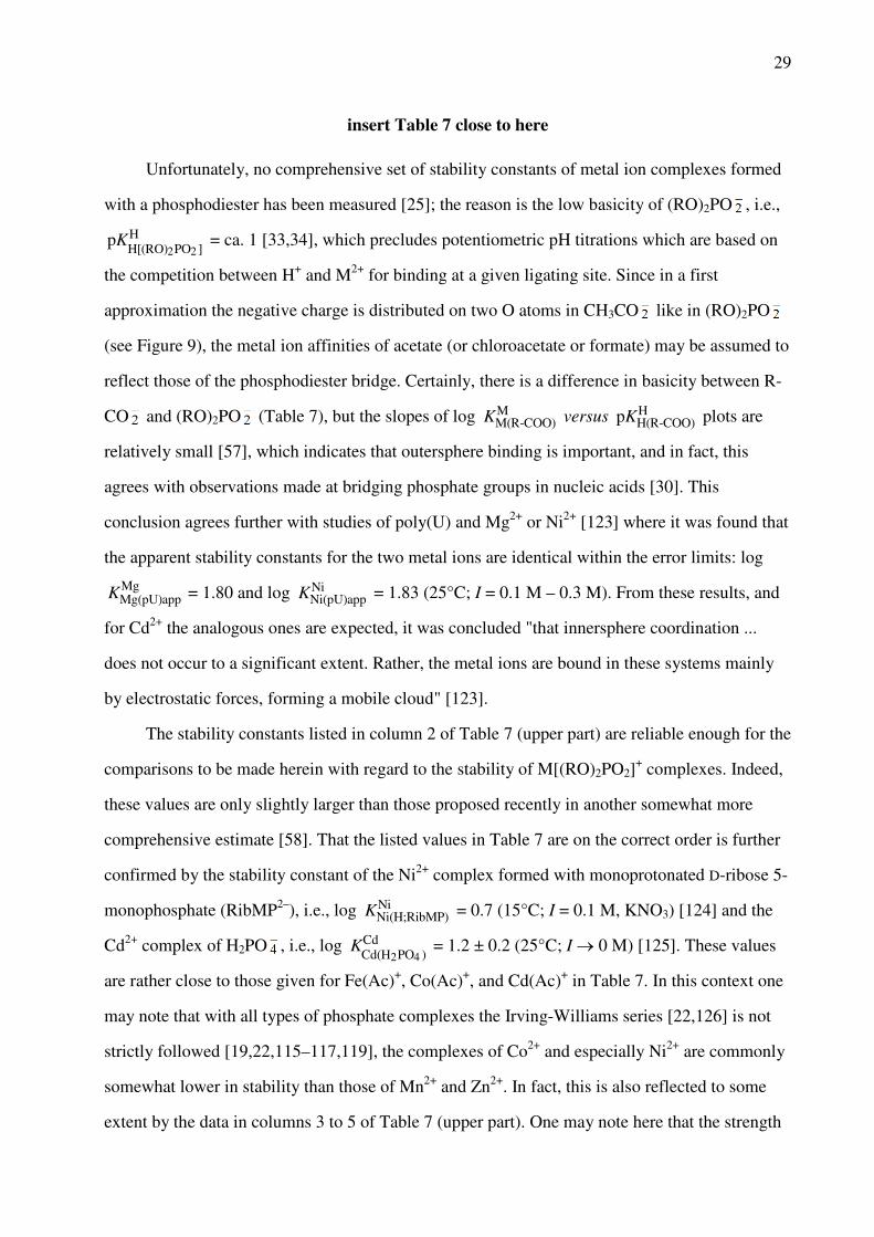
Unfortunately, no comprehensive set of stability constants of metal ion complexes formed
with a phosphodiester has been measured [25]; the reason is the low basicity of (RO)2PO , i.e.,
pK
H[(RO)2PO2 ]H = ca. 1 [33,34], which precludes potentiometric pH titrations which are based on
the competition between H+ and M2+ for binding at a given ligating site. Since in a first
approximation the negative charge is distributed on two O atoms in CH3CO like in (RO)2PO
(see Figure 9), the metal ion affinities of acetate (or chloroacetate or formate) may be assumed to
reflect those of the phosphodiester bridge. Certainly, there is a difference in basicity between R-
CO and (RO)2PO (Table 7), but the slopes of log KM(R-COO)
M versus pKH(R-COO)
H plots are
relatively small [57], which indicates that outersphere binding is important, and in fact, this
agrees with observations made at bridging phosphate groups in nucleic acids [30]. This
conclusion agrees further with studies of poly(U) and Mg2+ or Ni2+ [123] where it was found that
the apparent stability constants for the two metal ions are identical within the error limits: log
KMg(pU)app
Mg = 1.80 and log KNi(pU)app
Ni = 1.83 (25°C; I = 0.1 M – 0.3 M). From these results, and
for Cd2+ the analogous ones are expected, it was concluded "that innersphere coordination ...
does not occur to a significant extent. Rather, the metal ions are bound in these systems mainly
by electrostatic forces, forming a mobile cloud" [123].
The stability constants listed in column 2 of Table 7 (upper part) are reliable enough for the
comparisons to be made herein with regard to the stability of M[(RO)2PO2]+ complexes. Indeed,
these values are only slightly larger than those proposed recently in another somewhat more
comprehensive estimate [58]. That the listed values in Table 7 are on the correct order is further
confirmed by the stability constant of the Ni2+ complex formed with monoprotonated D-ribose 5-
monophosphate (RibMP2–), i.e., log KNi(H;RibMP)
Ni = 0.7 (15°C; I = 0.1 M, KNO3) [124] and the
Cd2+ complex of H2PO , i.e., log K
Cd(H2PO4 )Cd = 1.2 ± 0.2 (25°C; I → 0 M) [125]. These values
are rather close to those given for Fe(Ac)+, Co(Ac)+, and Cd(Ac)+ in Table 7. In this context one
may note that with all types of phosphate complexes the Irving-Williams series [22,126] is not
strictly followed [19,22,115–117,119], the complexes of Co2+ and especially Ni2+ are commonly
somewhat lower in stability than those of Mn2+ and Zn2+. In fact, this is also reflected to some
extent by the data in columns 3 to 5 of Table 7 (upper part). One may note here that the strength

30
of the metal ion-phosphate interaction is crucial for the activity of ribozymes: For example, the
catalytic cleavage rate of hammerhead ribozymes shows a direct correlation to the above
mentioned "irregular" phosphate affinities of the divalent metal ions [127].
To facilitate comparisons between the various types of complexes the log-stability
differences listed in the lower part of Table 7 have been prepared. As one would expect, increase
of the charge from minus one in Ac–/(RO)2PO to minus two in R-MP2– (Figure 9) increases the
stabilities of the complexes: The stability increases amount to about 0.9 (Ca2+) to 1.2 (Cd2+) log
units; only for the Cu2+ complexes the effect is a bit more pronounced with log ∆R-MP/Ac = 1.4
(lower part, column 2). In contrast, the stability increase of the complexes by going from M(R-
MP) to M(R-DP)– varies significantly from metal ion to metal ion, i.e., within the relatively large
span of about 1.45 to 2.4 log units. This changes again by going from M(R-DP)– to M(R-TP)2–,
then the stability increase is quite constant within the narrow range of about 0.8 to 1.0 log units,
if the special case of Cu2+ with its distorted octahedral coordination sphere [128] is ignored.
Overall this indicates in our view that outersphere species play a significant role in M(R-MP)
complexes, which also include six-membered semichelates with one of the terminal oxygens
innersphere and the other one outersphere [114]. Such outersphere interactions are hardly of
relevance in the corresponding di- and triphosphate species where two neighboring phosphate
units allow the formation of six-membered innersphere chelates. This is in accord with the log
KM(R-MP)
M versus pKH(R-MP)
H plots where the slopes are much lower (Ca2+: m = 0.131 ± 0.020;
Cd2+: 0.329 ± 0.019 [122] (thus indicating partial outersphere binding) than for the log KM(R-DP)
M
versus pKH(R-DP)
H plots (Ca2+: 0.379 ± 0.097; Cd2+: 0.945 ± 0.104 [115]). Clearly, for di- and
triphosphates the formation of innersphere complexes is more pronounced compared with
monophosphates, owing to the increased negative charge of these ligands. Overall, the situation
may be summarized with the earlier statement [129], "the lower the charge, the more
predominant are outersphere complexes".
6 Cadmium(II) Complexes of Nucleotides
6.1 Some General Considerations

31
The evaluations presented in this section are carried out in part in a manner analogous to our
previous review on Ni2+-nucleotide complexes [40]; of course, concentrating now on Cd2+ which
was not a part of the previous account. As is well known, nucleotides exist in solution mainly in
the so-called anti conformation. Two such examples are shown in Figure 10 [11–14,50]. It is the
phosphate residue in nucleotides which determines to a very large part the stability of the
complexes formed with labile metal ions including Cd2+ [74,117,119,130] independent of the
kind of nucleobase involved or whether a nucleoside mono-, di-, or triphosphate is considered.
insert Figure 10 close to here (width: 7 cm)
For purine-nucleoside 5'-phosphates the formation of macrochelates was proposed nearly
60 years ago [131] and more than 50 years ago it was concluded that they actually exist [132–
135], i.e., a metal ion coordinated to the phosphate residue of a purine nucleotide may also
interact in the dominating anti conformation with N7 of the purine moiety. Nowadays formation
of macrochelates in complexes formed by purine nucleotides with various metal ions including
Cd2+ is well established [117,120,136–138]. Clearly, the formation of such a macrochelate must
give rise to the intramolecular equilibrium (22):
(22)
As already discussed in Section 4.2, any kind of equilibrium between an 'open' isomer, M(L)op,
and a chelated or 'closed' isomer, M(L)cl, must be reflected in an increased complex stability
compared to the situation where only the open complex can form [51]. This stability
enhancement, log ∆M/L (eq. 14), can be evaluated by equations (16) to (20) (Section 4.2), which
furnish values for KI (eqs 16, 17) and thus for the formation degree of the closed species, %
M(L)cl (eq. 20).
One important aspect needs to be emphasized and kept in mind when dealing with purine
derivatives: All of them show a pronounced tendency for self-association, which occurs via
stacking of the purine rings [117,137,139,140]. Hence, experiments aimed at determining the
properties of monomeric metal ion complexes of purine nucleosides or their phosphates should

32
not be carried out in concentrations higher than 10–3 M. To be on the safe side, actually a
maximum concentration of only 5 × 10⁻⁴ M is recommended (cf., e.g., [122,136]). The
equilibrium constants discussed below were mostly obtained by potentiometric pH titrations and
the corresponding experimental conditions adhere to the above request.
6.2 Complexes of Nucleoside 5'-Monophosphates
6.2.1 Equilibrium Constants to Be Considered
A combination of the information provided in Figures 1 and 10 reveals that there are nucleoside
5'-monophosphates (NMP2–), which contain a nucleobase that may accept a proton, e.g., AMP2–,
whereas others contain a nucleobase that can only release one, e.g., UMP2–. Therefore, if one
neglects a twofold protonated phosphate group because this is not of relevance for a biological
system due to its large acidity (e.g., for H2(UMP) pK
H2 (UMP)H = 0.7 ± 0.3 [114]), one has to
consider eqs (3) and (7), next to equilibrium (23) (charges neglected):
H2(L) H(L) + H+ (23a)
K
H2 (L)H = [H(L)][H+]/[H2(L)] (23b)
Based on the structures of the various NMPs, it is clear that equilibria (23) and (3) hold for
H2(CMP)± and H2(AMP)±, and that equilibria (3) and (7) are of relevance for H(UMP)– and
H(dTMP)–, whereas all three equilibria (23), (3), and (7) are needed to describe the acid-base
properties of H2(IMP)± and H2(GMP)±. In all cases equilibrium (3) refers to the deprotonation of
the P(O)2(OH)– group in the H(NMP)– species.
Correspondingly, one has to consider the complexes M(H;NMP)+ and M(NMP), the
stabilities of which are defined in equilibria (24) and (2), respectively (charges neglected):
M2+ + H(L) M(H;L) (24a)
KM(H;L)
M = [M(H;L)]/([M2+][H(L)]) (24b)
At high pH values also Cd(NMP – H)– species (eq. 8) might form but at present no information
is available about these.
33
M(H;NMP)+ [= M(H;L)] complexes form with CMP2–, AMP2–, IMP2–, and GMP2–. In
these cases, M2+, including Cd2+, is mainly located at the nucleobase residue and the proton at
the phosphate group [50,130,141]. However, because the proton is lost with a pKa value of about
5 or below [50,130,141], these species are of relevance for biological systems only under very
special conditions. At the common physiological pH of about 7.5 they are not important and
therefore not considered further in the present context.
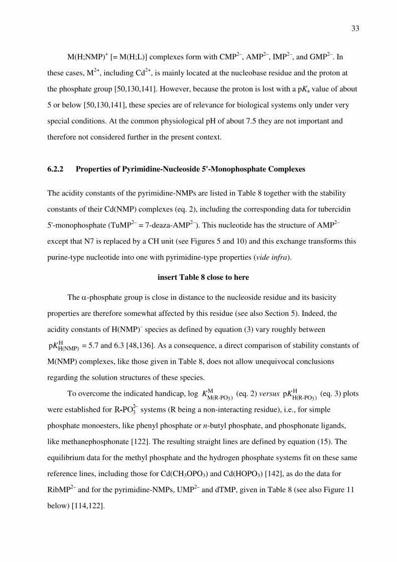
6.2.2 Properties of Pyrimidine-Nucleoside 5'-Monophosphate Complexes
The acidity constants of the pyrimidine-NMPs are listed in Table 8 together with the stability
constants of their Cd(NMP) complexes (eq. 2), including the corresponding data for tubercidin
5'-monophosphate (TuMP2– = 7-deaza-AMP2–). This nucleotide has the structure of AMP2–
except that N7 is replaced by a CH unit (see Figures 5 and 10) and this exchange transforms this
purine-type nucleotide into one with pyrimidine-type properties (vide infra).
insert Table 8 close to here
The α-phosphate group is close in distance to the nucleoside residue and its basicity
properties are therefore somewhat affected by this residue (see also Section 5). Indeed, the
acidity constants of H(NMP)– species as defined by equation (3) vary roughly between
pKH(NMP)
H = 5.7 and 6.3 [48,136]. As a consequence, a direct comparison of stability constants of
M(NMP) complexes, like those given in Table 8, does not allow unequivocal conclusions
regarding the solution structures of these species.
To overcome the indicated handicap, log K
M(R-PO3 )M (eq. 2) versus
pK
H(R-PO3 )H (eq. 3) plots
were established for systems (R being a non-interacting residue), i.e., for simple
phosphate monoesters, like phenyl phosphate or n-butyl phosphate, and phosphonate ligands,
like methanephosphonate [122]. The resulting straight lines are defined by equation (15). The
equilibrium data for the methyl phosphate and the hydrogen phosphate systems fit on these same
reference lines, including those for Cd(CH3OPO3) and Cd(HOPO3) [142], as do the data for
RibMP2– and for the pyrimidine-NMPs, UMP2– and dTMP, given in Table 8 (see also Figure 11
below) [114,122].

34
In the light of the results discussed in Sections 3.2 and 4.2, it is no surprise that the ribose
residue and the uracil and thymine moieties do not participate in metal ion binding in
M(RibMP), M(UMP), and M(dTMP) complexes [122]. How is the situation with Cd(CMP)? The
mentioned straight-line parameters [122] are summarized for Cd(R-PO3) in equation (25) [the
error limit (3σ) for a calculated log K
Cd(R-PO3 )Cd value is ±0.05 (for details see ref. [122]):
log K
Cd(R-PO3 )Cd = (0.329 ± 0.019)·
pK
H(R-PO3 )H + (0.399 ± 0.127) (25)
Application of pKH(CMP)
H = 6.19 (Table 8) to eq. (25) allows to calculate the stability of the
'open' isomer Cd(CMP)op in equilibrium (22), i.e., log K
Cd(R-PO3 )Cd = log
KCd(CMP)op
Cd = 2.44 ±
0.05. This calculated stability constant is within the error limits identical with the measured one
given in Table 8. Hence, there is no indication for an increased stability of the Cd(CMP)
complex, which means that equilibrium (22) is far on its left side and that no remarkable
amounts of macrochelates are formed. At first sight this result for Cd(CMP), which also holds
for Cu(CMP) and other M(CMP) species [50], may seem surprising because the ability of the N3
site of the cytosine residue to interact with metal ions is well known (Section 4.2; Table 5).
However, it should be recalled that CMP2– exists predominately in the anti conformation (Figure
10) in which N3 is pointing away from the metal ion coordinated at the phosphate group.
Evidently, for all divalent metal ions considered (Table 5) the anti-syn energy barrier, which has
been estimated to be on the order of about 7 kJ/mol [35], is too large to be overcome by
macrochelate formation.
6.2.3 Properties of Purine-Nucleoside 5'-Monophosphate Complexes
Purine-nucleosides represent a favored situation because not only N7 of the purine moiety is able
to bind metal ions (Section 4.1; Table 3) but also a metal ion coordinated at the phosphate
residue can reach N7 in the dominating anti conformation (Figure 10) as was already pointed out
in Section 6.1. Such a possible N7 interaction, depending on the kind of metal ion involved, may
give rise to macrochelate formation and if this occurs it must be reflected in an increased
complex stability. Indeed, a very significant stability enhancement is observed for the Cd(NMP)
complexes of AMP2–, IMP2–, and GMP2– (Figure 11), whereas that for the corresponding Ca2+

35
complexes is much smaller. This confirms the well known high affinity of Cd2+ for N sites, the
one of Ca2+ being much lower [22] (see also Section 2; Table 1).
insert Figure 11 close to here (width: 8 cm)
The vertical distances between M(NMP) data points and their corresponding straight-
reference line can easily be quantified. Application of the acidity constant pKH(NMP)
H to the
straight-line equation (15), the parameters of which are listed in refs [50,52,122], provides the
value of the intercept with the straight line and thus the stability constant of the 'open' species
(eq. 19). In other words, with equation (14) one may now define the stability enhancement, if
any, for the M2+ complexes of the purine-NMPs. Clearly, the calculated value for log K
M(R-PO3 )M
reflects the stability of the 'open' isomer in equilibrium (22), in which M2+ is only phosphate-
coordinated. Now, with known values for log ∆M/NMP (= log ∆; eq. 14), the evaluation procedure
defined in equations (16) through (20) (Section 4.2) can be applied and the formation degree of
the macrochelate which appears in equilibrium (22) can be calculated (eq. 20). The
corresponding results are summarized in Table 9 for the Cd(NMP) complexes of AMP2–, IMP2–,
and GMP2–, together with the analogous results of some other metal ions for comparisons.
insert Table 9 close to here
The results of column 6 in Table 9 demonstrate that in all M2+ 1:1 complexes with purine-
NMPs [with the possible exception of Ca(AMP)] at least some macrochelation occurs.
Macrochelation by an interaction of the phosphate-coordinated M2+ with N7 is thereby proven
with the Cd2+/TuMP2– system (Table 8). In TuMP2– a CH unit replaces the N7 of AMP2–
(Figures 5 and 10) which means that TuMP2– is no longer able to form the mentioned
macrochelates. Indeed, the stability constants of its M(TuMP) complexes fit on the reference
lines, thus proving that TuMP2– behaves like a simple phosphate monoester that binds metal ions
only to its phosphate group [50,136]. Similar to the situation with pyrimidine NMPs and their N3
site, also with purines the N1 site cannot be reached in the anti conformation.
The data assembled in Table 9 allow at least one more interesting conclusion: In all
instances the formation degree of the macrochelates increases in the order M(AMP)cl < M(IMP)cl
< M(GMP)cl. This increase for M(IMP)cl < M(GMP)cl is in accord with the increased basicity of

36
N7 in GMP2– compared with that of N7 in IMP2– [74]. However, with AMP2– the situation is
more complicated: 9-methylguanine has a pKa of 3.11 ± 0.06 [143,144] for its (N7)H+ site and
this value is rather close to the micro acidity constant for the same site in 9-methyladenine,
pkHgN7-N1N7-N1 = 2.96 ± 0.10 [143]. Hence, the N7 basicity cannot be responsible for the decreased
complex stability. Similar to the situation with pyrimidine derivatives as discussed in Section
4.2, the reduced stability by about 0.5 log unit of Cd(AMP), compared to that of Cd(GMP), is to
be attributed to the steric inhibition which (C6)NH2 exercises on a metal ion coordinated at N7
[71]. The (C6)O group does not have such an effect. In contrast, it rather promotes the stability
by forming outersphere bonds to a water molecule of the N7-bound metal ion [74].
Finally, a comment is needed about the effect of the ribose versus the 2'-deoxyribose
residue on the metal ion-binding properties of purine nucleotides. Unfortunately no data for Cd2+
complexes are available, but for those of Mg2+, Ni2+, Cu2+, and Zn2+ it was shown [121] that
complex stability and extent of macrochelate formation (eq. 22) between the phosphate-
coordinated metal ion and N7 of the purine residue is very similar (or even identical) for the
AMP2–/dAMP2– pair. This is different for the GMP2–/dGMP2– pair: The stabilities of the
M(dGMP) complexes are clearly higher, as are the formation degrees of the macrochelates
compared to the M(GMP) complexes. This can be attributed to the enhanced basicity (∆ pKa ca.
0.2 [31,121]) of N7 in the 2'-deoxy compound. It may be surmised that the complexes of Cd2+
also fit into the indicated picture. One may further add that the extent of macrochelation (eq. 22)
is altered, if the permittivity (dielectric constant) of the solvent is changed, e.g., by adding 1,4-
dioxane to an aqueous solution, as was shown for Cu2+ complexes of several purine-nucleotides
[145–147].
6.2.4 Complexes of Nucleoside 5'-Di- and Triphosphates
All the NDPs and NTPs considered here (Figures 1 and 10) form Cd(H;NDP) and Cd(H;NTP)–
complexes [115,116,119,130,148], the proton being mainly at the terminal phosphate group. For
the reasons already indicated in Section 6.2.1 for the M(H;NMP)+ species, the corresponding
Cd(H;NDP) and Cd(H;NTP)– complexes are also expected to play no or at best only a minor role
at physiological pH and will therefore not be discussed here.

37
In the NDP3– species the residue R (Figures 9 and 10) is still close enough to the terminal
β-phosphate group to have a small influence on the basicity of this group. Therefore log
KM(R-DP)
M versus pKH(R-DP)
H straight-line plots were constructed [115] to detect any possible
stability enhancements for the complexes of purine-nucleoside 5'-diphosphates (vide infra). The
important issue at this point is that in all instances the data points of the pyrimidine-nucleoside
5'-diphosphate complexes, including Cd(UDP)–, Cd(dTDP)–, and Cd(CDP)–, fall within the error
limits on the reference line constructed of ligands like phenyl diphosphate or n-butyl diphosphate
as can be seen in Figure 12. Hence, in none of these cases does the pyrimidine-nucleobase
interact with the metal ion coordinated at the phosphate residue. This corresponds to the
observations discussed in Section 6.2.2 for the complexes of the pyrimidine-NMPs.
insert Figure 12 close to here (width: 6.5 cm)
The situation for the complexes of the pyrimidine-nucleoside 5'-triphosphates is also quite
similar: Here all the H(NTP)3– species have the same acidity constant, i.e., pKH(NTP)
H = 6.50 ±
0.05 [31,37,116–120] (see also Section 5 and footnote "b" of Table 12, vide infra). Indeed, for a
given metal ion the stability constants of the M(UTP)2–, M(dTTP)2–, and M(CTP)2– complexes
are identical within the error limits [with the single exception of Cu(CTP)2– [116]). Thus, these
values can be averaged to obtain the stability of the 'open' complex in equilibrium (22), in which
M2+ is only coordinated to the phosphate chain [116,119].
With the above information at hand we are now in the position to investigate the situation
for the complexes of purine-NDPs and purine-NTPs in analogy to the procedure discussed in
Section 6.2.3 for M(NMP) complexes. Consequently, all the equations applied earlier, i.e.,
equations (16) to (20) as well as (2), (3), (7), (14), (23), and (24), are also valid here as long as L
is replaced by NDP3– or NTP4–.
It follows from the enhanced stabilities of the purine-NDP complexes seen in Figure 12
that the formation degree of the macrochelated species according to equilibrium (22) increases
within the series Cd(ADP)– ~ Cd(IDP)– < Cd(GDP)–. Indeed, this is confirmed by %
as listed in column 6 of Table 10. A similar observation is made for the Cd(NTP)2– complexes,
that is, Cd(ATP) < Cd(ITP) < Cd(GTP) , the data of which are listed in Table 11.
insert Tables 10 and 11 on opposite pages close to here

38
At this point it is interesting to compare the results assembled in Tables 9 to 11. Of the
many comparisons possible, only a few shall be indicated here. The extent of macrochelate
formation in the Cd2+ complexes of the adenine-nucleotides is within the error limits identical,
that is, Cd(AMP)cl ~ Cd(ADP) ~ Cd(ATP) . This means that the steric fit for a N7
interaction is about the same in all these complexes despite the fact that the overall stability
spans a range from about 2.7 to 5.3 log units. Interestingly, for the Co2+ and Zn2+ complexes it
holds M(AMP)cl > M(ADP) ~ ; this is possibly the effect of the smaller radii of
Co2+ and Zn2+ compared with the one of Cd2+ (Table 1). For situations with a lower formation
degree of macrochelates, like with Mg2+ and Ca2+, again M(AMP)cl ~ M(ADP) ~ M(ATP)
holds. For the ADP3–/dADP3– pair (just as described for AMP2–/dAMP2– in Section 6.2.3)
complex stability and extent of macrochelate formation between the phosphate-coordinated
metal ion and N7 of the adenine residue is very similar or even identical for the complexes of
Mg2+, Ni2+, Cu2+, and Zn2+ [121]. In the case of ATP4–/dATP4– the 2'-deoxy complexes are
somewhat more stable and show also a slightly enhanced tendency for macrochelate formation.
The same may be surmised for the corresponding Cd2+ complexes.
For all metal ion complexes, including those with Cd2+, and for all ribose-purine
nucleotides the extent of macrochelate formation increases in a first approximation for the
nucleobase residues within the series adenine < hypoxanthine < guanine (Tables 9–11). This
order is due to the steric influence of the (C6)NH2 group in adenine, the likely partial
(outersphere) participation of the (C6)O unit, and the higher basicity of N7 in guanine than in
hypoxanthine as discussed already for the NMPs in Section 6.2.3. The significant formation
degrees of the macrochelates involving N7 in the order of about 50 and 70% for and
, respectively (Table 11), lead to an acidification of the corresponding (N1)H sites,
which amounts to ∆ pKa = 0.95 and 1.40 for Zn(ITP)2– and Zn(GTP)2–, respectively [81].
Consequently, a similar acidification is to be expected for the Cd(ITP)2– and Cd(GTP)2–
complexes. Remarkably, the formation of ternary M(Bpy)(NTP)2– complexes with
intramolecular stacks (Section 8.3) between the pyridyl and purine residues [81,140], diminishes
the acidification considerably because of the removal of the metal ion from N7 [81].
There is one more point in the context of macrochelate formation. N7 may be innersphere

39
but also outersphere bound to the already phosphate-coordinated metal ion. For example, based
on several methods it was concluded for Ni(ATP)2– that about 30% are N7 innersphere, 25% are
N7 outersphere, and 45% exist as Ni(ATP) , i.e., the open form in equilibrium (22) [50,120].
In contrast, macrochelation occurs with Mg2+ only outersphere at N7 and with Cu2+ only
innersphere [120]. For Cd(ATP)2– it was concluded that about 30% are N7 innersphere, 20% N7
outersphere, and ca. 50% exist as Cd(ATP) [116,120]. Similarly, based on 1H NMR shift
experiments it became evident that Mg(ITP) and Mg(GTP) are of an outersphere type
[119,137], but that Cd(ITP) and Cd(GTP) are largely innersphere bound to N7. For the
three Mg(NDP)– complexes with ADP3–, IDP3–, and GDP3– again only outersphere binding to N7
is postulated, and for Cd(IDP) and Cd(GDP) it is concluded that they occur with nearly
100% innersphere coordination to N7 [149]. For Cd(ADP) also a significant N7 innersphere
binding is surmised [149].
Finally, it is well known that metal ions promote the dephosphorylation of NTPs [117,150]
and for the promoted hydrolysis of ATP at pH 7.5 the order Cu2+ > Cd2+ > Zn2+ >> Mn2+ ≥ Ni2+
> Mg2+ was established [29,150]. It should be noted that M2(NTP) complexes are especially
reactive in the hydrolysis reaction, which is actually a transphosphorylation to a water molecule;
the reactive coordination mode is M(α,β)-M(γ) because this facilitates the break between the β-
and γ-phosphate groups [29,117].
6.4 Complexes of Less Common Nucleotides
The structures of the nucleoside residues of the nucleotides considered in this section are shown
in Figures 1 (Ado), 5 (Tu), and 8 (Or–, Xao, U2S, U4S). The corresponding nucleoside
monophosphates result if the (C5)OH group is phosphorylated (see also Figure 10) with the
exception of 2'- and 3'AMP2– where the phosphate group is located at C2 or C3, respectively.
6.4.1 Tubercidin 5'-Monophosphate
Tubercidin 5'-monophosphate, an analogue of AMP2–, is synthesized by molds and fungi, as

40
already indicated in Section 4.3.1. The corresponding nucleoside is identical in its structure with
adenosine (Figures 1 and 5) except that N7 is substituted by CH [50,136]. As already indicated
in Section 6.2.2, TuMP2– behaves in its binding properties towards Cd2+ like a pyrimidine-NMP,
that is, like a simple phosphate monoester ligand. This conclusion is confirmed by calculating
the expected stability constant of the 'open' isomer (eqs 14, 19) for Cd(TuMP) by application of
equation (25) and pKH(TuMP)
H = 6.32 (Table 8): The result log KCd(TuMP)calc
Cd = 2.48 ± 0.05 is
within the error limits identical with the measured stability constant KCd(TuMP)exp
Cd = 2.42 ± 0.07
(Table 8), thus proving that equilibrium (22) is far to its left side. Because N7 is not present and
the N1 site cannot be reached in TuMP2– by a phosphate-coordinated metal ion, the tubercidin
moiety has no effect on the stability of M(TuMP) complexes [136]. The steric inhibition
exercised by the (C6)NH2 group on Cd2+ coordination to N1 has been quantified with o-
amino(methyl)pyridine-type ligands [59] (see also Figure 6). This is mentioned here because this
compound and derivatives thereof are often used as structural analogues of their corresponding
parent adenine compounds.
6.4.2 Nucleoside 2'- and 3'-Monophosphates
Other adenine-nucleotides, which occur in nature and which are important for nucleic acids, are
2'AMP2– and 3'AMP2–; several of their complexes have been studied [32,48]. What types of
macrochelates are possible with these two AMPs? By considering their structures (cf. with
Figure 10) it is evident that N7, though crucial for the properties of the M(5'AMP) complexes
(Section 6.2.3; Table 9), is for steric reasons not accessible to a metal ion already bound to either
the 2'- or 3'-phosphate group. Indeed, the stability enhancements observed for Cd(2'AMP) and
Cd(3'AMP) are with log ∆Cd/2'AMP = 0.06 ± 0.07 and log ∆Cd/3'AMP = 0.02 ± 0.06 (3σ) (eq. 14)
very small or even zero [48] (cf. with log ∆Cd/5'AMP = 0.30 ± 0.07; Table 9) and within the error
limits also identical allowing no sophisticated conclusions. Nevertheless, one might be tempted
to postulate some chelate formation with the neighboring OH groups of the ribose ring for both
AMPs.
However, in the light of the discussion in Sections 3.2 and 3.3 the above indication
appears as highly unlikely and based on the results obtained with Cu(2'AMP) and Cu(3'AMP) it

41
can actually be excluded: The steric conditions for 2'AMP2– and 3'AMP2– to form such 7-
membered chelates are identical and therefore equivalent properties for both complexes are
expected, yet the results are log ∆Cu/2'AMP = 0.26 ± 0.08 and log ∆Cu/3'AMP = 0.08 ± 0.08
corresponding to formation degrees of 45 ± 10% and 17 ± 16% for Cu(2'AMP)cl and
Cu(3'AMP)cl, respectively [48]. Thus, different structural qualities of the two AMPs must be
responsible for the different properties of the two complexes. The obvious conclusion is that in
Cu(2'AMP) partial macrochelate formation occurs by an interaction of the 2'-phosphate-
coordinated metal ion with N3 of the adenine residue. Actually, 2'AMP2– in its preferred anti
conformation is perfectly suited for this type of macrochelate formation [48], whereas in
Cu(3'AMP) an N3 interaction can only be achieved if the nucleotide adopts the less favored syn
conformation – and this costs energy and results thus in the observed lower formation degree of
the macrochelate.
The following results obtained for the Cd2+ complexes of 2'GMP2–, 3'GMP2–, and 2'-
deoxy-3'GMP2– [151] corroborate at least to some extent with the above conclusions:
log ∆Cd/2'GMP = 0.35 ± 0.07 % Cd(2'GMP)cl = 55 ± 7
log ∆Cd/3'GMP = 0.34 ± 0.09 % Cd(3'GMP)cl = 54 ± 9
log ∆Cd/2'd3'GMP = 0.29 ± 0.09 % Cd(2'd3'GMP)cl = 49 ± 10
The stability enhancements for Cd(2'GMP) and Cd(3'GMP) are of about the same size,
indicating that for the larger Cd2+ (compared with Cu2+) the anti/syn transformation is apparently
of lower relevance. The most important conclusion from this list, however, is the fact that
removal of the 2'-hydroxy group from the ribose ring does not affect the stability enhancement
and the degree of chelate formation. Hence, one may conclude that the 2'-OH group is not
involved in metal ion binding and that an interaction with N3 is responsible for the stability
enhancement. It should be added here that it is becoming increasingly clear from solution [152–
154] as well as X-ray crystal structure studies that the N3 site of a purine may bind metal ions
[155–160]. For example, Pt2+ [155] and Cd2+ [156] bind via N3 to adenine derivatives, Pd2+ to
N6',N6',N9-trimethyladenine [157], Pt2+ to a guanine derivative [158], as well as Cu2+ [159] and
Ni2+ [160] to neutral adenine.

42
6.4.3 Orotidinate 5'-Monophosphate
In Figure 8 (top, left) the structure of orotidine, the nucleoside of the above nucleotide, is shown;
a stability constant of its Cd2+ complex has been measured [93]. Orotidinate 5'-monophosphate
(OMP3–) is involved in the biosynthesis of pyrimidine-nucleotides: OMP3– is decarboxylated to
UMP2– (cf. Figures 1 and 8), which is then further transformed, e.g., to UTP4– or CTP4– (Figure
10). OMP3– itself exists predominately in the syn conformation, which is depictured for H(Or) in
Figure 8 (top, left) (i.e., the (C2)O group being above the ribose ring).
The carboxylic acid group at C6 of OMP is very acidic and is deprotonated with pKa =
1.46 ± 0.10 [93]. Due to the resulting negative charge the deprotonation of the P(O)2(OH)– group
of H(OMP)2– occurs at a slightly higher pH, i.e., pKa = 6.40 ± 0.02 [93], than that of the
corresponding group of H(UMP)–, i.e., = 6.15 ± 0.01 (Table 8). Consequently, at the
physiological pH of 7.5 the species OMP3– and UMP2– dominate. For Cd(UMP) we have seen
that the uracil moiety does not participate in metal ion binding (Section 6.2.2) and that its
stability constant fits on the reference line (Figure 11). This is different for the stability of
Cd(OMP)– which is by log ∆Cd/OMP = 0.49 ± 0.07 above this line and hence, this complex is more
stable than expected on the basis of the basicity of the phosphate group. However, this enhanced
complex stability is solely a charge effect of the non-coordinating (C6)COO– group as follows
from the average of the stability enhancements observed for 10 different M2+ ions in their
M(OMP)– complexes, i.e., log ∆M/OMP = 0.40 ± 0.06 (3σ) (defined according to eq. 14) [93]. At
higher pH values when the (N3)H site is becoming deprotonated, the different metal ions behave
differently due to their different affinity for an (N3)– site (Section 4.2; Figure 7): Now Cd(OMP
– H)2– exists as a macrochelate with a formation degree of 93 ± 2% (see equilibrium 22); the
phosphate-bound Cd2+ interacts with the (N3)– site [93], which is sterically easily reached (see
Figure 8, top, left).
6.4.4 Xanthosinate 5'-Monophosphate
Like OMP being an intermediate in the metabolism of pyrimidines, XMP is one in the
metabolism of purines (Section 4.3.3). It is most important to note that XMP exists at the

43
physiological pH of 7.5 as a xanthine-deprotonated (XMP – H)3– species, more exactly written as
(X – H·MP)3– (see Figure 13) [96]. This fact has so far mostly been overlooked [96,161] and
XMP is commonly written in textbooks in analogy to GMP2– (see Figures 1 and 10), which is not
correct. It must further be noted that in XMP the deprotonation of the xanthine moiety (pKa =
5.30 ± 0.02) takes place before that of the P(O)2(OH)– group (pKa = 6.45 ± 0.02) [96]. Naturally,
these acid-base properties affect the coordination chemistry of XMP [97,162,163].
insert Figure 13 close to here (width: 9 cm)
The M(XMP) complexes are quite special and they are better written as (M·X –
H·MP·H)± to indicate that the phosphate group carries a proton, and that the metal ion is
coordinated to N7 possibly also undergoing an (outersphere) interaction with (C6)O. With the
nine metal ions Ba2+, Sr2+, Ca2+, Mg2+, Mn2+, Co2+, Cu2+, Zn2+, and Cd2+ the average stability
enhancement for these complexes amounts to log ∆M/XMP = 0.46 ± 0.11 (3σ) [162]. These
stability enhancements are based on the basicity- and charge-corrected stabilities of the
M(xanthosinate)+ complexes [162]. This equality indicates that the interaction of the N7-bound
M2+ ions with the monoprotonated phosphate group occurs in an outersphere manner [97,162].
The stability enhancement for the (Cd·X – H·MP·H)± complex is at the lower limit of the
mentioned average, but with log ∆Cd/XMP = 0.30 ± 0.16 still within the error limits. From the
given stability enhancements formation degrees of 64 ± 9% (3σ) follow on average for the (M·X
– H·MP·H) [= M(XMP)cl] species (eq. 22), and 50 ± 18% if (Cd·X – H·MP·H) is
considered specifically, but again, the values overlap within the error limits [97,162].
Upon deprotonation of the phosphate group, which then becomes the primary binding site,
the metal ions show, as expected (see, e.g., Figure 11), their common individual properties.
Macrochelate formation according to equilibrium (22) involving N7 [and possibly also (C6)O)]
results for the M(XMP – H)– complexes, also written as (X – H·MP·M)– [162]. The stability
enhancement log ∆Cd(XMP – H) = 1.05 ± 0.17 (eq. 14) corresponds to a formation degree of 91 ±
3% for (X – H·MP·Cd) . The corresponding complexes with Cu2+ and Zn2+ form to 95 ± 2%
(log ∆Cu(XMP – H) = 1.33 ± 0.17) and 88 ± 5% (log ∆Zn(XMP – H) = 0.92 ± 0.17), respectively,
whereas for Mn2+ only to 48 ± 22% form (log ∆Mn(XMP – H) = 0.28 ± 0.18) [162]. It may be added
that by a different evaluation method [164], in which the various binding sites were considered

44
independently [97], it was concluded that the total amount of Cd(XMP – H) [= (X –
H·MP·Cd) ] (eq. 22) equals 87 ± 4% (which agrees within the error limits with the above result
of 93 ± 3%), but that there are two open isomers, one in which Cd2+ is coordinated to the
phosphate group of (XMP – H)3– and one where it is at the xanthinate residue (Figure 13), the
formation degrees being 9 and 14%, respectively [97].
6.4.5 Thiouracil Nucleotides
Though rare, thiouracil nucleotides occur in nature (see also Section 4.3.4), for example, 4-
thiouridine 5'-monophosphate (U4SMP2–) in bacterial and archaeal tRNA [100,101]. This
nucleotide and its relative, 2-thiouridine 5'-monophosphate (U2SMP2–), will be considered in
this section. The structure of both nucleotides is evident if the formula shown in Figure 8 are
combined with the content of Figure 10.
It is revealing to compare first the acidity constants of the two thiouracil nucleotides with
those of the parent compound, that is, uridine 5'-monophosphate (Figures 1 and 10):
Deprotonation of the monoprotonated phosphate groups in H(U2SMP)– and H(U4SMP)– occurs
with pKH(U2SMP)
H = 6.10 ± 0.02 and pKH(U4SMP)
H = 6.07 ± 0.03, respectively [105]; these values
are very similar to that of the parent H(UMP)– species, pKH(UMP)
H = 6.15 ± 0.01 [114]. This is
very different for the deprotonation of the (N3)H sites in these compounds: For U2SMP2– and
U4SMP2– pKU2SMPH = 8.30 ± 0.01 and pKU4SMP
H = 8.23 ± 0.02, respectively [165], hold,
showing that, if compared with UMP2–, i.e., pKUMPH = 9.45 ± 0.02 [114], the exchange of (C)O
by a (C)S group has a very pronounced effect.
The problem is that M(USMP) complexes, where USMP2– = U2SMP2– or U4SMP2–, form,
at least in part [105,165], but that these species are better written as [M·(US – H)MP·H]±, that is,
the proton is at the phosphate group and the metal ion at the deprotonated uracil residue; of
course, these species lose easily their proton from the P(O)2(OH)– group (pKa ca. 5.7 [165]). This
result is not surprising if one compares, e.g., the stability of the Cd(U – H)+ complexes in Figure
7 with that of Cu(UMP) in Figure 11; there is clearly the tendency visible that qualifies the (N3)-
deprotonated thiouracil group as the primary binding site.
For the above reason the open species of the thiouracilate and phosphate groups were

45
individually evaluated by considering also a potential chelate formation [105]. The result is that
for Cd[(U4S – H)MP]– the complex is to 99% present as [Cd·(U4S – H)MP]– (base-bound) and
only 1% exists as [(U4S – H)MP·Cd]– (phosphate-bound), hence, the Cd2+-thiouracilate isomer
dominates strongly; no indication for chelate formation was found. This is understandable
because the structure of [(U4S – H)MP]3– is such that a metal ion bound at the (C4)S/(N3)– unit
can hardly reach the phosphate group and vice versa (see Figure 8). Of course, the two open
species are also in equilibrium with each other.
For Cd[(U2S – H)MP]– exactly the same isomer distribution may be surmised [105]
because for the chelate effect [164] only 0.11 ± 0.15 log unit were derived [105]. Hence, within
the error limits the chelate effect is zero, meaning that no chelate forms. However, based on the
mentioned 0.11 log unit it could be that about 20% chelate, M[(U2S – H)MP] , exist and that
the remaining 80% of the open species are then present to ca. 79% with Cd2+ at the thiouracilate
residue and about 1% bound at the phosphate group [105]. Of course, chelate formation of a
metal ion coordinated at the (C2)S/(N3)– site with the phosphate group (or vice versa) is only
possible if the nucleotide is transformed into the syn conformer (Figure 8). In any case, it is
evident that replacement of (C)O by (C)S in the nucleobase residue enhances the stability of the
Cd2+ complexes considerably.
6.4.6 Flavin Mononucleotide
Flavin mononucleotide (FMN2–), also known as riboflavin 5'-phosphate, is a coenzyme of a large
number of enzymes which catalyze redox reactions in biological systems [90,91]. It is composed
of the 7,8-dimethyl isoalloxazine (i.e., the flavin) ring system, which is bound via N10 to the
methylene group of the sugar-related ribit, giving 7,8-dimethyl-10-ribityl-isoalloxazine, also
known as riboflavin or vitamin B2 [90,91], which carries at its 5' position a phosphate monoester
residue (see Figure 13, bottom). The corresponding P(O)2(OH)– group has an acidity constant of
the expected order, i.e., pKa = 6.18 ± 0.01 [166].
Surprisingly, the stability constants of the M(FMN) complexes, where M2+ = Mg2+, Ca2+,
Sr2+, Ba2+, Mn2+, Co2+, Ni2+, Cu2+, Zn2+, and Cd2+, are throughout slightly more stable than is
expected from the basicity of the phosphate group. This stability enhancement, as already

46
summarized in the context of a Ni2+ review [40], amounts on average to log ∆M/FMN = 0.16 ± 0.04
(3σ) (eq. 14) including also the value for the Cd(FMN) complex, log ∆Cd/FMN = 0.17 ± 0.06 (3σ)
[166]. This slight stability increase cannot be attributed to the formation of a 7-membered chelate
according to equilibrium (5) (see Section 3.3) involving the ribit-hydroxyl group at C4'. This is
demonstrated by the stabilities of the M(G1P) complexes which contain the same structural unit
(Figure 13) but show no enhanced stability as already discussed in Section 3.3. Hence, in
agreement with the results of a kinetic study [167] one has to conclude that the slight stability
increase of the M(FMN) complexes has to be attributed to the isoalloxazine ring.
However, the equality of the stability increase of the complexes for all ten mentioned
metal ions precludes its attributions to an interaction with a N site of the neutral isoalloxazine
ring and makes a specific interaction with an O site also rather unlikely (note, deprotonation of
(N3)H of FMN2– occurs only with pKa = 10.08 ± 0.05 [166]). Furthermore, carbonyl oxygens in
aqueous solution are no favored binding sites in macrochelate formation (see Section 3.3).
Maybe an outersphere interaction could be proposed – but to which site? The most likely
explanation is the earlier proposal [166] that the small but overall significant stability increase
originates from "bent" M(FMN) species in which the hydrophobic flavin residue [168,169] is
getting close to the metal ion at the phosphate group, thereby lowering the intrinsic dielectric
constant (permittivity) in the microenvironment of the metal ion and thus promoting indirectly
the /M2+ interaction [166].
The above mentioned average stability enhancement, log ∆M/FMN = 0.16 ± 0.04, results in
KI = 0.45 ± 0.13 (3σ) and a formation degree of the "bent" M(FMN) species of 31 ± 6% (3σ)
[166]. It is amazing to note that this average formation degree is within the error limits identical
with the one calculated [166] for Ni(FMN)cl, i.e., 38 (±10)%, based on kinetic results [167].
Moreover, the stability data of Stuehr et al. [167], also for the Ni(FMN) complex, lead to log
∆Ni/FMN = 0.17 and yield 32% for Ni(FMN)cl. To conclude, though the given explanation for the
enhanced stability of the M(FMN) complexes remains somewhat uncertain, the occurrence of
such a stability enhancement for the M(FMN) complexes including Cd2+ is definite.
7 Cadmium(II) Complexes of Nucleotide Analogues

47
The use of structurally altered nucleotides as probes is one way to study reactions of enzymes
which involve nucleotides as substrates. Another goal for structural alterations is the hope to
obtain compounds with useful pharmaceutical properties. In fact, over the years all three parts of
nucleotides have been systematically modified, i.e., the nucleobases, the ribose, and the
phosphate residues [170,171]. In this section we shall discuss (at least) one example each by
concentrating on such derivatives for which also the Cd2+ complexes have been studied.
Therefore, the (N1)-oxides of the adenosine and inosine 5'-monophosphates will not be covered,
though it needs to be pointed out that upon N-oxide formation the nucleobase residues become
more acidic, e.g., pKAdoH = 16.7 [34,113] versus
pKAdogNOH = 12.86 [172], and their metal ion-
binding properties increase dramatically upon the loss of a proton. As a consequence, e.g., Zn2+
switches at pH 6.9 from the phosphate group to the deprotonated o-amino N-oxide unit [173].
The analogous properties are expected for Cd2+. The properties of both (N1)-oxides have been
reviewed [40,174]. Otherwise the organization of this section follows to a great deal one in
which recently the properties of Ni2+ were summarized [40], but now, of course, the focus is on
Cd2+.
7.1 Properties of 1,N6-Ethenoadenosine and of Its Phosphates
Formation of a 1,N6-etheno bridge at the adenine residue leads to a 1,10-phenanthroline-like
metal ion binding site in adenosine and gives so-called ε-adenosine (ε-Ado) [171]. It is thus no
surprise that the stability of the Cd2+ complexes increases by a factor of more than 10 by going
from adenosine (log KCd(Ado)
Cd = 0.64; Table 3) to ε-adenosine (log KCd(ε-Ado)
Cd = ca. 1.8 [175]). It
may be added that N6 of ε-Ado can be protonated and that this proton is released with
pKH(ε-Ado)H = 4.05 ± 0.01 [169]. For the most likely resonance structure of H(ε-Ado)+ ref. [175]
should be consulted.
In accord with the above are the acid-base properties of H2(ε-AMP)± (for the structure see
Figure 14, top [176,171]) which releases the proton from (N6)H+ with pK
H2 (ε-AMP)H = 4.23 ±
0.02 and the one from P(O)2(OH)– with pKH(ε-AMP)
H = 6.23 ± 0.01 [136,178]. Hence, in the

48
neutral pH range complexes of the type M(ε-AMP) are expected and actually observed [136,178]
though M(H;ε-AMP)+ species are also known [178]. The stability enhancement for the Zn(ε-
AMP) complex, compared with a sole Zn2+-phosphate binding (eq. 14), amounts to
approximately 1 log unit meaning that the macrochelated species (equilibrium 22) dominate with
a formation degree of about 90% [136]. Unfortunately, no data about the corresponding Cd2+
complex are known, but it is to be expected that the stability enhancement for Cd(ε-AMP) is at
least as large as the one for Zn(ε-AMP).
insert Figure 14 close to here (width: 9 cm)
In Section 6.3 we have seen that Cd(ATP)2– experiences a stability enhancement of log
∆Cd/ATP = 0.27 ± 0.04 (Table 11) due to N7 binding of the phosphate-coordinated Cd2+ and that
the macrochelate reaches a formation degree of about 45% (Table 11). There is no value known
for Cd(ε-ATP)2–, but the extra stability enhancement for Zn(ε-ATP)2– by going from Zn(ATP)2–
(log ∆Zn/ATP = 0.27; Table 11) to Zn(ε-ATP)2– (log ∆Zn/ε-ATP = 0.62 [179]) amounts to about 0.35
log unit; hence, one estimates for Cd(ε-ATP)2– a stability enhancement of about 0.6 (= log ∆Cd/ε-
ATP) meaning that the formation degree of amounts to ca. 75% or more. The Cd2+-
promoted hydrolysis of ε-ATP has not been studied but from the results obtained with Cu2+ and
Zn2+ [180] it is clear that the dephosphorylation reactions of ε-ATP differ significantly from
those of ATP [150]. For example, the reaction in the Cu2+/ATP system proceeds via a dimeric
complex as the dependence of the reaction rate on the square of the concentration shows [150],
whereas in the hydrolysis of the Cu2+/ε-ATP system only a monomeric species is involved [180].
To conclude, it is evident that great care needs to be exercised if ε-adenosine phosphates
are employed as probes for adenosine phosphates in the presence of Cd2+ or other divalent 3d-
transition metal ions.
7.2 Complexes of Nucleoside 5'-O-Thiomonophosphates
The phosphate group of nucleotides has been altered in many ways [170]. The most popular
alteration probably is the substitution of one of the oxygens by a sulfur atom. Such derivatives
are then commonly employed as probes in ribozymes [28,181]. Nevertheless, quantitative

49
information on Cd2+ binding to groups is scarce. There are only two studies, one
involving adenosine 5'-O-thiomonophosphate (AMPS2–) [170] and the other uridine 5'-O-
thiomonophosphate (UMPS2–) (see Figure 14; bottom, left) [182]. Into the latter study also
methyl thiophosphate (MeOPS2–) was included for reasons of comparison. Thereby it was
proven, in accord with the discussion in Section 6.2.2, that the uracil residue is not involved in
metal ion binding in M(UMPS) complexes. It may be added that thiophosphates are considerably
less basic than phosphates with pKa values of about 4.8 [182,183]. It was further concluded that
one of the two negative charge units in a thiophosphate group is located on the sulfur atom
whereas monoprotonation occurs at one of the two terminal oxygen atoms [183].
The stability constants of the Cd(PS) complexes, where PS2– = MeOPS2–, UMPS2– or
AMPS2–, are listed in column 2 of Table 12, together with the corresponding data for the Mg(PS)
insert Table 12 close to here
and Zn(PS) species. For the M(AMPS) complex it is assumed that macrochelate formation
occurs in the same extent as in the M(AMP) complexes (see Section 6.2.3). In this way the
stability enhancement due to macrochelation can be corrected for (Table 12, column 3) and one
obtains then stability values which reflect only the influence of the sulfur atom on metal ion
binding in M(AMPS) species (column 4). Application of the previously defined straight-line
plots (see Figure 11) allows then, together with the acidity constants of the H(PS)– species (see
footnote "b" of Table 12), to calculate the stability of the complexes due to a (thio)phosphate-
M2+ coordination (Table 12, column 5). Together with these values the stability difference
according to equation (25) (which is analogous to eq. 14) can be calculated and in this way the
effect of the sulfur substitution can be quantified:
log ∆M/PS = log KM(PS)
M – log KM(PS)calc
M (25)
From the results listed in column 6 of Table 12 it is evident that there is no difference in
stability between the Mg2+ complexes of phosphate ( R-PO32− ) or thiophosphate monoesters
(PS2–) if the different basicities of R-PO32− and PS2– are taken into account. On the other hand,
all three Zn(PS) complexes show within the error limits the same remarkable enhanced complex
stability proving that the S atom of the thiophosphate group participates in Zn2+ binding.

50
However, for the Cd(PS) complexes a very dramatic stability enhancement is observed, which is
within the error limits also identical for the Cd(MeOPS), Cd(UMPS), and Cd(AMPS) species.
Interestingly, if one applies the procedure described in refs [170] and [182], one calculates for
the Zn(PS) complexes from the average value, log ∆Zn/PS = 0.63, that about 77% of Zn2+ is S-
coordinated and 23% is O-coordinated to the −OP(S)(O)22− residue. Yet, for the Cd(PS) species
it follows from the average value, log ∆Cd/PS = 2.41 (Table 12, column 6), that 99.6% of the
complexes have a Cd2+–S coordination and that only traces of Cd2+–O coordination remain.
Hence, it is clear that Cd2+ is considerably more thiophilic than Zn2+, which is in accord with the
solubility products of CdS and ZnS [170] and also the well known properties of metallothionein,
which has an approximately 10000 times larger affinity for Cd2+ compared to that for Zn2+ [184].
Evidently Cd2+ is ideal for so-called "rescue" experiments, which are often applied in ribozyme
chemistry [28,30] (see also Section 10.1).
7.3 Complexes of Acyclic Nucleotide Analogues
One way to alter the cyclic ribose residue of a nucleotide is to "delete" it. Indeed, acyclic
nucleotide phosphonates, i.e., analogues of (2'-deoxy)nucleoside 5'-monophosphates, have been
increasingly studied during the past 25 years. One of these compounds, 9-[2-
phosphonomethoxy)ethyl]adenine (PMEA; Adefovir; see Figure 14, bottom, right) was approved
in 2002/2003 in the form of its bis(pivaloyloxymethyl)ester for use in the therapy of hepatitis B,
a disease evoked by a DNA virus. Diphosphorylated PMEA2–, i.e., PMEApp4–, is initially
recognized by nucleic acid polymerases as an excellent substrate, but after insertion into the
growing nucleic acid chain, transcription is terminated due to the lack of a 3'-hydroxyl group.
Based on the metal ion-binding properties of PMEApp4– it can be explained why the ether
oxygen in the aliphatic chain, R–CH2–O–CH2–PO3pp4–, is compulsory for a useful biological
activity. The systematic progress made over the past 20 years [122] in understanding the
coordination chemistry of these compounds can be followed in the reviews [153] (1995), [185]
(1999), and [186] (2004) as well as in other recent works [187–189].
It can be seen from Figure 11 that the metal ion-binding properties of PMEA2– and AMP2–

51
differ, if one considers the stability of the Ca2+ complexes: Ca(PMEA) is evidently somewhat
more stable than Ca(AMP)! By inclusion of the dianion of (phosphonylmethoxy)ethane (PME2–)
it could be shown that this increased stability is solely due to the formation of a 5-membered
chelate involving the ether oxygen as expressed in equilibrium (26):
CORH
HPO
O–
M2+
O–
R
PO–C
O
M2+
O–
OH2
(26)
The percentage of the closed species amounts for Ca(PMEA) to 22 ± 13% (log ∆Ca/PMEA =
0.11 ± 0.07) [122]. For Mn(PMEA) and Zn(PMEA) the situation is similar and the enhanced
complex stabilities, log ∆Mn/PMEA = 0.21 ± 0.08 and log ∆Zn/PMEA = 0.30 ± 0.10 (eq. 14)
[122,185], are solely explained with equilibrium (26). Exactly the same applies for the
Cd(PMEA) complex with log ∆Cd/PMEA = 0.33 ± 0.06 [122], which corresponds to a formation
degree of the closed species of 53 ± 7% in equilibrium (26), with no hint for the coordination of
any of the N sites of the adenine residue [185,186].
However, it ought to be mentioned that detailed studies over the years revealed that for
Cu2+ [190] (and Ni2+ [191]) the situation is more complicated and that the four M(PMEA)
isomers indicated in scheme (27) are in equilibrium with each other:
M2+ + PMEA2– M(PMEA)op
KI/O
KI/N7
M(PMEA)cl/N7
KMM(PMEA)op
M(PMEA)cl/O
KI/O/N3
M(PMEA)cl/O/N3
(27)
This means, there is an 'open' species in which M2+ is only phosphonate-coordinated,
M(PMEA)op, and which then interacts either with N7 or the ether O atom giving the chelated
isomers, M(PMEA)cl/N7 or M(PMEA)cl/O, respectively. Finally, the latter species may, in addition
to the 5-membered chelate, also form a 7-membered ring by binding the coordinated M2+ also to
N3, M(PMEA)cl/O/N3. The formation degrees of the four isomers M(PMEA)op, M(PMEA)cl/N7,
(eq. 22), M(PMEA)cl/O (eq. 26), and M(PMEA)cl/O/N3 are about 17, 8, 34, and 41%, respectively,
for the Cu2+ system [186,190–192].

52
Evidently, the dominating isomers in the case of Cu2+ are also those involving the ether O.
In fact, Cu(PMEA)cl/O and Cu(PMEA)cl/O/N3 amount together to 75%. This result confirms the
rather high stability of the 5-membered chelates already indicated above in equilibrium (26). In
contrast, the corresponding 6-membered chelate that could be formed with the dianion of 9-[2-
(phosphonoethoxy)ethyl]adenine (PEEA2–) [one more CH2 group is inserted between O and
PO32− (Figure 14)] is considerably less stable, what agrees with the observations described in
Section 3.3. Indeed, Cu(PEEA)cl/O and Cu(PEEA)cl/O/N3 amount together now to only 61%, but
the formation degree of the macrochelate according to equilibrium (22), Cu(PEEA)cl/N7, is now
enhanced and amounts to about 18%. Even more revealing, with Ca2+ and Mg2+ the 6-membered
chelate does not form at all, that is, log ∆Ca/PEEA = –0.05 ± 0.06 and log ∆Mg/PEEA = –0.01 ± 0.07
[193]. Also in the Cd2+ system M(PEEA)cl/O does not occur, only Cd(PEEA)clN7 (eq. 22) occurs
with 29 ± 11%, next to the open species with a formation degree of 71 ± 11% [193].
Three lessons are to be learned here: (i) Five-membered chelates are more stable than 6-
membered ones (see also Section 3.3), and this observation offers an explanation why PMEA is a
nucleotide analogue with excellent antiviral properties and PEEA is not. In the latter case the
second metal ion needed for the nucleotidyl transfer is not properly adjusted at the triphosphate
residue [186,193] (see also the last paragraph in Section 6.3). (ii) From the Cu(PMEA) system it
follows that if properly pre-orientated by another primary binding site, N3 of a purine is well
able to coordinate metal ions (see also Section 6.4.2). (iii) The small structural difference as it
occurs between PMEA2– (Figure 14) and PEEA2– changes the coordinating properties of Cd2+
towards these nucleotide analogues completely: With PMEA2– only the Cd(PMEA)cl/O isomer
forms, whereas with PEEA2– only Cd(PEEA)cl/N7 occurs.
7.4 Cadmium(II) Binding to Nucleotides Containing a Platinum(II)-
Coordinated Nucleobase Residue
A further way to alter nucleotides via their nucleobases (cf. Section 7.1) is to "fix" a kinetically
inert metal ion to the nucleobase. Several such examples have been studied with cis-(NH3)2Pt2+
coordinated to N3 of dCMP2– [141] or to N7 of dGMP2– [194]. The main outcome of these

53
studies is that the acid-base properties of the phosphate groups are only relatively little affected
[195] and that this is also true for binding of divalent metal ions to the phosphate residues
[141,194].
To give a specific example: Coordination of cis-(NH3)2Pt(dGuo-N7)2+ to the N7 site of
dGMP2– results in the complex cis-(NH3)2Pt(dGuo-N7)(dGMP-N7) which may be protonated at
its phosphate group. Comparison of the corresponding acidity constant, pKH[Pt(dGuo)(dGMP)]
H =
5.85 ± 0.04, with pKH(dGMP)
H = 6.29 ± 0.01 reveals an acidification of the P(O)2(OH)– group due
to charge repulsion by the N7-coordinated Pt(II) of ∆ pKa = 0.44 ± 0.04 [196]. The average
acidification of cis-(NH3)2Pt2+ in the same complex on the two (N1)H sites is somewhat more
pronounced (as one would expect) and amounts to ∆ pKa/av = 0.78 ± 0.11 [196]. However,
coordination of Mg2+, Cu2+ or Zn2+ to the phosphate group of cis-(NH3)2Pt(dGuo-N7)(dGMP-
N7) is inhibited on average only by about 0.2 log unit [196]. The same may be surmised for Cd2+
binding. This moderate effect of –0.2 log unit suggests with regard to DNA or RNA that cis-
(NH3)2Pt2+, if coordinated to N7 of a guanine residue, affects metal ion binding to the nucleic
acid phosphate backbone only little.
Another interesting case concerns the acyclic nucleotide analogue PMEA2– (Figure 12;
bottom, left), already discussed in Section 7.3. Coordination of (Dien)Pt2+ either to N1 [197] or
to N7 [198] leads, if monoprotonated, to the complexes H[(Dien)Pt(PMEA-N1)]+ and
H[(Dien)Pt(PMEA-N7)]+. Acidification of the P(O)2(OH)– group by Pt2+ at N1 gives ∆ pKa/N1 =
0.21 ± 0.03 (based on = 6.90 ± 0.01) and by Pt2+ at N7 ∆ pKa/N7 = 0.44 ± 0.01 [197].
The higher acidity of H[(Dien)Pt(PMEA-N7)+ provides evidence that in the (Dien)Pt(PMEA-N7)
complex in aqueous solution an intramolecular, outersphere macrochelate is formed through
hydrogen bonds between the residue of PMEA2– and a Pt(II)-coordinated (Dien)-NH2
group. This formation amounts to about 40% and is in accord with several other related
observations [197].
Unfortunately, complex stability with Cd2+ has only been measured for (Dien)Pt(PMEA-
N7) as ligand: log KCd[(Dien)Pt(PMEA-N 7)]
Cd = 2.13 ± 0.09 [198]. If this value is compared with the
expected, i.e., calculated, stability constant based on the straight-line plot seen in Figure 11 and
the acidity constant pKH[(Dien)Pt(PMEA-N 7)]
H = 6.46 ± 0.01, that is, log KCd[(Dien)Pt(PMEA-N 7)]calc
Cd =

54
2.52 ± 0.05, one obtains the stability reduction log ∆Cd/(Dien)Pt(PMEA-N7) = –0.39 ± 0.10 (in analogy
to eq. 14), which is due to the repulsion between Pt2+ coordinated at N7 and Cd2+ bound at the
PO32− group (cf. Figure 14). This stability reduction is identical with the one observed for the
corresponding complexes formed with Mg2+, Ca2+, Mn2+, Co2+, Ni2+ (and Cd2+), which is on
average log ∆M/(Dien)Pt(PMEA-N7)/av = –0.42 ± 0.04 [198]. This result indicates that in all these
complexes the metal ion is only coordinated to the phosphonate group of the (Dien)Pt(PMEA-
N7) ligand.
Overall, these results confirm the above conclusion that a purine-coordinated Pt2+ unit
affects metal ion binding at the phosphate group of the same nucleotide only relatively little.
This is of relevance for multiple metal ion binding to nucleotides and nucleic acids as is the
already discussed result (Section 4.1) regarding (Dien)Pt(9EtG-N7)2+ and its metal ion-binding
properties, which are, despite the Pt2+ at N7 of the guanine residue, still quite remarkable at the
(N1)– site [82] (see also ref. [69]).
8 A Short Appraisal of Mixed Ligand Complexes Containing a Nucleotide
8.1 Some Definitions and General Comments
In following previous considerations and pathways [40,117,139,189] we define that a mixed
ligand or ternary complex of the kind to be considered here is composed of a metal ion and two
different coordinated ligands. There are various ways to quantify the stability of such ternary
complexes [199,200]. First we consider complexes in which a nucleotide (= NP = nucleoside
phosphate) is bound in the coordination sphere of M2+ and to which a second ligand L binds,
leading thus to the ternary M(NP)(L)2+ complex and equilibrium (28) (charges deleted):
M(NP)2+ + L M(NP)(L)2+ (28a)
KM(NP)(L)
M(NP) = [M(NP)(L)2+]/([M(NP)2+][L]) (28b)
Evidently, this equilibrium is best compared with the following one (see also eq. 2):
M2+ + L M(L)2+ (29a)

55
KM(L)
M = [M(L)2+]/([M2+][L]) (29b)
The difference ∆ log KM/NP/L (eq. 30),
∆ log KM/NP/L = log KM(NP)(L)
M(NP) – log KM(L)
M (30a)
= log KM(L)(NP)
M(L) – log KM(NP)
M (30b)
characterizes the coordination tendency of L towards M(NP)2+ (eq. 28) relative to M(aq)2+ (eq.
29) and vice versa (eq. 30b) [199,200]. The latter point is important because it means that the
difference ∆ log KM/NP/L (eq. 30) also equals the one resulting from the following two equilibria:
M(L)2+ + NP M(NP)(L)2+ (31a)
KM(NP)(L)
M(L) = [M(NP)(L)2+]/([M(L)2+][NP]) = KM(L)(NP)
M(L) (31b)
M2+ + NP M(NP)2+ (32a)
KM(NP)
M = [M(NP)2+]/([M2+][NP]) (32b)
Factors which arise through direct [139,189,201,202] or indirect (i.e., metal ion-mediated)
[56,199,200,203,204] ligand-ligand interactions, being either favorable or unfavorable, will show
up in this description.
One should note that ∆ log KM/NP/L is the difference between two log stability constants
and thus has to be a constant itself. Indeed, it quantifies the position of equilibrium (33):
M(NP)2+ + M(L)2+ M(NP)(L)2+ + M2+ (33a)
10∆ logKM/NP/L = (33b)
Since more coordination positions are available for binding of L to M2+ (eq. 29) than to M(NP)2+
(eq. 28), one expects on statistical grounds [199,200] and in accord with the general rule [22]
that KM(L)
M > K
M(L)2M(L) negative values for ∆ log KM/NP/L. For example, for the coordination of a
bidentate ligand followed by a monodentate one in a regular octahedral (oh) coordination sphere
like the one of Cd2+, statistical considerations [205] provide the value ∆ log K2:1/stat/oh = log
(4:1/6:1) = –0.18. For two different bidentate ligands ∆ log K2:2/stat/oh = log (5/12) = –0.38 [199]
results. Correspondingly, for the Jahn-Teller distorted octahedral coordination sphere of Cu2+ a

56
statistical value is more difficult to assess, but on the basis of previously advanced arguments
[194] one obtains the values ∆ log K2:1/stat/Cu = –0.5 and ∆ log K2:2/stat/Cu = –0.9 [199,205].
The available information on mixed ligand complexes formed with Cd2+ and a nucleotide
is rather limited. In addition, we shall concentrate below mainly on ternary complexes containing
NTPs because in these instances the available equilibrium constants are relatively well defined.
8.2 Ternary Cadmium(II) Complexes Containing ATP4–
and a Buffer
Molecule
Considering that many experiments in biochemistry are carried out in buffers to stabilize the pH
of a solution, it is important to indicate possible drawbacks of this procedure (as was done, e.g.,
in refs [206–208]). In fact, the Zn2+-promoted dephosphorylation of ATP is inhibited by acetate,
Tris or borate buffers [150,209]. Similarly, buffers composed of imidazole [210] or phosphate
[29,211] also inhibit the reactivity of M2+/ATP systems. Corresponding effects must be
anticipated for Cd2+/NTP systems.
Tris is one of the most popular buffers used in biochemical studies probably because its
buffer region encompasses the physiological to slightly alkaline pH range [212]. The same is true
for Bistris which buffers in the pH range 6 to 7.5 [213]. Another widely employed buffer is
Bicine [214,215] which is employed in the pH range 7.5 to 9. The structures of the three
mentioned buffers are shown in Figure 15.
insert Figure 15 close to here (width: 8 cm)
Because Bicine is derived from glycine, it was expected already nearly 50 years ago that
this buffer forms complexes with metal ions [216]. For Tris and Bistris the awareness of metal
ions interactions is much lower, and the fact that also mixed ligand complexes may form
[212,213], has hardly been realized. Therefore, the stabilities of the ternary Cd2+ and some other
metal ion complexes formed between these buffers and ATP4– are briefly summarized. The
stability constants according to equilibria (28) and (29) are listed in Table 13 together with the
stability differences defined in equation (30).
insert Table 13 close to here

57
The stabilities of the binary M2+ complexes of Tris and Bistris are quite large (Table 13,
column 3) and this fact has been attributed to chelate formation with the hydroxyl groups
[212,213]. The significant role of the hydroxyl group of these buffers is also confirmed by a
comparison of the stability constants of the M(Tris)2+ complexes with those of the corresponding
M(Bistris)2+ species (Table 13, column 3). The stability increase is between 0.05 and about 1.5
log units despite the fact that the basicity of the nitrogen in Bistris is about 1.5 pK units lower
(see footnote "b" in Table 13). This confirms the earlier conclusion that most of the hydroxyl
groups of Bistris participate in metal ion binding [213]. The rather high stability of the
Ca(Bistris)2+ complex is kind of a surprise but it has been verified [206]. The reason is probably
that Ca2+ with its relatively large ionic radius fits well into the "pocket" provided by Bistris [57].
Moreover, it is known that Ca2+ has an exceptionally high affinity towards hydroxyl groups [57].
The stability of Cd(Bistris)2+ is also relatively large, but again, it has been confirmed [208].
The hydroxyl groups also play an important role in the ternary complexes containing ATP4–
as is evident from the in general small negative values observed for ∆ log KM/ATP/L (Table 13,
column 5). Indeed, the role of the hydroxyl groups for the stability of the M(ATP)(Tris)2–
complexes follows, e.g., from a comparison with the stability constant of the Cu(ATP)(NH3)2–
complex (log K
Cu(ATP)(NH3 )Cu(ATP) = 3.4 [27]). This constant is somewhat smaller than that of the
corresponding Cu(ATP)(Tris)2– complex, log KCu(ATP)(Tris)
Cu(ATP) = 3.5 (Table 13, column 4), despite
the much lower basicity of Tris ( pKH(Tris)
H = 8.13 [212]) compared to that of NH3 ( pK
NH4
H = 9.38
[27]). On the other hand, for the Cd2+ complexes the situation is less clearcut (log K
Cd(ATP)(NH3 )Cd(ATP)
≤ 2.2 [27] versus log KCd(ATP)(Tris)
Cd(ATP) = 1.17), but overall it follows that the OH groups play a role,
though it is unclear to which extent they participate in different ternary complexes and if they do
this via direct metal ion binding or via hydrogen bonding to the phosphate oxygens of the
coordinated ATP4–.
However, the values for ∆ log KM/ATP/Bistris as observed for the Ca2+ and Co2+ complexes are
relatively small (i.e., not strongly negative). Because the coordination spheres of these metal ions
are already quite "saturated" by ATP4– and the nitrogen of Bistris, the most plausible explanation
for the relatively high complex stability is the additional formation of hydrogen bonds between
some OH groups of the coordinated Bistris and phosphate oxygens of the also coordinated ATP4–.

58
That ∆ log KCu/ATP/Bistris is with –1.65 log units rather low, is no surprise because of the Jahn-Teller
distorted coordination sphere of Cu2+ [128]. The value of ∆ log KCd/ATP/Bistris = –1.33 implies again
that the Cd(ATP)2–/OH interactions are not very pronounced as already indicated by the
Cd(ATP)(Tris)2– data.
The stability constants of the two examples for binary M(Bicinate)+ and ternary
M(ATP)(Bicinate)3– complexes (no value for Cd2+ is known) provide an order and speak for
themselves. These values are so large (Table 13) that Bicine, if used as buffer in the presence of
metal ions, will certainly complex a very significant amount of metal ions present. It is further
clear that the hydroxyl groups of Bicinate participate at least to some extent in metal ion binding.
To put the observed negative values for ∆ log KM/ATP/L overall into perspective, it may be helpful
to recall that the expected statistical value for a regular octahedral coordination sphere of M2+
(e.g., Co2+) and the coordination of two different but simple and symmetrical tridentate ligands
amounts already to ∆ log K3:3/stat/oh = –1.03 [213].
8.3 Mixed Ligand Complexes Containing a Nucleotide and a Further
Monodentate or Bidentate Ligand. Release of Purine-N7 and Formation
of Stacks
About 40 years ago intramolecular aromatic-ring stacking was described for the first time
[217,218] between heteroaromatic N bases (Arm), i.e., 2,2'-bipyridine or 1,10-phenanthroline,
and AMP2–, ADP3– or ATP4–, both ligands being bridged by a metal ion [200,219–221].
Originally the interactions had been proven by UV spectrophotometry [217–220] and 1H NMR
shift measurements [201,222,223]. Later, complexes of the type M(Arm)(adenine nucleotide)
were also isolated in the solid state and the intramolecular stacking interaction was proven by X-
ray crystal structure studies (e.g., refs [11,224–226]. The formation degrees of these
intramolecular stacks in aqueous solution were largely determined by potentiometric pH
titrations. A few results are summarized in Table 14 below.
From the results discussed in Section 6.3 it is evident that the "weak" point in macrochelate
formation of M(ATP)2– complexes is the coordination of N7 of the adenine residue. Indeed, by
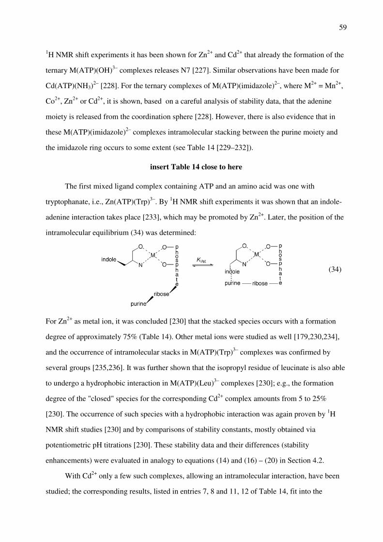
59
1H NMR shift experiments it has been shown for Zn2+ and Cd2+ that already the formation of the
ternary M(ATP)(OH)3– complexes releases N7 [227]. Similar observations have been made for
Cd(ATP)(NH3)2– [228]. For the ternary complexes of M(ATP)(imidazole)2–, where M2+ = Mn2+,
Co2+, Zn2+ or Cd2+, it is shown, based on a careful analysis of stability data, that the adenine
moiety is released from the coordination sphere [228]. However, there is also evidence that in
these M(ATP)(imidazole)2– complexes intramolecular stacking between the purine moiety and
the imidazole ring occurs to some extent (see Table 14 [229–232]).
insert Table 14 close to here
The first mixed ligand complex containing ATP and an amino acid was one with
tryptophanate, i.e., Zn(ATP)(Trp)3–. By 1H NMR shift experiments it was shown that an indole-
adenine interaction takes place [233], which may be promoted by Zn2+. Later, the position of the
intramolecular equilibrium (34) was determined:
(34)
For Zn2+ as metal ion, it was concluded [230] that the stacked species occurs with a formation
degree of approximately 75% (Table 14). Other metal ions were studied as well [179,230,234],
and the occurrence of intramolecular stacks in M(ATP)(Trp)3– complexes was confirmed by
several groups [235,236]. It was further shown that the isopropyl residue of leucinate is also able
to undergo a hydrophobic interaction in M(ATP)(Leu)3– complexes [230]; e.g., the formation
degree of the "closed" species for the corresponding Cd2+ complex amounts from 5 to 25%
[230]. The occurrence of such species with a hydrophobic interaction was again proven by 1H
NMR shift studies [230] and by comparisons of stability constants, mostly obtained via
potentiometric pH titrations [230]. These stability data and their differences (stability
enhancements) were evaluated in analogy to equations (14) and (16) – (20) in Section 4.2.
With Cd2+ only a few such complexes, allowing an intramolecular interaction, have been
studied; the corresponding results, listed in entries 7, 8 and 11, 12 of Table 14, fit into the

60
general picture: From entries 1 and 2 in Table 14 it is evident that the size of the aromatic-ring
systems (uracil versus adenine) matters, whereas a replacement of AMP2– by ATP4– in the
ternary complex (entry 3) does hardly affect the stacking intensity, even though the overall
stability of the latter complex (log KCu(Phen)(ATP)
Cu(Phen) = 6.88 ± 0.07 [223] is by a factor of about 1000
larger (log KCu(Phen)(AMP)
Cu(Phen) = 3.89 ± 0.01 [232]). As expected, replacement of the 3-ring Phen by
Bpy reduces the formation degree of the intramolecular stack (entries 4, 5). It is also expected
that the stacking interaction between the indole moiety of tryptophanate (Trp–) and the adenine
residue in Zn(ATP)(Trp)3– is more pronounced than the hydrophobic interaction between the
isopropyl residue of leucinate (Leu–) and adenine in Zn(ATP)(Leu)3– (entries 9, 10). The
Cd(ATP)(Leu)3– complex fits into the picture. Maybe here it should be mentioned that the 1H
NMR experiments allow a direct proof of intramolecular adduct formation, but from
potentiometric stability data usually the more exact formation degrees are obtained. The last
entry (no. 12) in Table 14 refers to Cd(ATP)(imidazole)2– for which, just like for
Zn(ATP)(imidazole)2– [229], stack formation between the adenine residue and the coordinated
imidazole could be proven [229]. This result is in line with a suggestion [237] based on
thermodynamic parameters that in Zn(ATP)(histamine)2– stacking occurs.
Finally, the above discussion should not distract from the fact that certain ligand
combinations, as discovered nearly 50 years ago [238], can give rise to enhanced complex
stabilities, i.e., the combination of a heteroaromatic N base (Arm) and an O donor ligand in
combination with a transition metal ion leads to indirect effects being mediated by the metal ion
[56,200,203,204]. For example, the combinations Cu(Bpy/Phen)2+/HPO [239],
Mn(Bpy)2+/malonate [203] or M(Arm)2+/pyrocatecholate [240] give rise to enhanced complex
stabilities. Clearly, the direct (as discussed in this section) and indirect ligand-ligand interactions
[200] that occur in mixed ligand complexes are of relevance regarding the discrimination and
selectivity observed in nature.
9 Cadmium(II) Binding in Dinucleotides and Dinucleoside Monophosphates
One of the important questions in metal ion-nucleic acid coordination is to find out to what

61
extent neighboring nucleotide units affect binding of a metal ion at a given site [58]. Some
answers to this question can be found by studying ligands of the kind shown in Figure 16.
Unfortunately, the information regarding Cd2+ complexes and dinucleotides in general is limited
to the nucleotide/nucleoside derivatives shown in Figure 16.
insert Figure 16 close to here (width: 9 cm)
9.1 The Phosphodiester Link
If one recalls from Section 6.2.2 that the uracil moiety is not involved in metal ion binding,
meaning that the log stability constants of the Cd(UMP) complex fits on the straight-reference
line seen in Figure 11, one may expect that this "indifference" of the uracil residue also holds for
the pUpU3– ligand. Indeed, if one applies the acidity constant pKH(pUpU)
H = 6.44 (Table 15,
footnote "b") to the straight-line equation (25), one obtains log K
Cd(R-PO3 )Cd = 2.52 ± 0.05, which
is 0.23 ± 0.05 log unit [33] lower than the measured stability constant log KCd(pUpU)
Cd = 2.75 ±
0.03 (Table 15 [241–243]).
Because a metal ion coordinated at the terminal phosphate group of pUpU3– will "feel" the
negative charge of the phosphodiester bridge even if no 10-membered chelate is formed, some
stability enhancement (eq. 14) is expected. Interestingly, for the complexes M(pUpU)– of Mg2+,
Mn2+, and Cd2+ the stability enhancement is within the error limits identical giving on average
log ∆M/pUpU/charge = 0.24 ± 0.04. This result for the three metal ions having rather different
properties can only mean that the given value represents indeed the charge effect and that none
of the three metal ions forms a 10-membered chelate with the diester bridge (a chelate occurs
with Zn2+ and Pb2+ [33]) or interacts with the uracil moiety.
insert Table 15 close to here
9.2 The Guanine Residue in a Dinucleotide
If one views the data for the Cd[d(pGpG)]– complex (second entry of Table 15) with the

62
presented result for the Cd(pUpU) complex in mind, it is immediately evident that a new
situation occurs: log ∆Cd/d(pGpG) = 1.45 ± 0.08 [241] is far beyond the indicated charge effect
(Section 9.1). In fact, the charge effect of the bridging phosphodiester unit is expected to be the
same with pUpU3– and d(pGpG)3–; furthermore, if coordinated to the terminal phosphate group
Cd2+ does not interact with the diester bridge as seen from Cd(pUpU)–, thus, it follows that in
Cd[d(pGpG)]– a guanine-N7 interaction (eq. 22) must take place for which the charge-corrected
stability enhancement log ∆Cd/d(pGpG)/charge,cor = (1.45 ± 0.08) – (0.24 ± 0.04) = 1.21 ± 0.09, holds.
Application of equations (14) and (16) – (20) leads to a formation degree for Cd[d(pGpG)]
of 94 ± 1.5% [241]. Are both guanine residues involved in this macrochelate formation or only
one? A tentative answer may be obtained if one considers the stability enhancement for the
Cd(dGMP) complex, which is unfortunately not known, but which may be estimated because the
information for both Zn(GMP) [50] and Zn(dGMP) [244] exists. This estimate is justified
because Zn(GMP) and Cd(GMP) reach within the error limits the same formation degree [50] for
M(GMP)cl/N7 (ca. 82%), despite the different overall stability constants. Because of the
somewhat higher basicity of N7 in dGMP, compared with that in GMP [245], one expects a
somewhat larger formation degree of the chelate for the deoxy complex. Indeed, this follows
from the given reasonings:
log ∆Cd/dGMP = log ∆Cd/GMP + log ∆Zn/dGMP – log ∆Zn/GMP
= (0.79 ± 0.06) + (0.84 ± 0.08) – (0.69 ± 0.07)
= 0.94 ± 0.12
This result corresponds to 88.5 ± 3.2% for Cd(dGMP)cl/N7. This stability enhancement for the
Cd(dGMP) complex is by 0.27 ± 0.15 log unit smaller than the one obtained for Cd[d(pGpG)]–
indicating that in the dinucleotide complex both guanine residues (Figure 16) are involved to
some extent.
It may be added that for the Cd[d(pGpG)]– system also the monoprotonated species, that is,
Cd[H;d(pGpG)], was observed [241], where the proton is located at the terminal phosphate
group and Cd2+ at guanine-N7. From other purine systems it is known that a metal ion bound to
N7 in a nucleotide complex may form macrochelates by also interacting with the P(O)2(OH)–
residue [97,162]. Indeed, a detailed evaluation provided a stability enhancement of log

63
∆Cd/H;d(pGpG) = 0.27 ± 0.18 for Cd[H;d(pGpG)] and this corresponds to a formation degree for the
chelated species of 46 ± 22% [241]. Hence, taken together, all these results prove that metal ions
are able to bridge the distance between a phosphate group and an N7 site in a d(GMP) unit. This
is of relevance for the interaction of metal ions with nucleic acids.
9.3. The Non-bridging Sulfur of the Thiophosphodiester Link
Considering the already previously indicated importance of so-called "rescue" experiments in
ribozyme chemistry (Sections 7.2 and 10.1), it is interesting to see that for Cd(pUp(s)U)– a
stability enhancement of log ∆Cd/pUp(s)U = 0.68 ± 0.09 is observed (Table 15 [242]) (Figure 16). If
this value is charge-corrected in the way explained above, one obtains log ∆Cd/pUp(s)U/charge,cor =
(0.68 ± 0.09) – (0.24 ± 0.04) = 0.44 ± 0.10. Since no interaction with an oxygen in the diester
bridge occurs in Cd(pUpU)–, the whole charge-corrected stability enhancement needs to be
attributed to a Cd2+/S interaction with the non-bridging S of the thiophosphodiester link. The
corresponding 10-membered chelate reaches a formation degree of 64 ± 8% [242].
It is worth to note that no stability enhancement is observed for the Mg(pUp(s)U)– and
Mn(pUp(s)U)– complexes. Clearly, this observation has consequences for the "rescue"
experiments in ribozyme chemistry [28].
9.4 Dinucleoside Monophosphates
Another interesting molecule in the present context is IpI– (Figure 16) for which the Cd2+
complexes Cd(IpI)+ and Cd(IpI – H) have been studied [75]. Since the stability constant of
Cd(IpI)+ could only be estimated, we will not consider this complex further, though it ought to
be mentioned that it is transformed to Cd(IpI – H) with pKCd(IpI)
H = 6.7 ± 0.3, meaning that the
deprotonation reaction occurs close to the physiological pH range. By using the Cd2+ complexes
of inosine as a basis a stability enhancement of log ∆Cd/(IpI – H) = 0.40 ± 0.30 was estimated for the
Cd(IpI – H) complex and this corresponds to a formation degree of 60% (with lower and upper
limits of 21 and 80%, respectively) [75] for the chelate.

64
Which is the structure of the closed or chelated species? Since N7 of a 5'-purine unit is in
general somewhat more basic than the one of a 3' unit [38,48], one may assume that the initial
complex formation in IpI– occurs preferably at N7 of the 5'-inosine residue. This binding mode
also allows an outersphere interaction with the (C6)O site, especially after deprotonation of
(N1)H [74,119,162], as well as a maximal electrostatic interaction with the negatively charged
phosphoryl bridge and possibly even macrochelate formation to a certain extent, as has been
suggested before for an intermediate Pt(II) complex with a related dinucleoside monophosphate
[246]. Such a structure is also well known for M(IMP) and related complexes (Section 6.2.3)
[40,50,117]. Hence, one may conclude that two types of macrochelates are possible: (i) the one
indicated above between N7 and the phosphodiester bridge or (ii) one that involves both N7 sites
of (IpI – H)2–. Of course, both types of macrochelates may occur in equilibrium with each other,
and then the formation degree mentioned above encompasses both species.
Considering that Mg2+ does not form significant amounts of Mg(IpI – H)cl macrochelates
[75] (whereas Co2+ and Zn2+ do), we favor N7/N7 macrochelates. However, in doing so, one has
to answer the question why no significant amounts of macrochelates are observed with the
Cd(GpG – H) and Cd[d(GpG – H)] systems (Figure 16) [38]. If one applies the indicated
evaluation procedure [75] to the data of ref. [38], one notes that the log ∆Cd/(GpG – H) and log
∆Cd/d(GpG – H) values are zero within their error limits. This agrees with the earlier conclusion [38]
that "no hint for the formation of significant amounts of intramolecular chelates involving both
N7 sites" have been found, though low concentrations could still occur, of course. A possible
explanation could be that the intramolecular stacking interaction in (IpI – H)2– is smaller than in
(GpG – H)2–, and in fact, this is expected [137,247,248]. Consequently, (IpI – H)2– is more
flexible for adapting to the configuration needed in aqueous solution for an N7/N7 macrochelate
which is known to form with the kinetically inert cis-(NH3)2Pt2+ and both dinucleotide
monophosphates [249,250]. One should emphasize here that the amount of free energy involved
to shift the situation from one side to another is very small; for example, a formation degree of
20% of a macrochelate at 25°C corresponds only to a stability enhancement of log ∆M/ligand = 0.1
(eq. 14) and a change in free energy of ∆G0 = –0.57 kJ/mol [52].

65
10 Cadmium(II) Binding to Nucleic Acids
Interest in the interaction of cadmium(II) with nucleic acids was triggered, at least in part, by the
discovery of Wacker and Vallee [7] more than 50 years ago that RNA from the horse kidney
cortex contained not only essential metal ions like those of Mg, Ca, Mn, Fe or Zn, but also Cd.
The toxicity of cadmium(II) is well established for many years (see Chapters 1, 14, and 15) and
it appears that it is primarily due to Cd(II)-protein interactions. However, due to its high affinity
for R–S– groups Cd2+ may affect thiolate-disulfide equilibria and thus the redox state of the cell.
This in turn means that Cd2+ may stimulate the production of reactive oxygen species (ROS) and
these may lead to DNA damage [251]. In addition, cadmium(II) interferes with DNA repair
pathways of the cell by directly inhibiting the relevant enzymes or by hampering their binding to
aberrant DNA sites [251].
Despite the obvious toxic effects on living cells, Cd2+ is often used in in vitro studies as a
probe of Mg2+ or Ca2+ (see Chapter 6) having the advantage of a higher affinity towards most
ligands, including nucleotides and their constituents, as described in the previous sections. Below
we consider now larger entities.
10.1 Cadmium(II)-Rescue Experiments
As indicated above in Section 7.2, Cd2+ binding to phosphorothioate nucleotides can be exploited
in so-called rescue experiments where one takes advantage of the higher affinity of Cd2+ towards
sulfur ligands compared to Mg2+ [170,252]. Such a rescue is also part of so-called nucleotide
analogue interference mapping/suppression (NAIM/NAIS) experiments, which are used to
identify catalytically crucial atoms within a RNA and/or tertiary contacts [253–255]. This
technique has been widely applied to study metal ion binding sites and their influence on
catalytic RNAs, i.e., ribozymes [256–262]. Major limitations of this method are the fact that (i)
T7 RNA polymerase, which is generally used for the transcription of defined RNA sequences,
inserts only Sp thionucleotides under inversion of their conformation to Rp. Consequently, in such
T7-transcribed RNA molecules only the Rp configuration can be studied. (ii) Sulfur has a slightly

66
larger radius compared to oxygen, which can lead to small perturbations in local geometry of a
metal ion-binding pocket or a tertiary interaction within the global fold of the RNA. In such
cases, the sulfur substitution cannot be rescued by the addition of Cd2+.
The principle of cadmium(II) rescue experiments is as follows (see also refs [8,10]):
Phosphorothioate nucleotides are inserted into a RNA at single, defined sites. In cases of
significantly decreased or abolished enzymatic activity of the RNA and subsequent restoration of
activity upon addition of soft metal ions like Cd2+, one assumes that the respective phosphate
moiety coordinates a metal ion which is relevant for the catalytic activity of the ribozymes.
The technique was first applied to the enzymes arginine kinase and creatinine kinase.
Studies on ATPγS and on the stereospecificity for diastereomers of ATPαS and ATPβS in the
presence of Mg2+ and Cd2+ [263,264] proved that metal ions coordinate to the respective
phosphate groups at certain steps of the catalytic cycle. Later, as the enzymatic nature of some
RNA molecules was discovered, the technique proved powerful in shedding light on their
catalytic mechanism as is exemplified with the following few examples.
The different metal ion-binding modes of the two classes of RNAse P, a ribonuclease and
ribozyme, could be revealed [265,266]. For group II introns, it was shown that a metal ion is
required to stabilize the leaving group in both steps of the splicing reaction [267]. In addition, the
results of this study were consistent with the findings on the catalysis of the eukaryotic
spliceosome [267,268], thus further corroborating a common ancestry of these two machineries.
In the case of the group I intron, another large ribozyme, cadmium(II) rescue experiments helped
to develop a very detailed understanding of the catalytic mechanism. It was found that three
metal ions are required for the splicing mechanism, which bind in the active site and
simultaneously contact the attacking nucleophile and the scissile phosphate [269,270].
Cadmium(II) binding was also intensively used to study metal ion requirements for the
catalysis of the self-cleaving hammerhead ribozyme. Also here, phosphorothioate studies helped
to prove the coordination of divalent metal ions during catalysis [271,272]. Cd2+ binds with a KD
of 25 µM in the ground state thereby increasing the activity 104-fold. The KD of Cd2+ in the
transition state was calculated to be 2.5 nM indicating that the metal ion recruits at least one
more ligand in the transition state. Confusion was caused by the observation that this Cd2+

67
binding site was situated at a 20 Å distance from the scissile phosphate; this discrepancy
originated in the fact that earlier studies were performed with a minimal ribozyme construct that
lacked important tertiary contacts [273]. The Cd2+ interactions were also investigated using 31P
and 15N NMR, Cd2+ binding leading to an upfield shift of the thiophosphate-31P or guanine-15N7
resonance, respectively [274–277]. The catalytic activity of the hammerhead ribozyme was also
shown to be in principle independent of divalent metal ions [278]: In the presence of
unphysiologically high, i.e., molar, concentrations of monovalent cations, activity can be partly
rescued, thus confirming that a direct M2+ binding to the active site is not mandatory but that also
M+ ions can be catalytically competent. Such an exchange of low concentration of divalent ions
versus high concentrations of monovalent metal ions is well known and is explained by their
differences in the binding affinities [127,279,280].
10.2 Crystal Structures of RNA or DNA-Protein Complexes Containing Cd2+
More detailed information on cadmium(II) binding to nucleic acids is available from a small
number of crystal structures of RNA as well as RNA- or DNA-protein complexes. There are
only 23 entries in the Protein Data Base that contain Cd2+ ions interacting with at least one atom
of a DNA or RNA and of these 18 structures represent different parts of the 50s subunit of the
prokaryotic ribosome; the other five are structures of a Diels-Alder ribozyme (1YKQ [281]), the
product complex of the hammerhead ribozyme (488D [282]), a prokaryotic enzyme mediating
DNA bending called "integration host factor" in complex with a DNA substrate (1IHF [283],
2HT0 [284]) and DNA polymerase β in complex with DNA and ATP (8ICE [285]).
Most of the binding sites identified in these structures confirm what is stated in the
previous sections on Cd2+ binding to isolated nucleotides. Innersphere contacts are almost
exclusively formed by guanine N7 or, to a lesser extent, by adenine N7 or one of the non-
bridging phosphate oxygens (OP1 and OP2). Additional outersphere contacts are highly
abundant. In case of innersphere binding the Cd2+–ligand distance is about 2 Å. If guanine N7 is
innersphere coordinated, then O6 of the same guanine is always in a distance to enable an
outersphere interaction (usually at a distance of about 3 Å). One example of such a coordination

68
pattern is found in the hammerhead ribozyme, where Cd2+ strongly influences catalytic activity
(see Section 10.1). The sites G10.1 N7 and A9 OP2 constitute the innersphere contacs with the
Cd2+ ion while numerous further atoms of five different nucleotides interact by outersphere
coordination with the Cd2+ ion from distances between 2.9 and 5.6 Å (Figure 17) [282,286].
insert Figure 17 in color close to here (width: 10 cm)
Apart from the above mentioned coordination pattern, Cd2+ contacts to nearly any nitrogen
or oxygen atom in nucleotides can be observed. The total number of potential interactions to a
single Cd2+ ion varies greatly depending on the binding site. The same is true for the number of
involved residues that ranges from 2 to 6. In general, one nucleobase (mostly the one coordinated
via N7) provides most further atoms to bind the Cd2+ ion while the residues at the n+1 and n–1
position and, if available, its base pairing partners provide few additional liganding atoms.
The only Cd2+-containing RNA-protein complex deposited in the Protein Data Bank is the
afore mentioned 50s subunit of the ribosome. Surprisingly for such a giant molecular assembly,
the maximum number is eight Cd2+ atoms bound in one structure (1MMS [287]), seven of which
comprise contacts to RNA. Two special Cd2+ coordination sites are detected in this 50s RNA. In
the first case, uracil O4 is the only innersphere contact to Cd2+ with no N7 or phosphate oxygen
being within 5 Å. In two further binding sites, one of which comprises numerous liganding
atoms from six different nucleotides, rare innersphere contacts between Cd2+ and the ribose O2'
are observed. These special cases illustrate how increasingly complex molecular assemblies open
up coordination options by providing a complex and defined overall geometry. This allows for a
summation of contributions of several weak interactions or facilitates weak interactions by a
spatial pre-arrangement of strong interactions.
To the best of our knowledge there is no structure available of only DNA with bound
Cd(II) ions. The structures of the prokaryotic DNA-bending protein IHF (integration host factor)
with doubly nicked DNA (PDB 2HT0) or singly-nicked DNA (PDB 1IHF) mostly display
conservative binding modes involving guanine and adenine N7 or phosphate oxygens. Often, a
combination of amino acids, DNA nucleotides, and water molecules constitute the binding sites
for the single Cd2+ ions.
The PDB structure of DNA polymerase with 7 base pairs of DNA and ATP bound

69
(PDB 8ICE) contains just one binding site in which DNA is involved. The cadmium(II) has
contacts to four different amino acids and two phosphate oxygens; it is also in coordination
distance to O5' of adenine and O3' of a guanine. Additionally, four oxygen atoms of
pyrophosphate coordinate the Cd2+. This is a good example of how DNA-Cd2+ contacts can be
facilitated by another main binding site. In this case, the pyrophosphate is required to bind a
metal ion in this site as in its absence the site remains unoccupied [285].
10.3. Cadmium(II) as Probe in EPR and NMR Spectroscopy
Cadmium(II)-induced EPR silencing was applied to evaluate the affinity of Mn2+ to several
distinct binding sites in the Diels-Alder ribozyme [288]. As Cd2+ is EPR-silent, it was used to
saturate four of five binding sites for divalent metal ions of the ribozyme to selectively study the
affinity of Mn2+ for each binding site separately. This method relies on the fact that a Cd2+ ion
once bound to the ribozyme cannot be substituted by Mn2+ since its affinity to all five binding
sites is higher.
As mentioned above in Section 10.1, Cd2+ is readily applied in combination with
thiophosphate derivatives using 31P NMR making advantage of the chemical shift dispersion of
the thiophosphate group as well as the higher affinity [274–277]. Similarly, Cd2+ can also be
applied as mimic of Mg2+ in chemical shift titration experiments, its higher affinity being of
advantage for better spectral quality [289]. In addition, 111Cd or 113Cd are very good NMR nuclei
and regularly used in proteins to investigate Cd2+ binding [290,291] (see also Chapter 6). In
larger nucleic acids, such studies are not as straight forward as with proteins due to the much
lower affinity and faster exchange rates. Consequently, no distinct signal for 111/113Cd is observed
upon binding. We are aware of only one example where a change in chemical shift of the 113Cd
is observed upon addition to the central core of a group II intron ribozyme [292].
11 Concluding Remarks
Cadmium(II) is a metal ion that displays distinct coordinating properties with a mostly
70
octahedral coordination sphere. It is commonly classified as a relatively soft ion and it prefers
thus aromatic-nitrogen sites, especially N7 of purine residues. However, interactions with
phosphate oxygens including those of the phosphodiester bridge are also not negligible and,
provided a suitably located primary binding site is available, also hydroxyl and carbonyl groups
may coordinate. Overall, one can conclude that the coordination chemistry of Cd2+ with
nucleotides and the corresponding substituents is relatively well understood today; this includes
the relations between the toxic Cd2+ and the essential Zn2+, Ca2+, and Mg2+ ions, for which it is
often employed as a probe. However, with regard to less common nucleotides, which also occur
in Nature, like thio-pyrimidine or -purine derivatives, our knowledge is very scarce and
considerably more work is needed. This is of high relevance because Cd2+-thio interactions are
very stable, meaning that such an interaction can totally change the properties of a nucleic acid
complex because commonly metal ion-nucleic acid interactions are weak compared to those of
proteins.
Indeed, Cd2+ coordinates to nucleic acids often not only directly, like with purine-N7 sites,
but also indirectly to oxygen and (other) nitrogen sites as is exemplified by many outersphere
interactions found in nucleic acid structures. Most important in this respect is also the high
affinity of Cd2+ towards sulfur ligands. These characteristics make Cd2+ on the one hand a toxic
element that interferes especially with proteins in numerous metabolic pathways, but on the other
hand it is also an interesting mimic for other metal ions in in vitro studies. Especially as a
replacement for Ca2+ and Mg2+ it is regularly applied in nucleic acid research.
The coordination chemistry of Cd2+ towards the building blocks of nucleic acids is well
established and the intrinsic affinities to almost all possible coordinating atoms are known. This
knowledge should provide a perfect basis to investigate in the future Cd2+ binding also to larger
and more complex nucleic acid structures. The higher intrinsic affinity of Cd2+ compared to
many other divalent metal ions, especially Mg2+ and Ca2+, allows in many cases a more detailed
investigation of the coordination pattern. In the case of ribozymes, a differing catalytic rate in the
presence of Cd2+ versus that of Mg2+ should in principle allow to draw conclusions on Mg2+
binding. However, such investigations are in many instances severely hampered by the fact that
even with Cd2+ it is often highly challenging to characterize a binding pocket down to the single

71
coordinating atoms. This is due to the size of the RNA itself and the limited chemical diversity
of its building blocks, which make it very difficult to distinguish individual nucleotides. Highly
promising in this respect are combinations of 13C, 15N, and 113Cd NMR, which allow to observe
both the RNA liganding sites and the metal ion simultaneously. Although hampered by the fast
exchange of the metal ion in nucleic acids, the increasing sensitivity of NMR probeheads, also
for hetero nuclei, will certainly lead to new developments and experiments in the future.
Abbreviations and Definitions
The abbreviations for the nucleobases and nucleosides are defined in Figure 1 and the labeling
systems for the di- and triphosphate residues in Figure 9. Other abbreviations are defined below.
Ac– acetate (Figure 9)
AcP2– acetyl phosphate (Figure 3)
Ade adenine (Figure 1)
Ado adenosine (Figure 1)
ADP3– adenosine 5'-diphosphate (Figure 10)
AMP2– adenosine 5'-monophosphate (Figure 3)
AMPS2– adenosine 5'-O-thiomonophosphate
AnP2– acetonylphosphonate (Figure 3)
ATP4– adenosine 5'-triphosphate (Figure 10)
Bicine N,N-bis(2-hydroxyethyl)glycine (Figure 15)
Bistris 2-[bis(2-hydroxyethyl)amino]-2-(hydroxymethyl)-1,3-propanediol (Figure
15)
Bpy 2,2'-bipyridine
C2S 2-thiocytidine
CDP3– cytidine 5'-diphosphate (Figure 10)
CMP2– cytidine 5'-monophosphate (Figure 10)
CTP4– cytidine 5'-triphosphate (Figure 10)

72
Cyd cytidine (Figure 1)
Cyt cytosine (Figure 1)
dCMP2– 2'-deoxycytidine 5'-monophosphate
dGMP2– 2'-deoxyguanosine 5'-monophosphate
d(GpG)– 2'-deoxyguanylyl(3'→5')-2'-deoxyguanosine (Figure 16)
dGuo 2'-deoxyguanosine
DHAP2– dihydroxyacetone phosphate (Figure 3)
Dien diethylenetriamine (= 1,4,7-triazaheptane)
DMBI 1,4-dimethylbenzimidazole (= 6,9-dimethyl-1,3-dideazapurine) (Figure 5)
d(pGpG)3– 2'-deoxyguanylyl(5'→3')-2'-deoxy-5'-guanylate (Figure 16)
dTDP3– thymidine [= 1-(2-deoxy-β-D-ribofuranosyl)thymine] 5'-diphosphate
dThd thymidine [= 1-(2-deoxy-β-D-ribofuranosyl)thymine (Figure 1)
dTMP2– thymidine [= 1-(2-deoxy-β-D-ribofuranosyl)thymine] 5'-monophosphate
dTTP4– thymidine [= 1-(2-deoxy-β-D-ribofuranosyl)thymine] 5'-triphosphate
ε dielectric constant (or permittivity)
ε-Ado ε-adenosine (= 1,N6-ethenoadenosine)
ε-AMP2– ε-adenosine 5'-monophosphate (= 1,N6-ethenoadenosine 5'-monophosphate)
(Figure 14)
En ethylenediamine (= 1,2-diaminoethane)
EPR electron paramagnetic resonance
9EtG 9-ethylguanine
FMN2– flavin mononucleotide (= riboflavin 5'-phosphate) (Figure 13)
G1P2– glycerol 1-phosphate (Figures 3 and 13)
GDP3– guanosine 5'-diphosphate
GMP2– guanosine 5'-monophosphate
GpG– guanylyl(3'→5')guanosine (Figure 16)
GTP4– guanosine 5'-triphosphate
Gua guanine (Figure 1)
Guo guanosine (Figure 1)

73
HMP2– hydroxymethylphosphonate (Figure 4)
HOAc– hydroxyacetate (Figure 4)
HOMPy o-(hydroxymethyl)pyridine (Figure 4)
H(Or) orotidine (Figure 8)
Hyp hypoxanthine (Figure 1)
I ionic strength
IDP3– inosine 5'-diphosphate
IHF integration host factor
Im imidazole
IMP2– inosine 5'-monophosphate
Ino inosine
IpI– inosylyl(3'→5')inosine (Figure 16)
ITP4– inosine 5'-triphosphate
KI intramolecular equilibrium constant
Ka general acidity constant
L general ligand
Leu leucine
Leu– leucinate
M2+ divalent metal ion
1MBI 1-methylbenzimidazole (Figure 5)
9MeA 9-methyladenine
9MeHyp 9-methylhypoxanthine
9MeG 9-methylguanine
MeOPS2– methyl thiophosphate
1MIm 1-methylimidazole (Figure 5)
2MPy 2-methylpyridine (Figure 5)
NAIM/NAIS nucleotide analogue interference mapping/suppression
NB nucleobase derivative
NDP3– nucleoside 5'-diphosphate

74
NMP2– nucleoside 5'-monophosphate
NP nucleoside phosphate
NTP4– nucleoside 5'-triphosphate
oh octahedral
OMP3– orotidinate 5'-monophosphate
oPyN ortho-aminopyridine-type ligand (Figure 7)
Or– orotidinate (Figure 8)
PDB Protein Data Bank
PEEA2– dianion of 9-[2-(phosphonoethoxy)ethyl]adenine
Phen 1,10-phenanthroline
PME2– (phosphonomethoxy)ethane = ethoxymethanephosphonate
PMEA2– dianion of 9-[2-(phosphonomethoxy)ethyl]adenine (= Adefovir) (Figure 14)
PMEApp4– diphosphorylated PMEA2–
PS2– thiophosphate monoester
pUpU3– uridylyl-(5'→3')-[5]uridylate (Figure 16)
Py pyridine
PyN pyridine-type ligand (Figure 7)
R-DP3– diphosphate monoester (Figure 9)
RibMP2– D-ribose 5-monophosphate (cf. Figure 1)
R-MP2– monophosphate monoester (Figure 9)
(RO)2PO phosphodiester unit (Figure 9)
monophosphate monoester and/or phosphonate ligand
R-TP4– triphosphate monoester (Figure 9)
Thy thymine (Figure 1)
Tris 2-amino-2-(hydroxymethyl)-1,3-propanediol (Figure 15)
Trp tryptophan
Trp– tryptophanate
Tu tubercidin (Figure 5)
TuMP2– tubercidin 5'-monophosphate ( = 7-deaza-AMP2–)

75
U uridine-type ligand
(U – H)– uridinate-type ligand
U2S 2-thiouridine (Figure 8)
U4S 4-thiouridine (Figure 8)
UDP3– uridine 5'-diphosphate
UMP2– uridine 5'-monophosphate
UMPS2– uridine 5'-O-thiomonophosphate (Figure 14)
Ura uracil (Figure 1)
Urd uridine (Figure 1)
UTP4– uridine 5'-triphosphate
Xao xanthosine (Figure 8)
XMP3– xanthosinate 5'-monophosphate = (XMP – H)3– = (X – H·MP)3– (Figure 13)
Species written in the text without a charge do not carry one or represent the species in general
(i.e., independent of the protonation degree); which of the two possibilities applies is always
clear from the context. In formulas such as M(H;NMP)+ the H+ and NMP2– are separated by a
semicolon to facilitate reading; yet they appear within the same parenthesis to indicate that the
proton is at the ligand without defining its location. A formula like (NB – H)– means that the
ligand, here a nucleobase residue, has lost a proton and it is to be read as NB minus H+. The term
(aq) is used to indicate that water is acting as a ligand.
Acknowledgments
Financial support from the Swiss National Science Foundation (R.K.O.S.), the European
Research Council (ERC Starting Grant to R.K.O.S.), the Universities of Zürich (R.K.O.S.) and
Basel (H.S.), and within the COST Action CM1105 (R.K.O.S.) are gratefully acknowledged.
References
1. J. K. Dunnick, B. A. Fowler, in Handbook on Toxicity of Inorganic Compounds, Eds H. G.

76
Seiler, H. Sigel, A. Sigel, Marcel Dekker, New York, 1988, pp. 155–175.
2. C. E. Housecroft, A. G. Sharpe, Inorganic Chemistry, Pearson Education, Harlow, UK,
2001, p. 579.
3. L. Thunus, R. Lejeune, in Handbook on Metals in Clinical and Analytical Chemistry, Eds
H. G. Seiler, A. Sigel, H. Sigel, Marcel Dekker, New York, 1994, p. 668.
4. R. F. M. Herber, in Handbook on Metals in Clinical and Analytical Chemistry, Eds H. G.
Seiler, A. Sigel, H. Sigel, Marcel Dekker, New York, 1994, pp. 283–297.
5. Y. Xu, L. Feng, P. D. Jeffrey, Y. Shi, F. M. M. Morel, Nature 2008, 452, 56–61.
6. T. W. Lane, M. A. Saito, G. N. George, I. J. Pickering, R. C. Prince, F. M. M. Morel,
Nature, 2005, 435, 42.
7. W.E. C. Wacker, B. L. Vallee, J. Biol. Chem. 1959, 234, 3257–3262.
8. M. C. Erat, R. K. O. Sigel, Met. Ions Life Sci. 2011, 9, 37–100.
9. D. Donghi, J. Schnabl, Met. Ions Life Sci. 2011, 9, 197–234.
10. M. Pechlaner, R. K. O. Sigel, Met. Ions Life Sci. 2012, 10, 1–42.
11. K. Aoki, Met. Ions Biol. Syst. 1996, 32, 91–134.
12. R. B. Martin, Y. H. Mariam, Met. Ions Biol. Syst. 1979, 8, 57–124.
13. R. Tribolet, H. Sigel, Eur. J. Biochem. 1987, 163, 353–363.
14. D. B. Davies, P. Rajani, H. Sadikot, J. Chem. Soc., Perkin Trans. 2, 1985, 279–285.
15. R. B. Martin, Met. Ions Biol. Syst. 1986, 20, 21–65.
16. P. B. Hammond, E. C. Foulkes, Met. Ions Biol. Syst. 1986, 20, 157–200.
17. R. B. Martin, in Handbook on Toxicity of Inorganic Compounds, Eds H. G. Seiler, H.
Sigel, A. Sigel, Marcel Dekker, New York, 1988, pp. 9–25.
18. R. B. Martin, Met. Ions Biol. Syst. 1984, 17, 1–49.
19. E. Freisinger, R. K. O. Sigel, Coord. Chem. Rev. 2007, 251, 1834–1851.
20. R. D. Shannon, Acta Cryst. 1976, A32, 751–767.
21. H. Sigel, R. B. Martin, Chem. Soc. Rev. 1994, 23, 83–91.
22. H. Sigel, D. B. McCormick, Acc. Chem. Res. 1970, 3, 201–208.
23. C. F. Baes, Jr., R. E. Mesmer, The Hydrolysis of Cations, Krieger Publishing Co., Malabar,
Florida, 1976, pp. 1–496.

77
24. A. M. Pyle, Met. Ions Biol. Syst. 1996, 32, 479–520.
25. NIST Critically Selected Stability Contstants of Metal Complexes, Reference Database 46,
Version 8.0; data collected and selected by R. M. Smith, A. E. Martell; U. S. Department
of Commerce, National Institute of Standards and Technology, Gaithersburg, MD, 2004.
26. R. K. O. Sigel, H. Sigel, in Bioinorganic Fundamentals and Applications; Metals in
Natural Living Systems and Metals in Toxicology and Medicine, Eds V. L. Pecoraro, T.
Hambley, in Volume 3 of Comprehensive Inorganic Chemistry II, Eds J. Reedijk, K.
Poeppelmeier, Elsevier, Oxford, UK, 2012, in press.
27. N. Saha, H. Sigel, J. Am. Chem. Soc. 1982, 104, 4100–4105.
28. R. K. O. Sigel, Eur. J. Inorg. Chem. 2005, 2281–2292.
29. H. Sigel, F. Hofstetter, R. B. Martin, R. M. Milburn, V. Scheller-Krattiger, K. H. Scheller,
J. Am. Chem. Soc. 1984, 106, 7935–7946.
30. R. K. O. Sigel, A. M. Pyle, Chem. Rev. 2007, 107, 97–113.
31. A. Mucha, B. Knobloch, M. Jeżowska-Bojczuk, H. Kozłowski, R. K. O. Sigel, Chem. Eur.
J. 2008, 14, 6663–6671.
32. H. Sigel, Chem. Soc. Rev. 1993, 22, 255–267.
33. B. Knobloch, D. Suliga, A. Okruszek, R. K. O. Sigel, Chem. Eur. J. 2005, 11, 4163–4170.
34. B. Lippert, Prog. Inorg. Chem. 2005, 54, 385–447.
35. B. Knobloch, H. Sigel, J. Biol. Inorg. Chem. 2004, 9, 365–373.
36. L. E. Kapinos, B. P. Operschall, E. Larsen, H. Sigel, Chem. Eur. J. 2011, 17, 8156–8164.
37. H. Sigel, E. M. Bianchi, N. A. Corfù, Y. Kinjo, R. Tribolet, R. B. Martin, J. Chem. Soc.,
Perkin Trans. 2, 2001, 507–511.
38. C. P. Da Costa, H. Sigel, Inorg. Chem. 2003, 42, 3475–3482.
39. H. Pezzano, F. Podo, Chem. Rev. 1980, 80, 365–401.
40. R. K. O. Sigel, H. Sigel, Met. Ions Life Sci. 2007, 2, 109–180.
41. K. Aoki, K. Murayama, Met. Ions Life Sci. 2012, 10, 43–102.
42. D. M. L. Goodgame, I. Jeeves, C. D. Reynolds, A. C. Skapski, Nucleic Acids Res. 1975, 2,
1375–1379.
43. S. Yano, M. Otsuka, Met. Ions Biol. Syst. 1996, 32, 27–60.

78
44. H. Kozłowski, P. Decock, I. Olivier, G. Micera, A. Pusino, L. D. Pettit, Carbohydrate Res.
1990, 197, 109–117.
45. B. Song, P. Mehrkhodavandi, P. Buglyó, Y. Mikata, Y. Shinohara, K. Yoneda, S. Yano, C.
Orvig, J. Chem. Soc., Dalton Trans. 2000, 1325–1333.
46. H. Reinert, R. Weiss, Hoppe-Seylers Z. Physiol. Chem. 1969, 350, 1321–1326.
47. Y.-Y. H. Chao, D. R. Kearns, J. Am. Chem. Soc. 1977, 99, 6425–6434.
48. S. S. Massoud, H. Sigel, Eur. J. Biochem. 1989, 179, 451–458.
49. G. S. Padiyar, T. P. Seshadri, J. Biomolecular Structure & Dynamics 1998, 15, 803–821.
50. H. Sigel, B. Song, Met. Ions Biol. Syst. 1996, 32, 135–205.
51. R. B. Martin, H. Sigel, Comments Inorg. Chem. 1988, 6, 285–314.
52. H. Sigel, L. E. Kapinos, Coord. Chem. Rev. 2000, 200–202, 563–594.
53. G. Liang, D. Chen, M. Bastian, H. Sigel, J. Am. Chem. Soc. 1992, 114, 7780–7785.
54. M. Furler, B. Knobloch, R. K. O. Sigel, Inorg. Chim. Acta 2009, 362, 771–776.
55. H. Sigel, C. P. Da Costa, B. Song, P. Carloni, F. Gregán
56. C. P. Da Costa, B. Song, F. Gregán
ˇ, J. Am. Chem. Soc. 1999, 121,
6248–6257.
57. F. M. Al-Sogair, B. P. Operschall, A. Sigel, H. Sigel, J. Schnabl, R. K. O. Sigel, Chem.
Rev. 2011, 111, 4964–5003.
ˇ, H. Sigel, J. Chem. Soc., Dalton Trans. 2000, 899–904.
58. R. K. O. Sigel, H. Sigel, Accounts Chem. Res. 2010, 43, 974–984.
59. L. E. Kapinos, H. Sigel, Inorg. Chim. Acta 2002, 337, 131–142; ICA issue in honor of K.
E. Wieghardt.
60. L. E. Kapinos, B. Song, H. Sigel, Chem. Eur. J. 1999, 5, 1794–1802.
61. A. Sigel, B. P. Operschall, H. Sigel, Coord. Chem. Rev. 2012, 256, 260–278.
62. A. T. Perrotta, M. D. Been, Biochemistry 2007, 46, 5124–5130.
63. A. S. Burton, N. Lehman, Biochimie 2006, 88, 819–825.
64. N. Lehman, G. F. Joyce, Nature 1993, 361, 182–185.
65. N. Lehman, G. F. Joyce, Curr. Biol. 1993, 3, 723–734.
66. M. C. Erat, R. K. O. Sigel, J. Biol. Inorg. Chem. 2008, 13, 1025–1036.
67. M. Steiner, K. S. Karunatilaka, R. K. O. Sigel, D. Rueda, Proc. Natl. Acad. Sci. U.S.A.

79
2008, 105, 13853–13858.
68. M. Steiner, D. Rueda, R. K. O. Sigel, Angew. Chem., Int. Ed. 2009, 48, 9739–9742.
69. R. Griesser, G. Kampf, L. E. Kapinos, S. Komeda, B. Lippert, J. Reedijk, H. Sigel, Inorg.
Chem. 2003, 42, 32–41.
70. L. E. Kapinos, B. Song, H. Sigel, Inorg. Chim. Acta 1998, 280, 50–56; ICA issue in
memory of Mark E. Vol’pin.
71. L. E. Kapinos, A. Holy
72. L.-n. Ji, N. A. Corfù, H. Sigel, J. Chem. Soc., Dalton Trans. 1991, 1367–1375.
´, J. Günter, H. Sigel, Inorg. Chem. 2001, 40, 2500–2508.
73. L. E. Kapinos, H. Sigel, results to be published.
74. H. Sigel, S. S. Massoud, N. A. Corfù, J. Am Chem. Soc. 1994, 116, 2958–2971.
75. B. Knobloch, A. Okruszek, H. Sigel, Inorg. Chem. 2008, 47, 2641–2648.
76. J. C. Morales, E. T. Kool, Nat. Struct. Biol. 1998, 5, 950–954.
77. H. Sigel, N. A. Corfù, L.-n. Ji, R. B. Martin, Comments Inorg. Chem. 1992, 13, 35–59.
78. M. A. Salam, K. Aoki, Inorg. Chim. Acta 2001, 314, 71–82.
79. H. Sigel, Pure Appl. Chem. 2004, 76, 1869–1886.
80. B. Song, J. Zhao, R. Griesser, C. Meiser, H. Sigel, B. Lippert, Chem. Eur. J. 1999, 5,
2374–2387.
81. H. Sigel, J. Am. Chem. Soc. 1975, 97, 3209–3214.
82. B. Knobloch, R. K. O. Sigel, B. Lippert, H. Sigel, Angew. Chem. Int. Ed. 2004, 43, 3793–
3795.
83. Y Kinjo, L.-n. Ji, N. A. Corfù, H. Sigel, Inorg. Chem. 1992, 31, 5588–5596.
84. J. Hogle, M. Sundaralingam, G. H. Y. Lin, Acta Crystallogr., Sect. B 1980, 36, 564–570.
85. K. Aoki, W. Saenger, J. Inorg. Biochem. 1984, 20, 225–245.
86. G. R. Clark, J. D. Orbell, Acta Crystallogr., Sect. B. 1978, 34, 1815–1822.
87. B. Knobloch, C. P. Da Costa, W. Linert, H. Sigel, Inorg. Chem Commun. 2003, 6, 90–93.
88. B. Knobloch, W. Linert, H. Sigel, Proc. Natl. Acad. Sci. U.S.A. 2005, 102, 7459–7464.
89. H. Follmann, R. Pfeil, H. Witzel, Eur. J. Biochem. 1977, 77, 451–461.
90. H. R. Mahler, E. H. Cordes, Biological Chemistry, Harper & Row, New York, 1966.
91. J. D. Rawn, Biochemistry, Neil Patterson Publ., Burlington, North Carolina, USA, 1989.
80
92. M. Bastian, H. Sigel, Inorg. Chim. Acta 1990, 178, 249–259.
93. M. Bastian, H. Sigel, J. Coord. Chem. 1991, 23, 137–154.
94. R. Hille, Met. Ions Life Sci. 2009, 6, 395–416.
95. Y. Kinjo, R. Tribolet, N. A. Corfù, H. Sigel, Inorg. Chem. 1989, 28, 1480–1489.
96. S. S. Massoud, N. A. Corfù, R. Griesser, H. Sigel, Chem. Eur. J. 2004, 10, 5129–5137.
97. H. Sigel, B. P. Operschall, R. Griesser, Chem. Soc. Rev. 2009, 38, 2465–2494.
98. R. B. Martin, Met. Ions Biol. Syst. 1996, 32, 61–89.
99. H. Sierzputowska-Gracz, E. Sochacka, A. Malkiewicz, K. Kuo, C. W. Gehrke, P. F. Agris,
J. Am. Chem. Soc. 1987, 109, 7171–7177.
100. L. Aravind, E. V. Koonin, Trends Biochem. Sci. 2001, 26, 215–217.
101. K. C. Rogers, A. T. Crescenzo, D. Söll, Biochimie, 1995, 77, 66–74.
102. T. Kowalik-Jankowska, H. Kozlowski, I. Sóvágó, B. Nawrot, E. Sochacka, A. J.
Malkiewicz, J. Inorg. Biochem. 1994, 53, 49–56.
103. T. Kowalik-Jankowska, K. Varnagy, J. Swiatek-Kozlowska, A. Jon, I. Sóvágó, E.
Sochacka, A. Malkiewicz, J. Spychala, H. Kozlowski, J. Inorg. Biochem. 1997, 65, 257–
262.
104. (a) R. B. Martin, in Encyclopedia of Inorganic Chemistry, Vol. 4, Ed R. B. King, Wiley,
Chichester, UK, 1994, pp. 2185–2196. (b) R. B. Martin, in Encyclopedia of Molecular
Biology and Molecular Medicine, Vol. 11, Ed R. A. Meyer, VCH, Weinheim, Germany,
1996, pp. 125–134.
105. A. Odani, H. Kozlowski, J. Swiatek-Kozlowska, J. Brasún, B. P. Operschall, H. Sigel, J.
Inorg. Biochem. 2007, 101, 727–735.
106. P. T. Selvi, M. Murali, M. Palaniandavar, M. Köckerling, G. Henkel, Inorg. Chim. Acta
2002, 340, 139–146.
107. E. Dubler, Met. Ions Biol. Syst. 1996, 32, 301–338.
108. R. Leipuviene, Q. Qian, G. R. Björk, J. Bacteriol. 2004, 186, 758–766.
109. I. Lukovits, E. Kalman, F. Zucchi, Corrosion, 2001, 57, 3–8.
110. A. R. Van Rompay, A. Norda, K. Lindén, M. Johansson, A. Karlsson, Mol. Pharmacol.
2001, 59, 1181–1186.

81
111. C. R. Raj, S. Behera, Biosens. Bioelectron. 2005, 21, 949–956.
112. J. Brasún, A. Matera, E. Sochacka, J. Swiatek-Kozlowska, H. Kozlowski, B. P. Operschall,
H. Sigel, J. Biol. Inorg. Chem. 2008, 13, 663–674.
113. R. Stewart, M. G. Harris, Can. J. Chem. 1977, 55, 3807–3814.
114. S. S. Massoud, H. Sigel, Inorg. Chem. 1988, 27, 1447–1453.
115. S. A. A. Sajadi, B. Song, F. Gregán
116. H. Sigel, R. Tribolet, R. Malini-Balakrishnan, R. B. Martin, Inorg. Chem. 1987, 26, 2149–
2157.
ˇ, H. Sigel, Inorg. Chem. 1999, 38, 439–448.
117. H. Sigel, R. Griesser, Chem. Soc. Rev. 2005, 34, 875–900.
118. H. Sigel, Pure Appl. Chem. 2004, 76, 375–388.
119. H. Sigel, E. M. Bianchi, N. A. Corfù, Y. Kinjo, R. Tribolet, R. B. Martin, Chem. Eur. J.
2001, 7, 3729–3737.
120. H. Sigel, Eur. J. Biochem. 1987, 165, 65–72.
121. A. Mucha, B. Knobloch, M. Jeżowska-Bojczuk, H. Kozłowski, R. K. O. Sigel, Dalton
Trans. 2008, 5368–5377.
122. H. Sigel, D. Chen, N. A. Corfù, F. Gregáň, A. Holý
123. H. Diebler, F. Secco, M. Venturini, Biophys. Chem. 1987, 26, 193–205.
´, M. Strašák, Helv. Chim. Acta 1992,
75, 2634–2656.
124. J. C. Thomas, C. M. Frey, J. E. Stuehr, Inorg. Chem. 1980, 19, 501–504.
125. K. J. Powell, P. L. Brown, R. H. Byrne, T. Gajda, G. Hefter, A.-K. Leuz, S. Sjöberg, H.
Wanner, Pure Appl. Chem. 2011, 83, 1163–1214.
126. H. M. Irving, R. J. P. Williams, J. Chem. Soc. 1953, 3192–3210.
127. J. Schnabl, R. K. O. Sigel, Curr. Opin. Chem. Biol. 2010, 14, 269–275.
128. H. Sigel, R. B. Martin, Chem. Rev. 1982, 82, 385–426.
129. H. Brintzinger, G. G. Hammes, Inorg. Chem. 1966, 5, 1286–1287.
130. E. M. Bianchi, S. A. A. Sajadi, B. Song, H. Sigel, Chem. Eur. J. 2003, 9, 881–892.
131. A. Szent-Györgyi, in Enzymes; Units of Biological Structure and Function, Ed O. H.
Gaebler, Academic Press, New York, 1956, p. 393.
132. M. Cohn, T. R. Hughes, Jr., J. Biol. Chem. 1962, 237, 176–181.

82
133. P. W. Schneider, H. Brintzinger, H. Erlenmeyer, Helv. Chim. Acta 1964, 47, 992–1002.
134. (a) H. Sternlicht, R. G. Shulman, E. W. Anderson, J. Chem. Phys. 1965, 43, 3133–3143.
(b) R. G. Shulman, H. Sternlicht, J. Mol. Biol. 1965, 13, 952–955.
135. (a) H. Sigel, Experientia 1966, 22, 497–499. (b) H. Sigel, H. Erlenmeyer, Helv. Chim. Acta
1966, 49, 1266–1274.
136. H. Sigel, S. S. Massoud, R. Tribolet, J. Am. Chem. Soc. 1988, 110, 6857–6865.
137. K. H. Scheller, F. Hofstetter, P. R. Mitchell, B. Prijs, H. Sigel, J. Am. Chem. Soc. 1981,
103, 247–260.
138. B. Knobloch, A. Mucha, B. P. Operschall, H. Sigel, M. Jeżowska-Bojczuk, H. Kozlowski,
R. K. O. Sigel, Chem. Eur. J. 2011, 17, 5393–5403.
139. O. Yamauchi, A. Odani, H. Masuda, H. Sigel, Met. Ions Biol. Syst. 1996, 32, 207–270.
140. N. A. Corfù, A. Sigel, B. P. Operschall, H. Sigel, J. Indian Chem. Soc. 2011, 88, 1093–
1115. JICS issue to commemorate Sir Prafulla Chandra Ray.
141. B. Song, G. Feldmann, M. Bastian, B. Lippert, H. Sigel, Inorg. Chim. Acta 1995, 235, 99–
109.
142. A. Saha, N. Saha, L.-n. Ji, J. Zhao, F. Gregán
143. G. Kampf, L. E. Kapinos, R. Griesser, B. Lippert, H. Sigel, J. Chem. Soc., Perkin Trans. 2,
2002, 1320–1327.
ˇ , S. A. A. Sajadi, B. Song, H. Sigel, J. Biol.
Inorg. Chem. 1996, 1, 231–238.
144. R. K. O. Sigel, E. Freisinger, B. Lippert, J. Biol. Inorg. Chem. 2000, 5, 287–299.
145. G. Liang, H. Sigel, Inorg. Chem. 1990, 29, 3631–3632.
146. E. M. Bianchi, R. Griesser, H. Sigel, Helv. Chim. Acta 2005, 88, 406–425.
147. B. P. Operschall, E. M. Bianchi, R. Griesser, H. Sigel, J. Coord. Chem. 2009, 62, 23–39.
148. (a) E. M. Bianchi, Comparison of the Stability and Solution Structures of Metal Ion
Complexes Formed with 5’-Di- and 5’-Triphosphates of Purine Nucleotides (Ph. D. Thesis,
University of Basel), Logos Verlag, Berlin, 2003, pp. 1–216. (b) H. Sigel, E. M. Bianchi, et
al. details to be published.
149. K. H. Scheller, H. Sigel, J. Am. Chem. Soc. 1983, 105, 5891–5900.
150. H. Sigel, Coord. Chem. Rev. 1990, 100, 453–539.

83
151. H. Sigel, M. C. F. Magalhães, J. Zhao, B. Song, results to be published.
152. W. Wirth, J. Blotevogel-Baltronat, U. Kleinkes, W. S. Sheldrick, Inorg. Chim. Acta 2002,
339, 14–26.
153. H. Sigel, Coord. Chem. Rev. 1995, 144, 287–319.
154. C. A. Blindauer, A. H. Emwas, A. Holy
155. C. Meiser, E. Freisinger, B. Lippert, J. Chem. Soc., Dalton Trans. 1998, 2059–2064.
´, H. Dvořáková, E. Sletten, H. Sigel, Chem. Eur. J.
1997, 3, 1526–1536.
156. M. A. Shipman, C. Price, A. E. Gibson, M. R. J. Elsegood, W. Clegg, A. Houlton, Chem.
Eur. J. 2000, 6, 4371–4378.
157. C. Meiser, B. Song, E. Freisinger, M. Peilert, H. Sigel, B. Lippert, Chem. Eur. J. 1997, 3,
388–398.
158. G. Raudaschl-Sieber, H. Schöllhorn, U. Thewalt, B. Lippert, J. Am. Chem. Soc. 1985, 107,
3591–3595.
159. E. Bugella-Altamirano, D. Choquesillo-Lazarte, J. M. González-Pérez, M. J. Sánchez-
Moreno, R. Marín-Sanchez, J. D. Martín-Ramos, B. Covelo, R. Carballo, A. Castiñeiras, J.
Niclós-Gutiérrez, Inorg. Chim. Acta 2002, 339, 160–170.
160. (a) A. Ciccarese, D. A. Clemente, A. Marzotto, M. Rosa, G. Valle, J. Inorg. Biochem.
1991, 43, 470. (b) A. Marzotto, D. A. Clemente, A. Ciccarese, G. Valle, J. Cryst.
Spectroscop. Res. 1993, 23, 119–131.
161. E. Kulikowska, B. Kierdaszuk, D. Shugar, Acta Biochim. Polonica 2004, 51, 493–531.
162. H. Sigel, S. S. Massoud, B. Song, R. Griesser, B. Knobloch, B. P. Operschall, Chem. Eur.
J. 2006, 12, 8106–8122.
163. H. Sigel, B. P. Operschall, S. S. Massoud, B. Song, R. Griesser, Dalton Trans. 2006,
5521–5529.
164. M. J. Sánchez-Moreno, A. Fernández-Botello, R. B. Gómez-Coca, R. Griesser, J. Ochocki,
A. Kotyński, J. Niclós-Gutiérrez, V. Moreno, H. Sigel, Inorg. Chem. 2004, 43, 1311–1322.
165. J. Swiatek-Kozlowska, J. Brasún, A. Dobosz, E. Sochacka, A. Glowacka, J. Inorg.
Biochem. 2003, 93, 119–124.
166. H. Sigel, B. Song, G. Liang, R. Halbach, M. Felder, M. Bastian, Inorg. Chim. Acta 1995,

84
240, 313–322.
167. J. Bidwell, J. Thomas, J. Stuehr, J. Am. Chem. Soc. 1986, 108, 820–825.
168. M. Bastian, H. Sigel, Inorg. Chem. 1997, 36, 1619–1624.
169. M. Bastian, H. Sigel, Biophys. Chem. 1997, 67, 27–34.
170. R. K. O. Sigel, B. Song, H. Sigel, J. Am. Chem. Soc. 1997, 119, 744–755.
171. H. Sigel, Chimia, 1987, 41, 11–26.
172. D. D. Perrin, J. Am. Chem. Soc. 1960, 82, 5642–5645.
173. H. Sigel, H. Brintzinger, Helv. Chim. Acta 1964, 47, 1701–1717; see also H. Sigel, B.
Prijs, Helv. Chim. Acta 1967, 50, 2357–2362.
174. H. Sigel, Met. Ions Biol. Syst. 1979, 8, 125–158.
175. K. H. Scheller, H. Sigel, J. Am. Chem. Soc. 1983, 105, 3005–3014.
176. C. A. Blindauer, A. Holy
177. C. H. Schwalbe, W. Thomson, S. Freeman, J. Chem. Soc., Perkin Trans. 1 1991, 1348–
1349.
´, H. Dvořáková, H. Sigel, J. Chem. Soc., Perkin Trans. 2 1997,
2353–2363.
178. H. Sigel, K. H. Scheller, Eur. J. Biochem. 1984, 138, 291–299.
179. H. Sigel, K. H. Scheller, V. Scheller-Krattiger, B. Prijs, J. Am. Chem. Soc. 1986, 108,
4171–4178.
180. V. Scheller-Krattiger, H. Sigel, Inorg. Chem. 1986, 25, 2628–2634.
181. L. Claus, S. Vortler, F. Eckstein, Methods Enzymol. 2000, 317, 74–91.
182. C. P. Da Costa, A. Okruszek, H. Sigel, ChemBioChem 2003, 4, 593–602.
183. B. Song, R. K. O. Sigel, H. Sigel, Chem. Eur. J. 1997, 3, 29–33.
184. M. Vas
185.H. Sigel, Pure Appl. Chem. 1999, 71, 1727–1740.
ák, J. H. R. Kägi, Met. Ions Biol. Syst. 1983, 15, 213–273; cf. p. 261.
186. H. Sigel, Chem. Soc. Rev. 2004, 33, 191–200.
187. E. De Clercq, A. Holy
188. T. Tichy
´, Nature Rev. Drug Discovery 2005, 4, 928–940.
´, G. Andrei, M. Dračínský, A. Holy
189. R. B. Gómez-Coca, C. A. Blindauer, A. Sigel, B. P. Operschall, H. Sigel, Chem.
´, J. Balzarini, R. Snoeck, M. Krečmerová,
Bioinorg. Med. Chem. 2011, 19, 3527–3539.
85
Biodiversity 2012, in press.
190. R. B. Gómez-Coca, L. E. Kapinos, A. Holy
191. R. B. Gómez-Coca, A. Holy
´, R. A. Vilaplana, F. González-Vilchez, H.
Sigel, J. Chem. Soc., Dalton Trans. 2000, 2077–2084.
192. R. B. Gómez-Coca, L. E. Kapinos, A. Holy
´, R. A. Vilaplana, F. González-Vilchez, H. Sigel, Bioinorg.
Chem. Appl. 2004, 2, 331–352.
193. A. Fernández-Botello, R. Griesser, A. Holy
´, R. A. Vilaplana, F. González-Vilchez, H.
Sigel, J. Biol. Inorg. Chem. 2004, 9, 961–972.
194. B. Song, G. Oswald, J. Zhao, B. Lippert, H. Sigel, Inorg. Chem. 1998, 37, 4857–4864.
´, V. Moreno, H. Sigel, Inorg. Chem. 2005, 44,
5104–5117.
195. B. Song, G. Oswald, M. Bastian, H. Sigel, B. Lippert, Metal-Based Drugs 1996, 3, 131–
141.
196. H. Sigel, B. Song, G. Oswald, B. Lippert, Chem. Eur. J. 1998, 4, 1053–1060.
197. G. Kampf, M. S. Lüth, L. E. Kapinos, J. Müller, A. Holy
198. G. Kampf, M. S. Lüth, J. Müller, A. Holy
´, B. Lippert, H. Sigel, Chem. Eur.
J. 2001, 7, 1899–1908.
199. H. Sigel, Angew. Chemie, Internat. Ed. Engl. 1975, 14, 394–402.
´, B. Lippert, H. Sigel, Z. Naturforsch. 2000, 55b,
1141–1152.
200. H. Sigel, in Coordination Chemistry-20, Ed D. Banerjea, published by IUPAC through
Pergamon Press, Oxford and New York, 1980, pp. 27–45.
201. B. E. Fischer, H. Sigel, J. Am. Chem. Soc. 1980, 102, 2998–3008.
202. A. Fernández-Botello, A. Holy
203. H. Sigel, B. E. Fischer, B. Prijs, J. Am. Chem. Soc. 1977, 99, 4489–4496.
´, V. Moreno, H. Sigel, J. Inorg. Biochem. 2004, 98, 2114–
2124.
204. H. Sigel, Inorg. Chem. 1980, 19, 1411–1413.
205. R. Malini-Balakrishnan, K. H. Scheller, U. K. Häring, R. Tribolet, H. Sigel, Inorg. Chem.
1985, 24, 2067–2076.
206. P. Champeil, T. Menguy, S. Soulié, B. Juul, A. Gomez de Gracia, F. Rusconi, P. Falson, L.
Denoroy, F. Henao, M. le Maire, J. V. Møller, J. Biol. Chem. 1998, 273, 6619–6631.
86
207. C. Montigny, P. Champeil, Analyt. Biochem. 2007, 366, 96–98.
208. P. Leverrier, C. Montigny, M. Garrigos, P. Champeil, Analyt. Biochem. 2007, 371, 215–
228.
209. P. E. Amsler, H. Sigel, Eur. J. Biochem. 1976, 63, 569–581.
210. D. H. Buisson, H. Sigel, Biochim. Biophys. Acta 1974, 343, 45–63.
211. H. Sigel, P. E. Amsler, J. Am. Chem. Soc. 1976, 98, 7390–7400.
212. B. E. Fischer, U. K. Häring, R. Tribolet, H. Sigel, Eur. J. Biochem. 1979, 94, 523–530.
213. K. H. Scheller, T. H. J. Abel, P. E. Polanyi, P. K. Wenk, B. E. Fischer, H. Sigel, Eur. J.
Biochem. 1980, 107, 455–466.
214. N. A. Corfù, B. Song, L.-n. Ji, Inorg. Chim. Acta 1992, 192, 243–251.
215. H. Sigel, Coord. Chem. Rev. 1993, 122, 227–242.
216. N. E. Good, G. D. Winget, W. Winter, T. N. Connolly, S. Izawa, R. M. M. Singh,
Biochemistry 1966, 5, 467–477.
217. C. F. Naumann, B. Prijs, H. Sigel, Eur. J. Biochem. 1974, 41, 209–216.
218. C. F. Naumann, H. Sigel, J. Am. Chem. Soc. 1974, 96, 2750–2756.
219. P. Chaudhuri, H. Sigel, J. Am. Chem. Soc. 1977, 99, 3142–3150.
220. P. R. Mitchell, H. Sigel, J. Am. Chem. Soc. 1978, 100, 1564–1570.
221. E. M. Bianchi, S. A. A. Sajadi, B. Song, H. Sigel, Inorg. Chim. Acta 2000, 300–302, 487–
498.
222. P. R. Mitchell, B. Prijs, H. Sigel, Helv. Chim. Acta 1979, 62, 1723–1735.
223. R. Tribolet, R. Malini-Balakrishnan, H. Sigel, J. Chem. Soc., Dalton Trans. 1985, 2291–
2303.
224. K. Aoki, J. Am. Chem. Soc. 1978, 100, 7106–7108.
225. P. Orioli, R. Cini, D. Donati, S. Mangani, J. Am. Chem. Soc. 1981, 103, 4446–4452.
226. W. S. Sheldrick, Z. Naturforsch. 1982, 37b, 863–871.
227. H. Sigel, K. H. Scheller, R. M. Milburn, Inorg. Chem. 1984, 23, 1933–1938.
228. R. Tribolet, R. B. Martin, H. Sigel, Inorg. Chem. 1987, 26, 638–643.
229. H. Sigel, R. Tribolet, O. Yamauchi, Comments Inorg. Chem. 1990, 9, 305–330.
230. H. Sigel, B. E. Fischer, E. Farkas, Inorg. Chem. 1983, 22, 925–934.

87
231. S. S. Massoud, H. Sigel, Inorg. Chim. Acta 1989, 159, 243–252.
232. S. S. Massoud, R. Tribolet, H. Sigel, Eur. J. Biochem. 1990, 187, 387–393.
233. C. F. Naumann, H. Sigel, FEBS Lett. 1974, 47, 122–124.
234. H. Sigel, C. F. Naumann, J. Am. Chem. Soc. 1976, 98, 730–739.
235. J.-J. Toulmé, Bioinorg. Chem. 1978, 8, 319–329.
236. R. Basosi, E. Gaggelli, E. Tiezzi, J. Chem. Res. (S’) 1977, 278–279.
237. G. Arena, R. Calì, V. Cucinotta, S. Musumeci, E. Rizzarelli, S. Sammartano, J. Chem.
Soc., Dalton Trans. 1984, 1651–1658.
238. H. Sigel, Chimia, 1967, 21, 489–500.
239. J. Zhao, B. Song, N. Saha, A. Saha, F. Gregán
240. B. E. Fischer, H. Sigel, Inorg. Chem. 1979, 18, 425–428.
ˇ , M. Bastian, H. Sigel, Inorg. Chim. Acta
1996, 250, 185–188.
241. B. Knobloch, H. Sigel, A. Okruszek, R. K. O. Sigel, Chem. Eur. J. 2007, 13, 1804–1814.
242. B. Knobloch, B. Nawrot, A. Okruszek, R. K. O. Sigel, Chem. Eur. J. 2008, 14, 3100–3109.
243. B. Knobloch, H. Sigel, A. Okruszek, R. K. O. Sigel, Organic Biomol. Chem. 2006, 4,
1085–1090.
244. B. Song, H. Sigel, Inorg. Chem. 1998, 37, 2066–2069.
245. A. Mucha, B. Knobloch, M. Jeżowska-Bojczuk, H. Kozłowski, R. K. O. Sigel, Chem. Eur.
J. 2008, 14, 6663–6671.
246. (a) J. Kozelka, G. Barre, Chem. Eur. J. 1997, 3, 1405–1409. (b) T. Weber, F. Legendre, V.
Novozamsky, J. Kozelka, Metal-Based Drugs 1999, 6, 5–16.
247. N. A. Corfù, H. Sigel, Eur. J. Biochem. 1991, 199, 659–669.
248. H. Sigel, Biol. Trace Elem. Res. 1989, 21, 49–59.
249. J. C. Chottard, J. P. Girault, G. Chottard, J. Y. Lallemand, D. Mansuy, J. Am. Chem. Soc.
1980, 102, 5565–5572.
250. J. C. Chottard, J. P. Girault, G. Chottard, J. Y. Lallemand, D. Mansuy, Nouv. J. Chim.
1978, 2, 551–553.
251. G. Bertin, D. Averbeck, Biochimie 2006, 88, 1549–1559.
252. V. L. Pecoraro, J. D. Hermes, W. W. Cleland, Biochemistry 1984, 23, 5262–5271.

88
253. M. D. Erlacher, N. Polacek, Methods Mol. Biol. 2012, 848, 215–226.
254. S. Basu, M. J. Morris, C. Pazsint, Methods Mol. Biol. 2012, 848, 275–296.
255. O. Fedorova, A. M. Pyle, EMBO J. 2005, 24, 3906–3916.
256. P. M. Gordon, R. Fong, J. A. Piccirilli, Chem. Biol. 2007, 14, 607–612.
257. P. M. Gordon, J. A. Piccirilli, Nat. Struct. Biol. 2001, 8, 893–898.
258. P. M. Gordon, E. J. Sontheimer, J. A. Piccirilli, Biochemistry 2000, 39, 12939–12952.
259. S.-O. Shan, A. V. Kravchuk, J. A. Piccirilli, D. Herschlag, Biochemistry 2001, 40, 5161–
5171.
260. S.-O. Shan, A. Yoshida, S. Sun, J. A. Piccirilli, D. Herschlag, Proc. Natl. Acad. Sci. USA
1999, 96, 12299–12304.
261. M. Boudvillain, A. de Lencastre, A. M. Pyle, Nature 2000, 406, 315–318.
262. M. Boudvillain, A. M. Pyle, EMBO J. 1998, 17, 7091–7104.
263. M. Cohn, N. Shih, J. Nick, J. Biol. Chem. 1982, 257, 7646–7649.
264. P. M. Burgers, F. Eckstein, J. Biol. Chem. 1980, 255, 8229–8233.
265. J. M. Warnecke, J. P. Fürste, W. D. Hardt, V. A. Erdmann, R. K. Hartmann, Proc. Natl.
Acad. Sci. USA 1996, 93, 8924–8928.
266. J. M. Warnecke, R. Held, S. Busch, R. K. Hartmann, J. Mol. Biol. 1999, 290, 433–445.
267. E. J. Sontheimer, P. M. Gordon, J. A. Piccirilli, Genes Dev. 1999, 13, 1729–1741.
268. S.-L. Yean, G. Wuenschell, J. Termini, R.-J. Lin, Nature 2000, 408, 881–884.
269. L. B. Weinstein, B. C. N. M. Jones, R. Cosstick, T. R. Cech, Nature 1997, 388, 805–808.
270. A. Yoshida, S. Sun, J. A. Piccirilli, Nature Struct. Mol. Biol. 1999, 6, 318–321.
271. E. C. Scott, O. C. Uhlenbeck, Nucleic Acids Res. 1999, 27, 479–484.
272. A. Peracchi, L. Beigelman, E. C. Scott, O. C. Uhlenbeck, D. Herschlag, J. Biol. Chem.
1997, 272, 26822–26826.
273. N. Kisseleva, A. Khvorova, E. Westhof, O. Schiemann, RNA 2005, 11, 1–6.
274. M. Maderia, L. M. Hunsicker, V. J. DeRose, Biochemistry 2000, 39, 12113–12120.
275. E. M. Osborne, W. L. Ward, M. Z. Ruehle, V. J. DeRose, Biochemistry 2009, 48, 10654–
10664.
276. K.-i. Suzumura, K. Yoshinari, Y. Tanaka, Y. Takagi, Y. Kasai, M. Warashina, T.

89
Kuwabara, M. Orita, K. Taira, J. Am. Chem. Soc. 2002, 124, 8230–8236.
277. Y. Tanaka, C. Kojima, E. H. Morita, Y. Kasai, K. Yamasaki, A. Ono, M. Kainosho, K.
Taira, J. Am. Chem. Soc. 2002, 124, 4595–4601.
278. J. B. Murray, A. A. Seyhan, N. G. Walter, J. M. Burke, W. G. Scott, Chem. Biol. 1998, 5,
587–595.
279. H. Sigel, R. Tribolet, J. Inorg. Biochem. 1990, 40, 163–179.
280. H. Sigel, Inorg. Chim. Acta 1992, 198–200, 1–11.
281. A. Serganov, S. Keiper, L. Malinina, V. Tereshko, E. Skripkin, C. Höbartner, A.
Polonskaia, A. T. Phan, R. Wombacher, R. Micura, Z. Dauter, A. Jäschke, D. J. Patel,
Nature Struct. Mol. Biol. 2005, 12, 218–224.
282. J. B. Murray, H. Szöke, A. Szöke, W. G. Scott, Mol. Cell 2000, 5, 279–287.
283. P. A. Rice, S.-w. Yang, K. Mizuuchi, H. A. Nash, Cell 1996, 87, 1295–1306.
284. K. K. Swinger, P. A. Rice, J. Mol. Biol. 2007, 365, 1005–1016.
285. H. Pelletier, M. R. Sawaya, Biochemistry 1996, 35, 12778–12787.
286. R. Koradi, M. Billeter, K. Wüthrich, J. Mol. Graphics 1996, 14, 29–32, 51–55.
287. B. T. Wimberly, R. Guymon, J. P. McCutcheon, S. W. White, V. Ramakrishnan, Cell
1999, 97, 491–502.
288. N. Kisseleva, S. Kraut, A. Jäschke, O. Schiemann, HFSP J. 2007, 1, 127–136.
289. B. Knobloch, M. C. Erat, R. K. O. Sigel, to be submitted for publication.
290. J. Loebus, E. A. Peroza, N. Bluthgen, T. Fox, W. Meyer-Klaucke, O. Zerbe, E. Freisinger,
J. Biol. Inorg. Chem. 2011, 16, 683–694.
291. E. A. Peroza, A. Al Kaabi, W. Meyer-Klaucke, G. Wellenreuther, E. Freisinger, J. Inorg.
Biochem. 2009, 103, 342–353.
292. D. Donghi, M. Pechlaner, B. Knobloch, C. Finazzo, R. K. O. Sigel, 2012, submitted for
publication.

90 Table 1 Collection of some physicochemical properties of metal ions (M2+), that is, preferred coordination numbers with the corresponding radii,
preferred ligand atoms, pKa/aq values of bound water molecules (eq. 1), and stability constants (eq. 2) of 1:1 complexes formed with acetate, CH3COO–
(Ac–), and ammonia, NH3, together with apparent stability constants (eq. 4) of the M(NH3)2+ complexes at pH 7.5.
Metal ion Coordination number Preferred ligand
atomsd pKa/aq
e log KM(Ac)
M f log K
M(NH3 )M i log
K
M(NH3 )appM j
4a 6a 8a
Mg2+ 57 72 89 O 11.44 0.51 ± 0.05 0.24 ± 0.01 –1.65
Ca2+ 100 112 O 12.85 0.55 ± 0.05 0.0 ± 0.1 –1.89
Fe2+ 63 78b 92 O, N 9.5 0.83g 1.68 ± 0.11 –0.21
Co2+ 58 74 90 N, O, S 9.65 0.86 ± 0.05 2.08 ± 0.03 0.19
Cu2+ 57 73 N, O 7.5 1.79 ± 0.06 4.18 ± 0.03 2.29
Zn2+ 60 74c 90 N, O, S 8.96 0.98 ± 0.13h 2.41 ± 0.09 0.52
Cd2+ 78 95 110 S, N, O 10.08 1.26 ± 0.07h 2.67 ± 0.03 0.78 a Effective ionic radii (pm); those with the more common coordination numbers are printed in italics. The values are taken from refs [15] and [20]. b High-spin value; the low-spin value is 61 pm. c Zn2+ has a chameleon-type coordination sphere; coordination number 4 (tetrahedral) is common
as well [21]. d According to refs [15,22] and our own intuition. e From ref. [23] (see also [24]). f From ref. [25]; 25°C; I = 0.1 M; pKH(Ac)
H =
4.56 ± 0.03 [25]. g Estimated value [26]; we estimate the error limit as ±0.06 log unit. h For the Zn(Ac)+ complex the listed stability constant
(with an estimated error) is the average of those given for I = 0.1 and 0.5 M (25°C) in ref. [25]. The listed value for Cd(Ac)+ refers to I = 0.5 M
(25°C) [25]. i The values for M(NH3)2+, where M2+ = Co2+, Cu2+, Zn2+ or Cd2+, are from Table 1 in ref. [27]; 25°C, I = 0.1 M;
pK
H(NH3 )H = 9.38 ±
0.01 [27]. The value for Fe(NH3)2+ is an estimate based on log
K
Mn(NH3 )Mn = 1.27 ± 0.10 [27] and log
K
Co(NH3 )Co = 2.08 ± 0.03. The values for
Mg(NH3)2+ (I = 2.0 M, NH4NO3) and Ca(NH3)
2+ (I = 1.0 M, NH4NO3) are from ref. [25]. j The apparent stability constants valid for pH = 7.5 were
calculated with eq. (4) and the stability constants listed in column 8 and the acidity constant given in footnote "i".

91 Table 2 Comparison of the stability constants (eq. 2), the stability enhancements, log ∆M/L, and the percentage of the chelated or closed (cl) species,
% M(L)cl, for the 1:1 complexes formed by hydroxymethylphosphonate (HMP2–), hydroxyacetate (HOAc–) or o-(hydroxymethyl)pyridine (HOMPy–)
and several metal ions in aqueous solution (25°C)a,b
M2+
HMP2– (I = 0.1 M) HOAc– (I = 0.1 M)a HOMPy– (I = 0.5 M)
log
KM(HMP)M
log
∆M/HMP
%
M(HMP)cl
log
KM(HOAc)M
log
∆M/HOAc
%
M(HOAc)
log
KM(HOMPy)M
log
∆M/HOMPy
%
M(HOMPy)
Mg2+ 1.84 ± 0.10 0.12 ± 0.10 24 (5/40)c
0.92 ± 0.06 0.54 ± 0.07 71 ± 4
Ca2+ 1.68 ± 0.06 0.13 ± 0.08 26 ± 13 1.11 ± 0.06 0.78 ± 0.08 83 ± 3
Cu2+ 3.53 ± 0.06 0.30 ± 0.08 50 ± 10 2.40 ± 0.06 0.79 ± 0.08 84 ± 3 3.56 ± 0.02 2.48 ± 0.09 99.7 ± 0.1
2.43 ± 0.03 0.77 ± 0.05 83 ± 2
Zn2+ 1.98 ± 0.04 1.04 ± 0.06 91 ± 1 2.03 ± 0.15 1.92 ± 0.17 98.8 ± 0.5
1.72 ± 0.06 1.05 ± 0.09 91 ± 2
Cd2+ 1.51 ± 0.02 0.50 ± 0.08 68 ± 6 1.65 ± 0.10 1.03 ± 0.13 90.7 ± 2.9
Solvent: Water containing 50% (v/v) 1,4-dioxane (25°C; I = 0.1 M, NaClO4)d
Cu2+ 3.96 ± 0.06 1.10 ± 0.06 92 ± 1
Zn2+ 3.26 ± 0.06 1.24 ± 0.07 94 ± 1
a Note, the values printed in italics refer to an ionic strength (I) of 2 M. b All values are collected from various tables in ref. [57]. The acidity
constants (25°C) of the ligands are pKH(HMP)
H = 6.97, pKH(HOAc)
H = 3.62 ± 0.03 (I = 0.1 M) and 3.74 ± 0.03 (I = 2 M), and pKH(HOMPy)
H = 4.95 [57]. c The values in parenthesis are the lower and upper limits of % Mg(HMP)cl, respectively. d Abstracted from Table 14 in ref. [57];
pKH(HOAc)
H = 4.88
± 0.03.

92
Table 3 Negative logarithms of the acidity constants (eq. 3) of some monoprotonated purine
nucleobase (NB) derivatives and logarithms of the stability constants (eq. 2) of the corresponding
Cd(NB)2+ and Cd(NB – H)+ complexes as determined by potentiometric pH titration in aqueous
solution at 25°C and I = 0.5 M (entries 1–7) or 0.1 M (entries 8 – 10) (NaNO3), together with data for
1-methylimidazole (1MIm) or pyridine (Py) derivatives (see Figure 5)a
H(NB)+ pKa for log KCd(NB)
Cd log KCd(NB − H)
Cd Ref. (N7)H+ (N1)H+/0
1b H(1MIm)+ 7.20 ± 0.02 2.76 ± 0.01 [70]
2b H(1MBI)+ 5.67 ± 0.01 2.10 ± 0.06 [60]
3b H(DMBI)+ 5.78 ± 0.02 0.72 ± 0.06 [71]
4c H(Py)+ 5.34 ± 0.02 1.51 ± 0.05 [59]
5c H(2MPy)+ 6.14 ± 0.02 0.68 ± 0.03 [59]
6c H(Tu)+ 5.27 ± 0.02 0.71 ± 0.07 [59,72]
7c H(Ado)+ (2.15 ± 0.15)d 3.64 ± 0.02 0.64 ± 0.03 [32,73]
8 H(Ino)+ 1.06 ± 0.06 8.76 ± 0.03e 0.85 ± 0.2f 2.6 ± 0.25f [74,75]
9 H(Guo)+ 2.11 ± 0.04 9.22 ± 0.02e [74]
10 H(dGuo)+ 2.34 ± 0.03 9.25 ± 0.02e 1.53 ± 0.07 3.15 ± 0.03 [38] a The errors given are three times the standard error of the mean value or the sum of the probable
systematic errors, whichever is larger. b In H(1MIm)+, H(1MBI)+, and H(DMBI)+ the proton is at
N3. c The proton is released from the positively charged (N1)H+ site. d Micro acidity constant,
pkHgN7-N1N7-N1 , for the (N7)H+ site of Ado under conditions where N1 does not carry a proton [32]. e The
proton is released from the neutral (N1)H site (see Figure 1). f Estimated values (I ca. 0.5 – 1 M)
[75].

93
Table 4 Extent of the (N1)H acidification by (N7)-coordinated Cd2+ at inosine (Ino) and 2'-
deoxyguanosine (dGuo) as defined by equation (12). The corresponding data of the Ni2+
complexes including those for 9-ethylguanine (9EtG) are given for comparison (aqueous
solution; 25°C; I = 0.1 M, NaNO3)a
M(NB)2+ pKNBH
(eq. 7)
log KM(NB)
M
(eq. 2)
log KM(NB − H)
M
(eq. 8) pKM(NB)
H b,c
(eqs 9,11)
∆ pKa
(eq. 12)
Cd(Ino)2+ 8.76 ± 0.03 0.85 ± 0.2 2.6 ± 0.25 7.01 ± 0.32b 1.75 ± 0.32
Cd(dGuo)2+ 9.25 ± 0.02 1.53 ± 0.07 3.15 ± 0.03 7.63 ± 0.08b 1.62 ± 0.08 Ni(Ino)2+ 8.76 ± 0.03 1.15 ± 0.13 2.8 ± 0.2 7.11 ± 0.24c 1.65 ± 0.24
Ni(dGuo)2+ 9.24 ± 0.03 1.53 ± 0.09 3.20 ± 0.18 7.57 ± 0.20c 1.67 ± 0.21
Ni(9EtG)2+ 9.57 ± 0.05 1.76 ± 0.10 3.48 ± 0.13 7.85 ± 0.17c 1.72 ± 0.18 a For the error limits see footnotes "a" of Table 3; the error limits of the derived data were
calculated according to the error propagation after Gauss. b Values for pKCd(NB)
H were
calculated according to equation (11b) with the values listed above in columns 2–4 and which
are taken from Table 3. c The entries for the Ni2+ complexes are taken from Table 2 in ref. [40]
(see also [80]).

94
Table 5 Comparison of the measured stability constants, KM(Cyd)
M (eqs 2, 18), of the M(Cyd)2+
complexes with the stability constants, KM(Cyd)op
M (= KM(oPyN)
M ; eq. 19), of the corresponding
isomers with a sole N3 coordination of M2+, and extent of the total chelate formation according
to equilibrium (13) in the M(Cyd)2+ complexes in aqueous solution at 25°C and I = 0.5 M
(NaNO3) as expressed by KI (eqs 16, 17) and the percentages of M(Cyd) (eq. 20)a
M2+ log KM(Cyd)
M log KM(Cyd)op
M log ∆M/Cyd KI % M(Cyd)
Mg2+ 0.12 ± 0.04 –0.06 ± 0.06 0.18 ± 0.07 0.51 ± 0.25 34 ± 11
Ca2+ 0.18 ± 0.06 –0.13 ± 0.13 0.31 ± 0.14 1.04 ± 0.67 51 ± 16 Co2+ 0.03 ± 0.08 0.03 ± 0.08 0.00 ± 0.11 0.00 ± 0.26 ~0
Cu2+ 1.56 ± 0.06 0.79 ± 0.07 0.77 ± 0.09 4.89 ± 1.25 83 ± 4
Zn2+ 0.20 ± 0.11 0.04 ± 0.08 0.16 ± 0.14 0.45 (0.05/1.00)b 31(5/50)b Cd2+ 0.91 ± 0.07 0.53 ± 0.09 0.38 ± 0.11 1.40 ± 0.63 58 ± 11 a For the error limits see footnotes "a" of Tables 3 and 4. The values of column 2 are from ref.
[83], all the other values were calculated as described in the text based on the straight-line
parameters (eq. 15) listed in ref. [59] and pKH(Cyd)
H = pKH(oPyN)
H = 4.24 ± 0.02 [83] (for details
see [35]. b The values in parentheses correspond to the lower and upper limits.

95
Table 6 Negative logarithms of the acidity constants (eq. 3) of uridine (Figure 1) and its thio
derivatives (U) shown in Figure 8, together with the stability constant comparisons (log ∆; eq.
14) for several M(U – H)– complexes between the measured stability constants (eq. 2) and those
calculated based on pKUH , equation (15) and the corresponding straight-line parameters given in
ref. [88]a
No. U pKUH M2+ log KM(U − H)
M log KM(U − H)calcM log ∆
1a Urd 9.18 ± 0.02 Cu2+ 4.13 ± 0.20 4.13 ± 0.21 0
b Cd2+ 3.16 ± 0.04 3.21 ± 0.05 –0.05 ± 0.06 2a U2S 8.05 ± 0.04 Cu2+ 5.91 ± 0.06 3.62 ± 0.21 2.29 ± 0.22
b Cd2+ 4.11 ± 0.03 2.77 ± 0.05 1.34 ± 0.06 3b U4S 8.01 ± 0.01 Cd2+ 4.34 ± 0.01 2.75 ± 0.05 1.59 ± 0.05 a The above data are abstracted from Table 1 in ref. [105]. The values in entries 1 are from ref.
[88] (25°C; I = 0.1 M, NaNO3) (3σ). The stability constant for Cu(Urd – H)+ (entry 1a) is
calculated from the log KCu(U − H)
Cu versus pKUH plot [88]; this result agrees well with constants
found in the literature (for details see [83]). Entry 2a is from ref. [102] and the values of entries
2b and 3b are from ref. [103] (25°C; I = 0.2 M, KCl) (1σ). The error limits listed above (σ =
standard deviation) are those given in the various studies. The error limits of the derived data, in
the present case for log ∆, were calculated according to the error propagation after Gauss.

96
Table 7 Comparison of the stability constants (eq. 2) of M2+ complexes formed with acetate
(Ac–), as a simple mimic of the phosphodiester bridge (Figure 9), mono- (R-MP2–), di- (R-DP3–),
and triphosphate monoesters (R-TP4–) in aqueous solution at 25°C and I = 0.1 M (NaNO3),
together with the corresponding stability differences as they follow from the data listed in
neighboring columnsa,b,c
M2+ log KM(L)
M (eq. 2) for L =
Ac– R-MP2– R-DP3– R-TP4–
H+ (4.56 ± 0.03)d,e 6.20d 6.40d 6.50d
Mg2+ 0.51 ± 0.05 1.56 ± 0.03 3.30 ± 0.03 4.21 ± 0.04
Ca2+ 0.55 ± 0.05 1.45 ± 0.05 2.91 ± 0.03 3.84 ± 0.05
Fe2+ 0.83 ± 0.06 2.05 ± 0.10f 3.92 ± 0.10f 4.85 ± 0.10f Co2+ 0.86 ± 0.05 1.94 ± 0.06 3.72 ± 0.05 4.76 ± 0.03
Cu2+ 1.46 ± 0.13b 2.87 ± 0.06 5.27 ± 0.04 5.86 ± 0.03
Zn2+ 0.98 ± 0.13 2.12 ± 0.06 4.12 ± 0.03 5.02 ± 0.02 Cd2+ 1.26 ± 0.07 2.44 ± 0.05 4.27 ± 0.03 5.07 ± 0.03
sM2+ log ∆R-MP/Ac log ∆R-DP/R-MP log ∆R-TP/R-DP
Mg2+ 1.05 ± 0.06 1.74 ± 0.04 0.91 ± 0.05
Ca2+ 0.90 ± 0.07 1.46 ± 0.06 0.93 ± 0.06
Fe2+ 1.22 ± 0.12 1.87 ± 0.14 0.93 ± 0.14 Co2+ 1.08 ± 0.08 1.78 ± 0.08 1.04 ± 0.06
Cu2+ 1.41 ± 0.14 2.40 ± 0.07 0.59 ± 0.05
Zn2+ 1.14 ± 0.14 2.00 ± 0.07 0.90 ± 0.04 Cd2+ 1.18 ± 0.09 1.83 ± 0.06 0.80 ± 0.04 a For the error limits see footnotes "a" of Tables 3 and 4. b The constants for the M(Ac)+
complexes are from column 7 of Table 1, with the exception of the value for the Cu2+ complex
which corresponds to the Cu(chloroacetate)+ complex because pKH(ClAc)
H = 2.7 [25] is closer
to pK
H[(RO)2P(O)(OH))H = ca 1 [33,121]. The basicity effect in the other instances is small.e
c The values for the M(R-MP) complexes (column 3) were calculated with pKH(R-MP)
H = 6.20
[114,115] and the straight-line equations given in refs [50,52,74,122]. The values for the M(R-
DP)– complexes (column 4, including the acidity constant) are from Table 7 in Ref. [115], and
for the values of the M(R-TP)2– species it holds: Those for the alkaline earth ion complexes are
from Table 2 in ref. [119] and all the others from Table IV of ref. [116] (see also [117]). d These values are the acidity constants of the monoprotonated ligands and refer to
pKH(L)
H (eq.
3). e The pKa value of (RO)2P(O)(OH) is ca. 1; despite the pKa difference acetate mimics the
complex stabilities of the M[(RO)2(PO2]+ species relatively well [58] (see also text). f The
values for the Fe2+ complexes are estimates as are the error limits; they are taken from the
terminating paragraph of ref. [115].

97
Table 8 Acidity constants of protonated pyrimidine-NMPs (analogous to eqs 3, 7) and stability
constants of their Cd(NMP) complexes (eq. 2). The corresponding data are also given for
H2(TuMP)± (aqueous solution; 25°C, I = 0.1 M, NaNO3)
a
Acid pKH2 (NMP)H
pKH(NMP)
H pKNMPH log
KCd(NMP)
Cd Ref.
H2(CMP)± 4.33 ± 0.04b 6.19 ± 0.02 2.40 ± 0.08 [114]c
H(UMP)– 6.15 ± 0.01 9.45 ± 0.02d 2.38 ± 0.04 [114]
H(dTMP)– 6.36 ± 0.01 9.90 ± 0.03d 2.42 ± 0.03 [114]
H2(TuMP)± 5.28 ± 0.02e 6.32 ± 0.01 2.42 ± 0.07 [136]
a For the error limits see footnotes "a" of Tables 3 and 4. b This proton is released from the
(N3)H+ site (see Figure 10). c See also ref. [141]. d The proton is released from the (N3)H
site (see Figure 1). e This proton is released from the (N1)H+ site of the 7-deaza-purine
residue (see Figures 1 and 5).

98
Table 9 Logarithms of the stability constants of several M(NMP) complexes as determined by
potentiometric pH titration, together with the calculated stability constants of the 'open' forms, as
well as with the enhanced complex stabilities, log ∆M/NMP (eq. 14), and the extent of
intramolecular macrochelate formation according to equilibrium (22) for aqueous solutions at
25°C and I = 0.1 M (NaNO3)a,b
M(NMP) log
KM(NMP)
M
(eqs 2, 18)
log KM(NMP)op
M
(eq. 19)c
log ∆M/NMP
(eq. 14)
KI
(eqs 16, 17)
%
(eqs 20, 22)
Mg(AMP) 1.62 ± 0.04 1.56 ± 0.03 0.06 ± 0.05 0.15 ± 0.13 13 ± 10
Ca(AMP) 1.48 ± 0.03 1.45 ± 0.05 0.03 ± 0.06 0.07 ± 0.14 7 ± 13
Co(AMP) 2.30 ± 0.04 1.94 ± 0.06 0.36 ± 0.07 1.29 ± 0.38 56 ± 7
Zn(AMP) 2.38 ± 0.07 2.13 ± 0.06 0.25 ± 0.09 0.78 ± 0.38 44 ± 12
Cd(AMP) 2.74 ± 0.05 2.44 ± 0.05 0.30 ± 0.07 1.00 ± 0.32 50 ± 8
Mg(IMP) 1.68 ± 0.05 1.57 ± 0.03 0.11 ± 0.06 0.29 ± 0.17 22 ± 10
Ca(IMP) 1.51 ± 0.03 1.45 ± 0.05 0.06 ± 0.06 0.15 ± 0.15 13 ± 12
Co(IMP) 2.74 ± 0.03 1.94 ± 0.06 0.80 ± 0.07 5.31 ± 0.97 84 ± 2
Zn(IMP) 2.59 ± 0.04 2.13 ± 0.06 0.46 ± 0.07 1.88 ± 0.48 65 ± 6
Cd(IMP) 3.02 ± 0.05 2.45 ± 0.05 0.57 ± 0.07 2.72 ± 0.60 73 ± 4
Mg(GMP) 1.73 ± 0.03 1.57 ± 0.03 0.16 ± 0.04 0.45 ± 0.14 31 ± 7
Ca(GMP) 1.57 ± 0.03 1.45 ± 0.05 0.12 ± 0.06 0.32 ± 0.18 24 ± 10
Co(GMP) 2.99 ± 0.04 1.95 ± 0.06 1.04 ± 0.07 9.96 ± 1.82 91 ± 2
Zn(GMP) 2.83 ± 0.03 2.14 ± 0.06 0.69 ± 0.07 3.90 ± 0.76 80 ± 3
Cd(GMP) 3.25 ± 0.03 2.46 ± 0.05 0.79 ± 0.06 5.17 ± 0.83 84 ± 2 a For the error limits see footnotes "a" of Tables 3 and 4. The values for AMP are from ref.
[130], those for IMP and GMP from ref. [50]. b The acidity constants are for H2(AMP)±
pK
H2 (AMP)H = 3.84 ± 0.02 (eq. 23) [(N1)H+ site] and
pKH(AMP)
H = 6.21 ± 0.01 (eq. 3), for
H2(IMP)± pK
H2 (IMP)H = 1.30 ± 0.10 (eq. 23) [mainly (N7)H+; see Figure 2 in ref. [74] for the
micro acidity constants], pKH(IMP)
H = 6.22 ± 0.01 (eq. 3) and pKIMPH = 9.02 ± 0.02 (eq. 7), and for
H2(GMP)± pK
H2 (GMP)H = 2.48 ± 0.04 (eq. 23) [(N7)H+ site],
pKH(GMP)
H = 6.25 ± 0.02 (eq. 3) and
pKGMPH = 9.49 ± 0.02 (eq. 7) [74]. c The values in this column were calculated with
pKH(NMP)
H and eq. (15) by using the straight-line parameters listed in refs [50,52,122].

99
Table 10 Comparison of the measured stability constants, KM(NDP)
M , of the M(ADP)–, M(IDP)–
and M(GDP)– complexes with the stability constants, KM(NDP)op
M , of the corresponding isomers
with a sole diphosphate coordination of M2+, and extent of the intramolecular macrochelate
formation according to equilibrium (22) in the mentioned M(NDP)– complexes in aqueous
solution at 25°C and I = 0.1 M (NaNO3)a,b
M(NDP)– log
KM(NDP)
M
(eqs 2, 18)
log KM(NDP)op
M
(eq. 19)c
log ∆M/NDP
(eq. 14)
KI
(eqs 16, 17)
%
(eqs 20, 22)
Mg(ADP)– 3.36 ± 0.03 3.30 ± 0.03 0.06 ± 0.04 0.15 ± 0.11 13 ± 9
Ca(ADP)– 2.95 ± 0.02 2.91 ± 0.03 0.04 ± 0.04 0.10 ± 0.09 9 ± 8
Co(ADP)– 3.92 ± 0.02 3.72 ± 0.05 0.20 ± 0.05 0.58 ± 0.20 37 ± 8
Zn(ADP)– 4.28 ± 0.05 4.12 ± 0.03 0.16 ± 0.06 0.44 ± 0.19 31 ± 9
Cd(ADP)– 4.63 ± 0.04 4.27 ± 0.03 0.36 ± 0.05 1.29 ± 0.26 56 ± 5 Mg(IDP)– 3.33 ± 0.03 3.29 ± 0.03 0.04 ± 0.04 0.10 ± 0.11 9 ± 9
Ca(ADP)– 2.96 ± 0.06 2.90 ± 0.03 0.06 ± 0.07 0.15 ± 0.18 13 ± 13
Co(ADP)– 4.16 ± 0.05 3.70 ± 0.05 0.46 ± 0.07 1.88 ± 0.47 65 ± 6
Zn(IDP)– 4.34 ± 0.05 4.09 ± 0.03 0.25 ± 0.06 0.78 ± 0.24 44 ± 8
Cd(IDP)– 4.63 ± 0.06 4.25 ± 0.03 0.38 ± 0.07 1.40 ± 0.37 58 ± 6 Mg(GDP)– 3.39 ± 0.04 3.29 ± 0.03 0.10 ± 0.05 0.26 ± 0.14 21 ± 9
Ca(GDP)– 3.05 ± 0.05 2.90 ± 0.03 0.15 ± 0.06 0.41 ± 0.19 29 ± 10
Co(GDP)– 4.31 ± 0.05 3.70 ± 0.05 0.61 ± 0.07 3.07 ± 0.66 75 ± 4
Zn(GDP)– 4.52 ± 0.03 4.09 ± 0.03 0.43 ± 0.04 1.69 ± 0.26 63 ± 4
Cd(GDP)– 4.86 ± 0.03 4.25 ± 0.03 0.61 ± 0.04 3.07 ± 0.40 75 ± 2 a For the error limits see footnotes "a" of Tables 3 and 4. The values for ADP are from ref. [130],
those for IDP and GDP from ref. [148]. b The acidity constants [130,148] are for H2(ADP)–
pK
H2 (ADP)H = 3.92 ± 0.02 (eq. 23) [(N1)H+ site] and
pKH(ADP)
H = 6.40 ± 0.01 (eq. 3), for H2(IDP)–
pK
H2 (IDP)H = 1.82 ± 0.03 (eq. 23) [mainly (N7)H+],
pKH(IDP)
H = 6.38 ± 0.02 (eq. 3) and pKIDPH =
9.07 ± 0.02 (eq. 7), and for H2(GDP)– pK
H2 (GDP)H = 2.67 ± 0.02 (eq. 23) [(N7)H+ site],
pKH(GDP)
H
= 6.38 ± 0.01 (eq. 3) and pKGDPH = 9.56 ± 0.03 (eq. 7). c The values in this column were
calculated with pKH(NDP)
H and eq. (15) by using the straight-line parameters listed in ref. [115].

100
Table 11 Comparison of the measured stability constants, KM(NTP)
M , of the M(ATP)2–, M(ITP)2–
and M(GTP)2– complexes with the stability constants, KM(NTP)op
M , of the corresponding isomers
with a sole coordination of M2+ to the triphosphate chain, and extent of the intramolecular
macrochelate formation according to equilibrium (22) in the mentioned M(NTP)2– complexes in
aqueous solution at 25°C and I = 0.1 M (NaNO3)a,b
M(NTP)2– log
KM(NTP)
M
(eqs 2, 18)
log KM(NTP)op
M
(eq. 19)c
log ∆M/NTP
(eq. 14)
KI
(eqs. 16, 17)
%
(eqs. 20, 22)
Mg(ATP)2– 4.29 ± 0.03 4.21 ± 0.04 0.08 ± 0.05 0.20 ± 0.14 17 ± 10
Ca(ATP)2– 3.91 ± 0.03 3.84 ± 0.05 0.07 ± 0.06 0.17 ± 0.16 15 ± 12
Co(ATP)2– 4.97 ± 0.09 4.76 ± 0.03 0.21 ± 0.09 0.62 ± 0.34 38 ± 13
Zn(ATP)2– 5.16 ± 0.06 5.02 ± 0.02 0.14 ± 0.06 0.38 ± 0.19 28 ± 10 Cd(ATP)2– 5.34 ± 0.03 5.07 ± 0.03 0.27 ± 0.04 0.86 ± 0.17 46 ± 5 Mg(ITP)2– 4.29 ± 0.04 4.21 ± 0.04 0.08 ± 0.06 0.20 ± 0.17 17 ± 11 Ca(ITP)2– 3.93 ± 0.05 3.84 ± 0.05 0.09 ± 0.07 0.23 ± 0.20 19 ± 13
Co(ITP)2– 5.08 ± 0.07 4.76 ± 0.03 0.32 ± 0.08 1.09 ± 0.38 52 ± 9
Zn(ITP)2– 5.32 ± 0.06 5.02 ± 0.02 0.30 ± 0.06 1.00 ± 0.28 50 ± 7 Cd(ITP)2– 5.62 ± 0.05 5.07 ± 0.03 0.55 ± 0.06 2.55 ± 0.49 72 ± 4 Mg(GTP)2– 4.31 ± 0.04 4.21 ± 0.04 0.10 ± 0.06 0.26 ± 0.17 21 ± 11 Ca(GTP)2– 3.96 ± 0.03 3.84 ± 0.05 0.12 ± 0.06 0.32 ± 0.18 24 ± 10
Co(GTP)2– 5.34 ± 0.05 4.76 ± 0.03 0.58 ± 0.06 2.80 ± 0.53 74 ± 4
Zn(GTP)2– 5.52 ± 0.05 5.02 ± 0.02 0.50 ± 0.05 2.16 ± 0.36 68 ± 4 Cd(GTP)2– 5.82 ± 0.05 5.07 ± 0.03 0.75 ± 0.06 4.62 ± 0.78 82 ± 2 a For the error limits see footnotes "a" of Tables 3 and 4. The values for ATP are from refs
[116,119], those for ITP and GTP from ref. [119]. b The acidity constants [37] are for
H2(ATP)2– pK
H2 (ATP)H = 4.00 ± 0.01 (eq. 23) [(N1)H+ site] and
pKH(ATP)
H = 6.47 ± 0.01 (eq. 3), for
H2(ITP)2– pK
H2 (ITP)H = 2.19 ± 0.05 (eq. 23) [mainly (N7)H+; see [33]],
pKH(ITP)
H = 6.47 ± 0.02
(eq. 3) and pKITPH = 9.11 ± 0.03 (eq. 7), and for H2(GTP)2–
pK
H2 (GTP)H = 2.94 ± 0.02 (eq. 23)
[(N7)H+ site], pKH(GTP)
H = 6.50 ± 0.02 (eq. 3) and pKGTPH = 9.57 ± 0.02 (eq. 7). c log
KM(NTP)op
M = KM(PyNTP)
M , where PyNTP4– = pyrimidine-nucleoside 5'-triphosphate; this means,
for each metal ion the stability constants of the M(UTP)2–, M(dTTP)2–, and M(CTP)2– complexes
have been averaged, with the exception for Cu2+, where only the values for Cu(UTP)2– and
Cu(dTTP)2– have been used [116] (see also [119]).

101
Table 12 Stability constant comparisons for the M(PS) complexes formed by Mg2+, Cd2+, or
Zn2+ and methyl thiophosphate (MeOPS2–), uridine 5'-O-thiomonophosphate (UMPS2–) or
adenosine 5'-O-thiomonophosphate (AMPS2–) between the measured stability constants, log
KM(PS)
M (eq. 2), or the stability constants of M(AMPS) corrected for the macrochelate effect, log
KM(AMPS)
M/cor , and the calculated stability constants, log KM(PS)calc
M , together with the stability
difference (eq. 25) for aqueous solutions at 25°C and I = 0.1 M (NaNO3)a,b
M(PS) log KM(PS)M log ∆M/AMP
c log KM(AMPS)M/cor log KM(PS)calc
M d log ∆M/PS
Mg(MeOPS) 1.33 ± 0.07 1.30 ± 0.03 0.03 ± 0.08
Mg(UMPS) 1.24 ± 0.05 1.27 ± 0.03 –0.03 ± 0.06
Mg(AMPS) 1.28 ± 0.04 0.06 ± 0.05 1.22 ± 0.06 1.25 ± 0.03 –0.03 ± 0.07
Cd(MeOPS) 4.50 ± 0.06 2.03 ± 0.05 2.47 ± 0.08
Cd(UMPS) 4.37 ± 0.08 1.97 ± 0.05 2.40 ± 0.09
Cd(AMPS) 4.62 ± 0.12 0.30 ± 0.07 4.32 ± 0.14 1.95 ± 0.05 2.37 ± 0.15
Zn(MeOPS) 2.34 ± 0.05 1.69 ± 0.06 0.65 ± 0.08
Zn(UMPS) 2.21 ± 0.06 1.63 ± 0.06 0.58 ± 0.08
Zn(AMPS) 2.52 ± 0.18 0.25 ± 0.09 2.27 ± 0.20 1.61 ± 0.06 0.66 ± 0.21 a For the error limits see footnotes "a" of Tables 3 and 4. The values for AMPS are from ref.
[170], those for MeOPS and UMPS from ref. [182]. b The acidity constants are for
H(MeOPS)– pKH(MeOPS)
H = 4.96 ± 0.02 (eq. 3), for H(UMPS)– pKH(UMPS)
H = 4.78 ± 0.02 (eq. 3)
and pKUMPSH = 9.47 ± 0.02 (eq. 7) [182], and for H2(AMPS)±
pK
H2 (AMPS)H = 3.72 ± 0.03 (eq. 23)
and pKH(AMPS)
H = 4.83 ± 0.02 (eq. 3) [183]; the micro acidity constants for H2(AMPS)±, the
second one being needed for the straight-line calculation,d are pkHgAMPgH
AMPSgH = 3.84 ± 0.02 [(N1)H+
deprotonation] and pkAMPSgH
AMPS = 4.71 ± 0.04 (deprotonation of the thiophosphate group) [183]. c See Table 9 in Section 6.2.3 or ref. [170]. d The values in this column were calculated with
the pKH(PS)
H values given aboveb and eq. (15) by using the straight-line parameters listed in refs.
[50,52,122].

102
Table 13 Stability constants of some ternary M(ATP)(L) (eq. 28) and binary M(L) complexes
(eq. 37), where L = Tris [212], Bistris [213], or Bicinate [214], as determined by potentiometric
pH titrations [212,213] or spectrophotometric measurements [214] in aqueous solutions at 25°C,
together with the stability differences according to eq. (30)a,b
L M2+ log KM(L)M log KM(ATP)(L)
M(ATP) ∆ log KM/ATP/L
Tris Ca2+ ≤0.7
Co2+ 1.73 ± 0.02 1.57 ± 0.05 –0.16 ± 0.05
Cu2+ 4.05 ± 0.02 3.50 ± 0.05 –0.55 ± 0.05
Cd2+ 1.94 ± 0.02 1.17 ± 0.05 –0.77 ± 0.05
Bistris Ca2+ 2.25 ± 0.02 1.85 ± 0.09 –0.40 ± 0.09
Co2+ 1.78 ± 0.03 1.33 ± 0.03 –0.45 ± 0.04
Cu2+ 5.27 ± 0.01 3.62 ± 0.03 –1.65 ± 0.03
Cd2+ 2.47 ± 0.02 1.14 ± 0.07 –1.33 ± 0.07
Bicinate Co2+ 5.08 ± 0.13 4.53 ± 0.22 –0.55 ± 0.26
Cu2+ 8.24 ± 0.09 6.57 ± 0.32 –1.67 ± 0.33 a For the error limits see footnotes "a" of Tables 3 and 4. b The acidity constants and
employed ionic strengths (I) are: (i) pKH(Tris)
H = 8.13 ± 0.01; I = 0.1 M, KNO3 [212]. (ii)
pKH(Bistris)
H = 6.72 ± 0.01; I = 1.0 M, KNO3 [213]. For the acidity constant of H(Bistris)+ at 25°C
and I = 0.1 M (KNO3) it holds pKH(Bistris)
H = 6.56 ± 0.04 [213]. (iii) The acidity constants of
H2(Bicinate)+ as determined by potentiometric pH titration are pK
H2 (Bicinate)H = 2.13 ± 0.06 and
pKH(Bicinate)
H = 8.33 ± 0.03; I = 1.0 M, KNO3 [214].

103
Table 14 Extent of intramolecular aromatic-ring stacking or hydrophobic adduct formation in
ternary M(N)(L) complexes, where N = nucleotide and L = another ligand, as depicted for
example in equilibrium (34) and as calculated from stability constants determined via
potentiometric (pot.) pH titrations: Stability enhancement log ∆M/N/L (analogous to eq. 14),
intramolecular and dimensionless equilibrium constant KI/st (eq. 34), and percentage of the
"closed" M(N)(L)cl species in aqueous solution at 25°C and I = 0.1 M (NaClO4 or KNO3).a,b For
comparison some results obtained from 1H NMR shift experiments are also given.
No. M(L)(N) or
M(N)(L) log ∆M/N/L KI/st
%M(N)(L)st Ref.
[139,140] pot.b NMR
1 Cu(Phen)(UMP) 0.33 ± 0.07 1.14 ± 0.34 53 ± 7 [231]
2 Cu(Phen)(AMP) 0.99 ± 0.08 8.77 ± 1.81 90 ± 2 [232]
3 Cu(Phen)(ATP)2– 1.07 11 92 ± (2) [233]
4 Zn(Phen)(ATP)2– ≥95c [222]
5 Zn(Bpy)(ATP)2– 0.54 2.5 70 55c [222,227]
6 Zn(Bpy)(UTP)2– 0.45 1.8 65 40c [222,227]
7 Cd(Bpy)(ATP)2– 0.38 1.4 60 [227]
8 Cd(Bpy)(CTP)2– 0.37 1.3 55 [227] 9 Zn (ATP)(Trp)3– 0.59 ± 0.06 2.89 ± 0.51 74 ± 3 40 ± 15c [179,230]
10 Zn(ATP)(Leu)3– 0.02 ± 0.09 0.05 (0/0.29)d 5 (0/22)d 30 (20/75)d,e [230]
11 Cd(ATP)(Leu)3– 10 (5/25)d,e [230]
12 Cd(ATP)(Im)2– 35 (15/50)c,d,f [229] a For the error limits (where given) see footnotes "a" of Tables 3 and 4. b The calculations
were done in analogy to equations (14) and (16) to (20) given in Section 4.2 (see also refs, e.g.,
[51,221,230,231]. c These NMR experiments were carried out in D2O at 27°C and I = 0.1 M
(NaNO3). d In parentheses the lower and upper limits are given, respectively. e Based on
NMR experiments carried out in H2O at 34°C and I = 0.1 – 1.5 M (KNO3). f The same result
has been obtained for Zn(ATP)(imidazole)2– [229].

104
Table 15 Comparison of the stability constants for the Cd(pUpU)–, Cd[d(pGpG)]–, and
Cd(pUp(s)U)– complexes (Figure 16) between the measured stability constants (eq. 2) and the
calculated stability constants for M(R-PO3) species,a based on the basicity of the terminal
phosphate groupb of the corresponding dinucleotide and the reference-line equation (25) with its
corresponding parameters [50,52,122], together with the stability differences log ∆M/L as defined
in equation (14) (aqueous solution; 25°C and I = 0.1 M, NaNO3)a,b
Ligand (L) log KCd(L)
Cd log KCd(R-PO3 )Cd log ∆Cd/L
pUpU3– 2.75 ± 0.03 2.52 ± 0.05 0.23 ± 0.05
d(pGpG)3– 4.01± 0.06 2.56 ± 0.05 1.45 ± 0.08
pUp(s)U3– 3.16 ± 0.07 2.48 ± 0.05 0.68 ± 0.09 a For the error limits see footnotes "a" of Tables 3 and 4. The values for the M(pUpU)–,
M[d(pGpG)]–, and M(pUp(s)U)– systems are from refs [33], [241], and [242], respectively. b Acidity constants (eq. 3) for the deprotonation of the monoprotonated terminal phosphate
groups of the dinucleotides (Figue 16): pKH(pUpU)
H = 6.44 ± 0.02 [33,243], pKH[d(pGpG)]
H = 6.56 ±
0.03 [241,243], and pKH(pUp(s)U)
H = 6.32 ± 0.03 [242]. For the other acidity constants of the three
dinucleotides see the given refs.

105
LEGENDS FOR THE FIGURES
Figure 1 Chemical structures of the nucleobases (NB) (R = H) occurring in RNA: adenine
(Ade), guanine (Gua), cytosine (Cyt), and uracil (Ura). Hypoxanthine (Hyp) is a rare nucleobase
and shown for comparison with guanine. Thymine (Thy), which occurs in DNA, can also be
addressed as 5-methyluracil. The corresponding nucleosides (Ns) (R = ribosyl residue) are
adenosine (Ado), inosine (Ino), guanosine (Guo), cytidine (Cyd), uridine (Urd), and thymidine
(dThd); in thymidine R is the 2'-deoxyribosyl residue. The dominating conformation of the
nucleosides is the anti one [11–14]; this conformation is obtained if the substitution at C1' of the
ribosyl residue is done in the way the bases are depicted within the plane.
Figure 2 Interdependencies between several elements relevant for mammals. An arrow from
element A → B indicates that administration of element A may reduce toxicity due to element B.
Note, there may be further interdependencies which are not shown (see also text in Section 2).
This figure is based on information provided by Martin in Figure 2 and the connected text of ref.
[17].
Figure 3 Chemical structures of dihydroxyacetone phosphate (DHAP2–), glycerol 1-phosphate
(G1P2–), acetyl phosphate (AcP2–), and acetonylphosphonate (AnP2–).
Figure 4 Chemical structures of hydroxymethylphosphonate (HMP2–), hydroxyacetate
(HOAc–), and o-(hydroxymethyl)pyridine.
Figure 5 Chemical structures of 1-methylimidazole (1MIm) derivatives, i.e., 1-
methylbenzimidazole (1MBI) and 1,4-dimethylbenzimidazole (DMBI = 6,9-dimethyl-1,3-
dideazapurine), as mimics of the purine N7 site, and of pyridine (Py) derivatives, i.e., 2-
methylpyridine and tubercidin for R = ribosyl residue (see Figure 1) (Tu = 7-deazaadenosine), as
mimics of the purine N1 site. For tubercidin, like for adenosine (Figure 1), the anti conformation
is dominant [13].

106
Figure 6 Evidence for the varying coordinating properties of cytidine () depending on the
metal ion involved. This observation is based on the log KM(L)
M versus pKH(L)
H relationship for
simple pyridine-type () as well as ortho-aminopyridine-type (⊗) ligands; the reduced stability
of the complexes formed with the latter ligands reflects the steric inhibition due to an ortho-
amino (or -methyl) group. The least-squares straight-reference lines for the simple pyridine-type
ligands are defined by the equilibrium constants for the systems containing () (at the top from
left to right) 3-chloropyridine (3ClPy), 4-bromopyridine (4BrPy), 4-(chloromethyl)pyridine
(4ClMPy), pyridine (Py), β-picoline (= 3-methylpyridine, 3MPy), and 3,5-lutidine (= 3,5-
dimethylpyridine, 3,5DMPy) and those for the ortho-aminopyridine-type ligands by the
constants for the systems containing () (at the bottom from left to right) 2-methyl-5-
bromopyridine (2M5BrPy), 2-amino-5-bromopyridine (2A5BrPy), tubercidin (= 7-
deazaadenosine, Tu), α-picoline (= 2-methylpyridine, 2MPy) and 2-aminopyridine (2APy). All
plotted equilibrium constants refer to aqueous solution at 25°C and I = 0.5 M (NaNO3); the data
for Cyd are from [83] and those for the pyridine derivatives from [59].
Figure 7 Comparison of the log KM(U − H)
M versus pKU H relationships (+) for Ca2+, Co2+, and
Cd2+ [88] with the corresponding log KM(L)
M versus pKH(L)
H relationships [35] for simple
pyridine-type (PyN) () and sterically inhibited ortho-amino(methyl)pyridine-type ligands
(oPyN) (). For the definition of the data points of the PyN and oPyN systems see legend of
Figure 6 (compare from left to right) (25ºC; I = 0.5 M, NaNO3). The straight-reference line for
the uridinate-type complexes (+) is defined (from left to right) by 5-fluorouridinate, 5-chloro-2'-
deoxyuridinate, uridinate, and thymidinate (25ºC; I = 0.1M, NaNO3; the corresponding
equilibrium constants are listed in ref. [88]).
Figure 8 Chemical structures of some less common nucleosides, that is, orotidine [H(Or)],
xanthosine (Xao), 2-thiouridine (U2S), 4-thiouridine (U4S), and 2-thiocytidine (C2S). US = U2S
and/or U4S. Orotidine exists in solution mostly in the syn conformation [89]; for all the other
nucleosides the anti conformation dominates.

107
Figure 9 Chemical structures of simple monoesters of triphosphate (R-TP4–), diphosphate (R-
DP3–), and monophosphate (R-MP2–). R represents a residue which does not affect metal ion
coordination at the phosphate, i.e., neither in a positive nor negative sense. (RO)2PO represents
the phosphodiester bridge of a nucleic acid; acetate (Ac–) is used as a primitive and simple
mimic of such a singly negatively charged situation (see also [58]).
Figure 10 Chemical structures of adenosine 5'-monophosphate (n = 1: AMP2–), adenosine 5'-
diphosphate (n = 2: ADP3–), and adenosine 5'-triphosphate (n = 3: ATP4–) as well as of cytidine
5'-monophosphate (n = 1: CMP2–), cytidine 5'-diphosphate (n = 2: CDP3–), and cytidine 5'-
triphosphate (n = 3: CTP4–) in their dominating anti conformation [11–14,50]. Note, the
triphosphate chain in nucleoside 5'-triphosphates (NTP4–) is labeled α, β, and γ, where γ refers to
the terminal phosphate group (see also Figure 9); for nucleoside 5'-diphosphates (NDP3–) the
situation is analogous with α and β (see Figure 9). The adenine and cytosine residues in the
nucleotide structures shown above may be replaced by one of the other nucleobase residues
shown in Figure 1; if this substitution is done in the way the bases are depicted within the plane
(Figure 1), then the anti conformation will also result for the corresponding nucleoside 5'-
phosphates. The abbreviations AMP2–, ADP3–, ATP4–, IMP2–, etc. in this text always represent
the 5'-derivatives; 2'- and 3'-derivatives are defined by 2'AMP2–, 3'AMP2–, etc.; in a few
instances where uncertainties might otherwise occur, the abbreviations 5'AMP2–, 5'ADP3–, etc.
are also used.
Figure 11 Evidence for an enhanced stability of the Cd2+ and Ca2+ 1:1 complexes (eq. 2) of
AMP2– (), IMP2– (), and GMP2– (), based on the relationship between log K
M(R-PO3 )M and
pK
H(R-PO3 )H for M(R-PO3) complexes of some simple phosphate monoester and phosphonate
ligands ( ) (): 4-nitrophenyl phosphate (NPhP2–
), phenyl phosphate (PhP2–), uridine 5'-
monophosphate (UMP2–), D-ribose 5-monophosphate (RibMP2–), thymidine [= 1-(2-deoxy-β-D-
ribofuranosyl)thymine] 5'-monophosphate (dTMP2–), n-butyl phosphate (BuP2–),
methanephosphonate (MeP2–), and ethanephosphonate (EtP2–) (from left to right). The least-

108
squares lines (eq. 15) are drawn through the corresponding 8 data sets () taken from ref. [114]
for the phosphate monoesters and from ref. [122] for the phosphonates. The points due to the
equilibrium constants for the M2+/NMP systems (, , ) are based on the values listed in
Table 9. The equilibrium data for the M2+/PMEA systems () are from ref. [122]. These results
are discussed in Section 7.3. The vertical (broken) lines emphasize the stability differences from
the reference lines; they equal log ∆M/NMP and log ∆M/PMEA, as defined in equation (14), for the
M(NMP) complexes. All the plotted equilibrium constants refer to aqueous solutions at 25°C and
I = 0.1 M (NaNO3).
Figure 12 Evidence for an enhanced stability of the Cd2+ as well as the Ca2+, Mg2+, and Co2+ 1:1
complexes (eq. 2) of ADP3– (), IDP3– (), and GDP3– (), based on the relationship between
log KM(R-DP)
M and pKH(R-DP)
H for simple M(R-DP)– complexes (), where R-DP3– = phenyl
diphosphate (PhDP3–), methyl diphosphate (MeDP3–), uridine 5'-diphosphate (UDP3–), cytidine
5'-diphosphate (CDP3–), thymidine [= 1-(2-deoxy-β-D-ribofuranosyl)thymine] 5'-diphosphate
(dTDP3–), and n-butyl diphosphate (BuDP3–) (from left to right). The least-squares lines (eq. 15)
are drawn through the indicated six data sets taken from ref. [115]. The points due to the
M2+/NDP systems are based on the values listed in Table 10. The vertical broken lines
emphasize the stability differences from the reference lines; they equal log ∆M/NDP as defined in
analogy to equation (14) (see also Table 10, column 4). All the plotted equilibrium constants
refer to aqueous solutions at 25°C and I = 0.1 M (NaNO3).
Figure 13 Chemical structures of xanthosinate 5'-monophosphate, which exists at the
physiological pH of about 7.5 as (XMP – H)3–, with a tautomeric equilibrium between (N1)–
/(N3)H and (N1)H/(N3)– sites [96,97,162], and flavin mononucleotide (FMN2– = riboflavin 5'-
phosphate) as well as for comparison of glycerol 1-phosphate (G1P2–). (XMP – H)3– is shown in
its dominating anti conformation [11–13].
Figure 14 Chemical structures of 1,N6-ethenoadenosine 5'-monophosphate (ε-AMP2–) and of
uridine 5'-O-thiomonophosphate (UMPS2–), as well as of the dianion of the acyclic nucleotide

109
analogue 9-[2-(phosphonomethoxy)ethyl]adenine (PMEA2–). To facilitate comparisons between
ε-AMP2– and AMP2– the conventional atom numbering for adenines is adapted, a procedure
which is common [171]. The purine-nucleotide ε-AMP2– [11–13] and the pyrimidine-nucleotide
UMPS2– [11,14] are shown in their dominating anti conformation. 1H-NMR shift measurements
have shown [176] that in solution PMEA2– adopts an orientation which is similar to the anti
conformation of AMP2–; this conclusion is in accord with a crystal structure study [177] of the
H2(PMEA)± zwitterion.
Figure 15 Chemical structures of 2-amino-2-(hydroxymethyl)-1,3-propanediol (Tris), 2-[bis(2-
hydroxyethyl)amino]-2-(hydroxymethyl)-1,3-propanediol (Bistris), and N,N-bis(2-
hydroxyethyl)glycine (Bicine).
Figure 16 Chemical structures of three dinucleotides, i.e., uridylyl-(5'→3')-5'-uridylate (pUpU3–),
p-thiouridylyl-(5'→3')-5'-uridylate (pUp(s)U3–), and 2'-deoxyguanylyl(5'→3')-2'-deoxy-5'-
guanylate [d(pGpG)3–], as well as of three dinucleoside monophosphates, i.e.,
guanylyl(3'→5')guanosine (GpG–), 2'-deoxyguanylyl(3'→5')-2-deoxyguanosine [d(GpG)–], and
inosylyl(3'→5')inosine (IpI–).
Figure 17 Cd2+ coordination within the hammerhead ribozyme. Two innersphere contacts of Cd2+
(green sphere) to N7 of G10.1 (blue) and OP2 of A9 (red) are shown. Further ligands for
outersphere interactions at a distance less than 5 Å to Cd2+ are shown as blue (nitrogen) and red
(oxygen) spheres. The figure was prepared with MOLOL [286] using PDB entry 488D [282].

110
Figure 1
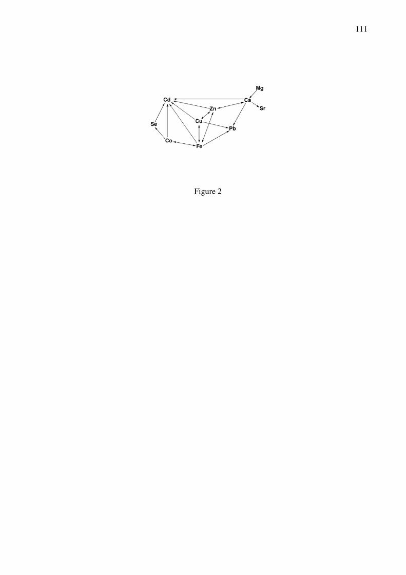
111
Figure 2

112
Figure 3

113
Figure 4

114
Figure 5
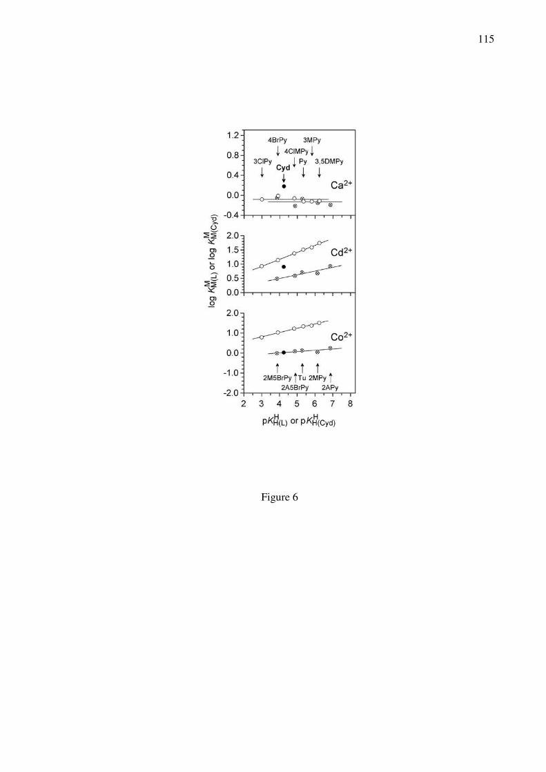
115
Figure 6

116
Figure 7

117
Figure 8

118
Figure 9

119
Figure 10

120
Figure 11

121
Figure 12

122
Figure 13

123
Figure 14

124
Figure 15

125
Figure 16

126
Figure 17

127
Equation 5

128
Equation 6

129
Equation 10

130
Equation 21

131
Equation 22

132
Equation 26

133
M2+ + PMEA2– M(PMEA)op
KI/O
KI/N7
M(PMEA)cl/N7
KMM(PMEA)op
M(PMEA)cl/O
KI/O/N3
M(PMEA)cl/O/N3
Equation 27
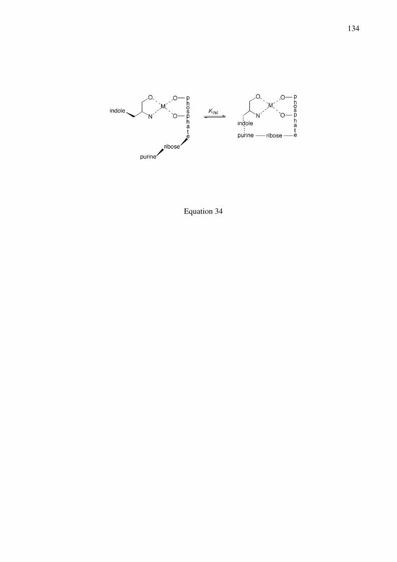
134
Equation 34