computational planning in facial surgery - thieme … · anthropometry digital patient ......
TRANSCRIPT

Computational Planning in Facial SurgeryStefan Zachow, PhD1
1Zuse Institute Berlin (ZIB), Berlin, Germany
Facial Plast Surg 2015;31:446–462.
Address for correspondence Stefan Zachow, PhD, Zuse Institute Berlin(ZIB), Mathematics for Life and Materials Sciences, Takustraße 7,14195 Berlin, Germany (e-mail: [email protected]).
Reconstructive facial surgery aims at the restoration of facialmalformations caused by trauma or tumor-related morpho-logical changes. In contrast, aesthetic or cosmetic surgeryaims at the “improvement” of a facial appearancewith respectto a particular objective. Facial surgery in cases of congenitaldysmorphisms are somehow in between having no uniqueobjective rather being oriented on establishing a naturalfunction and a natural as well as harmonious facial appear-ance. Thus, we can distinguish between reconstruction andconstruction of a face. In the former case, the surgical inten-tion is to restore a previous state as close as possible, and thelatter case is focused on changing and improving a facialappearance with respect to some idealized target. Typicalobjectives are symmetry, facial proportions within a normalrange, gender specific and cultural attributes, social trends,and manymore. An objective could try to preserve individualcharacteristics and personality or to obtain a “new” face.Hence, a surgical procedure must be thoroughly planned inadvance, where different options are tested, and possible
outcomes are visually communicated to the patient in ahighly conceivable manner to agree on a desired outcome.Within this article, computational planning approaches incranio-maxillofacial surgery are reviewed. A survey on exist-ing solutions is given and future perspectives of an improvedcomputerized planning of facial surgery are presented.
Facial Surgery Planning—The Past and thePresent
Preoperative preparation of a treatment concept for thesurgical correction of craniofacial deformities requires acomprehensive knowledge about normally developed ana-tomical structures (anatomical atlases). Anthropometric andcephalometric studies provide helpful insights to normalproportions of the body or face and the individual (agerelated), inter-individual (e.g., gender), and inter-culturalvariability.1–3 For a successful reconstruction or establish-ment of normal function, taking a harmonious shape of the
Keywords
► model-guided surgery► computer-aided
planning► anthropometry► digital patient models► surgical template
models► statistical shape
models
Abstract This article reflects the research of the last two decades in computational planning forcranio-maxillofacial surgery. Model-guided and computer-assisted surgery planning hastremendously developed due to ever increasing computational capabilities. Simulatorsfor education, planning, and training of surgery are often compared with flightsimulators, where maneuvers are also trained to reduce a possible risk of failure.Meanwhile, digital patient models can be derived from medical image data withastonishing accuracy and thus can serve for model surgery to derive a surgical templatemodel that represents the envisaged result. Computerized surgical planning ap-proaches, however, are often still explorative, meaning that a surgeon tries to find atherapeutic concept based on his or her expertise using computational tools that aremimicking real procedures. Future perspectives of an improved computerized planningmay be that surgical objectives will be generated algorithmically by employingmathematical modeling, simulation, and optimization techniques. Planning systemsthus act as intelligent decision support systems. However, surgeons can still use theexisting tools to vary the proposed approach, but they mainly focus on how to transferobjectives into reality. Such a development may result in a paradigm shift for futuresurgery planning.
Issue Theme The Facial Profile; GuestEditors, Werner J. Heppt, MD, and StefanZachow, PhD
Copyright © 2015 by Thieme MedicalPublishers, Inc., 333 Seventh Avenue,New York, NY 10001, USA.Tel: +1(212) 584-4662.
DOI http://dx.doi.org/10.1055/s-0035-1564717.ISSN 0736-6825.
446
Thi
s do
cum
ent w
as d
ownl
oade
d fo
r pe
rson
al u
se o
nly.
Una
utho
rized
dis
trib
utio
n is
str
ictly
pro
hibi
ted.

head or face into account while retaining individual charac-teristics, careful treatment planning using all available dataand resources is essential (►Fig. 1).
Based on a graphical analysis of lateral cephalometricradiographs (►Fig. 2, center), first computerized planningtools were used in the 1970s. Repositioning of bony struc-tures were performed in a facial profile view by exemptionand displacement of two-dimensional (2D) image segments.4
Since the mid-1980s, dedicated analysis and planning soft-ware was developed that enables a 2D planning based ondigitized lateral cephalograms or cephalometric landmarks
that are measured on a patient’s head, simultaneously usingempirically derived ratios to estimate a resulting soft-tissueprofile.5 With the availability of 2D image processing soft-ware, it became also possible to distort a profile photoaccording to a displacement of image features so that apossible postoperative outcome was visually communicatedin a rough approximation. The validity of these methods,however, becomes highly limited with increasing complexityof changes of the facial skeleton, and hence the visual com-munication of a possible facial outcome becomes less con-vincing or even questionable.
Fig. 1 Complex congenital craniofacial dysplasia (left) malocclusion of Angle class III, (center) hemifacial microsomia, (right) malocclusion ofAngle class II.
Fig. 2 Facial profile analysis based on lateral cephalograms.
Facial Plastic Surgery Vol. 31 No. 5/2015
Computational Planning in Facial Surgery Zachow 447
Thi
s do
cum
ent w
as d
ownl
oade
d fo
r pe
rson
al u
se o
nly.
Una
utho
rized
dis
trib
utio
n is
str
ictly
pro
hibi
ted.
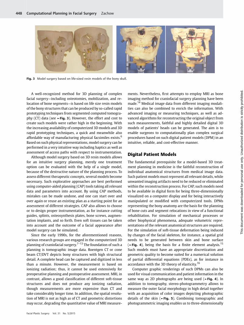
A well-recognized method for 3D planning of complexfacial surgery—including osteotomies, mobilization, and re-location of bone segments—is based on life size resin modelsof the bony structures that can be produced by so-called rapidprototyping techniques from segmented computed tomogra-phy (CT) data (see ►Fig. 3). However, the effort and cost tocreate such models were rather high in the beginning. Withthe increasing availability of computerized 3Dmodels and 3Drapid prototyping techniques, a quick and meanwhile alsoaffordable way of manufacturing physical facsimiles exists.6
Based on such physical representations, model surgery can beperformed in a very intuitive way including haptics as well asassessment of access paths with respect to instrumentation.
Although model surgery based on 3D resin models allowsfor an intuitive surgery planning, merely one treatmentoption can be evaluated with the help of a single model,because of the destructive nature of the planning process. Toassess different therapeutic concepts, several models becomenecessary. Such explorative approaches are best performedusing computer-aided planning (CAP) tools taking all relevantdata and parameters into account. By using CAP methods,mistakes can be made undone, and one can start over andover again or reuse an existing plan as a starting point for anassessment of different strategies. CAP also allows to chooseor to design proper instrumentation, as for instance cuttingguides, splints, osteosynthesis plates, bone screws, augmen-tation implants, and so forth. Even soft tissues can be takeninto account and the outcome of a facial appearance aftermodel surgery can be simulated.
Since the early 1990s, for the aforementioned reasons,various research groups are engaged in the computerized 3Dplanning of craniofacial surgery.7–13 The foundation of such aplanning is tomographic image data. Roentgen CT or conebeam CT/DVT depicts bony structures with high structuraldetail. A complete head can be captured and digitized in lessthan a minute. However, the measurement is based onionizing radiation; thus, it cannot be used extensively forpreoperative planning and postoperative assessment. MRI, incontrast, allows a good classification of different soft-tissuestructures and does not produce any ionizing radiation,though measurements are more expensive than CT andtake considerably longer time. In addition, the spatial resolu-tion of MRI is not as high as of CT and geometric distortionsmay occur, degrading the quantitative value of MRI measure-
ments. Nevertheless, first attempts to employ MRI as boneimaging method for craniofacial surgery planning have beenmade.14 Medical image data from different imaging modali-ties can also be combined to enrich the information. Withadvanced imaging or measuring techniques, as well as ad-vanced algorithms for reconstructing the original object fromsuch measurements, faithful and highly detailed digital 3Dmodels of patients’ heads can be generated. The aim is toenable surgeons to computationally plan complex surgicalprocedures based on such digital patient models (DPM) in anintuitive, reliable, and cost-effective manner.
Digital Patient Models
The fundamental prerequisite for a model-based 3D treat-ment planning in medicine is the faithful reconstruction ofindividual anatomical structures from medical image data.Such patientmodelsmust represent all relevant details, whileunwanted imaging artifacts need to be reduced or eliminatedwithin the reconstruction process. For CAP, suchmodels needto be available in digital form for being three-dimensionallyvisualized on a computer display and for being interactivelymanipulated or modified with computerized tools. DPMsrepresenting the bony anatomy are the basis for the planningof bone cuts and segment relocations in view of a functionalrehabilitation. For simulation of mechanical processes orother biophysical phenomena, adequate volumetric repre-sentations of the relevant anatomical structures are required.For the simulation of soft-tissue deformation being inducedby changes of the facial skeleton, for instance, a spatial gridneeds to be generated between skin and bone surface(►Fig. 4), being the basis for a finite element analysis.15
Such models must have an appropriate discretization andgeometric quality to become suited for a numerical solutionof partial differential equations (PDEs), as for instance inaccordance with the 3D theory of elasticity.16
Computer graphic renderings of such DPMs can also beused for visual communication and patient information in thesame way as 2D photographs are being used (►Fig. 4). Inaddition to tomography, stereo-photogrammetry allows tomeasure the outer facial morphology in high detail togetherwith an acquisition of color images depicting photographicdetails of the skin (►Fig. 5). Combining tomographic andphotogrammetric imaging enables us to three-dimensionally
Fig. 3 Model surgery based on life-sized resin models of the bony skull.
Facial Plastic Surgery Vol. 31 No. 5/2015
Computational Planning in Facial Surgery Zachow448
Thi
s do
cum
ent w
as d
ownl
oade
d fo
r pe
rson
al u
se o
nly.
Una
utho
rized
dis
trib
utio
n is
str
ictly
pro
hibi
ted.
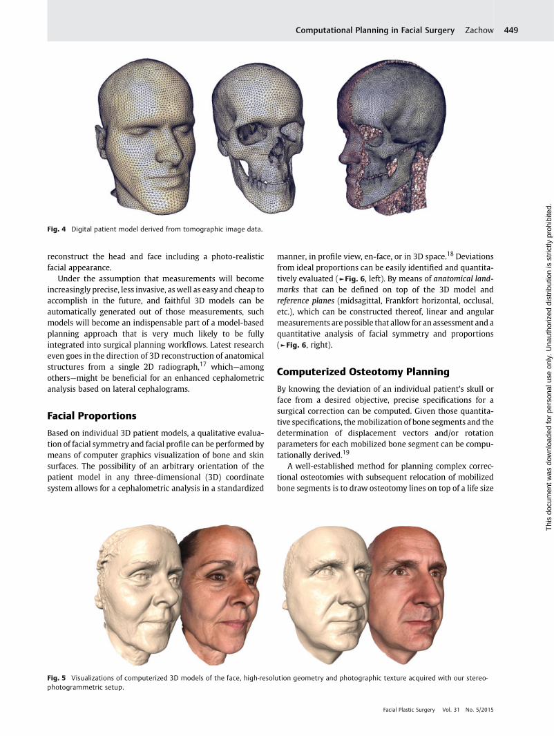
reconstruct the head and face including a photo-realisticfacial appearance.
Under the assumption that measurements will becomeincreasingly precise, less invasive, aswell as easy and cheap toaccomplish in the future, and faithful 3D models can beautomatically generated out of those measurements, suchmodels will become an indispensable part of a model-basedplanning approach that is very much likely to be fullyintegrated into surgical planning workflows. Latest researcheven goes in the direction of 3D reconstruction of anatomicalstructures from a single 2D radiograph,17 which—amongothers—might be beneficial for an enhanced cephalometricanalysis based on lateral cephalograms.
Facial Proportions
Based on individual 3D patient models, a qualitative evalua-tion of facial symmetry and facial profile can be performed bymeans of computer graphics visualization of bone and skinsurfaces. The possibility of an arbitrary orientation of thepatient model in any three-dimensional (3D) coordinatesystem allows for a cephalometric analysis in a standardized
manner, in profile view, en-face, or in 3D space.18 Deviationsfrom ideal proportions can be easily identified and quantita-tively evaluated (►Fig. 6, left). By means of anatomical land-marks that can be defined on top of the 3D model andreference planes (midsagittal, Frankfort horizontal, occlusal,etc.), which can be constructed thereof, linear and angularmeasurements are possible that allow for an assessment and aquantitative analysis of facial symmetry and proportions(►Fig. 6, right).
Computerized Osteotomy Planning
By knowing the deviation of an individual patient’s skull orface from a desired objective, precise specifications for asurgical correction can be computed. Given those quantita-tive specifications, themobilization of bone segments and thedetermination of displacement vectors and/or rotationparameters for each mobilized bone segment can be compu-tationally derived.19
A well-established method for planning complex correc-tional osteotomies with subsequent relocation of mobilizedbone segments is to draw osteotomy lines on top of a life size
Fig. 4 Digital patient model derived from tomographic image data.
Fig. 5 Visualizations of computerized 3D models of the face, high-resolution geometry and photographic texture acquired with our stereo-photogrammetric setup.
Facial Plastic Surgery Vol. 31 No. 5/2015
Computational Planning in Facial Surgery Zachow 449
Thi
s do
cum
ent w
as d
ownl
oade
d fo
r pe
rson
al u
se o
nly.
Una
utho
rized
dis
trib
utio
n is
str
ictly
pro
hibi
ted.

resin model of the patient’s skull and to cut this modelaccordingly (►Fig. 3 and ►Fig. 7, top left). To become anaccepted planning tool in clinical practice, a computerized 3Dplanning must be performed in a similar and intuitive man-ner. Hence, a reasonable computerized planning approachmust allow to draw osteotomy lines on top of a DPM of theskull, while the model can be freely rotated. Such osteotomyplanning can be easily achieved using dedicated input devicessuch as stylus and a touch-sensitive display as provided withmodern tablet computers (►Fig. 8).20An evenmore advancedapproach automatically projects osteotomy lines for stan-dardized procedures onto the respective skull model. Pro-posed osteotomy lines can afterward be interactivelymodified to meet individual requirements.
In ►Fig. 7, an example of the planning of a bimaxillaryosteotomy for combined upper and lower jawdisplacement isdepicted. After osteotomy lines have been defined, the re-
sulting, arbitrarily shaped cut surfaces are automaticallygenerated from the drawn contours and visualized, revealingthe course of the cut within the bone.20 On top of thesesurfaces, the original image data can be visualized(►Fig. 7, bottom). That way, the planned osteotomy can beassessed with respect to internal structures, such as nerves,vessels, roots of teeth, and so forth, without having thosestructures explicitly segmented from medical image data.
Orthognathic Surgery—Remodeling theMaxillofacial Skeleton
Since anatomical planes or local coordinate systems can beeasily derived from anatomical landmarks, geometric devia-tions of a patient’s anatomy with respect to symmetry can beassessed. Mirroring of parts of a healthy contralateral sidemay serve as a target for the adjustment of the position and
Fig. 6 Computerized profile and en-face analysis based on digital 3D patient models.
Fig. 7 Osteotomy planning based on a resin model, and on digital 3D skull models, (a) conventional, (b) high, and (c) quadrangular Le Fort Iosteotomy.
Facial Plastic Surgery Vol. 31 No. 5/2015
Computational Planning in Facial Surgery Zachow450
Thi
s do
cum
ent w
as d
ownl
oade
d fo
r pe
rson
al u
se o
nly.
Una
utho
rized
dis
trib
utio
n is
str
ictly
pro
hibi
ted.

orientation of malpositioned or malformed bony structures.Mobilized segments of the digital skull model can be freelypositioned to assess a modified facial skeletonwith respect tonatural proportions, symmetry, functional rehabilitation,19
as well as a harmonious facial appearance under consider-ation of facial soft tissue.21,22 The spatial transformation ofmobilized bone segments can be interactively applied viastandard techniques of 3D computer graphics and human–computer interaction (HCI). Parts can be translated in 3Dspace or rotated around a predefined center or axis (►Fig. 9).Transformations can be limited to particular degrees offreedom as well as analyzed regarding colliding adjacentstructures, or even be restricted via collision prevention
techniques (►Fig. 10). Based on a computerized approach,various osteotomies with segment relocations can be as-sessed in view of a resulting spatial arrangement of the facialskeleton.
During the preoperative planning phase, it is crucial that asurgeon has all options to modify the model at his or her ownwill. Helpful toolsmay simplify the separation of parts, spatialalignment, and measurement to assist in the task of anexplorative but goal-oriented design of a regular facial skele-ton. At any time, a modified skull is vividly visualized in threedimensions and can be interactively rotated to understandcomplex spatial relationships. As a specialty of computerizedplanning, parts of the DPM can be removed or visualized in a
Fig. 8 Intuitive osteotomy planning based on a computerized model.20
Fig. 9 Relocation of mobilized bone segments. Maxillary advancement and rotation (top), maxillary advancement and mandibular setback(bottom).
Facial Plastic Surgery Vol. 31 No. 5/2015
Computational Planning in Facial Surgery Zachow 451
Thi
s do
cum
ent w
as d
ownl
oade
d fo
r pe
rson
al u
se o
nly.
Una
utho
rized
dis
trib
utio
n is
str
ictly
pro
hibi
ted.

semitransparentmanner to reveal hidden structures to assessa new arrangement and to identify possible complicationsthat may result from a modification. By actively performingthese remodeling steps, a surgeon already becomes betterprepared.
Orthodontics—Dentition and DentalOcclusion
Surgical and orthodontic treatment planning belong togetherin maxillofacial surgery to achieve an optimal functional andaesthetically pleasing result.23 Teeth need to be reconfiguredto achieve a proper dental occlusion in accordance with thenew skeletal arrangement and especially the incisors have animpact on the facial appearance. Hence, the computationalplanning of orthodontic treatments and its integration intofacial surgery planning concepts is of importance.24
An orthodontist’s primary goal is to achieve/obtain aproper dental occlusion based on a sufficient dentition.Although the occlusal plane can be roughly determinedfrom tomographic data, detailed geometric informationabout dental fissures and occlusal surfaces cannot be derivedfrom tomography. A combination of tomographic data anddigitized dental casts enables us to integrate a highly detailedmodel of the dentition into the DPM25 (►Fig. 10, left). Thecombinedmodel can then be used for a spatial rearrangementof jaw segments allowing an improved geometric analysis oftooth contacts (►Fig. 10, right). The contact zones, as shownin ►Fig. 10, are useful indicators for planning an orthodontictreatment together with the orthognathic surgery to considernormal dental occlusion.
Because dental occlusion is such an important factor forfunctional rehabilitation, advanced CAP systems for orthog-nathic surgery planning must provide a computerized ana-logue to a dental articulator as shown in ►Fig. 3. Otherwise,orthodontic planning needs to be done in a conservativemanner based on dental casts, and the parameters of thearticulator must be transferred into the CAP system for arespective adjustment of the jaw segments of the DPM.26
Even more advanced CAP systems for orthodontic treatmentplanning could automatically propose proper positions for
the jaw segments, or even individual teeth. Using a mathe-matical modeling, simulation, and optimization (MSO) ap-proach, they could also provide decision support for anoptimal bracket design, taking forces and moments intoaccount that are acting on the respective configuration ofthe teeth.
By assessing the modified DPM, a decision for an appro-priate surgical procedure can be derived within the preoper-ative planning phase. The surgical template model (STM)serves as a kind of blueprint for the envisaged surgical result.An STM may also serve as a basis for the design of surgicalsplints, surgical cutting guides, or any other type of custom-ized instrumentation.27
Osteosynthesis and Implant Design
The STM—that is, the altered DPM—represents the plannedfacial skeleton as it is supposed to be after surgery. That way itis an ideal basis for a preoperative selection and positioning ofosteosynthesis plates up to the fully customized design ofosteosyntheses28,29 or other implants that will secure thedesired positions of mobilized bone segments for stablefusion and proper bone healing (►Fig. 11, left). Meanwhile,commercial software and services become available for cus-tomized implant design and patient-specific instrumentationthat are currently under evaluation by clinical researchgroups.30
Besides a design or configuration of osteosyntheses, anSTM can also be used for the selection or design of boneaugmentations.31 The optimal shape of an augmentationimplant is given by the difference between the targeted andthe actual shape of the bone. A planned maxillary advance-ment, for instance, leads to an STM that differs locally fromthe PDM. The volumetric difference of the two models in thatregion yields an estimate for an augmentation implant. Suchimplants need to have a good fit to the bony support; thus, theDPM can be seen as a local molding or casting form(►Fig. 11, center and right). On the soft tissue side, suchimplants need to be smooth without introducing unwantededges. Since the shape of the implant will determine the facialappearance, its shape design is of utmost importance.
Fig. 10 Analysis of dental occlusion for orthodontic treatment planning.25
Facial Plastic Surgery Vol. 31 No. 5/2015
Computational Planning in Facial Surgery Zachow452
Thi
s do
cum
ent w
as d
ownl
oade
d fo
r pe
rson
al u
se o
nly.
Una
utho
rized
dis
trib
utio
n is
str
ictly
pro
hibi
ted.
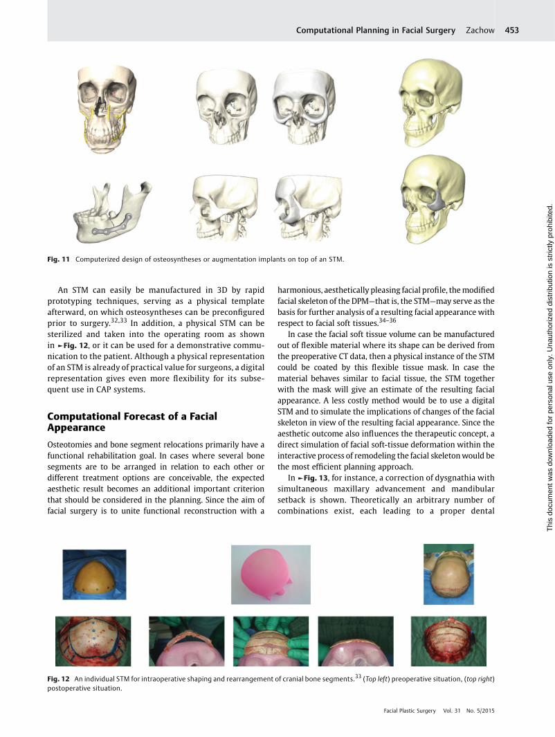
An STM can easily be manufactured in 3D by rapidprototyping techniques, serving as a physical templateafterward, on which osteosyntheses can be preconfiguredprior to surgery.32,33 In addition, a physical STM can besterilized and taken into the operating room as shownin ►Fig. 12, or it can be used for a demonstrative commu-nication to the patient. Although a physical representationof an STM is already of practical value for surgeons, a digitalrepresentation gives even more flexibility for its subse-quent use in CAP systems.
Computational Forecast of a FacialAppearance
Osteotomies and bone segment relocations primarily have afunctional rehabilitation goal. In cases where several bonesegments are to be arranged in relation to each other ordifferent treatment options are conceivable, the expectedaesthetic result becomes an additional important criterionthat should be considered in the planning. Since the aim offacial surgery is to unite functional reconstruction with a
harmonious, aesthetically pleasing facial profile, themodifiedfacial skeleton of the DPM—that is, the STM—may serve as thebasis for further analysis of a resulting facial appearance withrespect to facial soft tissues.34–36
In case the facial soft tissue volume can be manufacturedout of flexible material where its shape can be derived fromthe preoperative CT data, then a physical instance of the STMcould be coated by this flexible tissue mask. In case thematerial behaves similar to facial tissue, the STM togetherwith the mask will give an estimate of the resulting facialappearance. A less costly method would be to use a digitalSTM and to simulate the implications of changes of the facialskeleton in view of the resulting facial appearance. Since theaesthetic outcome also influences the therapeutic concept, adirect simulation of facial soft-tissue deformation within theinteractive process of remodeling the facial skeletonwould bethe most efficient planning approach.
In ►Fig. 13, for instance, a correction of dysgnathia withsimultaneous maxillary advancement and mandibularsetback is shown. Theoretically an arbitrary number ofcombinations exist, each leading to a proper dental
Fig. 11 Computerized design of osteosyntheses or augmentation implants on top of an STM.
Fig. 12 An individual STM for intraoperative shaping and rearrangement of cranial bone segments.33 (Top left) preoperative situation, (top right)postoperative situation.
Facial Plastic Surgery Vol. 31 No. 5/2015
Computational Planning in Facial Surgery Zachow 453
Thi
s do
cum
ent w
as d
ownl
oade
d fo
r pe
rson
al u
se o
nly.
Una
utho
rized
dis
trib
utio
n is
str
ictly
pro
hibi
ted.

occlusion with neutral bite. A simulation of the resultingarrangement of facial soft tissues and thus the facialappearance allows an assessment of the planning from anaesthetic point of view.
Basis for a reliable forecast of the soft-tissue arrangementwith respect to the modified DPM is an adequate geometricmodel of the soft-tissue volumewith all embedded structureson the one hand, and on the other hand a physical deforma-tion model that describes the mechanical properties ofbiological soft tissue in close approximation.37 The latter isbased on the theory of elasticity, which is used in manyengineering disciplines for stress analysis. The mathematicalcalculation of deformation is based on the given displace-ments of bony structures by means of the finite elementmethod.38 Taking the displacements as boundary conditions,the respective system of PDEs is solved on the entire volu-metric mesh that represents the facial soft tissue. In contrastto approaches that take into account heuristically deter-mined, local displacement ratios for soft tissue, a simulationbased on continuum mechanics yields a deformation for any
point on the facial surface. Such a computation can beperformed on conventional computers in a few minutes.
In ►Fig. 14, a DPM of a patient with dysgnathia of Angleclass III is shown. There are different options of surgicalcorrection varying from maxillary advancement, mandibularsetback, or a combination of the two. For the patient onwhombimaxillary osteotomy was planned, the DPM was modifiedaccordingly, and facial outcomes were tested by full advance-ment of themaxilla, a full setbackof themandibular segment,as well as a gradual advancement and setback of maxilla andmandible, respectively, considering dental occlusion.21 Eachresulting STM serves as input for the soft-tissue simulation.Single or combined bone segment relocations can be contin-uously adjusted and the resulting effect on the facial appear-ance can be displayed in three dimensions from any viewingangle. The simulation can even be visualized in photorealisticquality using photographic textures as shown in ►Fig. 14.Animated image sequences of the simulation contribute in afar more intuitive enlightenment of a patient, as it waspreviously the case in preoperative patient information.
Fig. 13 Computerized planning of a bimaxillary osteotomy with maxillary advancement and mandibular setback including a simulation of theresulting facial appearance.
Fig. 14 DPM showing the preoperative situation (left) simulation of maxillary advancement and mandibular setback based on the modified DPM.
Facial Plastic Surgery Vol. 31 No. 5/2015
Computational Planning in Facial Surgery Zachow454
Thi
s do
cum
ent w
as d
ownl
oade
d fo
r pe
rson
al u
se o
nly.
Una
utho
rized
dis
trib
utio
n is
str
ictly
pro
hibi
ted.
Intraoperative Transfer of the Plan
Having planned facial surgery and having derived a thera-peutic concept, themain challenge that needs to be addressedis how to surgically achieve the planned result: that is, how toexactly reproduce osteotomies and bone segment relocationson the patient within the operating room?
To reproduce the planned osteotomies, individual or navigat-ed cutting guides of very precise laser or piezoelectric osteoto-mies are conceivable.39,40 For the accurate reproduction of theplanned relocation of mobilized bone segments, different ap-proaches have been tested. One is the design and the fabricationof individualized surgical splints.41,42 Such splints help in adjust-ing mobilized bone segments (such as mandible andmaxilla) toeach other or in adjusting mobilized segments to unaffectedregions of the skull base. Another approach is to use the relativetransformations of mobilized bone segments resulting from theplan and to reproduce these transformations in a registeredcoordinate system of the patient using navigation techniques.The latter method leads to a fully computerized planningapproach, thus avoiding the production of surgical splints.
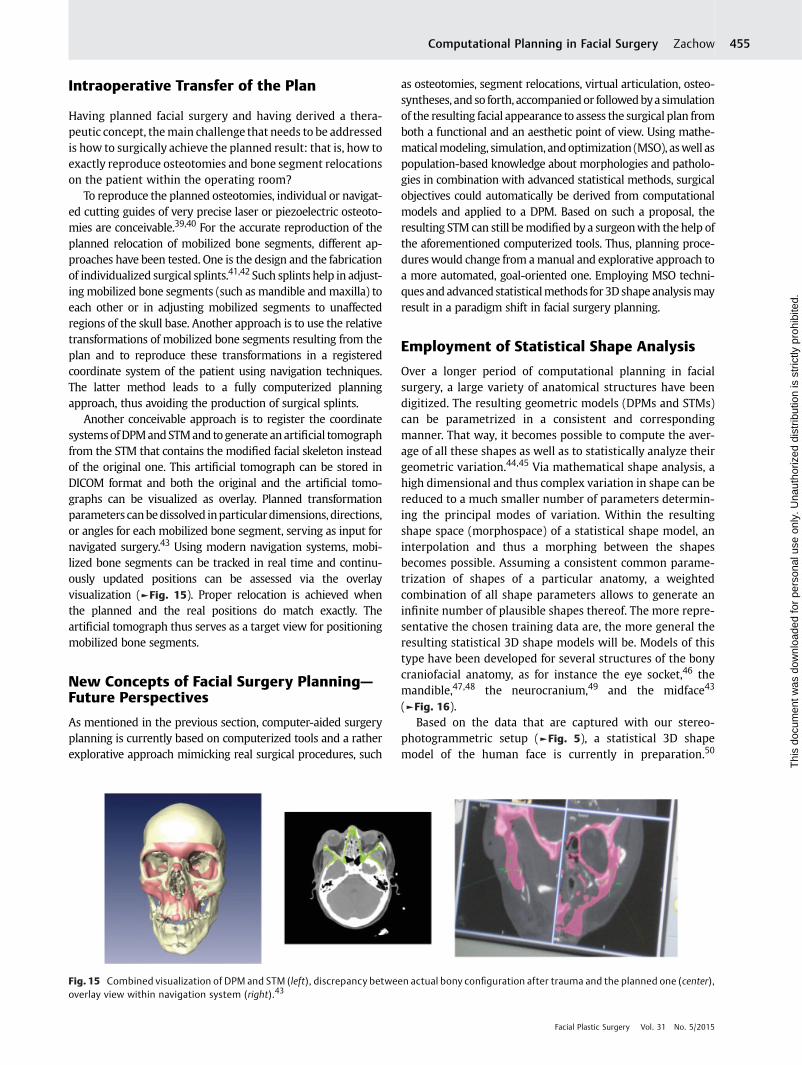
Another conceivable approach is to register the coordinatesystemsofDPMandSTMand togenerate anartificial tomographfrom the STM that contains the modified facial skeleton insteadof the original one. This artificial tomograph can be stored inDICOM format and both the original and the artificial tomo-graphs can be visualized as overlay. Planned transformationparameters canbedissolved inparticulardimensions, directions,or angles for each mobilized bone segment, serving as input fornavigated surgery.43 Using modern navigation systems, mobi-lized bone segments can be tracked in real time and continu-ously updated positions can be assessed via the overlayvisualization (►Fig. 15). Proper relocation is achieved whenthe planned and the real positions do match exactly. Theartificial tomograph thus serves as a target view for positioningmobilized bone segments.
New Concepts of Facial Surgery Planning—Future Perspectives
As mentioned in the previous section, computer-aided surgeryplanning is currently based on computerized tools and a ratherexplorative approach mimicking real surgical procedures, such
as osteotomies, segment relocations, virtual articulation, osteo-syntheses, and so forth, accompaniedor followedbya simulationof the resulting facial appearance to assess the surgical plan fromboth a functional and an aesthetic point of view. Using mathe-maticalmodeling, simulation, andoptimization (MSO), aswell aspopulation-based knowledge about morphologies and patholo-gies in combination with advanced statistical methods, surgicalobjectives could automatically be derived from computationalmodels and applied to a DPM. Based on such a proposal, theresulting STM can still bemodified by a surgeonwith the help ofthe aforementioned computerized tools. Thus, planning proce-dures would change from amanual and explorative approach toa more automated, goal-oriented one. Employing MSO techni-ques andadvanced statisticalmethods for 3D shape analysismayresult in a paradigm shift in facial surgery planning.
Employment of Statistical Shape Analysis
Over a longer period of computational planning in facialsurgery, a large variety of anatomical structures have beendigitized. The resulting geometric models (DPMs and STMs)can be parametrized in a consistent and correspondingmanner. That way, it becomes possible to compute the aver-age of all these shapes as well as to statistically analyze theirgeometric variation.44,45 Via mathematical shape analysis, ahigh dimensional and thus complex variation in shape can bereduced to a much smaller number of parameters determin-ing the principal modes of variation. Within the resultingshape space (morphospace) of a statistical shape model, aninterpolation and thus a morphing between the shapesbecomes possible. Assuming a consistent common parame-trization of shapes of a particular anatomy, a weightedcombination of all shape parameters allows to generate aninfinite number of plausible shapes thereof. The more repre-sentative the chosen training data are, the more general theresulting statistical 3D shape models will be. Models of thistype have been developed for several structures of the bonycraniofacial anatomy, as for instance the eye socket,46 themandible,47,48 the neurocranium,49 and the midface43
(►Fig. 16).Based on the data that are captured with our stereo-
photogrammetric setup (►Fig. 5), a statistical 3D shapemodel of the human face is currently in preparation.50
Fig. 15 Combined visualization of DPM and STM (left), discrepancy between actual bony configuration after trauma and the planned one (center),overlay view within navigation system (right).43
Facial Plastic Surgery Vol. 31 No. 5/2015
Computational Planning in Facial Surgery Zachow 455
Thi
s do
cum
ent w
as d
ownl
oade
d fo
r pe
rson
al u
se o
nly.
Una
utho
rized
dis
trib
utio
n is
str
ictly
pro
hibi
ted.

Currently, approximately 100 faces have been integrated intoour statistical 3D shape model of the face (►Fig. 17). Thismodel is analyzed in view of population-based facial charac-teristics that may serve as an objective for a classification ofindividual types of facial morphology or any individualdeviation from normal proportions.
A combined statistical 3D shape model of the skull and theface is our envisaged goal. Such a model could be used forcorrelation analysis between the shape of the skull and theshape of the face to study and classify dysmorphisms orpathologically developed structures. Such a model wouldalso be a very valuable tool in forensic medicine.
A statistical shape model enables us to investigate shapecharacteristics via multivariate statistical analysis and to deter-mine sets of deformation parameters. With an increasingamount of shape data, population-based studies will becomepossible and variation in shape can be analyzed with respect togender, age, body mass, or other demographic information.Owing to a normally distributed variation in shape in a largepopulation, it is not surprising that the averaged shape of a verylarge set of randomly selected anatomical structures such asskulls or faces represents a kind of normally developed anatomy.Normality relates to the average and to the variation withincertain limits around that average. Thus, statistical 3D shapemodels of anatomical structuresmay become the foundation foradvanced anatomical atlases that not only represent an averageshape per anatomical structure but also its normal or evenpathological variation in shape. Such atlases are only accessiblein their full power in a digital form. However, representatives ofanatomical structures can always be manufactured.
Since statistical shape models can be analyzed with re-spect to demographic attributes, a population-based analysismay be introducedwithin the planning process. From such ananalysis, objectives for the alteration of an individual head orface can be derived,whichwill become input for a subsequent
therapy planning. With the availability of representative,normally pronounced reference anatomies in the form ofstatistical 3D shape models that can be adapted to individualanatomical landmarks, it becomes possible to generate ther-apeutic proposals even for highly asymmetric malformationsand severe disfigurations,47 up to the design of missing partsor structures that need to be fully reconstructed, for instancefacial epitheses for the nose, eye, or ear.51 A shape-basedclassification is also conceivable for an early detection ofcraniofacial syndromes when applied to regularly acquiredstereo-photogrammetric measurements of infant heads, forexample, in routinely performed well-child visits.
Instead of testing outcomes in an explorative manner by amodification of a DPM and subsequent assessment of simu-lations that are driven by such modifications, the planningprocedure could start with a facial target that might bederived from anthropometric analysis based on an individu-alized statistical shape model of the face. Having a desiredfacial outcome as an objective, surgeon and patient will agreeon, the remaining task is to determine the appropriatesurgical procedure that leads to the envisaged result. Thereare already mathematical methods on algorithmic determi-nation of how an underlying skull surface needs to be modi-fied to achieve a desired outcome of a facial appearance.52 Infacial surgery, for instance, it could either be the modificationof the facial skeleton or the design of augmentation implantsor a combination of the two to alter the appearance of theface. Having a proposal for a modified DPM, a planningapproach reduces to the task of deciding how the proposedalteration can be achieved surgically.
Facial Expressions
An important issue that has only marginally been addressedin facial surgery planning so far is the consideration of facial
Fig. 16 Statistical shape models of craniofacial anatomy (neurocranium, orbit, midface, mandible).
Facial Plastic Surgery Vol. 31 No. 5/2015
Computational Planning in Facial Surgery Zachow456
Thi
s do
cum
ent w
as d
ownl
oade
d fo
r pe
rson
al u
se o
nly.
Una
utho
rized
dis
trib
utio
n is
str
ictly
pro
hibi
ted.

expressions, which are affected by a surgical modification of aface. In surgery planning, it is important to consider and tocommunicate the postoperative facial appearance of a patientfor varying facial expressions. First attempts to simulate facialexpressions by applying mechanistic approaches based onthe concept of facial action units that has been proposed byPaul Ekman and Wallace Friesen53 did not lead to convincingresults.54,55 Using advanced photogrammetric measurementmethods in combination with high detail surface reconstruc-tion, as shown in ►Fig. 5, faces of different persons withvarying facial expressions can be captured and respectivestatistical 3D shape and appearancemodels of thehuman facecan be established from such measurements.
A statistical analysis of intra- or inter-individual facialmorphology under different facial expressions enables us toidentify clusters of similar expressions from a set of measure-ments of an individual person or a larger cohort and to derivecharacteristic facial deformation patterns. These deformationpatterns can be classified and main modes of facial variationcan be statistically extracted and transferred to an individualDPM (►Fig. 18). That way it becomes possible to assess a facialappearance for a surgically altered DPM in view of differentfacial expressions. Considering patients with facial palsy,where facial muscles are not properly innervated, simulation
and assessment of individual facial expressions may help inestablishing a suitable therapeutic concept for muscle re-innervation to achieve, for instance, a natural smile.56
Craniofacial Growth
An additional and also important issue that needs to beconsidered in craniofacial surgery is growth. When congen-ital dysmorphisms of the head and face are to be treatedsurgically, growth must be taken into account. Growth canbe statistically analyzed with respect to its morphometriceffects. A statistical analysis of morphometric measure-ments of skulls and faces in the ages between birth andadolescence yield that the most significant variation inshape is affected by growth. However, growth is not just alinear increase of size (►Fig. 19). Instead it depends onmanyfactors and occurs in age intervals.57,58 Different parts of thehuman body grow with different velocity starting on differ-ent time points. Thus, one cannot simply scale an infant’sskull linearly between birth and adolescence to forecast itsthree-dimensional shape. The final shape of an anatomicalstructure is genetically predetermined. Thus, a thoroughmorphological analysis of anatomical growth and the result-ing shapes has to be performed. In the following, two
Fig. 17 Statistical shape model of facial skin morphology (Columns: 1st three modes of facial variation showing characteristic types of faceswithin a set of captured faces. The middle row depicts the average face, top and bottom rows depict the range of variation along the resp. mode.)
Facial Plastic Surgery Vol. 31 No. 5/2015
Computational Planning in Facial Surgery Zachow 457
Thi
s do
cum
ent w
as d
ownl
oade
d fo
r pe
rson
al u
se o
nly.
Una
utho
rized
dis
trib
utio
n is
str
ictly
pro
hibi
ted.

examples are given that are influenced by growth: thereshaping of infant skulls in cases of craniosynostosis33
(►Fig. 12) as well as the surgical correction of dysgnathiafor juvenile patients22 (►Fig. 1, right).
For a surgical treatment of craniosynostosis—that is, thereshaping of the neurocranium—it is quite obvious that anongoing increase in size needs to be taken into account.59 Thisalso holds—and is even more obvious—in cases of helmettherapies, where helmets are individually designed in such away that growth is limited in certain directions but desired inothers. The success of both therapies will strongly benefit ifwe know where and when regions of the neurocraniumincrease in size and to what extent. In addition, it is alsobeneficial to knowhowgrowth locally affects the shape of theneurocranium. A statistical 3D shape analysis gives muchmore detailed information about cranial growth than justmeasurements of the circumference of the head. Understand-ing growth and development in a better way enables us toprovide improved therapy plans that, in the case of treatingcraniosynostosis, take a natural increase in volume and therespective change in shape into account.
For juvenile patients with severe congenital dysmor-phisms of the jaws, where chewing, swallowing, and evenbreathing are negatively affected, a surgical interventionmight be indicated. However, mandibular or maxillary
osteotomies with pronounced relocation of jaw segmentshave a negative effect on later growth. Thus, differenttherapeutic strategies, such as distraction osteogenesis,are better suited.60 Within the preoperative planningphase, not only suitable osteotomies are to be determinedbut also positions and orientations of the distraction de-vices to achieve proper distraction vectors. These vectors(directions) of appropriate lengths (distance) determinethe resulting shape of the respective anatomical structure.However, the final shape will also be influenced by ongoinggrowth that needs to be considered within the therapeuticconcept, too.
Hence, results of the analysis of growth patterns need to betaken into account for craniofacial surgery planning. After therespective parameters of a statistical 3D shape model havebeen identified, they can be isolated and transferred to anindividual model. A variation of this parameter will thencreate characteristic growth effects in combination with anindividual patient’s craniofacial morphology.
Facial Aging
The facial appearance is also influenced by aging. Especially incosmetic facial surgery, undesired aging effects such aswrinkles up to deep folds, lacrimalis, loss of lip volume,
Fig. 19 Nonlinear morph of aging faces.58
Fig. 18 Different facial expressions synthesized via a statistical 3D shape and appearance model.
Facial Plastic Surgery Vol. 31 No. 5/2015
Computational Planning in Facial Surgery Zachow458
Thi
s do
cum
ent w
as d
ownl
oade
d fo
r pe
rson
al u
se o
nly.
Una
utho
rized
dis
trib
utio
n is
str
ictly
pro
hibi
ted.

and sagging, are subject to treatment. The typical objective isto establish a facial appearancewith a juvenile look. Therapiesrange from the application of fillers or botulinum toxin tosurgical “face lifts” to strengthen and firm facial soft tissues.
There are mechanistic approaches to simulate skin ag-ing;61–63 however, suchmethods are neither applicable for anaccurate forecast of an individual patient’s appearance nor arethey for any kind of treatment planning. Instead, statisticalmethods may become important in the future when skinaging effects can be quantified using high detail reconstruc-tion of stereo-photogrammetric measurements (►Fig. 5).Similar to the aforementioned future perspectives, agingeffects can also be statistically analyzed with respect togeometric effects. If the respective parameters of the statisti-
cal shape model are identified, they can again be isolated andtransferred to an individual face. A variation of this parameterwill then create characteristic aging effects, such as wrinklesand folds to communicate a possible aging process. Analo-gously, such amodel can also invert this process, demonstrat-ing howa personmight look younger. By quantitative analysisof changes that result from the variation of age-related shapeparameters, therapeutic proposals can be derived.
In addition to an age-related alteration of geometricfeatures, skin also exhibits increasing signs of aging due topigmental moles leading to an inhomogeneous, unevenlytoned appearance of the skin. Hence, a combined statisticalshape and appearance model is developed that covers thegeometry and the image of the facial surface. The averaged
Fig. 20 Comparison of the simulated facial profile to photographs.
Fig. 21 Comparison of the simulated facial appearance to postoperative CT data.
Facial Plastic Surgery Vol. 31 No. 5/2015
Computational Planning in Facial Surgery Zachow 459
Thi
s do
cum
ent w
as d
ownl
oade
d fo
r pe
rson
al u
se o
nly.
Una
utho
rized
dis
trib
utio
n is
str
ictly
pro
hibi
ted.
appearance of the skin results in a smooth and even-tonedimage just as the averaged shape does with respect toroughness or geometric texture. Both characteristics canbe analyzed independently, although both are correlatedwith age. A variation of age-related parameters of theindividualized statistical shape and appearance model com-municates an individual aging or rejuvenation process—aconcept that is, although still under research, conceivablewith advanced CAP tools.
Conclusion
Computerized facial surgery planning including soft-tissuesimulation has been developed at Zuse Institute Berlin (ZIB)since 1999 andwas successfully applied inmore than 50 casesin cooperations with clinics in Munich, Basel, Leipzig, Erlan-gen, Vienna, Hannover, Berlin, Stockholm, and Zurich. Pre-dominantly, malformations of the jaws and highlyasymmetric malformations of the facial skeletonwere treatedusing our planning approaches.
A comparison of the profile line of simulation results withpostoperative photographs already shows a very good corre-lation between the planning and the simulation results thathave been achieved (►Fig. 20). 3D comparisons of forecastswith postoperative CT data also demonstrate a very goodcorrelation and thus a prediction capability (►Fig. 21).37,64
Some Final Words on Planning
Model-based planning helps a lot in the preparation ofcomplex surgeries, where insufficient routine would endup in a higher risk of failure. Planning in general assists instructured thinking. Drawing or interactive computer-basedpracticing—that is, manipulation of computerized models insurgery simulation—teaches much more than just mentalpreparation. A descriptive visualization helps in memorizingthings and also in communicating therapeutic strategies tothe patient, which is beneficial for motivation and compli-ance. Computer-aided design, modeling, simulation, andoptimization, all being common practice in mechanicalengineering, will become common practice in surgery inthe near future.
AcknowledgmentsOur research in facial surgery planning was mainly fundedby Zuse Institute Berlin. Aspects of our research werefunded by DFN CoDisp (grant # TK 602 NT 202.2), MedicalUniversity of Vienna, Klinik Professor Sailer, DFG ResearchCenter Matheon, and 1000shapes GmbH. Our research instudying facial morphology is funded by the DFG cluster ofexcellence “Bild-Wissen-Gestaltung,” Special thanks go toall cooperating surgeons and especially to Prof. Zeilhofer(University Hospital Basel), Prof. Sader (University Hospi-tal Frankfurt), Priv. Doz. Dr. Westermark (formerly atKarolinska Hospital Stockholm), and Priv. Doz. Dr. Haberl(University Hospital Ulm, formerly at Charité Berlin), as
well as to all patients who have given their explicit consentto the publication of their images.
References1 Powell N, Humphreys B. Proportions of the Aesthetic Face. Thieme
Medical Publishers; 19842 Steinhäuser EW. Proportionen des ästhetischen Gesichts im Ver-
gleich zur bildenden Kunst. In: Fortschritte in der Kiefer- undGesichtschirurgie; Thieme Medical Publishers; 1986:1–4
3 Farkas LG. Anthropometry of the Head and Face. 2nd ed. NewYork:Raven Press; 1994
4 Ricketts RM, Bench RW, Hilgers JJ, Schulhof R. An overview ofcomputerized cephalometrics. Am J Orthod 1972;61(1):1–28
5 Landes CA, Zachar R, Diehl T, Kovács AF. Introduction of a three-dimensional anthropometry of the viscerocranium. Part II: evalu-ating osseous and soft tissue changes following orthognathicsurgery. J Craniomaxillofac Surg 2002;30(1):25–34
6 Bibb R, Eggbeer D, Paterson A, eds. Physical reproduction. In:Medical Modelling—The Application of Advanced Design andRapid Prototyping Techniques in Medicine. 2nd ed. WoodheadPublishing Series in Biomaterials. New York: Elsevier Ltd.; 2015:65–98
7 Cutting C, Bookstein FL, Grayson B, Fellingham L, McCarthy JG.Three-dimensional computer-assisted design of craniofacial sur-gical procedures: optimization and interaction with cephalomet-ric and CT-basedmodels. Plast Reconstr Surg 1986;77(6):877–887
8 Yasuda T, Hashimoto Y, Yokoi S, Toriwaki JI. Computer system forcraniofacial surgical planning based on CT images. IEEE Trans MedImaging 1990;9(3):270–280
9 Altobelli DE, Kikinis R, Mulliken JB, Cline H, Lorensen W, Jolesz F.Computer-assisted three-dimensional planning in craniofacialsurgery. Plast Reconstr Surg 1993;92(4):576–585, discussion586–587
10 Keeve E. Visualisierungs- und Simulationsverfahren zur interak-tiven Planung kraniofazialer Korrekturoperationen. PhD Disser-tation, Friedrich-Alexander-Universität Erlangen-Nürnberg; 1996
11 Sarti A, Gori R, Lamberti C. A physically based model to simulatemaxillofacial surgery from 3D CT images. Future Gener ComputSyst 1999;15(2):217–221
12 Koch RM. Methods for Physics Based Facial Surgery Prediction.PhD. Dissertation No. 13912, ETH Zürich; 2000
13 Zachow S, Gladilin E, Zeilhofer HF, Sader R. Improved 3D osteot-omy planning in cranio-maxillofacial surgery. Medical ImageComputing and Computer-Assisted Intervention—MICCAI 2001.Lecture Notes in Computer Science 2001;2208:473–481
14 Eley KA, Watt-Smith SR, Sheerin F, Golding SJ. “Black Bone”MRI: apotential alternative to CTwith three-dimensional reconstructionof the craniofacial skeleton in the diagnosis of craniosynostosis.Eur Radiol 2014;24(10):2417–2426
15 Zachow S, Zilske M, Hege HC. 3D reconstruction of individualanatomy from medical image data: segmentation and geometryprocessing. ZIB Report 07–41, 2007. Available at: https://opus4.kobv.de/opus4-zib/frontdoor/index/index/docId/1044
16 Deuflhard P. Differential equations in technology and medicine:computational concepts, adaptive algorithms, and virtual labs. In:Capasso et al., eds. Computational Mathematics Driven by Indus-trial Problems, Springer, Lecture Notes in Mathematics; 2006;1739:69–126. doi:10.1007/BFb0103918
17 Ehlke M, Ramm H, Lamecker H, Hege HC, Zachow S. Fast genera-tion of virtual X-ray images for reconstruction of 3D anatomy. IEEETrans Vis Comput Graph 2013;19(12):2673–2682
18 Swennen GRJ, Schutyser F, Hausamen JE, eds. Three-DimensionalCephalometry—A Color Atlas and Manual. New York: Springer;2005. doi:10.1007/3-540-29011-7
Facial Plastic Surgery Vol. 31 No. 5/2015
Computational Planning in Facial Surgery Zachow460
Thi
s do
cum
ent w
as d
ownl
oade
d fo
r pe
rson
al u
se o
nly.
Una
utho
rized
dis
trib
utio
n is
str
ictly
pro
hibi
ted.

19 ZinserMJ, Zachow S, Sailer HF. Bimaxillary ‘rotation advancement’procedures in patients with obstructive sleep apnea: a 3-dimen-sional airway analysis of morphological changes. Int J Oral Max-illofac Surg 2013;42(5):569–578
20 Zachow S, Gladilin E, Sader R, Zeilhofer HF. Draw & Cut: Intuitive3D Osteotomy Planning on Polygonal Bone Models. ComputerAssisted Radiology and Surgery 2003. Elsevier, Int. CongressSeries, 2003;1256:362–69. doi:10.1016/S0531-5131(03)00272-3
21 Westermark A, Zachow S, Eppley B. Three-dimensional osteotomyplanning inmaxillofacial surgery including soft tissue prediction. JCraniofac Surg 2005;16(1):100–104
22 Zachow S, Hege HC, Deuflhard P. Computer assisted planning incranio-maxillofacial surgery. CIT J Comput Inf Technol 2006;14(1):53–64
23 Bibb R, Eggbeer D, Paterson A, eds. Surgical applications case study5. In:MedicalModelling—The Application of Advanced Design andRapid Prototyping Techniques in Medicine. 2nd ed. WoodheadPublishing Series in Biomaterials, New York: Elsevier Ltd.; 2015:167–172
24 Nadjmi N, MollemansW, Daelemans A, Van Hemelen G, SchutyserF, Bergé S. Virtual occlusion in planning orthognathic surgicalprocedures. Int J Oral Maxillofac Surg 2010;39(5):457–462
25 Nkenke E, Zachow S, Benz M, et al. Fusion of computed tomogra-phy data and optical 3D images of the dentition for streak artefactcorrection in the simulation of orthognathic surgery. Dentomax-illofac Radiol 2004;33(4):226–232
26 Chapuis J, Zachow S, Langlotz F, Schramm A. Device for PlanningOrthodontics and/or Orthognathic Surgery. 2008. Patent WO/2008/080235, PCT/CH2007/00004, A61C 7/00 (2006.01)
27 Bibb R, Eggbeer D, Paterson A, eds. Surgical applications case study3. In:MedicalModelling—The Application of Advanced Design andRapid Prototyping Techniques in Medicine. 2nd ed. WoodheadPublishing Series in Biomaterials, New York: Elsevier Ltd.; 2015:145–154
28 Pereira C, Ventura F, Gaspar MC, Fontes R, Mateus A. Customizedimplant development for maxillo-mandibular reconstruction. In:Bártolo et al., eds. Virtual and Rapid Manufacturing. Philadelphia:Taylor & Francis; 2008:159–166
29 Cornelius CP, Smolka W, Giessler GA, Wilde F, Probst FA. Patient-specific reconstruction plates are the missing link in computer-assisted mandibular reconstruction: a showcase for technicaldescription. J Craniomaxillofac Surg 2015;43(5):624–629
30 Wilde F, Hanken H, Probst F, Schramm A, Heiland M, CorneliusCP. Multicenter study on the use of patient-specific CAD/CAMreconstruction plates for mandibular reconstruction. Int J CARS2015
31 Guevara Rojas G, Figl M, Schicho K, et al. Patient specific PEEKfacial implants in a computer-aided planning workflow. Inpreparation for Journal of Oral and Maxillofacial Surgery, avail-able at ResearchGate: http://www.researchgate.net/publication/260217023, 2015
32 Haberl H, Hell B, Zöckler MJ, et al. Technical aspects and results ofsurgery for craniosynostosis. Zentralbl Neurochir 2004;65(2):65–74
33 Zachow S, Lamecker H, Zöckler M, Haberl EJ. ComputergestütztePlanung zur chirurgischen Korrektur von frühkindlichen Schädel-fehlbildungen (Craniosynostosen). Face 02/09, Int. Mag. of Orofa-cial Esthetics, Oemus Journale Leipzig, 2009:48–53
34 Zachow S. Computergestützte 3D Osteotomieplanung unterBerücksichtigung der räumlichen Weichgewebeanordnung(in German). (English translation: Computer assisted osteotomyplanning in cranio-maxillofacial surgery under consideration offacial soft tissue changes.) PhD Dissertation, TU Berlin,Germany; 2005
35 MollemansW, Schutyser F, Nadjmi N, Maes F, Suetens P. Predictingsoft tissue deformations for a maxillofacial surgery planningsystem: from computational strategies to a complete clinicalvalidation. Med Image Anal 2007;11(3):282–301
36 Swennen GR, Mollemans W, Schutyser F. Three-dimensionaltreatment planning of orthognathic surgery in the era of virtualimaging. J Oral Maxillofac Surg 2009;67(10):2080–2092
37 Zachow S, Weiser M, Hege HC, Deuflhard P. Soft tissue predictionin computer-assisted maxillofacial surgery planning. In: Payan,ed. Biomechanics Applied to Computer-Assisted Surgery. Ontario,Canada: Research Signpost Publisher; 2005:277–298
38 Deuflhard P, Weiser M, Zachow S. Mathematics in facial surgery.AMS Notices 2006;53(9):1012–1016
39 Baek KW, Deibel W, Marinov D, et al. A comparative investigationof bone surface after cutting with mechanical tools and Er:YAGlaser. Lasers Surg Med 2015;47(5):426–432
40 Hennet P. Piezoelectric bone surgery: A review of the literatureand potential applications in veterinary oromaxillofacial surgery.Front Vet Sci 2015. Available at: http://journal.frontiersin.org/article/10.3389/fvets.2015.00008/full
41 Hernández-Alfaro F, Guijarro-Martínez R. New protocol for three-dimensional surgical planning and CAD/CAM splint generation inorthognathic surgery: an in vitro and in vivo study. Int J OralMaxillofac Surg 2013;42(12):1547–1556
42 Zinser MJ, Sailer HF, Ritter L, Braumann B, Maegele M, Zöller JE. Aparadigm shift in orthognathic surgery? A comparison of naviga-tion, computer-aided designed/computer-aided manufacturedsplints, and “classic” intermaxillary splints to surgical transfer ofvirtual orthognathic planning. J Oral Maxillofac Surg 2013;71(12):2151.e1–2151.e21
43 Zachow S, Kubiack K, Malinowski J, Lamecker H, Essig H, GellrichNC. Modellgestützte chirurgische Rekonstruktion komplexerMittelgesichtsfrakturen. Proc. BMT Biomed Tech 2010;55(Suppl 1):107–108
44 Lamecker H. Variational and Statistical Shape Modeling for 3DGeometry Reconstruction. PhD Dissertation, FU Berlin, Germany;2008
45 Lamecker H, Zachow S. Statistical shape modeling of musculoskel-etal structures and its applications, Lecture Notes in Computa-tional Vision and Biomechanics, Vol. 23. In: Zheng G, Li S eds.Computational Radiology for Orthopaedic Interventions. NewYork: Springer; 2016:1–21
46 Lamecker H, Kamer L, Wittmers A, et al. A method for the three-dimensional statistical shape analysis of the bony orbit. In:Freysinger et al., eds. Computer Aided Surgery around the Head.Berlin: Pro Business Verlag; 2007:94–97
47 Zachow S, Lamecker H, Elsholtz B, Stiller M. Reconstruction ofmandibular dysplasia using a statistical 3D shape model. In:Lemke et al, eds. Computer Assisted Radiology and Surgery, Int.Congress Series (1281), New York: Elsevier; 2005:1238–1243
48 Lamecker H, Zachow S,Wittmers A, et al. Automatic segmentationof mandibles in low-dose CT-data. Int J CARS 2006;1(1):393–395
49 Hochfeld M, Lamecker H, Thomale UW, Schulz M, Zachow S,Haberl H. Frame-based cranial reconstruction. J Neurosurg Pediatr2014;13(3):319–323
50 GreweM, ZachowS. ZIB projects: Camera Facialis (http://www.zib.de/projects/camera-facialis) and Facial Morphology (http://www.zib.de/projects/facial-morphology), 2015
51 Bibb R, Eggbeer D, Paterson A, eds.Maxillofacial rehabilitation casestudy 6 and 7. In: Medical Modelling—The Application of Ad-vancedDesign and Rapid Prototyping Techniques inMedicine. 2nded. Woodhead Publishing Series in Biomaterials, New York: Elsev-ier Ltd.; 2015:256–277
52 Lubkoll L, Schiela A, Weiser M. An optimal control problem inpolyconvex hyperelasticity. SIAM J Contr Optim 2014;52(3):1403–1422
53 Ekman P, Friesen WV. The Facial Action Coding System: A Tech-nique for theMeasurement of Facial Movement. San Francisco, CA:Consulting Psychologists Press; 1978
54 Gladilin E, Zachow S, Deuflhard P, Hege HC. Realistic prediction ofindividual facial emotion expressions for craniofacial surgerysimulations. Proc SPIE 2003;5029:520–527
Facial Plastic Surgery Vol. 31 No. 5/2015
Computational Planning in Facial Surgery Zachow 461
Thi
s do
cum
ent w
as d
ownl
oade
d fo
r pe
rson
al u
se o
nly.
Una
utho
rized
dis
trib
utio
n is
str
ictly
pro
hibi
ted.

55 Gladilin E, ZachowS, Deuflhard P, HegeHC. Anatomy- and physics-based facial animation for craniofacial surgery simulations. MedBiol Eng Comput 2004;42(2):167–170
56 Zachow S, Gladilin E, Hege HC, Deuflhard P. Towards patientspecific, anatomy based simulation of facial mimics for surgicalnerve rehabilitation. In: Lemke et al., eds. Computer AssistedRadiology and Surgery 2002. New York: Springer; 2005:3–6.doi:10.1007/978-3-642-56168-9_1
57 HermanussenM, Ed. Auxology—Studying HumanGrowth andDevel-opment. Stuttgart, Germany: Schweizerbart Science Publishers; 2013
58 Grewe CM, Lamecker H, Zachow S. Landmark-based statisticalshape analysis. In: Hermanussen M ed. Auxology—Studying Hu-man Growth and Development. Stuttgart, Germany: Schweizer-bart Science Publishers; 2013:199–201
59 Hayward R, Jones B, Dunaway D, Evans R, eds. The ClinicalManagement of Craniosynostosis. Vol. 163 of Clinics in Develop-mental Medicine, London: MacKeith Press; 2004
60 Klein C, Howaldt HP. Mandibular distraction osteogenesis as firststep in the early treatment of severe dysgnathia in childhood.J Orofac Orthop 1996;57(1):46–54
61 Magnenat-Thalmann N, Kalra P, Lévêque JL, Bazin R, BatisseD, Querleux B. A computational skin model: fold andwrinkle formation. IEEE Trans Inf Technol Biomed 2002;6(4):317–323
62 Kuwazuru O, Saothong J, Yoshikawa N. Mechanical approach toaging and wrinkling of human facial skin based on the multistagebuckling theory. Med Eng Phys 2008;30(4):516–522
63 Flynn C, McCormack BAO. Simulating the wrinkling and aging ofskin with a multi-layer finite element model. J Biomech 2010;43(3):442–448
64 Nadjmi N, Defrancq E, Mollemans W, Hemelen GV, Bergé S.Quantitative validation of a computer-aided maxillofacial plan-ning system, focusing on soft tissue deformations. Ann MaxillofacSurg 2014;4(2):171–175
Facial Plastic Surgery Vol. 31 No. 5/2015
Computational Planning in Facial Surgery Zachow462
Thi
s do
cum
ent w
as d
ownl
oade
d fo
r pe
rson
al u
se o
nly.
Una
utho
rized
dis
trib
utio
n is
str
ictly
pro
hibi
ted.