computer-based learning center - west virginia university
TRANSCRIPT

J Pharm Pharmaceut Sci (www. cspsCanada.org): 10(2) 180-202, 2007
180
Molecular imaging agents for clinical positron emission tomography in oncology other than fluorodeoxy-glucose (FDG): applications, limitations and potential John R. Mercer, Oncologic Imaging, Cross Cancer Institute, Edmonton AB, Canada T6G 1Z2. Dedicated to the memory of Dr. Antoine Noujaim Received December 20, 06; Revision January 31, 07; Accepted February 1, 07; Published June 14, 2007 ABSTRACT – Purpose: Recent efforts in radiopharmaceutical design for positron emission tomography (PET) imaging in clinical oncology have provided a library of tracers that have the potential to contribute to individualizing cancer patient management. These tracers can provide PET images that reveal aspects of the fundamental underlying biochemistry in the tumor before and during treatment. For a number of these PET tracers the cellular processing has been well described and they are now generally referred to as molecular imaging agents. Despite their recognized value in clinical oncology these tracers have not yet obtained widespread acceptance and are not generally available at centers performing PET scans. There are a number of barriers and challenges to the widespread use of these PET tracers that include; a limited clinical demand, challenges presented by the chemistry and formulation for clinical acceptability, the short physical half life of the PET radionuclides, regulatory issues and the overall costs associated with PET radiopharmaceutical production. In addition the interpretation of the PET images requires a clear understanding of the biochemical processes involved at the cellular level. A concerted effort is required among stakeholders including clinicians, scientists, industry and governmental agencies if the potential of these agents in clinical oncology is to be realized. INTRODUCTION 2-Deoxy-2-18F-fluorodeoxyglucose (18F-FDG) is by far the most widely used positron emitting tomography (PET) radiopharmaceutical in clinical oncology. Over the last two decades this agent has proven to be an almost universally applicable
imaging agent for human cancers (1) in addition to its clinical applications in cardiology and neurology. The growth of 18F-FDG in clinical oncology has resulted from a combination of clinical demand, based on its accepted value in the detection and assessment of cancer, and an increasing availability fostered by regional 18F-FDG production sites and commercial sales of the radiotracer. Although it can not be said to be globally available it is now in use throughout North America, Western Europe and Japan with extensive availability in a number of other regions. Its expansion is likely to increase into additional jurisdictions. The advent of clinical instruments that combine PET imaging with x-ray tomography scans (PET-CT) has added additional impetus for the growth of 18F-FDG PET imaging. Modern PET-CT instrumentation allows fused images that provide an exquisite combination of anatomical and functional information. The widespread use of 18F-FDG and its accepted utility in tumor imaging has not however slowed the research efforts to identify additional PET tracers for oncology. With the power of 18F-FDG PET in oncology we might ask why we would need to expend effort to find additional agents. We can identify two incentives that encourage the search for novel PET tracers. Firstly 18F-FDG is recognized to have a number of limitations in tumor imaging which include;
i) Normal uptake of 18F-FDG in metabolically active tissues including the brain makes tumors in or near these structures difficult to evaluate.
ii) Tumors that are slow growing or highly differentiated often do not take up 18F-FDG adequately to permit definitive imaging. This has been noted with prostate tumors and with a number of neuroendocrine tumors (2-4).
iii) 18F-FDG is excreted through the kidneys and urinary bladder making tumors in these organs hard to assess. This also complicates attempts to image prostate tumors.
Corresponding author: John R. Mercer, Oncologic Imaging, Cross Cancer Institute, 11560 University Avenue, Edmonton, AB Canada T6G 1Z2, [email protected]
J Pharm Pharmaceut Sci (www. cspsCanada.org): 10(2) 180-202, 2007
181
iv) A number of hepatocellular carcinomas show relatively low uptake of 18F-FDG related to the levels of cellular transport of the sugar by the GLUT transport system (5) or elimination of 18F-FDG through up regulation of the intracellular glucose-6-phosphatase enzyme (6).
v) 18F-FDG is relatively non-specific and also accumulates at sites of inflammation and infection. Although in the proper context this increases the imaging indications for this radiopharmaceutical (7) it can be a significant complication in oncologic imaging in a number of settings including in post surgery assessment of residual tumor where inflammation and/or infection are likely to be present (8).
The second limitation of 18F-FDG relates to its restricted applications in the growing field of molecular medicine. Molecular medicine is defined by the National Library of Medicine (9) as “A science that seeks to comprehend disease causes and mechanisms at the molecular level, and to apply
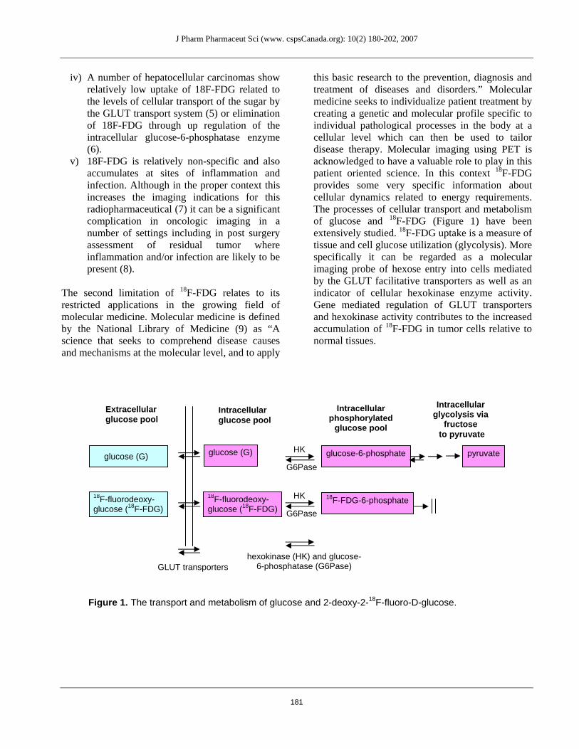
this basic research to the prevention, diagnosis and treatment of diseases and disorders.” Molecular medicine seeks to individualize patient treatment by creating a genetic and molecular profile specific to individual pathological processes in the body at a cellular level which can then be used to tailor disease therapy. Molecular imaging using PET is acknowledged to have a valuable role to play in this patient oriented science. In this context 18F-FDG provides some very specific information about cellular dynamics related to energy requirements. The processes of cellular transport and metabolism of glucose and 18F-FDG (Figure 1) have been extensively studied. 18F-FDG uptake is a measure of tissue and cell glucose utilization (glycolysis). More specifically it can be regarded as a molecular imaging probe of hexose entry into cells mediated by the GLUT facilitative transporters as well as an indicator of cellular hexokinase enzyme activity. Gene mediated regulation of GLUT transporters and hexokinase activity contributes to the increased accumulation of 18F-FDG in tumor cells relative to normal tissues.
Figure 1. The transport and metabolism of glucose and 2-deoxy-2-18F-fluoro-D-glucose.
pyruvateglucose-6-phosphate
18F-FDG-6-phosphate
glucose (G)
18F-fluorodeoxy- glucose (18F-FDG)
glucose (G)
18F-fluorodeoxy- glucose (18F-FDG)
Extracellular glucose pool
Intracellular glucose pool
GLUT transporters
HK
G6Pase
HK
G6Pase
hexokinase (HK) and glucose-6-phosphatase (G6Pase)
Intracellularphosphorylated
glucose pool
Intracellular glycolysis via
fructose to pyruvate

J Pharm Pharmaceut Sci (www. cspsCanada.org): 10(2) 180-202, 2007
182
Table 1. Targets and agents for PET molecular imaging and their relevance in clinical oncology
Cellular Target Relevance in Clinical Management of Cancer Patients
Potential, Clinically Relevant, PET Molecular Imaging Agent for Oncology
GLUT transporters, hexokinase activity
Tumor can be detected and graded through increased metabolism / energy utilization. Assessment of tumor response to therapy can be made.
18FDG, 11C-DG,
Amino acid transport and incorporation into polypeptides.
Tumors can be detected and graded through increased protein synthesis related to cell proliferation. Cellular growth kinetics and its alteration as a result of therapy can be measured.
11C-methionine, amino acid analogs of tyrosine, leucine, phenylalanine (18F-fluoroethyltyrosine)
Nucleoside transport, nucleoside phosphorylation, DNA synthesis
Cell proliferation can be determined as a measure of tumor growth and growth kinetics. Cellular responses to therapy can be measured by changes in proliferative ability.
18F-FLT, 11C-thymidine, 18F-FMAU, 124I-UdR
Choline transport and choline kinase activity, phospholipid synthesis
Phospholipid synthesis for intracellular phospholipids and cell membranes relates to increased cell growth and division in proliferating tissue. Prostate tumors have high phospholipid synthesis rates. Measurement of tumor growth and response to therapy can be made.
11C-choline, 18F-choline, 11C-acetate
Amine transport and decarboxylation
Amine precursor uptake and decarboxylation is a feature common to peptide producing endocrine tissue including neuroendocrine tumors. Tumors in this category can be detected and identified as having an endocrine origin.
18F-FDOPA, 11C-hydroxytryptophan
Intracellular reductive environment, hypoxia
Hypoxic tumor tissue can be resistant to both chemotherapy and radiotherapy. Tumor response to oxygenation strategies can be measured.
64Cu-ATSM, 18F-F-MISO, 18F-FAZA
Cellular receptors / tumor markers
The presence or absence of cell cytosolic receptors can predict response to hormone therapy in breast and prostate tumors. Tumor heterogeneity and response to therapy can be measured. Tumor markers can be imaged.
18F-FES, 18F-dihydroxy-testosterone, 68Ga-DOTATOC, EGFR inhibitors
Apoptotic changes in cells Cell death through apoptosis following therapy can be measured. Early assessment of imminent cell death can be made.
18F-annexin V
Elevated or unique products from gene expression including enzymes and RNA.
Gene therapy can be monitored using a reporter gene strategy. mRNA can be targeted as a surrogate marker of gene expression using oligonucleotides.
18F-gancyclovir/ HSV-1 tk reporter gene system, antisense oligonucleotides
Vascular endothelial factors related to angiogenesis
Angiogenesis accompanying tumor growth or therapy targeting microvascular development can be assessed.
Integrin/RDG peptide, VEGF/tk

J Pharm Pharmaceut Sci (www. cspsCanada.org): 10(2) 180-202, 2007
183
Table 2. Physical properties of selected positron emitting radionuclides relevant in human clinical imaging.
Radionuclide Mode of production Half-life
(Emissions)
β+ energy (Emax) MeV
Range of Emax β+
(mm in water)
Main chemistry used for radionuclide incorporation
Carbon-11 14N(p,α)11C (cyclotron) 20.4 min (β+ only) 0.97 4 Methylation, covalent
bond formation
Nitrogen-13 16O(p,α)13N (cyclotron) 9.9 min (β+ only) 1.20 5 C-N covalent bonds,
gas phase reactions
Oxygen-15 14N(d,n)15O (cyclotron) 122 sec (β+ only) 1.74 8 in target oxygen gases,
water production
Fluorine-18 18O(p,n)18F (cyclotron) 110 min (β+ only) 0.64 2.5
Nucleophilic, electrophilic substitution
Copper-62 63Cu(p,2n)62Zn, 62Zn → 62Cu (generator)
9.74 min (β+ only) 2.9 13 Metal chelation
Copper-64 64Ni(p,n)64Cu (cyclotron)
Zn(various)64Cu (cyclotron) 12.7 hr (β+, β-, gamma) 0.66 2.6 Metal chelation
Gallium-68 69Ga(p,2n)68Ge, 68Ge → 68Ga (generator)
68.3 min (β+, gamma) 1.9 9 Metal chelation
Bromine-76 76Se(p,n)76Br (cyclotron) 16.2 hr (β+, gamma) 3.94 20 Halide addition of
substitution
Iodine-124 124Te(p,n)124I (cyclotron) 4.15 day (β+, gamma) 2.1 10 Halide addition or
substitution
18F-FDG’s usefulness as a molecular imaging probe depends on the concept of bioisosterism in which the substitution of the fluorine group for the hydroxyl group produces a compound that has similar physicochemical and biological properties to the parent glucose. Efforts to produce a GLUT transportable glucose analog labeled with conventional single photon emitters such as 99mTc, 123I or 111In have not met with success. The GLUT transporters will have relatively stringent requirements for the molecules they transport. Similarly the substrate specificity of enzymatic systems like hexokinase will not tolerate substantial alteration of the hexose structure. An important distinction must be made between the behavior of 18F-FDG and glucose at the cellular level. 18F-FDG is metabolically trapped after phosphorylation to 18F-FDG-6-phosphate. Unlike glucose it is not a substrate for the subsequent enzymatic conversion to fructose-6-phosphate by phosphohexose
isomerase and thus does not further participate in the glycolytic pathway. Subtle differences between glucose and 18F-FDG may also be expected when they are transported by GLUTs and phosphorylated by hexokinase. 18F-FDG must be regarded as a surrogate marker for glucose although for some processes (transport and phosphorylation) the two molecules have very similar behavior. 18F-FDG AND OTHER PET MOLECULAR IMAGING AGENTS IN CLINICAL ONCOLOGY There are many molecular targets relevant to cancer that are not measured or assessed by 18F-FDG. Several recent reviews have addressed this issue (10,11). Table 1 provides a list of currently assessable molecular targets in tumor cells and tissues along with a description of their relevance in clinical management of cancer patients and a list of
J Pharm Pharmaceut Sci (www. cspsCanada.org): 10(2) 180-202, 2007
184
agents that have seen some significant clinical application as PET imaging agents or where clinical introduction of PET molecular imaging agents is imminent. Table 2 provides a list of PET radionuclides that are discussed in this paper along with their physical properties and method of production. At present the library of PET clinical tracers in oncology include; Agents Targeting GLUT Transporters and Hexokinase Enzyme Activity Rapidly growing cancer cells will exhibit high rates of metabolism accompanied by a requirement for glucose to support cellular energy processes. Uptake of glucose is mediated by the high level of expression of both the facilitated hexose transporter GLUT1 and the enzyme hexokinase. In this regard 18F-FDG represents a somewhat unique situation in that the transport and trapping mechanisms result in high concentrations of radioactivity in target tissues and consequently high tumor to background ratios which contribute to effective PET imaging. For many of the other molecular imaging agents there will be a much lower concentration of activity expected at the target site. In addition to 18F-FDG as discussed above there is at present no other suitable PET radiopharmaceutical to assess glycolysis. There is some incentive to explore additional sugar analogs based on the recognized alteration in GLUT expression and substrate specificity for transport in certain tumor tissues (12). 11C-glucose has been synthesized and examined in PET studies (13,14) but its application in oncology is hampered by the short physical half-life of the radionuclide which is not compatible with either the synthesis or biological disposition of 11C-glucose. The analysis of 11C glucose is further complicated by its conversion to labeled metabolites which show rapid efflux from tissues. Amino Acids for Transport and Incorporation into Polypeptides A number of amino acid analogs have been used as PET imaging agents in oncology. Amino acids are the building blocks for cellular proteins and other polypeptides that are produced to accompany cell proliferation and tissue expansion during tumor growth. Tumor cells can exhibit increased amino acid transport and increased protein synthesis rates which can serve as molecular imaging targets for radiolabeled amino acid analogs.
There are many examples of the synthesis and in vivo investigation of PET amino acid analogs (15) but only a few have any substantial impact in clinical oncology. Methyl-[11C]-L-methionine (11C-MET) has been used widely in PET imaging of brain tumors where it provides high contrast images. The very low level of amino acid utilization by normal brain permits the visualization of even relatively slow growing brain tumors (16). The short half life of the 11C label has stimulated the development of longer lived PET amino acid tracers of which the most widely used clinical product is O-(2-[18F]fluoroethyl)-L-tyrosine (18F-FET) first synthesized and used in humans in 1999 (17). A recent review explores the use of this tracer predominantly in brain tumors (18). FET is transported by amino acid transporters and accumulates in cells. However this amino acid analog does not appear to be involved in protein synthesis (18). Despite its inability to mimic amino acids and participate in macromolecule synthesis the uptake of 18F-FET has been shown to correlate closely with 11C-MET which shares the same transport system and which is readily incorporated into proteins (19). Nucleoside Analogs for Measurement of Cellular Nucleic Acid Utilization and Cell Proliferation Cell proliferation requires the expansion of the nucleic acid content in the cell prior to division. This is accomplished by the transport of nucleosides or their de novo synthesis as building blocks for DNA. Thymidine is a recognized marker for cell proliferation being moved into cells by the nucleoside transport system and being rapidly incorporated into DNA. Additionally thymidine, unlike the other three nucleoside building blocks is incorporated into DNA but not into RNA making it an accurate probe for DNA replication. 11C-thymidine was first described as an in vivo marker of DNA synthesis in 1972 (20) and it has been used sporadically as a clinical measure of cell proliferation. Because it is modified only by the substitution of 11C for 12C it will behave identically to the native nucleoside (21). The major problems encountered with 11C-thymidine relate to the short half-life of 11C and to the catabolism of thymidine, mediated by the enzyme thymidine phosphorylase, leading to the production of 11C-labeled metabolites (Figure 2).

J Pharm Pharmaceut Sci (www. cspsCanada.org): 10(2) 180-202, 2007
185
A recently developed 18F-fluorinated analog of thymidine, 3-deoxy-3-[18F]fluorothymidine (18F-FLT), has eliminated some of the problems encountered with 11C-labeled nucleoside analogs by having a longer radionuclide half-life and by not being a substrate for enzymatic catabolism (22-25). FLT, unlike thymidine, is not measurably incorporated into DNA. FLT uses the nucleoside transport system and is a substrate for thymidine kinase (TK1) but very little of the FLT-phosphate pool becomes incorporated into DNA (26). Despite the trapping of FLT at the phosphate stage a number of studies have shown that 18F-FLT accumulation in tissue corresponds to the proliferation rate of the tissue (27,28). What is actually measured by 18F-FLT uptake into tissues is cellular nucleoside transport and phosphorylation by thymidine kinase with a strong correlation with TK1 levels in cells (26,28).
Agents for the Measurement of Phospholipid Synthesis Cell membrane phospholipid synthesis can be regarded as an indirect measure of cell proliferation. Choline is a precursor for phospholipids through its incorporation into phosphatidylcholine, a primary component of cell membrane construction. Tumor cells have also demonstrated elevated levels of choline kinase. Following on the detection of elevated levels of choline in prostate tumors by magnetic resonance spectroscopy (29) the PET molecular imaging agent 11C-choline was developed (30) and has since had widespread application in the PET imaging of prostate tumors (31-33) in addition to bladder (34) and brain (35). The relationship between cell proliferation and 11C-choline incorporation in prostate cancer is particularly complex involving phosphatidylcholine production,
Figure 2. The transport and cellular utilization of thymidine and 3-deoxy-3-[18F]fluorothymidine (18F-FLT).
11C-thymidine (11C-TdR)
3-deoxy-3-18F-fluorothymidine (18F-FLT)
11C-thymidine (11C-TdR)
11C-TdR metabolites
3-deoxy-3-18F-fluorothymidine (18F-FLT)
11C-TdR metabolites
11C-TdR monophosphate
11C-TdR triphosphate
11C-TdR diphosphate
18F-FLT monophosphate
18F-FLT diphosphate
18F-FLT triphosphate
DNA
DNA X
Extracellular nucleoside pool
Intracellular nucleoside pool
Intracellular phosphorylated nucleoside pool
TK1
dN
TK1
dN
Equilibrative and concentrative nucleoside transporters
Thymidine kinase (TK1) and deoxynucleotidase (dN)

J Pharm Pharmaceut Sci (www. cspsCanada.org): 10(2) 180-202, 2007
186
phospholipid synthesis and upregulation of choline kinase and other enzymes. This has led to the conclusion that despite its recognized value in tumor visualization 11C-choline uptake does not correlate directly with cell proliferation (36). The accepted physical decay half-life advantage of 18F over 11C has resulted in the synthesis of several 18F-choline analogs including 18F-fluoro-ethylcholine (18F-FEC) and 18F-fluoromethylcholine (18F-fluorocholine; 18F-FCH) (37-39). 18F-FCH has shown a similar uptake to 11C-choline in animal studies and human prostate tumors (38) and has demonstrated its utility in prostate tumor imaging and staging in a number of clinical studies (40,41). Detailed studies of the relationship between cell proliferation and uptake of the fluorocholines have not been performed although their transport and interaction with choline kinase and other enzymes would account for the similarity to 11C-choline in uptake in prostate tumors. Amine Precursors Involved in Cell Uptake and Decarboxylation Amine precursor uptake and decarboxylation is a feature common to peptide producing endocrine tissue including neuroendocrine tumors. Tumors in this category can be detected and identified as having an endocrine origin. The two main clinical PET tracers that have found some application in imaging studies in this category are 18F-fluorodihydroxyphenylalanine (18F-FDOPA) and 11C-5-hydroxytryptophan. Endocrine tumors have the capacity to transport and accumulate these compounds as amino acid analogs following intercellular decarboxylation. Recent studies have explored the value of these agents in clinical studies of patients with neuroendocrine tumors (42-45). In clinical medical imaging of neuroendocrine lesions the PET agents will be compared with the more readily available single photon somatostatin receptor imaging agents such as 111In-pentreotide. A clear advantage over the conventional imaging would have to be demonstrated to justify the PET imaging study. For example 18F-FDOPA was shown to be a superior imaging agent for carcinoid tumors with a recommendation that it be used as the first-line scintigraphic imaging modality for these tumors while 111In-pentetreotide was superior as a scintigraphic imaging modality for noncarcinoid digestive tumors (42).
Agents to Assess Cellular Hypoxia Rapidly growing tumors can develop oxygen deficiency (hypoxia) and regions of dead cells (necrosis) by outstripping the process of angiogenesis which strives to provide adequate blood flow and consequently oxygen and nutrients to support tissue growth and metabolism in neoplastic tissue. Hypoxic tumor tissues can be resistant to chemotherapy and to radiotherapy. A number of agents exploit the low oxygen tension in hypoxic tissues to permit intercellular trapping by reductive mechanisms. The trapping mechanism is well understood for the nitroimidazoles, which are the compounds most often exploited for imaging hypoxia. After entry into a viable cell the nitroimidazole molecule undergoes a one electron reduction. When oxygen is present the reduced form of the nitroimidazole is reoxidized but in the absence of oxygen the molecule may undergo further reductive processes and the reactive species thus formed become trapped in the cell through covalent binding with intercellular molecules (46). Another class of hypoxia imaging compounds (Cu-labeled diacetyl-bis(N4-methylthiosemicarbazone; Cu-ATSM ) relies on reduction of a radioactive isotope of the chelated copper (eg. 64Cu) and its subsequent trapping in the cell (47). The main PET agents that have shown clinical utility to image hypoxia in tumors are 18F-fluoromisonidazole (18F-MISO), 64Cu-ATSM and the recently introduced 18F-fluoroazomycin-arabinoside (18F-FAZA). The characteristics of binding and the dependency primarily on cellular oxygen levels have been examined in detail for these compounds (47-52). The target for these compounds is an enzymatically active reductive environment within viable cells that maintain the capacity to transport substrates. A number of clinical studies have been published for 18F-FMISO (53,54) and those for 64Cu-ATSM are imminent based on clinical studies with the non-PET agent 60Cu-ATSM (55,56). Clinical studies with 18F-FAZA are in progress at several centers but at present unpublished. Cellular Receptor Imaging Agents The presence of cellular receptors on the cell surface or in the cytoplasm that may be unique to tumors or to subpopulations of cells within tumors presents a number of opportunities to exploit PET imaging agents for detection and as a guide to

J Pharm Pharmaceut Sci (www. cspsCanada.org): 10(2) 180-202, 2007
187
designing patient specific treatment. This is a very active area of research in imaging science and we can expect many clinical agents to be developed over the next few years. There are several examples where clinical PET imaging has already exploited cell receptors. One example is the cytoplasmic estrogen receptor which when activated by estrogen binding translocates to the nucleus. In the nucleus the activated receptor binds hormone specific DNA sequences resulting in the regulation of growth and differentiation of cells of the female and male reproductive systems and the growth and proliferation of breast cancer cells. Primary and metastatic breast cancer cells that express the estrogen receptor have been shown to respond to antiestrogen therapy with Tamoxifen. The PET imaging agent 16-α-[18F]-fluoroestradiol (18F-FES) was designed as an estrogen analog that recognizes and binds to the cytoplasmic estrogen receptor. The first patient studies with 18F-FES were performed in 1988 (57) and the agent continues to be investigated for human breast cancer patients (58,59). There appears to be a clear application for 18F-FES in identifying tumors that express estrogen receptors as a guide to selecting patients that will benefit from chemotherapy induced estrogen blockade. A recent study has also shown that quantitative PET studies with 18F-FES can predict patient responses to hormonal therapy and may thus assist in the selection of appropriate treatment (59). A second cell receptor molecular imaging agent, 16β-18F-fluoro-5α-dihydrotestosterone (18F-dihydrotestosterone; 18F-FDHT) has been synthesized (60) and used to image androgen receptors in prostate tumor cells in animal models (61) and in human clinical studies (62-64). Antiandrogen therapy is an accepted treatment modality for primary and metastatic prostate tumors but the effectiveness of therapy seems to be compromised by the emergence of tumor sub-populations that have lost their androgen dependency. In one study a reduction in the FDHT uptake in prostate tumors was noted after patients had been treated with the antiandrogen drug flutamide suggesting that 18F-FDHT uptake could stratify patients for androgen therapy (64). Recently there has been much interest in the targeting of PET radiopharmaceuticals to the family of somatostatin receptors on tumor cells. These receptors have been found on a variety of
neuroendocrine tumors, such as paragangliomas, carcinoids and breast tumors. There is a long history of somatostatin receptor imaging using the non-PET agent 111In-DTPA-octreotide and this agent has found widespread application in clinical nuclear medicine (65,66). PET imaging of the somatostatin receptor has been accomplished using 66Ga- and 68Ga-labeled [1,4,7,10-tetra-azacyclododecane-N,N′,N′′,N′′′-tetraacetic-acid-D-Phe1-Tyr3]-octreotide (66Ga-, 68Ga-DOTATOC) which primarily binds to somatostatin receptor 2 (SSTR2) (67-71). The first clinical PET studies in patients with neuroendocrine tumors were reported in 2003 using 68Ga-DOTATOC. Because the DOTATOC and related ligands also bind therapeutic radiometals including 90Y (72) a number of clinical PET imaging studies with 68Ga-DOTATOC have been performed in patients with metastatic neuroendocrine tumors scheduled for 90Y-DOTATOC therapy (70,71). The recognition of unique cell receptors or unique concentrations of receptors expressed by cancer cells is an area of intense research since these receptors provide opportunities for both detection and therapy of tumors. In this regard there have been exciting preclinical investigations with, for example, the family of epidermal growth factor receptors (73,74) including the Her2/neu receptor, a tyrosine kinase cell membrane receptor which is overexpressed in many breast cancers. It is relatively easy to develop antibody based imaging probes to cell surface receptors such as HER2/neu but the slow accumulation at the target and the persistent blood activity make antibodies generally unsuitable for PET imaging, especially with the shorter lived radionuclides. More promising PET probes are prepared from antibody fragments or peptide sequences that maintain the binding and specificity of intact antibodies. Recent work with diabodies and affibodies (75,76) serve to illustrate this approach. We should expect to see clinical PET imaging using these affinity molecules targeting HER2/neu and other cellular receptors in the near future. Agents Targeting Cells Undergoing Apoptosis A number of groups are investigating PET labeled probes that can non-invasively image apoptosis. The induction of apoptosis is a very early indication of the effectiveness of chemotherapy that can be assessed long before there are changes in tumor size

J Pharm Pharmaceut Sci (www. cspsCanada.org): 10(2) 180-202, 2007
188
or proliferative potential that can be assessed by other imaging modalities. At present preclinical PET investigations are proceeding with several analogs of annexin V labeled with either 18F (77) or 124I (78,79). Annexin V is one of a family of endogenous proteins that have a variety of functions including binding with calcium and phospholipids. Annexin V binds to cells with exposed surface phosphatidylserine (PS) groups. PS is normally restricted to the inner leaflet of cell membranes but becomes externalized at a very early stage in the apoptotic process presenting a target for annexin V binding. Although PET imaging in humans with annexin V analogs has not yet been published there have been several studies with cancer patients using 99mTc labeled annexin V in conventional imaging (80-83). The clinical value of the PET apoptosis imaging agents is yet to be established but the potential of obtaining a rapid assessment of the efficacy of anticancer treatment is a very strong motivation to continue to seek a PET tracer in this category. Agents Targeting Gene Expression With the modern understanding of the human genome and the genetic basis of cancer induction, progression and responses to therapy there is a growing interest in methods for imaging gene expression in vivo. This interest extends from endogenous genes that may be expressed in conjunction with the various phases of cancer progression or following therapy as well as to the imaging of genes transported into cells in gene therapy applications. Clinical PET has shown its potential for molecular imaging of gene therapy in oncology primarily in the area of advances in reporter gene detection as described in several recent reviews (84-86). The most widely used and successful reporter gene has been the herpes simplex virus 1 thymidine kinase gene (HSV1-tk). In this molecular imaging technique a therapeutic gene would be linked to the HSV1-tk gene using molecular biology methods and the combined gene would be delivered to target cells by a transfection methodology. Once inside the cell the HSV1-tk gene produces the herpes simplex virus 1 thymidine kinase enzyme (HSV1-TK) which allows for phosphorylation of nucleosides and nucleoside analogs. The essential feature of the HSV1-TK is that it has a much broader phosphorylation profile than endogenous mammalian thymidine kinase (TK). Thus nucleoside analogs that would not be
retained in cells expressing endogenous TK are trapped in cells possessing the mutant HSV1-TK enzyme due to selective phosphorylation (87). The PET probes that have shown potential in this imaging method include 2’-fluoro-2’-deoxy-1-β-arabinofuranosyl-5-[124I]iodouracil (124I-FIAU) (87,88), 2’-[18F]fluoro-2’-deoxy-5-methyl-1-β-arabinofuranosyluracil (18F-FMAU) (89,90), 8-[18F]-fluorogancyclovir (91,92), 9-(4-[18F]-fluoro-3-[hydroxymethyl]butyl)guanine (18F-FHBG) (93) and a number of others (84). These PET probes are not substrates for mammalian TK and after intravenous administration they would not be trapped by phosphorylation except in cells expressing the mutant HSV1-tk gene encoding for the less selective enzyme. Although the vast majority of reporter gene investigations have been carried out in vitro and in animal studies they have a great potential in human medicine and a number of clinical investigations with these imaging agents have been reported recently (94-98). Gene expression can also be theoretically imaged by the use of PET radionuclide labeled antisense oligonucleotides which can bind with gene encoded mRNA within cells. This approach has the potential to detect gene activity relative to cancer or to responses to cancer therapy although it has not yet been exploited in a clinical setting (99-101). Vascular Endothelial Factors Related to Angiogenesis The increased angiogenesis present in tumors relative to normal tissues represents a potential target for molecular imaging. This presents the possibility not only of tumor detection but also the assessment of anti-angiogenesis therapies which are proving to be a powerful tool in cancer chemotherapy. One approach to PET molecular imaging of angiogenesis, presently in preclinical investigation, has been to label vascular endothelial growth factor (VEGF) by using 64Cu-DOTA linked to VEGF (102). VEGF binding is specific to endothelial cells such as those present in rapidly growing microvasculature where it associates with two receptor tyrosine kinases Flt-1 and KDR (103). The receptors are greatly increased in the rapidly proliferating tumor endothelial cells when compared to the quiescent endothelial cells of normal vasculature.

J Pharm Pharmaceut Sci (www. cspsCanada.org): 10(2) 180-202, 2007
189
A second approach to imaging angiogenesis has targeted the dimeric transmembrane glycoprotein integrin αvβ3 which promotes angiogenesis and also plays a role in tumor cell growth and invasiveness (104). This integrin is virtually absent in mature vasculature and normal organs but is highly expressed in the endothelial cells of new vasculature produced during angiogenesis. The integrin αvβ3 is a receptor for extracellular matrix proteins which have the peptide sequence arginine:glycine:aspartic acid (RGD) (104). A number of RGD peptides have been developed to explore both therapy and preclinical imaging of active endothelial growth (105) and a PET agent, 18F-galacto-RGD, has exploited the selective binding between the integrin and the RGD tripeptide to perform imaging of angiogenesis in human cancer patients (106-108). A clear correlation has been noted between the accumulation of 18F-galacto-RGD in tissue and the degree of integrin αvβ3 expression (106). THE REALITY: Few agents can be said to be “routine” in clinical PET imaging in oncology With the exception of 18F-FDG there is no general supply of PET tracers through commercial sources. The great demand for 18F-FDG has encouraged commercial development through collaboration of existing companies such as the Schering – IBA venture in Western Europe or the development of commercial nuclear pharmacies such as Cardinal Health, Eastern Isotopes and PETnet in the United States. These companies supply only 18F-FDG as a PET imaging agent in oncology. Some simple molecule, gas phase PET tracers to measure perfusion, oxygen utilization and blood flow including 11C-CO, 11C-CO2, 15O-O2 and 15O-H2O are in general use but only at sites with in-house cyclotron facilities. We can additionally identify a handful of oncology tracers that are in widespread use and are produced routinely by PET centers for local or regional use. The list includes 11C-methionine, 11C-choline, 18F-FLT, 18F-FES, 18F-FDOPA, 18F-F-MISO and a few others. Notwithstanding the facile production of these PET agents they are not available to the majority of sites that have PET imaging capability but remain restricted to sites with in-house cyclotrons. The challenges to the widespread routine utilization of available PET tracers include the following:
i) Limited clinical or research demand;
ii) Chemistry challenges; iii) Clinical acceptability of PET
radiopharmaceuticals; iv) Physical half-life and other radionuclide
properties; v) Regulatory issues;
vi) Understanding of the molecular dynamics and pharmacokinetics;
vii) Cost. These items are explored in more detail below. Limited Clinical or Research Demand The demand for new tracers is attenuated by the wide applicability of 18F-FDG in clinical oncology. In routine PET scanning, where the aim will be to determine the extent of cancer progression (staging) or the evaluation of residual or recurrent disease in patients who have has cancer surgery or therapy, 18F-FDG is almost always effective. Possible exceptions would relate to the list of limitations presented earlier relating to specific anatomical sites or unusual biochemical situations in tumor cells. In much of the clinical imaging literature the terms PET and FDG-PET are synonymous. The clinical and research demand for new tracers also suffers because many physicians may be unaware of the specialized tracers that are available and the research literature that supports their utility in oncologic imaging. PET nuclear medicine physicians will have had specific training in the interpretation of PET images using 18F-FDG but will usually have no background in the interpretation of scans with other PET agents. It is not surprising that they might be unable or unwilling to incorporate new tracers into their practice. Chemistry Challenges The routine availability of PET tracers requires the resources and personnel to perform the chemistry and radiochemistry. The routine nature of 18F-FDG now allows this product to be produced with full automation and without the requirement for skilled radiochemistry personnel. Many of the other PET oncology tracers have not yet achieved this routine status. The aim with 18F radiopharmaceuticals and to an even greater extent with the shorter half-life 11C tracers is to perform the radiolabeling as the final step in the synthesis followed by a rapid simple purification procedure. With radiolabeling as the ultimate chemistry step there is the requirement

J Pharm Pharmaceut Sci (www. cspsCanada.org): 10(2) 180-202, 2007
190
to prepare a reactive precursor which may involve a complex and synthetically challenging series of chemistry steps. This will certainly be a consideration with the development of novel PET tracers and will require highly trained chemists. For many of the more common PET tracers in oncology the precursors are now available as GMP quality products from commercial suppliers. 18F and 11C and carbon remain the most used PET radionuclides for clinical products but a number of other radionuclides have had some application in clinical or preclinical studies. Table 2 lists the more common radionuclides in this category. This is a diverse group of elements including carbon, oxygen, nitrogen, halides (18F, 76Br, 124I) and a variety of radiometals (62Cu, 64Cu, 68Ga). Table 2 also indicates the main class of chemistry that is involved in the preparation of radiopharmaceuticals from each of the radionuclides. Diverse chemistry skills and specializations are required in the preparation or radiopharmaceuticals from these radionuclides. 18F radiopharmaceuticals are generally prepared by nucleophilic or electrophilic substitution reactions resulting in stable carbon fluorine covalent bonds. 11C is often incorporated into PET radiopharmaceuticals by methylation reactions using 11C-methyl iodide or triflate although a number of other reactive small molecule carbon compounds have been exploited to prepare PET products. The halides 76Br and 124I can be incorporated by electrophilic aromatic substitution or addition reactions. Radiometals will generally rely on chelation to appropriately functionalized molecules. Clinically acceptable PET radiopharmaceuticals will generally be prepared by automated chemistry. The advantages of automation include rapid synthesis, remote operation, the ability to isolate the automated units for radiation protection and for microbiological control (sterility), and the capacity to provide security, process control and data logging that makes the PET product acceptable to regulatory bodies. Commercial automated synthesis units (ASUs) are now available that prepare 18F-FDG as well as a number of the more common non-FDG PET tracers. The radiation containment will require specially constructed hot cells with appropriate radiation shielding and provision to maintain an appropriate internal environment suitable for the preparation of a pharmaceutical
product that will ultimately be injected into a patient to be imaged. Personnel issues must also be considered since the requirements for reagent preparation for the ASUs, the monitoring and adjustment of radiochemistry parameters and the final purification and quality control of the product will require a highly trained staff. Recognizing the need for trained radiochemistry and radiopharmacy staff and allocating the resources to hire them will go a long way to overcoming the challenges of radiopharmaceutical preparation. Manpower issues are complicated at present by a global shortage of scientists with suitable training in the fields of radiochemistry and radiopharmacy. A number of initiatives are underway to provide suitable opportunities for training additional practitioners in these fields. (For example: Nuclear Education Online run by the University of Arkansas and University of New Mexico [www.nuclearonline.org/nuclear_education /default.asp] and the Radiopharmaceutical Chemistry / Radiopharmacy postgraduate diploma course (PDC), organized by the Universities of Frankfurt am Main, Leipzig and ETH Zurich [www.pharma.ethz.ch/nd/].) Even with the availability of ASUs, hot cells and trained personnel it may not always be possible to perform the chemistry necessary to prepare a product routinely. There are instances where the complexity or sensitive nature of a synthetic process has been an impediment to its translation into a clinical oncology setting outside of a few selected laboratories. For example the elegant production of 11C-DOPA and 11C-5-hydroxy-tryptophan using a multienzymatic synthesis developed by Langstrom and co-workers (109) (Figure 3) does not appear to have been adopted by other laboratories despite the application of these tracers in imaging neuroendocrine malignancies (43-45). Clinical Acceptability of PET Radio-pharmaceuticals PET radiopharmaceuticals will generally be administered by intravenous injection. Any new radiopharmaceutical will therefore require testing for its suitability for human administration by this route. Most radiopharmaceuticals will be provided at high specific activity, and as tracers will be administered in chemical doses that will not elicit

J Pharm Pharmaceut Sci (www. cspsCanada.org): 10(2) 180-202, 2007
191
physiological responses in the patients. Issues of chemical toxicity are of less significance than standard therapeutic drug products but nonetheless there will be many quality control issues that will need to be addressed. Each new product will require a formalized series of quality control tests that will fall into the general categories of a) immediate release quality control tests (performed on each batch of product before release); b) retrospective quality control tests (performed on each batch of product after release) and; c) supporting characterization tests (performed periodically on selected batches after release). The actual tests performed will be product and institution specific but are designed to ensure both the quality and the safety of the product. As an example the three categories of tests for FDG as performed at the Edmonton PET Center are presented in Tables 3-5. When PET radiopharmaceuticals are purchased from a commercial supplier the responsibility for product testing and certification will be assumed by the manufacturer. The likely scenario for many of the new PET radiopharmaceuticals and certainly the 11C products is that they will continue to be produced in-house. Thus the responsibility for the quality control testing will be undertaken by personnel and using equipment that are part of the local PET program. In many instances the cost and complexity of assuring a quality product will be a significant barrier to the adoption of additional PET oncology tracers beyond FDG.
Physical Half-life and Other Radionuclide Properties The physical half-life of some PET radionuclides severely restricts their availability and their elaboration into routine clinical tracers in oncology. 13N (t1/2 = 9.9 m) and 15O (t1/2 = 122 s) can only be used where an in-house cyclotron is present and where the local chemistry is in place to provide suitable PET tracers (e.g. 13N-ammonia, 15O-water). 11C (t1/2 = 20.4 min) tracers can only be used locally for distribution to sites within a delivery radius achievable within a few half lives. 18F (t1/2 = 110 m) radiopharmaceuticals can be widely distributed where transportation options are available and this is well illustrated by the ability of commercial suppliers to provide 18F-FDG to almost any location in the United States and Western Europe. A number of longer lived PET radionuclides are being used in the development of oncology imaging agents including 64Cu (110) (t1/2 = 12.7 h), 76Br (111) (t1/2 = 16 h) and 124I (111) (t1/2 = 4.15 d) and several generator products mainly 68Ga (112) and 62Cu (113) can be widely distributed (Table 2). These radionuclides and their radiopharmaceuticals can not yet be considered to be routinely available and their impact on clinical imaging has to date been slight although their longer half-life continues to stimulate interest in the development of clinical imaging products.
Figure 3. A synthetic scheme for the production of the PET radiotracers 11C-DOPA and 11C-5-hydroxy-tryptophan involving multiple enzymatic steps (109).

J Pharm Pharmaceut Sci (www. cspsCanada.org): 10(2) 180-202, 2007
192
Table 3. Summary of typical immediate release quality control tests for 18F-FDG.
Test Method Preliminary Specification
Appearance Visual Inspection Clear, colourless, no visible matter
Radioactivity Assay Dose Calibrator > 150 MBq / mL
Radiochemical Purity TLC (area integration) > 90% 18F-FDG < 4% 18F-Fluoride
Radiochemical Identity TLC (Rf) Rf: +/- 0.05 Rf of FDG Std
pH pH paper pH: 4.5 – 8.5
Terminal Filter Integrity Test Bubble Point Passes Test
Radionuclidic Identity Half-life Determination 105 – 115 minutes
Chemical Purity: Residual Kryptofix
TLC (spot test) < 50 µg / mL
Table 4. Summary of typical retrospective quality control tests for 18F-FDG.
Test Method Specification
Endotoxin Test Bacterial Endotoxin Test USP on Pool < 175 EU / dose
Sterility Sterility Test USP on Pool Sterile
Table 5. Summary of typical supporting characterization tests for 18F-FDG.
Test Method Preliminary Specification
Radionuclidic Purity Gamma spectrometry > 99.5% 0.511 MeV +1.022 MeV + Compton scatter
Chemical Purity: Acetonitrile Ethanol Chlorodeoxyglucose FDG
GC GC HPLC HPLC
< 0.04% (w/v) < 0.5% (w/v) < 0.5 mg / dose < 10 mg / dose
Particulates Particulate Test USP Small volume parenteral USP
Osmolality Osmometer 260 – 300 mOs/kg

J Pharm Pharmaceut Sci (www. cspsCanada.org): 10(2) 180-202, 2007
1
Table 6. Structures of matched 11C and 18F radiopharmaceuticals recognizing equivalent molecular
Molecular target 11C product 18F product
Metabolism / energy utilization
11C-glucose
18F-fluorodeoxyglucose
Phospholipid synthesis
11C-choline
18F-fluoromethylcholine
Amine decarboxylation
11C-DOPA
18F-FDOPA
Cell proliferation / DNA synthesis
11C-thymidine
18F-fluorothymidine
In addition to the issues of delivery of the radionuclides or the radiopharmaceuticals that are synthesized from them the half-life can impact on other aspects of tracer production and use. The half-life of some of the PET radionuclides severely restricts their elaboration into suitable radiopharmaceuticals due to the time requirements of chemical processing, purification and quality control. The short physical half-life radionuclides will not be compatible with slow biological targeting or slow blood clearance. In a number of molecular imaging situations both 11C and 18F products have been developed. Where these products have the same biological behavior and
ability to assess similar molecular targets the longer lived radiopharmaceutical may be preferred. Table 6 gives a number of examples where both an 11C and an 18F PET oncology molecular imaging agent have been developed. Half-life and availability are the most important factors affecting the clinical acceptability of radionuclides and the radiopharmaceuticals derived from them. In addition the quality of PET images will be affected by the positron energy and by the presence of single photon emissions where the decay mode is not purely by positron emission. As Table 2 indicates the positrons from different

J Pharm Pharmaceut Sci (www. cspsCanada.org): 10(2) 180-202, 2007
194
radionuclides are emitted over a wide range of Emax values and the range of an Emax particle can be quite substantial. Positrons do not annihilate until they have lost most of their kinetic energy and thus the position of photon production will be different than the position of the decaying radionuclide. For a number of reasons the Emax particle range does not give an accurate indication of the actual magnitude of this separation. Firstly the average energy of positrons is much less than the Emax value and the mean positron range will be consequently much shorter (114). In addition the positron does not travel in a straight line but has a tortuous path that shortens the overall distance between radionuclide and annihilation event. For energies up to Emax of 1.5 MeV the influence of positron travel will be negligible (115,116). A recent analysis has discussed the influence of positron energy on spatial resolution in PET including calculations in various tissues (117). The positron energy is less significant than factors such as the non-colinearity of the coincident positrons and PET camera factors in determining the overall spatial resolution. PET images will also be degraded in the situation where single photons are produced in the decay process in addition to positrons. For example 76Br emits gamma rays at 0.559 and 0.657 MeV and 124I at 0.602 MeV that will fall within the normal energy detection window of PET scanners. Consequently, without correction for these prompt gamma rays, the image resolution for these two isotopes will be poorer than a pure beta emitter such as 18F (114). Regulatory Issues The use of PET tracers in clinical oncology will necessarily fall under the scrutiny of local and national regulatory agencies. Local committees will usually be required to assess human ethics for a new imaging agent. Approvals will depend on evidence of safety and efficacy of the radiopharmaceutical. In many countries new PET radiopharmaceuticals will be regarded as new drug entities and will be subjected to the same regulatory process as is used for the approval of therapeutic drug products. This regulatory process can be a severe impediment to bringing PET tracers into the clinical setting since the rigorous drug assessment is expensive and time consuming. Drug companies have shown little interest in working through the regulatory issues with novel PET tracers
recognizing that the small market for the PET tracers other than 18F-FDG means that these products are unlikely to be financially viable. Most countries will permit clinical trials to be carried out where a novel PET imaging agent is to be assessed or where an established PET tracer is to be used in a new patient population. Clinical trials will require regulatory approvals but are generally less burdensome that approvals for new drugs. Clinical trials will allow the assessment of a PET tracer to be carried out in a limited number of patients but will help to evaluate the drug as a candidate for a more extensive testing. A number of jurisdictions have now acknowledged the distinction between tracer radiopharmaceuticals and therapeutic drugs and will permit small trial studies to be performed with very minimal regulatory burden for the researchers. The regulatory process will also require strict adherence to quality control issues during radiopharmaceutical preparation. The specific requirements for radiopharmaceutical manufacture will vary with jurisdiction but in many cases they provide a significant commitment in terms of space and personnel and can discourage the in-house preparation and evaluation of novel tracers. Understanding of the Molecular Dynamics and Pharmacokinetics In order to use the full potential of PET molecular imaging agents in oncology there must be a thorough understanding of the molecular dynamics and tissue pharmacokinetics of the tracers. Unfortunately issues related to cellular delivery, transport, metabolism, trapping and intracellular localization of many PET tracers is poorly understood. Where PET tracers are to be used as markers of molecular processes there must be a clear understanding of the cellular biochemistry. An advantage with some of the 11C-labeled PET radiopharmaceuticals is that they are identical to native biological molecules for which the biochemical pathways for metabolism and catabolism have been thoroughly elucidated. This claim of equivalency for the PET tracers requires that the isotopic substitution of 11C for 12C causes negligible perturbation to biochemical processes. When analogs of naturally occurring biological molecules are used some care must be exercised to define the actual molecular target and cellular processes involved.

J Pharm Pharmaceut Sci (www. cspsCanada.org): 10(2) 180-202, 2007
195
As an example we can consider the use of 11C-thymidine and its analog 3-deoxy-3-[18F]fluorothymidine (18F-FLT) to assess cell proliferation. While both of these PET radiotracers are generally accepted as probes to assess cell proliferation, only 11C-thymidine is actually incorporated into the DNA of replicating cells (Figure 2). Because 11C-thymidine also undergoes extensive metabolism its distribution in tissues is complex and the analysis of tissue proliferation based on radioactivity measurement requires a multicompartmental mathematical analysis (118,119). 18F-FLT would seem to be an improvement over 11C-thymidine not only because of the longer physical half-life of 18F but also because of a simpler biological profile in the cell. 18F-FLT is a substrate for the nucleoside transporters, undergoes minimal degradation to metabolic products and is phosphorylated by thymidine kinase (TK-1). However 18F-FLT is not incorporated into DNA. The uptake of 18F-FLT in tissue may correlate with cell proliferation but strictly speaking the PET image obtained in proliferating tissue reflects increased nucleoside transport and increased activity of TK-1 in converting the nucleoside analog into the phosphate forms (27,28,120). Additional discussion of the appropriate interpretation of the in vivo distribution and PET images obtained with molecular imaging tracers has been published recently for the radiopharmaceutical pairings of 11C-glucose with glucose as well as 11C-thymidine with 18F-FLT (120). The biochemical complexity of cellular processing for PET radiopharmaceuticals does not have to be an impediment to their routine use. In many situations the simple interpretation of radiotracer distribution will be adequate to provide clinically useful and relevant information. Therefore for example if we consider 18F-FLT as a marker of tissue proliferation this will almost always be accurate and appropriate. It will only be in the unusual circumstance where transport and phosphorylation of 18F-FLT very poorly correlate with actual cell proliferation that the image interpretation may provide misleading information relevant to disease management. Cost PET imaging requires a substantial commitment of healthcare dollars. The basic infrastructure to use
18F-FDG will consist of a PET camera or more appropriately for modern imaging a combined PET-CT camera. Most PET imaging centers will use commercially supplied 18F-FDG making this the only additional cost without considering the clinical costs for performing and interpreting the study and the cost of specialized training for the physicians and nuclear medicine technologists. If a center wishes to use a molecular imaging tracer other than 18F-FDG there are substantial additional costs involved. Included in these costs can be: i) cyclotron, targetry and operating personnel; ii) chemistry costs including automated synthesis units, hot cells and trained radiochemists; iii) quality assurance costs including analytical instrumentation, construction of appropriate facilities for GMP radiopharmaceutical production and the provision of trained staff to perform quality control testing; iv) preparation of regulatory documents to support new drug submissions or clinical trials applications to local and national medical oversight bodies; v) training of clinical staff in the appropriate use of the PET tracers and in the interpretation of PET images. The substantial cost of PET studies in oncology has been the subject of intense scrutiny with the usual goal being to demonstrate the cost effectiveness of PET imaging. There are more than 130 publications listed by PubMed since 1994 that deal with cost effectiveness of PET in cancer management (121). PET with 18F-FDG can provide images that guide clinical decision making and can prevent ineffective treatment thereby generating substantial cost savings (e.g. (122,123)). Perhaps more importantly PET imaging can decrease patient morbidity from inappropriate treatments and contribute to patient quality of life in a palliative setting and to more effective and potentially curative strategies in a treatment setting. An understanding of the cost effectiveness of PET in oncology has contributed to reimbursement for specified applications by health administration bodies in many countries although this is far from universal and has required intense lobbying. When additional PET molecular imaging agents are introduced into clinical practice it will certainly take time and effort before reimbursement is possible. Thus the initial costs must be assumed by the hospitals and clinics that wish to use a PET tracer other than 18F-FDG.

J Pharm Pharmaceut Sci (www. cspsCanada.org): 10(2) 180-202, 2007
196
The seven items discussed above have combined to slow the introduction of additional PET agents into clinical oncology and they continue to present a formidable challenge to the scientists and clinicians who work in this field. CONCLUSION Non-FDG PET tracers can play a valuable role in defining the molecular basis of human cancer and in individualizing patient treatment and care. A number of the newer molecular imaging tracers are relevant to modern drug and radiotherapy treatment strategies targeting such processes as angiogenesis, unique cell cytosolic and surface markers and cell proliferation biochemistry. Determining the molecular-biochemical profile of a specific tumor can be combined with immediate and effective treatment decisions. The value of the molecular imaging agents is increased if we have a thorough understanding of their in vivo distribution, their molecular targets and their biochemical behavior at the cellular level. At present most new PET tracers that have a specific rather than a general application in oncologic imaging have remained restricted to one or only a few PET centers. Other than 18F-FDG only a handful of tracers can be regarded as having global acceptance and these will only be found where there are in-house or local resources for isotope production and PET radiochemistry. Clinical demand and commercial production have combined to make 18F-FDG routinely available in many countries and it remains to be seen if this can be repeated with the more popular non-FDG tracers. One potential stimulus for the development of novel PET tracers in oncology comes from the recognition that these agents can play a role in anticancer drug development. It is now accepted that molecular imaging can speed up drug development by providing direct evidence of drug action using both animal PET and human clinical trials. Drug companies are increasingly contracting with PET centers to carry out molecular imaging studies in conjunction with drug trials as a rapid assessment of efficacy. Although 18F-FDG has taken a leading role in this endeavor (124) the value of non-FDG tracers is now widely recognized (125). The development of PET molecular imaging probes is necessarily a multidisciplinary science using the
expertise of nuclear physicists for the production of novel radionuclides, radiochemists and radiopharmacists for the elaboration of the radiotracers, biologists and experimental oncologists to determine the details of targeting and molecular processing of the tracers and clinical scientists to effectively evaluate the human imaging. The future of this science will depend on collaborative efforts among PET centers and their supporting clinical colleagues, governmental agencies regulating the PET tracer development or providing research funding and companies with an interest in imaging instrumentation or drug development. ACKNOWLEDGEMENTS I acknowledge the support and scientific guidance given to me by Dr. Tony Noujaim throughout my scientific and academic career at the University of Alberta. He was a mentor, a colleague and a friend who is greatly missed. REFERENCES [1] Kostakoglu, L., Agress, H., Goldsmith, S.J.
Clinical role of FDG PET in evaluation of cancer patients. RadioGraphics, 23:315-340, 2003.
[2] Mottaghy, F.M. Reske, S.N. Functional imaging of neuroendocrine tumours with PET. Pituitary, 9:237-42, 2006.
[3] Adams, S., Baum, R., Rink, T., Schumm-Drager, P.M., Usadel, K.H., Hor, G. Limited value of fluorine-18 fluorodeoxyglucose positron emission tomography for the imaging of neuroendocrine tumours. Eur. J. Nucl. Med., 25:79–83, 1998.
[4] Adams, S., Baum, R.P., Hertel, A., Schumm-Drager, P.M., Usadel, K.H., Hor, G. Metabolic (PET) and receptor (SPET) imaging of well- and less well-differentiated tumours: comparison with the expression of the Ki-67 antigen. Nucl. Med. Commun., 19:641–647, 1998.
[5] Lee, J.D., Yang, W.I., Park, Y.N., Kim, K.S., Choi, J.S., Yun, M., Ko, D., Kim, T.S., Cho, A.E., Kim, H.M., Han, K.H., Im, S.S., Ahn, Y.H., Choi, C.W., Park, J.H. Different glucose uptake and glycolytic mechanisms between hepatocellular carcinoma and intrahepatic mass-forming cholangiocarcinoma with increased (18)F-FDG uptake. J. Nucl. Med., 46:1753-9, 2005.
[6] Torizuka, T., Tamaki, N., Inokuma, T., Magata, Y., Sasayama, S., Yonekura, Y., Tanaka, A., Yamaoka, Y., Yamamoto, K., Konishi, J. In vivo assessment of glucose metabolism in hepato-

J Pharm Pharmaceut Sci (www. cspsCanada.org): 10(2) 180-202, 2007
197
cellular carcinoma with FDG-PET. J. Nucl. Med., 36:1811-7, 1995.
[7] Zhuang, H., Alavi, A. 18-fluorodeoxyglucose positron emission tomographic imaging in the detection and monitoring of infection and inflammation. Semin. Nucl. Med., 32:47-59, 2002.
[8] Chang, J.M., Lee, H.J., Goo, J.M., Lee, H.Y., Lee, J.J., Chung, J.K., Im, J.G. False positive and false negative FDG-PET scans in various thoracic diseases. Korean J. Radiol., 7:57-69, 2006.
[9] United States, National Library of Medicine - National Institutes of Health, 8600 Rockville Pike, Bethesda, MD 20894. Url:http://www.nlm.nih.gov/
[10] Nanni, C., Rubello, D., Al-Nahhas, A., Fanti, S. Clinical PET in oncology: not only FDG. Nucl. Med. Commun., 27:685-8, 2006.
[11] Shiue, C.Y., Welch, M.J. Update on PET radiopharmaceuticals: life beyond fluorodeoxy-glucose. Radiol. Clin. North Am., 42:1033-53, 2004.
[12] Smith, T.A.D., Sharma, R.I., Thompson, A.M., Pauline, F.E.M. Tumor 18F-FDG incorporation is enhanced by attenuation of p53 function in breast cancer in vitro. J. Nucl. Med., 47:1525-1530. 2006.
[13] Ehrin, E., Stone-Elander, S., Nilsson, J.L., Bergstrom, M., Blomqvist, G., Brismar, T., Eriksson, L., Greitz, T., Jansson, P.E., Litton, J.E., Malmborg, P., af Ugglas, M., Widen, L. C-11-labeled glucose and its utilization in positron-emission tomography. J. Nucl. Med., 24:326-31, 1983.
[14] Stone-Elander, S., Nilsson, J.L., Blomqvist, G., Ehrin, E., Eriksson, L., Garmelius, B., Greitz, T., Johnstrom, P., Sjogren, I., Widen, L. 11C-2-deoxy-D-glucose: synthesis and preliminary comparison with 11C-D-glucose as a tracer for cerebral energy metabolism in PET studies. Eur. J. Nucl. Med., 10:481-6, 1985.
[15] Jager, P.L., Vaalburg, W., Pruim, J., de Vries, E.G., Langen, K.J., Piers, D.A. Radiolabeled amino acids: basic aspects and clinical applications in oncology. J. Nucl. Med., 42: 432- 445, 2001.
[16] Torii, K., Tsuyuguchi, N., Kawabe, J., Sunada, I., Hara, M., Shiomi, S. Correlation of amino-acid uptake using methionine PET and histological classifications in various gliomas. Ann. Nucl. Med., 19:677-83, 2005.
[17] Wester, H.J., Herz, M., Weber, W., Heiss, P., Senekowitsch-Schmidtke, R., Schwaiger, M., Stocklin, G. Synthesis and radiopharmacology of O-(2-[18F]fluoroethyl)-L-tyrosine for tumor imaging. J. Nucl. Med., 40:205-12, 1999.
[18] Langen, K.J., Hamacher, K., Weckesser, M., Floeth, F., Stoffels, G., Bauer, D., Coenen, H.H., Pauleit, D., O-(2-[18F]fluoroethyl)-L-tyrosine: uptake mechanisms and clinical applications. Nucl. Med. Biol., 33:287-94, 2006.
[19] Weber, W., Wester, H.J., Grosu, A.L., Herz, M., Dzewas, B., Feldmann, H.J., Molls, M., Stocklin, G., Schwaiger, M. O-(2-[18F] fluoroethyl)-L-tyrosine and L-[methyl-11C] methionine uptake in brain tumours: Initial results of a comparative study. Eur. J. Nucl. Med., 27:542–549, 2000.
[20] Christman, D., Crawford, E.J., Friedkin, M., Wolf, A.P. Detection of DNA synthesis in intact organisms with positron-emitting methyl-[C-11]-thymidine. Proc. Natl. Acad. Sci. U.S.A., 69:988– 92, 1972.
[21] Wells, P., Gunn, R.N., Alison, M., Steel, C., Golding, M., Ranicar, A.S., Brady, F., Osman, S., Jones, T., Price, P. Assessment of proliferation in vivo using 2-[11C]thymidine positron emission tomography in advanced intra-abdominal malignancies. Cancer Res., 62:5698–702, 2002.
[22] Shields, A.F., Grierson, J.R., Dohmen, B.M., Machulla, H.-J., Stayanoff, J.C., Lawhorn-Crews, J.M., Obradovich, J.E., Muzik, O. Mangner, T.J. Imaging proliferation in vivo with [F-18]FLT and positron emission tomography. Nature Medicine, 4:1334-1336, 1998.
[23] Muzi, M., Spence, A.M., O'Sullivan, F., Mankoff, D.A., Wells, J.M., Grierson, J.R., Link, J.M., Krohn, K.A. Kinetic analysis of 3'-deoxy-3'-18F-fluorothymidine in patients with gliomas. J. Nucl. Med., 47:1612-21, 2006.
[24] Grierson, J.R., Shields, A.F. Radiosynthesis of 30-deoxy-30-[(18)F]fluorothymidine: [(18)F]FLT for imaging of cellular proliferation in vivo. Nucl. Med. Biol., 27:143–56, 2000.
[25] Mankoff, D.A., Shields, A.F., Krohn, K.A. PET imaging of cellular proliferation. Radiol. Clin. North Am., 43:153–167, 2005.
[26] Toyohara, J., Waki, A., Takamatsu, S., Yonekura, Y., Magata, Y., Fujibayashi, Y.. Basis of FLT as a cell proliferation marker: comparative uptake studies with [3H]thymidine and [3H]arabino-thymidine, and cell-analysis in 22 asynchronously growing tumor cell lines. Nucl. Med. Biol., 29:281–7, 2002.
[27] Rasey, J.S., Grierson, J.R., Wiens, L.W., Kolb, P.D., Schwartz, J.L. Validation of FLT uptake as a measure of thymidine kinase-1 activity in A549 carcinoma cells. J. Nucl. Med., 43:1210–7, 2002.
[28] Barthel, H., Perumal, M., Latigo, J., He, Q., Brady, F., Luthra, S.K., Price, P.M., Aboagye, E.O. The uptake of 3'-deoxy-3'-[18F]fluorothymidine into L5178Y tumours in vivo is dependent on thymidine kinase 1 protein levels. Eur. J. Nucl. Med. Mol. Imaging, 32:257-63, 2005.
[29] Negendank, W. Studies of human tumors by MRS: a review. NMR Biomed., 5:303-24, 1992.
[30] Hara, T., Kosaka, N., Kishi, H. PET imaging of prostate cancer using carbon-11-choline. J. Nucl. Med., 39:990–995, 1998.

J Pharm Pharmaceut Sci (www. cspsCanada.org): 10(2) 180-202, 2007
198
[31] Kotzerke, J., Prang, J., Neumaier, B., Volkmer, B., Guhlmann, A., Kleinschmidt, K., Hautmann, R., Reske, S.N. Experience with carbon-11 choline positron emission tomography in prostate carcinoma. Eur. J. Nucl. Med., 27:1415–1419, 2000.
[32] Farsad, M., Schiavina, R., Castellucci, P., Nanni, C., Corti, B., Martorana, G., Canini, R., Grigioni, W., Boschi, S., Marengo, M., Pettinato, C., Salizzoni, E., Monetti, N., Franchi, R., Fanti, S. Detection and localization of prostate cancer: correlation of 11C-choline PET/CT with histo-pathologic step-section analysis. J. Nucl. Med., 46: 1642–1649, 2005.
[33] Reske, S.N., Blumstein, N.M., Neumaier, B., Gottfried, H.W., Finsterbusch, F., Kocot, D., Moller, P., Glatting, G., Perner, S. Imaging prostate cancer with 11C-choline PET/CT. J. Nucl. Med., 47:1249-54, 2006.
[34] Schoder, H., Larson, S.M. Positron emission tomography for prostate, bladder, and renal cancer. Semin. Nucl. Med., 34:274-92, 2004.
[35] Tian, M., Zhang, H., Oriuchi, N., Higuchi, T., Endo, K. Comparison of 11C-choline PET and FDG PET for the differential diagnosis of malignant tumors. Eur. J. Nucl. Med. Mol. Imaging, 31:1064-72, 2004.
[36] Breeuwsma, A.J., Pruim, J., Jongen, M.M., Suurmeijer, A.J., Vaalburg, W., Nijman, R.J., de Jong, I.J. In vivo uptake of [11C]choline does not correlate with cell proliferation in human prostate cancer. Eur. J. Nucl. Med. Mol. Imaging, 32:668-73, 2005.
[37] Hara, T., Kosaka, N., Kishi, H. Development of 18F-fluoroethylcholine for cancer imaging with PET: synthesis, biochemistry, and prostate cancer imaging. J. Nucl. Med., 43:187–199, 2002.
[38] DeGrado, T.R., Coleman, R.E., Wang, S., Baldwin, S.W., Orr, M.D., Robertson, C.N., Polascik, T.J., Price, D.T. Synthesis and evaluation of 18F labeled choline as an oncologic tracer for positron emission tomography: initial findings in prostate cancer. Cancer Res, 61:110–117, 2001.
[39] DeGrado, T.R., Baldwin, S.W., Wang, S., Orr, M.D., Liao, R.P., Friedman, H.S., Reiman, R., Price, D.T., Coleman, R.E. Synthesis and evaluation of (18)F-labeled choline analogs as oncologic PET tracers. J. Nucl. Med., 42:1805-14, 2001.
[40] Cimitan, M., Bortolus, R., Morassut, S., Canzonieri, V., Garbeglio, A., Baresic, T., Borsatti, E., Drigo, A., Trovo, M.G. [(18)F]fluorocholine PET/CT imaging for the detection of recurrent prostate cancer at PSA relapse: experience in 100 consecutive patients. Eur. J. Nucl. Med. Mol. Imaging, 33:1387-98, 2006.
[41] Kwee, S.A., Coel, M.N., Lim, J., Ko, J.P. Prostate
cancer localization with 18fluorine fluorocholine positron emission tomography. J. Urol., 173:252–255, 2005.
[42] Montravers, F., Grahek, D., Kerrou, K., Ruszniewski, P., de Beco, V., Aide, N., Gutman, F., Grange, J.D., Lotz, J.P., Talbot, J.N. Can fluorodihydroxyphenylalanine PET replace somatostatin receptor scintigraphy in patients with digestive endocrine tumors? J. Nucl. Med., 47: 1455-62, 2006.
[43] Becherer, A., Szabo, M., Karanikas, G., Wunderbaldinger, P., Angelberger, P., Raderer, M., Kurtaran, A., Dudczak, R., Kletter, K. Imaging of advanced neuroendocrine tumors with (18)F-FDOPA PET. J. Nucl. Med., 45:1161-7, 2004.
[44] Orlefors, H., Sundin, A., Garske, U., Juhlin, C., Oberg, K., Skogseid, B., Langstrom, B., Bergstrom, M., Eriksson, B. Whole-body (11)C-5-hydroxytryptophan positron emission tomography as a universal imaging technique for neuro-endocrine tumors: comparison with somatostatin receptor scintigraphy and computed tomography. J. Clin. Endocrinol. Metab., 90:3392-400, 2005.
[45] Eriksson, B., Bergstrom, M., Sundin, A., Juhlin, C., Orlefors, H., Oberg, K., Langstrom, B. The role of PET in localization of neuroendocrine and adrenocortical tumors. Ann. N.Y. Acad. Sci., 970: 159-69, 2002.
[46] Chapman, J.D., Baer, K., Lee, J. Characteristics of the metabolism-induced binding of misonidazole to hypoxic mammalian cells. Cancer Res., 43:1523-8, 1983.
[47] Obata, A., Yoshimi, E., Waki, A., Lewis, J.S., Oyama, N., Welch, M.J., Saji, H., Yonekura, Y., Fujibayashi, Y. Retention mechanism of hypoxia selective nuclear imaging/radiotherapeutic agent cu-diacetyl-bis(N4-methylthiosemicarbazone) (Cu-ATSM) in tumor cells. Ann. Nucl. Med., 15:499-504, 2001.
[48] Rasey, J.S., Nelson, N.J., Chin, L., Evans, M.L., Grunbaum, Z. Characteristics of the binding of labeled fluoromisonidazole in cells in vitro. Radiat. Res., 122:301-8, 1990.
[49] Rasey, J.S., Grunbaum, Z., Magee, S., Nelson, N.J., Olive, P.L., Durand, R.E., Krohn, K.A. Characterization of radiolabeled fluoro-misonidazole as a probe for hypoxic cells. Radiat. Res., 111:292-304, 1987.
[50] Lewis, J.S., McCarthy, D.W., McCarthy, T.J., Fujibayashi, Y., Welch, M.J. Evaluation of 64Cu-ATSM in vitro and in vivo in a hypoxic tumor model. J. Nucl. Med., 40:177-83, 1999.
[51] Sorger, D., Patt, M., Kumar, P., Wiebe, L.I., Barthel, H., Seese, A., Dannenberg, C., Tannapfel, A., Kluge, R., Sabri, O. [18F]Fluoroazomycin-arabinofuranoside (18FAZA) and [18F]Fluoro-misonidazole (18FMISO): a comparative study of
J Pharm Pharmaceut Sci (www. cspsCanada.org): 10(2) 180-202, 2007
199
their selective uptake in hypoxic cells and PET imaging in experimental rat tumors. Nucl. Med. Biol., 30:317-26, 2003.
[52] Piert, M., Machulla, H.J., Picchio, M., Reischl, G., Ziegler, S., Kumar, P., Wester, H.J., Beck, R., McEwan, A.J., Wiebe, L.I., Schwaiger, M. Hypoxia-specific tumor imaging with 18F-fluoroazomycin arabinoside. J. Nucl. Med., 46: 106-13, 2005.
[53] Rischin, D., Hicks, R.J., Fisher, R., Binns, D., Corry, J., Porceddu, S., Peters, L.J. Prognostic significance of [18F]-misonidazole positron emission tomography-detected tumor hypoxia in patients with advanced head and neck cancer randomly assigned to chemoradiation with or without tirapazamine: a substudy of Trans-Tasman Radiation Oncology Group Study 98.02. J. Clin. Oncol., 24:2098-104, 2006.
[54] Rajendran, J.G., Schwartz, D.L., O'Sullivan, J., Peterson, L.M., Ng, P., Scharnhorst, J., Grierson, J.R., Krohn, K.A. Tumor hypoxia imaging with [F-18] fluoromisonidazole positron emission tomography in head and neck cancer. Clin. Cancer Res., 12:5435-41, 2006.
[55] Dehdashti, F., Mintun, M.A., Lewis, J.S., Bradley, J., Govindan, R., Laforest, R., Welch, M.J., Siegel, B.A. In vivo assesment of tumor hypoxia in lung cancer with 60Cu-ATSM. Eur. J. Nucl. Med. Mol. Imaging, 30:844–850, 2003.
[56] Dehdashti, F., Grigsby, P.W., Mintun, M.A., Lewis, J.S., Siegel, B.A., Welch, M.J. Assessing tumor hypoxia in cervical cancer by positron emission tomography with 60Cu-ATSM: relationship to therapeutic response-a preliminary report. Int. J. Radiat. Oncol. Biol. Phys., 55:1233–1238, 2003.
[57] Mintun, M.A., Welch, M.J., Siegel, B.A., Mathias, C.J., Brodack, J.W., McGuire, A.H., Katzenellenbogen, J.A. Breast cancer: PET imaging of estrogen receptors. Radiology, 169:45-8, 1988.
[58] McGuire, A.H., Dehdashti, F., Siegel, B.A., Lyss, A.P., Brodack, J.W., Mathias, C.J., Mintun, M.A., Katzenellenbogen, J.A., Welch, M.J. Positron tomographic assessment of 16 alpha-[18F] fluoro-17 beta-estradiol uptake in metastatic breast carcinoma. J. Nucl. Med., 32:1526-31, 1991.
[59] Linden, H.M., Stekhova, S.A., Link, J.M., Gralow, J.R., Livingston, R.B., Ellis, G.K., Petra, P.H., Peterson, L.M., Schubert, E.K., Dunnwald, L.K., Krohn, K.A., Mankoff, D.A. Quantitative fluoroestradiol positron emission tomography imaging predicts response to endocrine treatment in breast cancer. J. Clin. Oncol., 24:2793-9, 2006.
[60] Choe, Y.S., Lidstrom, P.J., Chi, D.Y., Bonasera, T.A., Welch, M.J., Katzenellenbogen, J.A. Synthesis of 11 beta-[18F]fluoro-5 alpha
dihydrotestosterone and 11 beta-[18F]fluoro-19-nor-5 alpha-dihydrotestosterone: preparation via halofluorination-reduction, receptor binding, and tissue distribution. J. Med. Chem., 38:816–825, 1995.
[61] Bonasera, T.A., O’Neil, J.P., Xu, M., Dobkin, J.A., Cutler, P.D., Lich, L.L., Choe, Y.S., Katzenellenbogen, J.A., Welch, M.J. . Preclinical evaluation of fluorine-18-labeled androgen receptor ligands in baboons. J. Nucl. Med., 37: 1009–1015, 1996.
[62] Zanzonico, P.B., Finn, R., Pentlow, K.S., Erdi, Y., Beattie, B., Akhurst, T., Squire, O., Morris, M., Scher, H., McCarthy, T., Welch, M., Larson, S.M., Humm, J.L. PET-based radiation dosimetry in man of 18F-fluorodihydrotestosterone, a new radio-tracer for imaging prostate cancer. J. Nucl. Med., 45:1966-71, 2004.
[63] Larson, S.M., Morris, M., Gunther, I., Beattie, B., Humm, J.L., Akhurst, T.A., Finn, R.D., Erdi, Y., Pentlow, K., Dyke, J., Squire, O., Bornmann, W., McCarthy, T., Welch, M., Scher, H. Tumor localization of 16beta-18F-fluoro-5alpha-dihydro-testosterone versus 18F-FDG in patients with progressive, metastatic prostate cancer. J. Nucl. Med., 45:366-73, 2004.
[64] Dehdashti, F., Picus, J., Michalski, J.M., Dence, C.S., Siegel, B.A., Katzenellenbogen, J.A., Welch, M.J. Positron tomographic assessment of androgen receptors in prostatic carcinoma. Eur. J. Nucl. Med. Mol. Imaging, 32:344-50, 2005.
[65] de Herder, W.W., Kwekkeboom, D.J., Valkema, R., Feelders, R.A., van Aken, M.O., Lamberts, S.W., van der Lely, A.J., Krenning, E.P. Neuro-endocrine tumors and somatostatin: imaging techniques. J. Endocrinol. Invest., 28(11 Suppl): 132-6, 2005.
[66] Rufini, V., Calcagni, M.L., Baum, R.P. Imaging of neuroendocrine tumors. Semin. Nucl. Med., 36: 228-47, 2006.
[67] Heppeler, A., Froidevaux, S., Macke, H.R., Jermann, E., Behe, M., Powell, P., Hennig, M. Radiometal-labelled macrocyclic chelator derivatised somatostatin analogue with superb tumor targeting properties and potential for receptor mediated internal radiotherapy. Chem. Eur. J., 5:1974–1981, 1999.
[68] Ugur, O., Kothari, P.J., Finn, R.D., Zanzonico, P., Ruan, S., Guenther, I., Maecke, H.R., Larson, S.M. Ga-66 labeled somatostatin analogue DOTA-DPhe1-Tyr3-octreotide as a potential agent for positron emission tomography imaging and receptor mediated internal radiotherapy of somatostatin receptor positive tumors. Nucl. Med. Biol., 29:147-57, 2002.
[69] Kowalski, J., Henze, M., Schuhmacher, J., Macke, H.R., Hofmann, M., Haberkorn, U. Evaluation of
J Pharm Pharmaceut Sci (www. cspsCanada.org): 10(2) 180-202, 2007
200
positron emission tomography imaging using [68Ga]-DOTA-D Phe(1)-Tyr(3)-Octreotide in comparison to [111In]-DTPAOC SPECT. First results in patients with neuroendocrine tumors. Mol. Imaging Biol., 5:42-8, 2003.
[70] Koukouraki, S., Strauss, L.G., Georgoulias, V., Eisenhut, M., Haberkorn, U., Dimitrakopoulou-Strauss, A. Comparison of the pharmacokinetics of (68)Ga-DOTATOC and [(18)F]FDG in patients with metastatic neuroendocrine tumours scheduled for (90)Y-DOTATOC therapy. Eur. J. Nucl. Med. Mol. Imaging, 33:1115-22, 2006.
[71] Koukouraki, S., Strauss, L.G., Georgoulias, V., Schuhmacher, J., Haberkorn, U., Karkavitsas, N., Dimitrakopoulou-Strauss, A. Evaluation of the pharmacokinetics of 68Ga-DOTATOC in patients with metastatic neuroendocrine tumours scheduled for 90Y-DOTATOC therapy. Eur. J. Nucl. Med. Mol. Imaging, 33:460-6, 2006.
[72] Kaltsas, G.A., Papadogias, D., Makras, P., Grossman, A.B. Treatment of advanced neuro-endocrine tumours with radiolabelled somatostatin analogues. Endocr. Relat. Cancer, 12:683-99, 2005.
[73] Velikyan, I., Sundberg, A.L., Lindhe, O., Hoglund, A.U., Eriksson, O., Werner, E., Carlsson, J., Bergstrom, M., Langstrom, B., Tolmachev, V. Preparation and evaluation of (68)Ga-DOTA-hEGF for visualization of EGFR expression in malignant tumors. J. Nucl. Med., 46:1881-1888, 2005.
[74] Olafsen, T., Kenanova, V.E., Sundaresan, G., Anderson, A.-L., Crow, D., Yazaki, P.J., Li, L., Press, M.F., Gambhir, S.S., Williams, L.E., Wong, J.Y.C., Raubitschek, A.A., Shively, J.E., Wu1, A.M. Optimizing radiolabeled engineered anti-p185HER2 antibody fragments for in vivo imaging. Cancer Res., 65:5907-5916, 2005.
[75] Robinson, M.K., Doss, M., Shaller, C., Narayanan, D., Marks, J.D., Adler, L.P., Trotter, D.E.G., Adams, G.P. Quantitative immuno-positron emission tomography imaging of HER2-positive tumor xenografts with an iodine-124 labeled anti-HER2 diabody. Cancer Res., 65:1471-8, 2005.
[76] Orlova, A., Magnusson, M., Eriksson, T.L.J., Nilsson, M., Larsson, B., Guthenberg, I.H., Widstrom, C., Carlsson, J., Tolmachev, V., Stahl, S., Nilsson, F.Y. Tumor imaging using a picomolar affinity HER2 binding affibody molecule. Cancer Res., 66:4339-48, 2006.
[77] Grierson, J.R., Yagle, K.J., Eary, J.F., Tait, J.F., Gibson, D.F., Lewellen, B., Link, J.M., Krohn, K.A. Production of [F-18]fluoroannexin for imaging apoptosis with PET. Bioconjug. Chem., 15(2):373-9, 2004.
[78] Keen, H.G., Dekker, B.A., Disley, L., Hastings, D., Lyons, S., Reader, A.J., Ottewell, P., Watson, A.,
Zweit, J. Imaging apoptosis in vivo using 124I-annexin V and PET. Nucl. Med. Biol., 32:395-402, 2005.
[79] Dekker, B., Keen, H., Lyons, S., Disley, L., Hastings, D., Reader, A., Ottewell, P., Watson, A., Zweit, J. MBP-annexin V radiolabeled directly with iodine-124 can be used to image apoptosis in vivo using PET. Nucl. Med. Biol., 32:241-52, 2005.
[80] Kartachova, M.S., Valdes Olmos, R.A., Haas, R.L., Hoebers, F.J., van den Brekel, M.W., van Zandwijk, N., Herk, M., Verheij, M. Mapping of treatment-induced apoptosis in normal structures: 99mTc-Hynic-rh-annexin V SPECT and CT image fusion. Eur. J. Nucl. Med. Mol. Imaging, 33:893-9, 2006.
[81] Kartachova, M., Haas, R.L., Olmos, R.A., Hoebers, F.J., van Zandwijk, N., Verheij, M. In vivo imaging of apoptosis by 99mTc-Annexin V scintigraphy: visual analysis in relation to treatment response. Radiother. Oncol., 72:333-9, 2004.
[82] Belhocine, T., Steinmetz, N., Green, A., Rigo, P. In vivo imaging of chemotherapy-induced apoptosis in human cancers. Ann. N.Y. Acad. Sci., 1010:525-9, 2003.
[83] van de Wiele, C., Lahorte, C., Vermeersch, H., Loose, D., Mervillie, K., Steinmetz, N.D., Vanderheyden, J.L., Cuvelier, C.A., Slegers, G., Dierck, R.A. Quantitative tumor apoptosis imaging using technetium-99m-HYNIC annexin V single photon emission computed tomography. J. Clin. Oncol., 21:3483-7, 2003.
[84] Acton, P.D., Zhou, R. Imaging reporter genes for cell tracking with PET and SPECT. Q. J. Nucl. Med. Mol. Imaging, 49:349-60, 2005.
[85] Iyer, M., Sato, M., Johnson, M., Gambhir, S.S., Wu, L. Applications of molecular imaging in cancer gene therapy. Curr. Gene Ther., 5:607-18, 2005.
[86] Penuelas, I., Haberkorn, U., Yaghoubi, S., Gambhir, S.S. Gene therapy imaging in patients for oncological applications. Eur. J. Nucl. Med. Mol. Imaging, 32 Suppl 2:S384-403, 2005.
[87] Tjuvajev, J.G., Avril, N., Oku, T., Sasajima, T., Miyagawa, T., Joshi, R., Safer, M., Beattie, B., DiResta, G., Daghighian, F., Augensen, F., Koutcher, J., Zweit, J., Humm, J., Larson, S.M., Finn, R., Blasberg, R. Imaging herpes virus thymidine kinase gene transfer and expression by positron emission tomography. Cancer Res., 58, 4333-4341, 1998.
[88] Tjuvajev, J.G., Doubrovin, M., Akhurst, T., Cai, S., Balatoni, J., Alauddin, M.M., Finn, R., Bornmann, W., Thaler, H., Conti, P.S., Blasberg, R.G. Comparison of radiolabeled nucleoside probes (FIAU, FHBG, and FHPG) for PET

J Pharm Pharmaceut Sci (www. cspsCanada.org): 10(2) 180-202, 2007
201
imaging of HSV1-tk gene expression. J. Nucl. Med., 43, 1072-1083, 2002.
[89] Alauddin, M.M., Shahinian, A., Gordon, E M., Conti, P.S. Direct comparison of radiolabeled probes FMAU, FHBG, and FHPG as PET imaging agents for HSV1-tk expression in a human breast cancer model. Mol. Imaging, 3:76-84, 2004.
[90] Alauddin, M.M.; Shahinian, A., Gordon, E.M., Conti, P.S. Evaluation of 2’-deoxy-2’-flouro-5-methyl-1-β-D-arabinofuranosyluracil as a potential gene imaging agent for HSV-tk expression in vivo. Mol. Imaging, 1:74-81, 2002.
[91] Gambhir, S.S., Barrio, J.R., Phelps, M.E., Iyer, M., Namavari, M., Satyamurthy, N., Wu, L., Green, L.A., Bauer, E., MacLaren, D.C., Nguyen, K., Berk, A.J., Cherry, S.R., Herschman, H.R. Imaging adenoviral-directed reporter gene expression in living animals with positron emission tomography. Proc. Natl. Acad. Sci. U.S.A., 96:2333-8, 1999.
[92] Gambhir, S.S., Barrio, J.R., Herschman, H.R., Phelps, M.E. Assays for noninvasive imaging of reporter gene expression. Nucl. Med. Biol., 26: 481-90, 1999.
[93] Yaghoubi, S.S., Couto, M.A., Chen, C.C., Polavaram, L., Cui, G., Sen, L., Gambhir, S.S. Preclinical safety evaluation of 18F-FHBG: a PET reporter probe for imaging herpes simplex virus type 1 thymidine kinase (HSV1-tk) or mutant HSV1-sr39tk's expression. J. Nucl. Med., 47:706-15, 2006.
[94] Yaghoubi, S., Barrio, J.R., Dahlbom, M., Iyer, M., Namavari, M., Satyamurthy, N., Goldman, R., Herschman, H.R., Phelps, M.E., Gambhir, S.S. Human pharmacokinetic and dosimetry studies of [(18)F]FHBG: a reporter probe for imaging herpes simplex virus type-1 thymidine kinase reporter gene expression. J. Nucl. Med., 42:1225-34, 2001.
[95] Jacobs, A., Braunlich, I., Graf, R., Lercher, M., Sakaki, T., Voges, J., Hesselmann, V., Brandau, W., Wienhard, K., Heiss, W.D. Quantitative kinetics of [124I]FIAU in cat and man. J. Nucl. Med., 42:467-75, 2001.
[96] Peñuelas, I. Mazzolini, G., Boan, J.F., Sangro, B., Marti-Climent, J., Ruiz, M., Ruiz J, Satyamurthy, N., Qian, C., Barrio, J.R., Phelps, M.E., Richter, J.A., Gambhir, S.S., Prieto, J. Positron emission tomography imaging of adenoviral-mediated transgene expression in liver cancer patients. Gastroenterology, 128:1787-95, 2005.
[97] Voges, J., Reszka, R., Gossmann, A., Dittmar, C., Richter, R., Garlip, G., Kracht, L., Coenen, H.H., Sturm, V., Wienhard, K., Heiss, W.D., Jacobs, A.H. Imaging-guided convection-enhanced delivery and gene therapy of glioblastoma. Ann. Neurol., 54:479-87, 2003.
[98] Jacobs, A., Voges, J., Reszka, R., Lercher, M., Gossmann, A., Kraeht, L., Coenen, H.H., Sturm,
V., Wienhard, K., Heiss, W.D., Jacobs, A.H. Positron-emission tomography of vector-mediated gene expression in gene therapy for gliomas. Lancet, 358:727-9, 2001.
[99] Haberkorn, U., Mier, W., Eisenhut, M., Scintigraphic imaging of gene expression and gene transfer. Curr. Med. Chem., 12:779-94, 2005.
[100] Duatti, A. In vivo imaging of oligonucleotides with nuclear tomography. Curr. Drug Targets, 5:753-60, 2004.
[101] Tavitian, B. In vivo imaging with oligonucleotides for diagnosis and drug development. Gut, 52(Suppl 4):iv40-7, 2003.
[102] Cai, W., Chen, K., Mohamedali, K.A., Cao, Q., Gambhir, S.S., Rosenblum, M.G., Chen, X. PET of vascular endothelial growth factor receptor expression. J. Nucl. Med., 47:2048-2056, 2006.
[103] Sato, Y., Kanno, S., Oda, N., Abe, M., Ito, M., Shitara, K., Shibuya, M. Properties of two VEGF receptors, Flt-1 and KDR, in signal transduction. Ann. N.Y. Acad. Sci., 902:201–205, 2000.
[104] Hood, J.D., Cheresh, D.A. Role of integrins in cell invasion and migration. Nat. Rev. Cancer, 2:91–100, 2002.
[105] Liu, S. Radiolabeled multimeric cyclic RGD peptides as integrin alphavbeta3 targeted radiotracers for tumor imaging. Mol. Pharm., 3:472-87, 2006.
[106] Beer, A.J., Haubner, R., Sarbia, M., Goebel, M., Luderschmidt, S., Grosu, A.L., Schnell, O., Niemeyer, M., Kessler, H., Wester, H.J., Weber, W.A., Schwaiger, M. Positron emission tomography using [18F]Galacto-RGD identifies the level of integrin alpha(v)beta3 expression in man. Clin. Cancer Res., 12:3942-9, 2006.
[107] Haubner, R., Weber, W.A., Beer, A.J., Vabuliene, E., Reim, D., Sarbia, M., Becker, K.F., Goebel, M., Hein, R., Wester, H.J., Kessler, H., Schwaiger, M. Non-invasive visualization of the activated alphavbeta3 integrin in cancer patients by positron emission tomography and [18F]galacto-RGD. PLoS Medicine, 2:e70:244-52, 2005.
[108] Beer, A.J., Haubner, R., Goebel, M., Luderschmidt, S., Spilker, M.E., Wester, H.J., Weber, W.A., Schwaiger, M. Biodistribution and pharmacokinetics of the alphavbeta3 selective tracer 18F galacto-RGD in cancer patients. J. Nucl. Med., 46:1333-41, 2005.
[109] Hartvig, P., Lindner, K.J., Tedroff, J., Andersson, Y., Bjurling, P., Langstrom, B. Brain kinetics of 11 C-labelled L-tryptophan and 5-hydroxy-L-tryptophan in the rhesus monkey. A study using positron emission tomography. J. Neural. Transm. Gen. Sect., 88:1-10, 1992.
[110] Smith, S.V. Molecular imaging with copper-64. J. Inorg. Biochem., 98:1874-901, 2004.
[111] Glaser, M., Luthra, S.K., Brady, F. Applications of

J Pharm Pharmaceut Sci (www. cspsCanada.org): 10(2) 180-202, 2007
202
positron-emitting halogens in PET oncology (Review). Int. J. Oncol., 22:253-67, 2003.
[112] Nakayama, M., Haratake, M., Ono, M., Koiso, T., Harada, K., Nakayama, H., Yahara, S., Ohmomo, Y., Arano, Y. A new 68Ge/68Ga generator system using an organic polymer containing N-methylglucamine groups as adsorbent for 68Ge. Appl. Radiat. Isot., 58:9-14, 2003.
[113] Fukumura, T., Okada, K., Suzuki, H., Nakao, R., Mukai, K., Szelecsenyi, F., Kovacs, Z., Suzuki, K. An improved 62Zn/62Cu generator based on a cation exchanger and its fully remote-controlled preparation for clinical use. Nucl. Med. Biol., 33: 821-7, 2006.
[114] McQuade, P., Rowland, D.J., Lewis, J.S., Welch, M.J. Positron-emitting isotopes produced on biomedical cyclotrons. Curr. Med. Chem., 12:807-18, 2005.
[115] Phelps, M.E., Hoffman, E.J., Huang, S.C., Ter-Pogossian, M.M. Effect of positron range on spatial resolution. J. Nucl. Med., 16:649-52, 1975.
[116] Levin, C.S., Hoffman, E.J. Calculation of positron range and its effect on the fundamental limit of positron emission tomography system spatial resolution. Phys. Med. Biol., 44:781-99, 1999.
[117] Sanchez-Crespo, A., Andreo, P., Larsson, S.A. Positron flight in human tissues and its influence on PET image spatial resolution. Eur. J. Nucl. Med. Mol. Imaging, 31:44-51, 2004.
[118] Mankoff, D.A., Shields, A.F., Graham, M.M., Link, J.M., Eary, J.F., Krohn, K.A. Kinetic analysis of 2-[C-11]-thymidine PET imaging studies: compartmental model and mathematical analysis. J. Nucl. Med., 39:1043–55, 1998.
[119] Wells, J.M., Mankoff, D.A., Muzi, M., O’Sullivan,
F., Eary, J.F., Spence, A.M., Krohn, K.A. Kinetic analysis of 2-[11C]-thymidine PET imaging studies of malignant brain tumors: compartmental model investigation and mathematical analysis. Mol. Imaging, 3:145-50, 2002.
[120] Krohn, K.A., Mankoff, D.A., Muzi, M., Link, J.M., Spence, A.M. True tracers: comparing FDG with glucose and FLT with thymidine. Nucl. Med. Biol., 32:663-71, 2005.
[121] PubMed accessed at: www.pubmed.gov/ using the search terms “cost effectiveness” and “PET” and “cancer” with the limit of human studies.
[122] Sloka J.S., Hollett, P.D., Mathews, M. Cost-effectiveness of positron emission tomography in breast cancer. Mol. Imaging Biol., 7:351-60, 2005.
[123] Zubeldia, J.M., Bednarczyk, E.M., Baker, J.G., Nabi, H.A. The economic impact of 18FDG positron emission tomography in the surgical management of colorectal cancer with hepatic metastases. Cancer Biother. Radiopharm., 20:450-6, 2005.
[124] Kelloff, G.J., Hoffman, J.M., Johnson, B., Scher, H.I., Siegel, B.A., Cheng, E.Y., Cheson, B.D., O'Shaughnessy, J., Guyton, K.Z., Mankoff, D.A., Shankar, L., Larson, S.M., Sigman, C.C., Schilsky, R.L., Sullivan, D.C. Progress and promise of FDG-PET imaging for cancer patient management and oncologic drug development. Clin. Cancer Res., 11:2785-808, 2005.
[125] Hammond, L.A., Denis, L., Salman, U., Jerabek, P., Thomas, C.R. Jr., Kuhn, J.G. Positron emission tomography (PET): expanding the horizons of oncology drug development. Invest. New Drugs, 21:309-40, 2003.