cone-beam computed tomographic evaluation of root canal...
TRANSCRIPT
Research ArticleCone-Beam Computed Tomographic Evaluation of Root CanalMorphology of Maxillary Premolars in a Saudi Population
Abdullah Alqedairi ,1 Hussam Alfawaz,1 Yousef Al-Dahman ,2
Faisal Alnassar,3 Asma Al-Jebaly,4 and Sara Alsubait1
1Division of Endodontics, Department of Restorative Dental Sciences, College of Dentistry, King SaudUniversity, Riyadh, Saudi Arabia2Endodontist, Ministry of Health, Saudi Arabia3Endodontic Division, Restorative Department, Majmaah University, Saudi Arabia4General Practitioner, Riyadh, Saudi Arabia
Correspondence should be addressed to Abdullah Alqedairi; [email protected]
Received 19 May 2018; Accepted 9 August 2018; Published 15 August 2018
Academic Editor: Mirella Falconi
Copyright © 2018 Abdullah Alqedairi et al. This is an open access article distributed under the Creative Commons AttributionLicense, which permits unrestricted use, distribution, and reproduction in any medium, provided the original work is properlycited.
Objective. The aim of the study was to investigate the root canal morphology of maxillary first and second premolars in a Saudipopulation using Cone-Beam Computed Tomography (CBCT).Methods. This retrospective cross-sectional study assessed CBCTimages of 707 Saudi patients. The number of roots and canal configuration were identified based on Vertucci’s classification.Fisher’s exact Chi-square tests were performed to analyze the association between sex and number of roots and sex and root canalconfiguration. Results. Most teeth had two roots in maxillary first premolars (75.1%) and one root in maxillary second premolars(85.2%). Type IV was the most prevalent canal configuration in maxillary first premolars (69.1%), while Type I was the most inmaxillary second premolars (49.4%). All types of canal configurations were observed in maxillary premolars except Type VII forthe maxillary second premolar. Chi-square tests showed no significant association between gender and number of roots and sexand root canal configuration in both maxillary first and second premolars although higher number of roots was seen in men (P> 0.05). Conclusion. Most maxillary first premolars had two roots with Type IV being the most predominant canal configuration,while a single root with Type I canal configuration was the most frequently observed morphology in maxillary second premolars.In maxillary first premolars, 21.3% had one canal apically, 75.4% had two canals apically, and 3.3% had three canals apically. Inmaxillary second premolars, 80.2% had one canal apically, 18.9% had two canals apically, and 0.9% had three canals apically.
1. Introduction
Proper knowledge of the anatomy of the root canal systemand its morphological variations plays a significant rolein all the steps of endodontic treatment [1–3]. Therefore,the clinician should have a thorough understanding of thedetailed anatomy of the root canal in order to utilize the mostappropriate treatment techniques and protocols and therebyincrease the success rate [4].
The anatomical variations of the root canal system arecrucial in endodontic treatment. The untreated missing rootcanal will lead to persistent presence of microorganismsand necrotic tissue inside the canal, which may result indevelopment of apical pathosis [5].
Different classifications have described the root canalsystems of human permanent teeth including the Weine[6], Vertucci [1], and Gulabivala [7] classifications. Vertucci’sclassification is considered the most widely used and includeseight categories: Type I (1), Type II (2-1), Type III (1-2-1),Type IV (2), Type V (1-2), Type VI (2-1-2), Type VII (1-2-1-2), and Type VIII (3). Root canal treatment of premolar teethis reportedly very challenging due to anatomical variations inthe number of roots and types of canal configurations [3, 8–11].
The maxillary first premolars are considered among themost difficult teeth to be endodontically treated because ofthe number of roots and canals, the direction and longitu-dinal depressions of the roots, the various pulp cavity con-figurations, and the difficulties in visualizing the apical limit
HindawiBioMed Research InternationalVolume 2018, Article ID 8170620, 8 pageshttps://doi.org/10.1155/2018/8170620
2 BioMed Research International
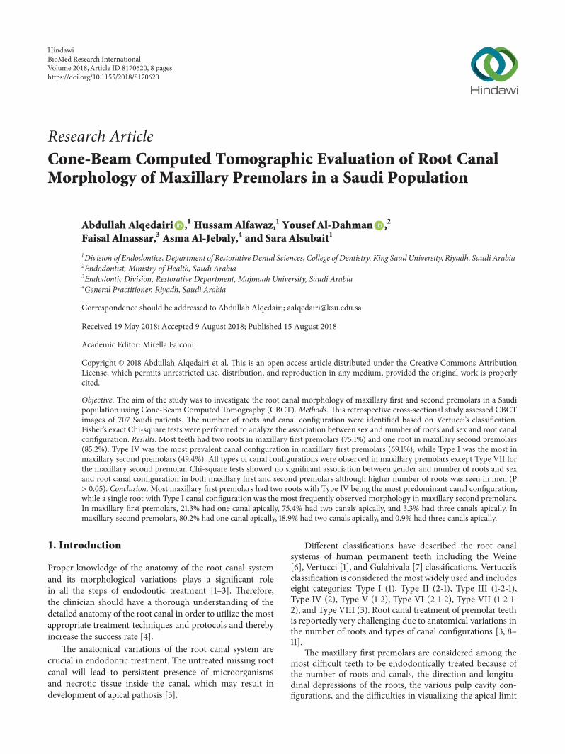
(a) (b) (c)
Figure 1: CBCT of maxillary first premolar with three roots: (a) the axial plane, (b) the coronal plane, and (c) the sagittal plane.
on periapical radiographs [12]. Moreover, high variability hasbeen reported in the root canal morphology of the maxillarysecond premolars [1, 13–15].
Different methods have been utilized to investigate rootcanal anatomy including in vivo and in vitro methods.The in vivo techniques include clinical evaluation duringroot canal treatment, retrospective assessment of patientrecords, conventional radiographic evaluation, and advancedradiographic techniques such as cone-beam computed radio-graphy (CBCT) [16–18], while the in vitro methods includecanal staining and tooth clearing [1, 19], root sectioning[6], microscopic examination, examination of conventionalradiographs, and using three-dimensional modalities such asmicrocomputed tomography (𝜇-CT) [20–22].
The CBCT technique has the ability to detect root canalmorphology as precisely as the root canal staining andclearing techniques which, in the past, were consideredsuperior to conventional techniques used for studying theroot canal system because of their ability to provide three-dimensional views and complete morphologic details [5].
To the best of our knowledge, no study has so far eval-uated the root canal morphology of maxillary premolars ofboth sexes in a Saudi population using CBCT. Therefore, theaim of the study was to investigate the root canal morphologyof maxillary first and second premolars in a Saudi populationusing CBCT.
2. Materials and Methods
Seven hundred and seven CBCT images of Saudi patients(396 female, 311 male) aged between 16 and 71 years, withaverage age of 41.5 years, seeking routine dental treatmentwho were referred to the Radiology Department of theCollege of Dentistry, King Saud University, between 2015 and2017 were collected.
The sampling was purposive where the presence of at leastone maxillary first and/or second premolar with fully devel-oped roots was the inclusion criterion. Unclear or distortedCBCT images, previously endodontically initiated or treatedteeth, teeth with posts or crowns, periapical lesions, andany physiological or pathological process such as immatureapex were excluded. The total final sample, consisting of 334
maxillary first premolars and 318maxillary second premolars,was evaluated in terms of number of roots and root canalconfiguration. The data were observed and recorded for thenumber of roots and canal configuration based on Vertucci’sclassification [1]. The sex of the patients was also recorded.
The CBCT images were accessed and evaluated at theRadiology Department of the College of Dentistry, King SaudUniversity, by two endodontists for the number of rootsand root canal configuration using the using the PlanmecaRomexis Viewer software (Planmeca, Roselle IL).The imageswere collected from different CBCT machines: CS9300 3Ddigital imaging system (Carestream, Rochester, NY) witha voxel size of 90 to 300 𝜇m and Planmeca ProMax 3D(Planmeca, Roselle IL) with a voxel size of ≤ 200 𝜇m.
To ensure the reliability of the results, inter- and intraex-aminer reliabilities were measured by identifying the rootcanal anatomy of maxillary premolars of 30 randomlyselected CBCT images according to the evaluation criteria.For intraexaminer reliability, the same images were evaluatedafter 1 week. Both inter- and intraexaminer reliabilitieswere calculated using the interclass correlation coefficient(ICC). Data were analyzed with Fisher’s exact test usingSPSS software version 22 (SPSS Inc., Chicago, IL, USA). Thesignificance was set at the 95% confidence level.
3. Results
For interexaminer reliability, the ICCwas 0.886 (excellent) forthe number of roots and 0.625 (good) for canal configuration.For intraexaminer reliability, the ICCwas 1 for the first exam-iner with regard to number of roots and canal configurationand 1 and 0.95 for the second examinerwith regard to numberof roots and canal configuration, respectively. The ICCsverified that the procedure was reliable for the evaluationsand measurements performed by the two observers.
The number of roots recorded in maxillary first andsecond premolars was up to three roots (Figure 1). Inmaxillary first premolars, most teeth had two roots (75.1%),while in maxillary second premolars most were single rooted(85.2%). Type IV canal configuration (69.1%) was the mostprevalent observation inmaxillary first premolars, while TypeI (49.4%) was the most prevalent configuration in maxillary
BioMed Research International 3
second premolars. The frequency and percentage of numberof roots and canal configuration in maxillary first and secondpremolars are shown in Tables 1 and 2, respectively.
A higher percentage of three-rooted maxillary first pre-molars was observed in men (2.3%) and none in women(0%), while the most predominant root morphology seen inboth sexes was two rooted, with no statistically significantdifference (P = 0.078). In canal configurations, there wereno statistically significant differences in all types between thesexes (P = 0.095).
In maxillary second premolars, there was no statisticallysignificant difference in number of roots between the sexes (P= 0.383). AlthoughType III, VI, andVIII canal configurationswere more frequent in female patients and Type II, IV, and Vinmale patients, the differencewas not statistically significant(P = 0.257).
Both right and left maxillary first premolars were presentin 170 patients. Symmetrical number of roots and canalconfiguration were seen in 91.2% of teeth, while 2.3% showedsymmetrical number of roots but different canal configura-tion and 0.6% showed symmetrical canal configuration butdifferent number of roots. Moreover, 5.9% of teeth did notshow any type of symmetry.
In 163 patients, right and left maxillary second pre-molars were present. Symmetrical numbers of roots andcanal configuration were seen in 85.3% of teeth, while 11%showed symmetrical number of roots but different canalconfiguration. Only 3.7% showed no type of symmetry.
4. Discussion
Patient ethnicity is an incontrovertible factor that may affectthe perception of the clinician for the suspected root canalanatomy. In the present study, maxillary premolars in aSaudi population, where the majority is Arab, presented withup to three roots and included most of Vertucci’s types ofcanal configuration with similarities and differences to theother reported studies in different populations. Moreover,sex predilection and symmetrical anatomy were investigatedto evaluate their significance in the prediction of root canalanatomy.
CBCT is reportedly an excellent tool for more accuratelydetecting root canal anatomy than intraoral periapical radio-graphy due to its ability to evaluate and assess root canalmorphology in three dimensions [5, 23–27]. The data of thisretrospective study were collected from a single center, KingSaud University Dental Hospital, which is considered oneof the largest governmental dental institutes providing freedental services to a large portion of the Saudi populationfrom different regions. To avoid exposing a large number ofpatients to unnecessary radiation, a CBCT imaging databasewas accessed regardless of the voxel size to achieve a largersample size.
In maxillary first premolars, the prevalence of one rootwas reported to be 22% to 66%, of two roots 33% to 84%,and of three roots 0% to 6% [28–34]. In maxillary secondpremolars, the prevalence of one root was reported to be69.6% to 90.3%, of two roots 9.7% to 29.7%, and of three roots0% to 1.6% [30, 34–37].
In maxillary first premolars in the present study, themost frequently observed root morphology was two rooted(75.1%), followed by single rooted (23.7%), and three rooted(1.2%). These findings are consistent with those reportedby Elkady and Allouba, using CBCT, who reported that,in maxillary first premolars in Saudi subpopulation, theprevalence of one root was 28.3% and of two roots 71.7%,while no three roots were observed [38]. In another studywith a Saudi population, using visual, digital radiography andtransverse sectioning methods, in maxillary first premolars,the prevalence of one root was reported to be 17.9%, of tworoots 80.9%, and of three roots 1.2% [16].
Regarding canal configuration, all types of Vertucci’sclassification were observed in this study, which it is in linewith Vertucci and Gegauff ’s findings [32]. Most patients hadType IV canal configurations, followed by Type I, Type II,and finally Type VII. In comparison to other studies includedin a literature review [39], Type IV canal configuration wasreported to be 65.3% in Saudi population, followed by TypeII (19.7%), Type I (7.6%), Type III (3.3%), Types V, VI, and VII(3.3%), and finally Type VIII (0.8%).
In maxillary second premolars, up to three-rooted teethwere observed. The highest prevalence was single rootedfollowed by double rooted and three rooted (0.3%). Addi-tionally, the majority exhibited Type I canal configuration,followed by Type II and Type IV.These findings coincide withthose of other studies conducted in Saudi Arabia, where oneroot was observed in 76.4% and 67%, two roots in 23.6% and30%, and three roots in 0% and 3% teeth [38, 40]. Moreover,Type I canal configuration was seen in 36.3% and 17%, Type IIin 10.9% and 7%, andType IV in 23.6% and 23% teeth [38, 40].
Compared to a recent CBCT study by Abella et al. [37]in Spanish population, in maxillary first premolars, the mostprevalent root morphology observed was two rooted (51.4%)and most of the teeth exhibited Type IV canal configuration(52.8%), while in maxillary second premolars, the mostprevalent root morphology seen was single rooted (82.9%)and most of these teeth exhibited Type I canal configuration(47.2%).Moreover, two-rooted maxillary first premolars werereported to be 33% with 51% having Type IV canal configu-ration in a Chinese population [41], 68.6% with 68% havingType I canal configuration in a Pakistani population [42],and 44.8% with 76.8% having Type IV canal configurationin a Turkish Cypriot population [43]. Additionally, single-rooted maxillary second premolars were reported to be 84%with 53.4% having Type I canal configuration in a Pakistanipopulation [42], and Type I canal configuration was the mostcommonly observed in a Turkish Cypriot population (49.4%)[43].
Sex predilection regarding the number of roots androot canal morphology has been reported [44–47]. Martinset al. [48] reported that female patients in a Portuguesepopulation had a lower number of roots in both maxillaryfirst and second premolars with a statistically significantdifference in the maxillary first premolars. Moreover, TypeIV canal configuration in maxillary first premolars was morefrequent in male patients and Type I canal configuration inmaxillary second premolars was more in female patients witha statistical significant difference (P < 0.05), while Abella

4 BioMed Research International
Table1:Th
efrequ
ency
andpercentage
ofnu
mbero
froo
tsandcanalcon
figurationin
maxillaryfirstprem
olar
teeth.
Num
bero
fRoo
tsOne
Root
TwoRo
ots
ThreeR
oots
Total
Frequenc
y(%
)Wom
en43
(26.5%
)119
(73.5%
)0(0%)
162(100
%)
Men
36(20.9%
)132(76.8%
)4(2.3%)
172(100
%)
Total
79(23.7%
)251(75.1%
)4(1.2%)
334(100
%)
Can
alCon
figuration
Type
I(1)
Type
II(2-1)
Type
III(1-2-1)
Type
IV(2)
Type
V(1-2)
Type
VI(2-1-2)
Type
VII(1-2-1-2)
Type
VIII(3)
Total
Frequenc
y(%
)Wom
en15
(9.3%)
17(10.5%
)5(3.1%
)112
(69.1
%)
9(5.6%)
3(1.8%)
0(0%)
1(0.6%
)162(100
%)
Men
21(12.2%
)11(6.4%)
1(0.6%
)124(72.1%
)4(2.3%)
4(2.3%)
1(0.6%
)6(3.5%)
172(100
%)
Total
36(10.8%
)28
(8.4%)
6(1.8%)
236(70.6%
)13
(3.9%)
7(2.1%
)1(0.3%
)7(2.1%
)334(100
%)

BioMed Research International 5
Table2:Th
efrequ
ency
andpercentage
ofnu
mbero
froo
tsandcanalcon
figurationin
maxillarysecond
prem
olar
teeth.
Num
bero
fRoo
tsOne
Root
TwoRo
ots
ThreeR
oots
Total
Frequenc
y(%
)Wom
en138(87.3
%)
20(12.7%
)0(0%)
158(100
%)
Men
133(
83.1%
)26
(16.3%
)1(0.6%
)159(100
%)
Total
271(85.2%)
46(14
.5%)
1(0.3%
)318(100
%)
Can
alCon
figuration
Type
I(1)
Type
II(2-1)
Type
III(1-2-1)
Type
IV(2)
Type
V(1-2)
Type
VI(2-1-2)
Type
VII(1-2-1-2)
Type
VIII(3)
Total
Frequenc
y(%
)Wom
en80
(50.6%
)38
(24.0%
)10
(6.3%)
15(9.5%)
8(5.1%
)5(3.2%)
0(0%)
2(1.3%)
158(100
%)
Men
77(48.1%
)44
(27.5
%)
6(3.8%)
22(13.8%
)10
(6.2%)
0(0%)
0(0%)
1(0.6%
)160(100
%)
Total
157(49.4
%)
82(25.8%
)16
(5%)
37(11.6
%)
18(5.7%)
5(1.6%)
0(0%)
3(0.9%)
318(100
%)
6 BioMed Research International
et al. [37] reported that there was no statistically significantcorrelation between the number of roots and the sexes inSpanish population. In this study, there was no statisticallysignificant correlation between sex and number of roots orsex and root canal configuration in both maxillary first andsecond premolars although a higher number of roots wereseen in men (P > 0.05).
The degree of bilateral symmetry in root canal morphol-ogy using CBCT has been reported in different studies. Formaxillary first premolars, bilateral symmetry was observedin 88.5% of teeth for the number of roots and in 77% forcanal configuration in Saudi patients [38], and symmetryof 64% was reported for both number of roots and canalconfiguration in a Chinese population [41]. In maxillarysecond premolars, bilateral symmetry was observed in 84%of teeth for the number of roots and in 76% for canalconfiguration [38]. The findings of the present study arein agreement with those of the previous studies where ahigh degree of symmetry in the number of roots and canalconfiguration was seen in both maxillary first and secondpremolars.
5. Conclusion
Within its limitations, in this study with a Saudi popu-lation, most maxillary first premolars had two roots withType IV being the most predominant canal configuration,while a single root with Type I canal configuration was themost frequently observed morphology in maxillary secondpremolars. Moreover, more than one root with differentcanal configurations was detected in some cases. Furtherstudies with larger sample sizes are recommended for thegeneralization of our results.
Data Availability
The CBCT images data used to support the findings of thisstudy are restricted by the local institutional review board atKing Saud University in order to protect patients’ privacy.
Ethical Approval
The study was independently reviewed and approved by thelocal institutional review board (No. E-18-3090) at King SaudUniversity and conducted in full accordance with the WorldMedical Association Declaration of Helsinki.
Consent
Consent was not required for this type of study. The data wasanonymised and deidentified prior to analysis.
Conflicts of Interest
The authors declare that they have no conflicts of interest.
Acknowledgments
The authors thank College of Dentistry Research Center,King Saud University, for their support in conducting this
project (CDRC no. FR 0440, IRB Research Project no. E-18-3090) and the Deanship of Scientific Research and RSSUat King Saud University for their technical support. Theauthors also thank Dr. Ahmed Qattan (General Practitioner,Thadiq General Hospital, Saudi Arabia) for his efforts in datacollection. There was no financial support for this study.
References
[1] F. J. Vertucci, “Root canal anatomy of the human permanentteeth,” Oral Surgery, Oral Medicine, Oral Pathology, Oral Radi-ology, and Endodontology, vol. 58, no. 5, pp. 589–599, 1984.
[2] F. J. Vertucci, “Root canal morphology and its relationship toendodontic procedures,” Endodontic Topics, vol. 10, no. 1, pp. 3–29, 2005.
[3] S. Nallapati, “Three canal mandibular first and second premo-lars: A treatment approach,” Journal of Endodontics, vol. 31, no.6, pp. 474–476, 2005.
[4] Y. Jou, B. Karabucak, J. Levin, and D. Liu, “Endodontic workingwidth: current concepts and techniques,”Dental Clinics of NorthAmerica, vol. 48, no. 1, pp. 323–335, 2004.
[5] P. Neelakantan, C. Subbarao, and C. V. Subbarao, “Com-parative evaluation of modified canal staining and clearingtechnique, cone-beam computed tomography, peripheral quan-titative computed tomography, spiral computed tomography,and plain and contrast medium-enhanced digital radiographyin studying root C,” Journal of Endodontics, vol. 36, no. 9, pp.1547–1551, 2010.
[6] F. S. Weine, H. J. Healey, H. Gerstein, and L. Evanson, “Canalconfiguration in the mesiobuccal root of the maxillary firstmolar and its endodontic significance,” Oral Surgery, OralMedicine, Oral Pathology, Oral Radiology, and Endodontology,vol. 28, no. 3, pp. 419–425, 1969.
[7] K. Gulabivala, T. H. Aung, A. Alavi, and Y.-L. Ng, “Root andcanal morphology of Burmese mandibular molars,” Interna-tional Endodontic Journal, vol. 34, no. 5, pp. 359–370, 2001.
[8] B. M. Cleghorn, W. H. Christie, and C. C. S. Dong, “The rootand root canal morphology of the human mandibular firstpremolar: a literature review,” Journal of Endodontics, vol. 33, no.5, pp. 509–516, 2007.
[9] B. M. Cleghorn, W. H. Christie, and C. C. S. Dong, “The rootand root canal morphology of the human mandibular secondpremolar: a literature review,” Journal of Endodontics, vol. 33, no.9, pp. 1031–1037, 2007.
[10] J. Kottoor, D. Albuquerque, N. Velmurugan, and J. Kuruvilla,“Root anatomy and root canal configuration of human per-manent mandibular premolars: a systematic review,” AnatomyResearch International, vol. 2013, Article ID 254250, 14 pages,2013.
[11] R. R. Slowey, “Root canal anatomy. Road map to successfulendodontics,”Dental Clinics of North America, vol. 23, no. 4, pp.555–573, 1979.
[12] J. D. Pecora, P. C. Saquy, M. D. Sousa Neto, and J. B. Woelfel,“Root form and canal anatomy of maxillary first premolars,”Brazilian Dental Journal, vol. 2, no. 2, pp. 87–94, 1992.
[13] F. Vertucci, A. Seelig, and R. Gillis, “Root canal morphologyof the human maxillary second premolar,” Oral Surgery, OralMedicine, Oral Pathology, Oral Radiology, and Endodontology,vol. 38, no. 3, pp. 456–464, 1974.
[14] S. Sert and G. S. Bayirli, “Evaluation of the root canal config-urations of the mandibular and maxillary permanent teeth by
BioMed Research International 7
gender in the Turkish population,” Journal of Endodontics, vol.30, no. 6, pp. 391–398, 2004.
[15] K. P. Sardar, N. H. Khokhar, and I. Siddiqui, “Frequency oftwo canals in maxillary second premolar tooth,” Journal of theCollege of Physicians and Surgeons–Pakistan: JCPSP, vol. 17, pp.12–14, 2007.
[16] M. A. Atieh, “Root and canal morphology of maxillary firstpremolars in a Saudi population,”The Journal of ContemporaryDental Practice, vol. 9, pp. 46–53, 2008.
[17] N. Pattanshetti, M. Gaidhane, and A. M. Al Kandari, “Rootand canal morphology of the mesiobuccal and distal rootsof permanent first molars in a Kuwait population—a clinicalstudy,” International Endodontic Journal, vol. 41, no. 9, pp. 755–762, 2008.
[18] S.H.G.DeOliveira, L. C.DeMoraes,H. Faig-Leite, S. E. AfonsoCamargo, and C. H. R. Camargo, “In vitro incidence of rootcanal bifurcation in mandibular incisors by radiovisiography,”Journal of Applied Oral Science, vol. 17, no. 3, pp. 234–239, 2009.
[19] L. Awawdeh, H. Abdullah, and A. Al-Qudah, “Root formand canal morphology of jordanian maxillary first premolars,”Journal of Endodontics, vol. 34, no. 8, pp. 956–961, 2008.
[20] C. Grover andN. Shetty, “Methods to study root canalmorphol-ogy: A review,” ENDO - Endodontic Practice Today, pp. 171–182,2012.
[21] B. M. Cleghorn, W. H. Christie, and C. C. S. Dong, “Root androot canal morphology of the human permanentmaxillary firstmolar: a literature review,” Journal of Endodontics, vol. 32, no. 9,pp. 813–821, 2006.
[22] G. Plotino, N. M. Grande, R. Pecci, R. Bedini, C. H. Pameijer,and F. Somma, “Three-dimensional imaging using microcom-puted tomography for studying tooth macromorphology,” TheJournal of the American Dental Association, vol. 137, no. 11, pp.1555–1561, 2006.
[23] R. P. Matherne, C. Angelopoulos, J. C. Kulild, and D. Tira,“Use of cone-beam computed tomography to identify root canalsystems in vitro,” Journal of Endodontics, vol. 34, no. 1, pp. 87–89,2008.
[24] J. D. Domark, J. F.Hatton, R. P. Benison, andC. F.Hildebolt, “Anex vivo comparison of digital radiography and cone-beam andmicro computed tomography in the detection of the number ofcanals in the mesiobuccal roots of maxillary molars,” Journal ofEndodontics, vol. 39, no. 7, pp. 901–905, 2013.
[25] K. M. P. Soares De Toubes, M. I. D. S. Cortes, M. A. D.A. Valadares, L. C. Fonseca, E. Nunes, and F. F. Silveira,“Comparative analysis of accessory mesial canal identificationin mandibular first molars by using four different diagnosticmethods,” Journal of Endodontics, vol. 38, no. 4, pp. 436–441,2012.
[26] D. Zhang, J. Chen, G. Lan et al., “The root canal morphology inmandibular first premolars: a comparative evaluation of cone-beam computed tomography and micro-computed tomogra-phy,” Clinical Oral Investigations, vol. 21, no. 4, pp. 1007–1012,2017.
[27] M. B. Vizzotto, P. F. Silveira, N. A. Arus, F. Montagner, B. P. F.A. Gomes, and H. E. D. da Silveira, “CBCT for the assessmentof second mesiobuccal (MB2) canals in maxillary molars teeth:effect of voxel size and presence of root filling,” InternationalEndodontic Journal, vol. 46, no. 9, pp. 870–876, 2013.
[28] E. J. Carns and A. E. Skidmore, “Configurations and deviationsof root canals of maxillary first premolars,” Oral Surgery, OralMedicine, Oral Pathology, Oral Radiology, and Endodontology,vol. 36, no. 6, pp. 880–886, 1973.
[29] H. S. Loh, “Root morphology of the maxillary first premolar inSingaporeans,”AustralianDental Journal, vol. 43, no. 6, pp. 399–402, 1998.
[30] N. Kartal, B. Ozcelik, and H. Cimilli, “Root canal morphologyof maxillary premolars,” Journal of Endodontics, vol. 24, no. 6,pp. 417–419, 1998.
[31] R. Bellizzi and G. Hartwell, “Radiographic evaluation of rootcanal anatomy of in vivo endodontically treated maxillarypremolars,” Journal of Endodontics, vol. 11, no. 1, pp. 37–39, 1985.
[32] F. J. Vertucci and A. Gegauff, “Root canal morphology ofthe maxillary first premolar,” Journal of the American DentalAssociation, vol. 99, no. 2, pp. 194–198, 1979.
[33] E. M. Senan, H. A. Alhadainy, T. M. Genaid, and A. A. Madfa,“Root form and canal morphology of maxillary first premolarsof a Yemeni population,” BMC Oral Health, vol. 18, article 94,2018.
[34] J. N. Martins, Y. Gu, D. Marques, H. Francisco, and J. Carames,“Differences on the Root and Root Canal Morphologiesbetween Asian and White Ethnic Groups Analyzed by Cone-beam Computed Tomography,” Journal of Endodontics, vol. 44,no. 7, pp. 1096–1104, 2018.
[35] J. D. Pecora, M. D. Sousa Neto, P. C. Saquy, and J. B. Woelfel,“In vitro study of root canal anatomy of maxillary secondpremolars,” Brazilian Dental Journal, vol. 3, no. 2, pp. 81–85,1993.
[36] L. Yang, X. Chen, C. Tian, T. Han, and Y. Wang, “Use ofCone-beam Computed Tomography to Evaluate Root CanalMorphology and Locate Root Canal Orifices of MaxillarySecond Premolars in a Chinese Subpopulation,” Journal ofEndodontics, vol. 40, no. 5, pp. 630–634, 2014.
[37] F. Abella, L. M. Teixido, S. Patel, F. Sosa, F. Duran-Sindreu,and M. Roig, “Cone-beam Computed Tomography Analysisof the Root Canal Morphology of Maxillary First and SecondPremolars in a Spanish Population,” Journal of Endodontics, vol.41, no. 8, pp. 1241–1247, 2015.
[38] A. Elkady and K. Allouba, “Cone beam computed tomographicanalysis of root and canal morphology of maxillary premolarsin Saudi subpopulation,” in Egyptian Dental Journal, vol. 59, pp.3419–3429, 2013.
[39] I. A. Ahmad, “Root and root canal morphology of SaudiArabian permanent dentition,” Saudi Endodontic Journal, vol.5, no. 2, pp. 99–106, 2015.
[40] M. Elnour, A. Khabeer, and E. AlShwaimi, “Evaluation of rootcanal morphology of maxillary second premolars in a SaudiArabian sub-population: An in vitro microcomputed tomogra-phy study,”The Saudi Dental Journal, vol. 28, no. 4, pp. 162–168,2016.
[41] Y.-. Tian, B. Guo, R. Zhang et al., “Root and canal morphologyof maxillary first premolars in a Chinese subpopulation eval-uated using cone-beam computed tomography,” InternationalEndodontic Journal, vol. 45, no. 11, pp. 996–1003, 2012.
[42] M. R. Nazeer, F. R. Khan, and R. Ghafoor, “Evaluation of rootmorphology and canal configuration of maxillary premolars ina sample of Pakistani population by using cone beam computedtomography,” Journal of the Pakistan Medical Association, vol.68, no. 3, pp. 423–427, 2018.
[43] B. Celikten, K. Orhan, U. Aksoy et al., “Cone-beam CT eval-uation of root canal morphology of maxillary and mandibularpremolars in a Turkish Cypriot population,” BDJ Open, vol. 2,article 15006, 2016.
[44] H. Arslan, H. Ertas, E. Tarim Ertas, F. Kalabalik, G. Saygili,and I. Davut Capar, “Evaluating root canal configuration of
8 BioMed Research International
mandibular incisors with cone-beam computed tomography ina Turkish population,” Journal of Dental Sciences, vol. 10, no. 4,pp. 359–364, 2015.
[45] B. V. Caputo, G. A. Noro Filho, D. M. R. de Andrade Salgado,C. Moura-Netto, E. M. Giovani, and C. Costa, “Evaluationof the Root Canal Morphology of Molars by Using Cone-beam Computed Tomography in a Brazilian Population: PartI,” Journal of Endodontics, vol. 42, no. 11, pp. 1604–1607, 2016.
[46] D. G. Bulut, E. Kose, G. Ozcan, A. E. Sekerci, E. M. Canger,and Y. Sisman, “Evaluation of root morphology and rootcanal configuration of premolars in the Turkish individualsusing cone beam computed tomography,” European Journal ofDentistry, vol. 9, no. 4, pp. 551–557, 2015.
[47] S. Burklein, R. Heck, and E. Schafer, “Evaluation of the rootcanal anatomy of maxillary and mandibular premolars in aselected german population using cone-beam computed tomo-graphic data,” Journal of Endodontics, vol. 43, no. 9, pp. 1448–1452, 2017.
[48] J. N. R. Martins, D. Marques, H. Francisco, and J. Carames,“Gender influence on the number of roots and root canal systemconfiguration in human permanent teeth of a Portuguesesubpopulation,” Quintessence International, vol. 49, no. 2, pp.103–111, 2018.

Hindawiwww.hindawi.com
International Journal of
Volume 2018
Zoology
Hindawiwww.hindawi.com Volume 2018
Anatomy Research International
PeptidesInternational Journal of
Hindawiwww.hindawi.com Volume 2018
Hindawiwww.hindawi.com Volume 2018
Journal of Parasitology Research
GenomicsInternational Journal of
Hindawiwww.hindawi.com Volume 2018
Hindawi Publishing Corporation http://www.hindawi.com Volume 2013Hindawiwww.hindawi.com
The Scientific World Journal
Volume 2018
Hindawiwww.hindawi.com Volume 2018
BioinformaticsAdvances in
Marine BiologyJournal of
Hindawiwww.hindawi.com Volume 2018
Hindawiwww.hindawi.com Volume 2018
Neuroscience Journal
Hindawiwww.hindawi.com Volume 2018
BioMed Research International
Cell BiologyInternational Journal of
Hindawiwww.hindawi.com Volume 2018
Hindawiwww.hindawi.com Volume 2018
Biochemistry Research International
ArchaeaHindawiwww.hindawi.com Volume 2018
Hindawiwww.hindawi.com Volume 2018
Genetics Research International
Hindawiwww.hindawi.com Volume 2018
Advances in
Virolog y Stem Cells International
Hindawiwww.hindawi.com Volume 2018
Hindawiwww.hindawi.com Volume 2018
Enzyme Research
Hindawiwww.hindawi.com Volume 2018
International Journal of
MicrobiologyHindawiwww.hindawi.com
Nucleic AcidsJournal of
Volume 2018
Submit your manuscripts atwww.hindawi.com