conformational symmetry and vibrational …iupac.org/publications/pac/pdf/2009/pdf/8103x0549.pdfsoni...
TRANSCRIPT

549
Pure Appl. Chem., Vol. 81, No. 3, pp. 549–570, 2009.doi:10.1351/PAC-CON-08-08-24© 2009 IUPAC
Conformational symmetry and vibrationaldynamics of polymers*
Poonam Tandon‡, Naresh Kumar, Vineet Gupta, Deepika Chaturvedi,Soni Mishra, and Vishwambhar D. Gupta
Department of Physics, University of Lucknow, Lucknow 226 007, India
Abstract: Polymers are an important class of materials, and their conformation dictates theirdynamical, thermodynamical, and hydrodynamical behavior. Several spectroscopic and othertechniques have been employed to characterize their conformation. However, little use hasbeen made of group-theoretical techniques except in the classification of symmetry species.In the present review, an attempt has been made to correlate normal modes and their disper-sion profiles with the conformation of the polymeric systems. This has been attempted in thecase of 2-, 3-, 4-fold and α-helical polymers.
Keywords: polymers; conformation; dispersion curves; heat capacity; density of states; vi-brational spectra.
INTRODUCTION
Polymers are important structural units of living and non-living systems, each by itself, often exercis-ing surprising and complicated functions. Polymers consist of chains built up from a relatively smallnumber of different types of monomer units. The most important of these polymers are proteins, whichinclude enzymes that catalyze and regulate metabolism and the nucleic acids, which carry genetic in-formation in the form of a control program for the biosynthesis of proteins [1]. Proteins and poly-peptides have a tendency to fold spontaneously into their native conformation both in vivo and in vitro.However, these can be transformed from one conformation to another by varying pH, temperature, orsurfactant solvent. Depending upon the size and type of interactions, the biopolymeric systems are ca-pable of existing in a variety of secondary structures such as helical (α, 3-fold, and 4-fold), β-pleatedsheet, planar zigzag, globular, bends, and random coil states [2]. Many of these structures are simulta-neously present in a given chain. Generally, α, β, and random coils occur more frequently because oftheir inherent thermodynamic stability. Random coil generally occurs when a polypeptide contains ad-jacent bulky residues such as isoleucine or charged residue such as glutamic acid, aspartic acid.Repulsion between these groups causes the polypeptide to assume a random coil configuration. Bragget al. [3] characterized another type of helical form 413 termed as ω-helix, which is distorted α-helix.The ω-helix has a 4-fold screw axis in which there are four amino acid residues per helix turn with aresidue translation of 1.3 Å and helix pitch of 5.2 Å. There are several other single-stranded intra-molecularly hydrogen-bonded helices which have been proposed and found to exist, e.g., 2.27, 310(δ-helix), 5.117 (γ-helix), 4.416 (π-helix), 185 (α-helix), etc.
*Paper based on a presentation at POLYCHAR 16: World Forum on Advanced Materials, 17–21 February 2008, Lucknow, India.Other presentations are published in this issue, pp. 389–570.‡Corresponding author: E-mail: [email protected]

Several techniques have been used to characterize the conformation of polymeric systems. Theseinclude infrared (IR) absorption, Raman spectra, inelastic neutron scattering (INS), X-ray diffraction(XRD), nuclear magnetic resonance (NMR), circular dichroism (CD), and optical rotatory dispersion(ORD). These techniques are based on characteristic features such as IR absorption due to amide bandsin a biopolymer and characteristic stretch modes in an industrial tactic polymer. However, none of thesetechniques reflects the characteristic dynamical feature within the first Brillouin zone of a vibratingpolymer. INS, which is free from all kinds of constraints imposed by selection rules, reflects this dy-namical feature through the density-of-states. However, it has its own problems in terms of resolutionand sensitivity.
Vibrational spectroscopy plays an important role in the elucidation of polymeric structure, and itis intimately related to the symmetry of the polymers. In the present communication, we have tried toshow how the dispersion profile of low-frequency modes reflects the symmetry of the polymer chain.This has been done in a variety of symmetry groups [4–16]. Strong supporting evidence of the spectralparameters (constants of IR and Raman) is obtained from quantum chemical [Hartree–Fock (HF), post-HF, and density functional theory (DFT)] [17–20] and molecular dynamical (MD) approaches. Theyhave the additional advantage of including interchain interaction and anharmonic effects. The MD evenenables modeling of disordered systems. The forgoing techniques have been widely used in a variety ofsystems [21–29].
An overall understanding of vibrational dynamics in a polymer involves calculation of the dis-persion curves. These curves also provide knowledge of the degree of uninterrupted sequence lengthsin an ordered conformation. The dynamics of a polymeric system is an order of magnitude more diffi-cult than usual molecular systems because the phase relationship between the adjacent units has to beconsidered. This in turn necessitates recasting of kinetic and potential fields as a function of phase re-lationship. The dispersion curves also facilitate correlation of the microscopic behavior in a long-chainmolecule with its macroscopic properties such as entropy, enthalpy, specific heat, etc.
THEORY
Molecular vibrations of finite molecules
In attempting to account for the observed IR and Raman spectra of real molecules, a certain simplifiedmodel is adopted and then the spectra that this model would exhibit are calculated. The model consistsof particles endowed with mass and held together by electrical forces such as dipole–dipole interactions,covalent, etc. Particles represent the atoms and are treated as if all the mass were concentrated at a point.For a molecule having N atoms there are 3N-6 vibrational degrees of freedom (or 3N-5 if the moleculeis linear). Using any set of generalized coordinates, the problem can be reduced to writing the secularequations to be solved to get eigen frequencies and eigen vectors. However, a brief description ofWilson’s GF matrix method [30,31] is being given. It uses internal coordinates that are bond stretches,in-plane angle bending, torsions, and waggings. The internal coordinates are easiest and simplest andcan best reflect the physical nature of molecular vibrations. They also permit the transferability of forceconstants from a group in one molecule to the corresponding group in another molecule in a similar en-vironment. The small amplitude and harmonic nature of molecular vibrations provide a linear transfor-mation between various displacement coordinates. The internal coordinates R are related to theCartesian coordinate X in matrix notation as follows.
R = BX (1)
where R is the internal coordinate and B is the transformation matrix and depends upon the geometryof the molecule. Then, the G-matrix or the inverse kinetic energy matrix is defined by the relation
P. TANDON et al.
© 2009 IUPAC, Pure and Applied Chemistry 81, 549–570
550

(2)
where mi is the mass of the ith atom.
G = BM–1 B'
The kinetic energy of vibration can be written in terms of the internal coordinates in the form
(3)
where R' denotes the transpose of R.The potential energy is given by the expression
2V = R'FR (4)
where F is the potential energy matrix and its elements are linear function of the force constants fk. Inpractice, it is computed through the basis matrix (z-matrix) where
The coefficients zijk (z-matrix) are determined by the geometry of the molecule. The molecularvibrational problem is then represented by the matrix equation
GFL = λL (5)
where the eigen values λ’s are related to the vibrational frequencies ν’s by λ = 4π2c2ν2 and the normalcoordinates Q are related to the internal coordinates R by
R = LQ (6)
To obtain the vibrational frequencies, one has to solve the secular equation
|GF – λI| = 0 (7)
where I is the unit matrix.In each normal mode of vibration, all the atoms in a molecule vibrate in phase with the same fre-
quency νk at a particular instant. They may have different amplitudes. The extant of vibrational cou-pling between the various internal displacement coordinates, in a given normal mode Qk is qualitativelydescribed by the elements of the eigenvector Lk belonging to the eigenvalues λk.
Because of the different dimensions of L vectors for stretching and bending coordinates, it ispreferable to use the potential energy distribution (PED) to characterize the energy distribution. ThePED is the fractional part of potential energy of the normal mode vibration contributed by each forceconstant. If each FijLikLjk term is divided by the total sum of FijLikLjk terms for this vibration (which isequal to λk), then
(8)
© 2009 IUPAC, Pure and Applied Chemistry 81, 549–570
Vibrational dynamics of polymers 551
G B B m k l nkl ki li ii
n
= = −( )=∑ / . . . . . . . . . . . ,, 1 3 61
3
G
G G G
G G G
G G G
n
n
n n nn
=
11 12 1
21 22 2
1 2
K
K
KKKKK
K
2 1T R G R=• − •
'
F z fij ijk kk
= ∑
PEL F
i
k ik ii
k( ) =
2
λ
where the L vectors have been defined previously. The PED is useful in making the assignment of var-ious frequencies.
Symmetry considerations in finite molecules
In this section, the importance of the symmetry of the molecule and group-theoretical ideas is eluci-dated. For a molecule having M atoms, the dimensions of the secular equation are (3M × 3M), and it isquite cumbersome to solve the problem even for a molecule of moderate size. But in the case of thesymmetric molecule, the secular equation can be factored into blocks of smaller dimensions by usinginternal symmetry coordinates [32]
(9)
where γ is the particular symmetry species, N is the normalization factor, O is the symmetry operation,XO(γ) is the character of the γ species for Oth operation, and ORm stands for the internal coordinate to
which Rm is transformed by the operation O.Applying group theory, the number of normal modes in a particular symmetry species (γ) is given
as
(10)
Here, H is the order of the point group, gj is the number of symmetry operations in the jth class, X j(γ) is
the character of the γ species in the jth class, and Xj is the character of the jth symmetry transformation
of the displacement coordinates.The symmetry of the molecule can be exploited in solving the secular equation by transforming
the kinetic and potential energy matrices in the space of symmetry coordinates. The dynamical matri-ces when transformed in the space of symmetry coordinates factor in blocks belongs to one of the sym-metry species. The internal coordinates are related to the symmetry coordinates. In the matrix notation
S = UR (11)
U is a transformation matrix obtained from the coefficients of symmetry coordinates and it is used todiagonalize into blocks, G and F matrices
GB = U 'GU (12)
FB = U 'FU (13)
U ' is the transpose of U. So the modified secular equations becomes
|G(γ) F(γ) – λI | = 0 (14)
These can be solved completely independent of each other for individual symmetry species.
P. TANDON et al.
© 2009 IUPAC, Pure and Applied Chemistry 81, 549–570
552
S N X ORm OO
mγ γ( ) ( )= ∑
n H g X Xj j jj
( ) ( )*/γ γ= ∑1

Molecular vibrations of polymer chains
The basic concepts discussed in the previous section can be applied to polymer chains but as the poly-mer molecule is of infinite length, the order of the matrices becomes infinite. In order to reduce theproblem to one of workable dimensions, the screw symmetry of the polymer chain is exploited. Thepolymer chains are built up of chemical units or monomers arranged in a regularly repeating fashion.Higgs [33] was the first to point out the importance of the helical symmetry present in polymer mole-cules and utilize it in order to adapt the Wilson’s GF matrix for polymeric systems. He considered in afairly general manner the vibrations of a helical molecule and used group-theoretical ideas to classifythe normal modes and derive the selection rules for Raman and IR spectra.
According to Higgs treatment, an infinite helical molecule is built of identical units linked to-gether in such a way that each unit is transformed geometrically into the next by the operation H(l, f).This operation is defined by a translation through a distance l along the axis plus a rotation through anangle f about the same axis. Thus, any group in the chain can be transformed into another group ±n unitsaway by operating with Hn (n is any integer positive or negative). This operation transforms one unitinto another and constitutes an infinite group that is simply isomorphic with the infinite cyclic groupC∞. Its irreducible representations are, therefore, all one-dimensional, and may be labeled Γ(θ) withparameter (θ) which runs through all values in the range –π ≤ θ ≤ +π the corresponding characters aregiven by
χ(θ, Hn) = exp(inθ) (15)
Every normal mode of vibration of molecule must belong to one of these representations. Thismeans that if in a normal mode a certain unit vibrates in some manner with an amplitude A, then the nth
unit further must vibrate in the same manner with an amplitude Aexp(–inθ). Each frequency νi of anisolated unit gives rise to a band of frequencies νi(θ) in the helically linked molecule, where θ is thephase difference between adjacent units. It can be shown that corresponding to each frequency νi(θ),there lies a frequency νi(–θ), which is equal to νi(θ). Thus, with the exception of νi(0) and θi(π), thefrequencies are degenerate in pairs belonging to Γ(θ) where (0 ≤ θ ≤ +π). Therefore, the correspondingcomplex normal modes can be combined to form two real ones in which the amplitudes vary as (A cosnθ) and (A sin nθ).
Coming to the solution of the vibrational secular equation, let Rin denote the ith internal displace-
ment coordinate belonging to the nth chemical unit. Then the potential interaction and the kinetic cou-pling between the ith internal coordinate and the nth unit and the kth internal coordinate of the nth unitare given by the matrix element Fik
nn' and Giknn'. Now the periodicity of the chain requires that these ma-
trix elements depend on n and n' only through their differences, that is,
Fiknn' = Fik
S (16)
Giknn' = Gik
S (17)
where S = (n–n'). In view of this, a particular portion of the infinite G matrix can be written in terms ofR’s as coordinates,
© 2009 IUPAC, Pure and Applied Chemistry 81, 549–570
Vibrational dynamics of polymers 553
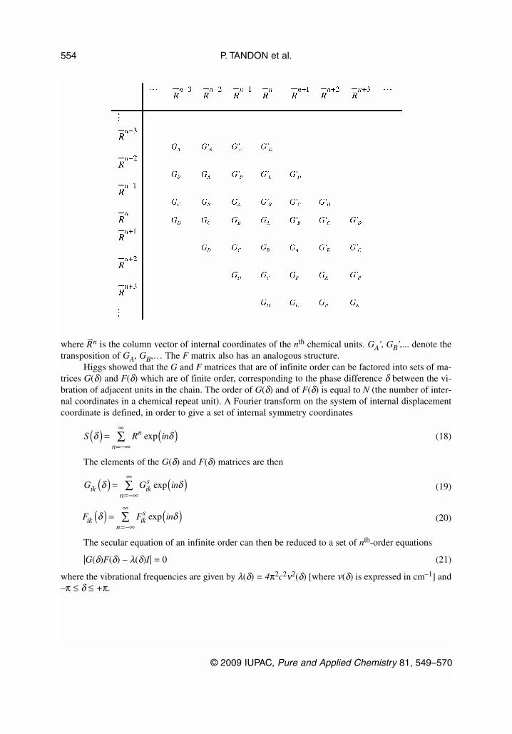
where R–n is the column vector of internal coordinates of the nth chemical units. GA', GB',... denote the
transposition of GA, GB,… The F matrix also has an analogous structure.Higgs showed that the G and F matrices that are of infinite order can be factored into sets of ma-
trices G(δ) and F(δ) which are of finite order, corresponding to the phase difference δ between the vi-bration of adjacent units in the chain. The order of G(δ) and of F(δ) is equal to N (the number of inter-nal coordinates in a chemical repeat unit). A Fourier transform on the system of internal displacementcoordinate is defined, in order to give a set of internal symmetry coordinates
(18)
The elements of the G(δ) and F(δ) matrices are then
(19)
(20)
The secular equation of an infinite order can then be reduced to a set of nth-order equations
|G(δ)F(δ) – λ(δ)I| = 0 (21)
where the vibrational frequencies are given by λ(δ) = 4π2c2ν2(δ) [where ν(δ) is expressed in cm–1] and–π ≤ δ ≤ +π.
P. TANDON et al.
© 2009 IUPAC, Pure and Applied Chemistry 81, 549–570
554
S R inn
nδ δ( ) = ( )
=−∞
∞∑ exp
G G inik iks
nδ δ( ) = ( )
=−∞
∞∑ exp
F F inik iks
nδ δ( ) = ( )
=−∞
∞∑ exp

Symmetry properties and selection rules
The vibrational frequencies calculated by eq. 21 are infinite in number but only a few of them are op-tically active in polymers. Selection rules for such molecules were first worked out by Higgs, in termsof the phase difference δ and the angle φ. It is well known that the only frequencies which appear as al-lowed fundamentals in IR absorption are those belonging to representation Γ(θ), and it is essential thatthese are contained in the representations Γ(M–) which has, as its basis, the components of the total mo-lecular electric dipole moment M
–. It turns out that IR absorption arises either from the a vibrations with
phase difference δ = 0 (the transition moment being parallel to the helix axis) or from the E(φ) vibra-tion with δ = φ (the transition moment being perpendicular to the axis).
In order to derive the Raman selection rules, one has to consider the total molecular electric po-larizability. It can be shown that Raman absorption arises from the vibrations with phase difference δ =0, ±φ, ±2φ.
Use of group-theoretical ideas
At this point, the importance of group-theoretical ideas in an understanding of polymer spectra needsto be described. This importance arises because the symmetry of a helical polymer can be described bya one-dimensional space-group. As mentioned earlier, only the normal modes in which all the unit cellsvibrate in the same phase are active in the IR and Raman scattering [34]. Thus, it is sufficient spectro-scopically to study only the factor group of the one-dimensional space group, which has the transla-tional subgroup as the unit element. Let us consider an infinite helical molecule in which the one-di-mensional crystallographic repeat unit contains n chemical units and m turns (each chemical repeat unitcontains p atoms). In such a molecule, the basic symmetry operation is defined by a rotation of 2mπ/nabout the axis of the helix followed by a translation along the axis of 1/n of the unit cell length. Thefactor group in question may be denoted by C2mπ/n, which is isomorphic with the point group Cn. Ananalysis of the factor group of the polymer is useful in predicting the number of normal modes, theirsymmetry properties, and IR and Raman activity (Table 1) [35]. Coupled with dichroic studies on poly-mer spectra, an analysis of the symmetry group of the polymer can help in the analysis and interpreta-tion of the spectra, even in the absence of complete normal vibration calculation. Liang and coworkers[36–41] have used these group-theoretical ideas to interpret the spectra of a large number of high poly-mers such as polyethylene, polytetrafluorethylene, polystyrene, polyvinyl chloride, etc. The work hasbeen reviewed by Krimm [42].
Table 1 Character table, numbers of normal modes, and selection rules (A: active, F: forbidden) for the helicalpolymer molecule under the group C2mπ/n [35].
n = odd
E C1 C2 … Cn–1 N IR Raman
A 1 1 1 … 1 3p(Tπ,Rπ) A AE1 1 ε ε2 … εn–1 3p(Tσ) A A
1 ε–1 ε–2 … ε–(n–1) 3p(Tσ) A AE2 1 ε2 ε4 … ε2(n–1) 3p F A
1 ε–2 ε–4 … ε–2(n–1) 3p F AE3 1 ε3 ε6 … ε3(n–1) 3p F F
1 ε–3 ε-6 … ε–3(n–1) 3p F F. . . . … . . . .. . . . … . . . .E(n–1)/2 1 ε(n–1)/2 ε[(n–1)/2]2 … ε[(n–1)/2](n–1) 3p F F
1 ε–(n–1)/2 ε–[(n–1)/2]2 … ε–[(n–1)/2](n–1) 3p F F
© 2009 IUPAC, Pure and Applied Chemistry 81, 549–570
Vibrational dynamics of polymers 555
(continues on next page)
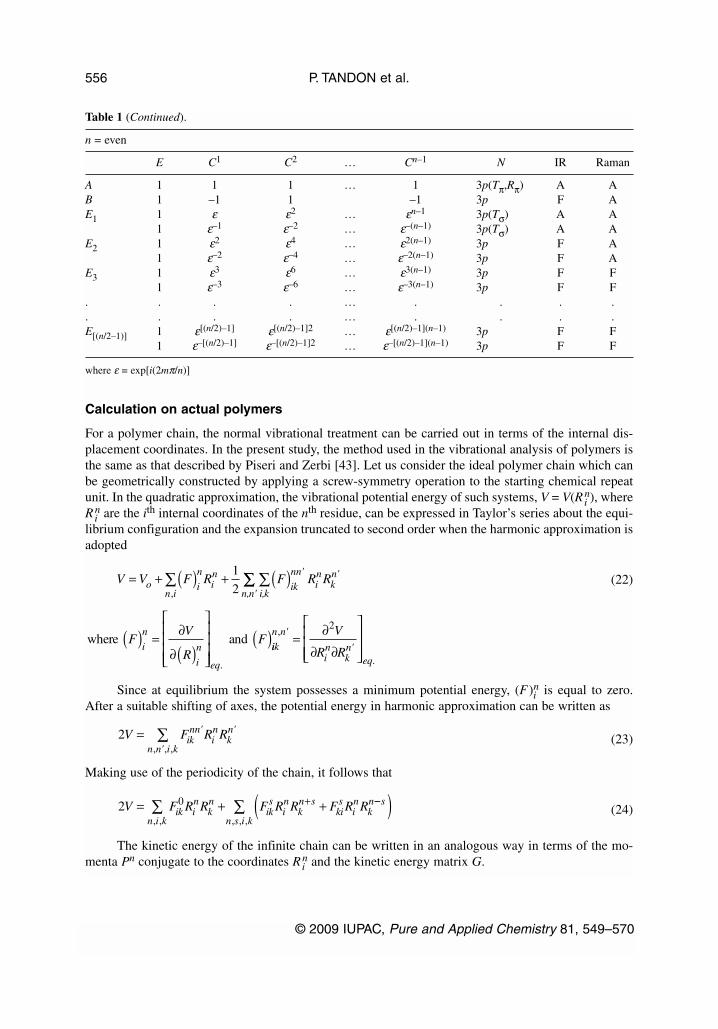
n = even
E C1 C2 … Cn–1 N IR Raman
A 1 1 1 … 1 3p(Tπ,Rπ) A AB 1 –1 1 –1 3p F AE1 1 ε ε2 … εn–1 3p(Tσ) A A
1 ε–1 ε–2 … ε–(n–1) 3p(Tσ) A AE2 1 ε2 ε4 … ε2(n–1) 3p F A
1 ε–2 ε–4 … ε–2(n–1) 3p F AE3 1 ε3 ε6 … ε3(n–1) 3p F F
1 ε–3 ε–6 … ε–3(n–1) 3p F F. . . . … . . . .. . . . … . . . .E[(n/2–1)] 1 ε[(n/2)–1] ε[(n/2)–1]2 … ε[(n/2)–1](n–1) 3p F F
1 ε–[(n/2)–1] ε–[(n/2)–1]2 … ε–[(n/2)–1](n–1) 3p F F
where ε = exp[i(2mπ/n)]
Calculation on actual polymers
For a polymer chain, the normal vibrational treatment can be carried out in terms of the internal dis-placement coordinates. In the present study, the method used in the vibrational analysis of polymers isthe same as that described by Piseri and Zerbi [43]. Let us consider the ideal polymer chain which canbe geometrically constructed by applying a screw-symmetry operation to the starting chemical repeatunit. In the quadratic approximation, the vibrational potential energy of such systems, V = V(Ri
n), whereRi
n are the ith internal coordinates of the nth residue, can be expressed in Taylor’s series about the equi-librium configuration and the expansion truncated to second order when the harmonic approximation isadopted
(22)
Since at equilibrium the system possesses a minimum potential energy, (F)in is equal to zero.
After a suitable shifting of axes, the potential energy in harmonic approximation can be written as
(23)
Making use of the periodicity of the chain, it follows that
(24)
The kinetic energy of the infinite chain can be written in an analogous way in terms of the mo-menta Pn conjugate to the coordinates Ri
n and the kinetic energy matrix G.
P. TANDON et al.
© 2009 IUPAC, Pure and Applied Chemistry 81, 549–570
556
Table 1 (Continued).
V V F R F R Ro i
n
n iin
ik
nnin
kn= + ( ) + ( )∑ ∑
,
1
2' '
i,kn,n'∑∑
where andFV
RF
i
n
i
n
eq
( ) = ∂
∂( )
( ).
iik
n n
in
kn
eq
V
R R
,
.
'
'= ∂
∂ ∂
2
2V F R Riknn
in
kn
n n i k= ∑ ' '
', ,,
2 0V F R R F R R F R Rik in
kn
n i kiks
in
kn s
kis
in
kn= + +∑ +
, ,
−−( )∑ s
n s i k, , ,

(25)
Hamiltonian’s equation of motion can be written using eqs. 24 and 25, thus leading to a systemof an infinite number of second-order differential equations in the Ri
n+s whose solution can be assumedto be planar wave
Rin+s = Ai exp [–i(λ1/2t + sδ)] (26)
Here, δ is phase shift between two equivalent internal displacement coordinates in adjacent units.Substitution of eq. 26 into the system of differential equations yields a system of 3p simultaneous ho-mogeneous linear equations (p is the number of atoms in a chemical repeat unit) in the unknowns Ai,whose nontrivial solutions are given by the equation
|G(δ)F(δ) – λ(δ)I| = 0 (27)
where λ(δ) = 4π2c2v2(δ) and
(28A)
(28B)
Equations 28A and 28B coincide with those first given by Higgs eqs. 19 and 20 through a Fouriertransform on the system of internal displacement coordinates. A calculation of ν(δ) as a function of δgives the dispersion curves. This function is periodic with a period equal to 2π, i.e., n(δ + 2π). Further,ν(–δ) = ν(δ) so that one can limit a study of the function to the range 0 ≤ δ ≤ π, which corresponds tohalf of the first Brillouin zone (reduced zone scheme). Optically active frequencies correspond to val-ues of δ = 0, φ, 2φ.
For any given phase difference δ (other than 0 or π), the G(δ) and F(δ) matrices are complex. Anumber of difficulties arise in handling complex numbers. In order to avoid these difficulties, certainmethods have been proposed to convert matrices into equivalent real matrices. This can be done by con-structing suitable linear combinations of coordinates, as mentioned in eq. 9. It can be shown that anycomplex matrix can be set to its real equivalent through a suitable similarity transformation. If C is acomplex matrix with a real part X and an imaginary part Y, it can be written as
(29)
It can be transformed into its real equivalent R by matrix
(30)
(31)
And I is the identity matrix of order 3p.In the present case, we can write G(δ) = GR(δ) + iG1(δ) and F(δ) = FR(δ) + iFl(δ), where GR(δ),
FR(δ), Gl(δ), Fl(δ) are the real and imaginary parts of G(δ) and F(δ). The product H(δ) = G(δ) F(δ) be-comes
© 2009 IUPAC, Pure and Applied Chemistry 81, 549–570
Vibrational dynamics of polymers 557
2 0T G P P G P P G P Pik in
n i kkn
iks
in
kn s
kis
in
kn= + +∑ +
, ,
−−( )∑ s
n s i k, , ,
G G G is G iss s
sδ δ δ( ) = + ( ) + −( )
∑0 exp exp'
F F F is F iss s
sδ δ δ( ) = + ( ) + −( )
∑0 exp exp'
CX iY
X iY=
+
−
0
0
R PCPX
Y
Y
X= =
−
−1
where PI
iI
I
iI=
−
1
2
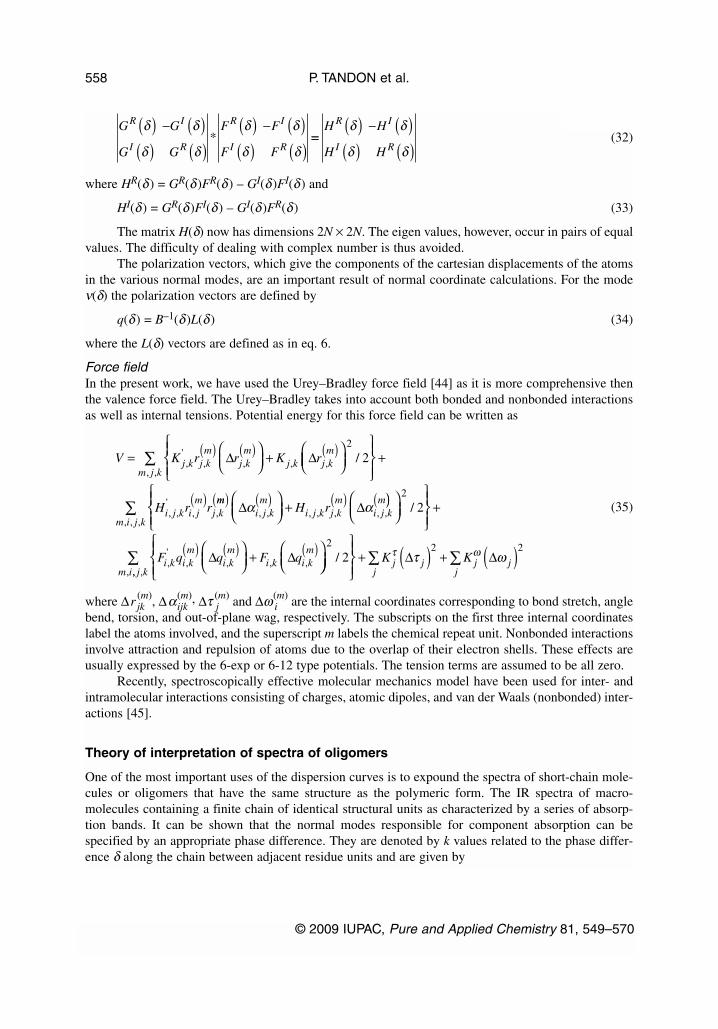
(32)
where HR(δ) = GR(δ)FR(δ) – GI(δ)FI(δ) and
HI(δ) = GR(δ)FI(δ) – GI(δ)FR(δ) (33)
The matrix H(δ) now has dimensions 2N × 2N. The eigen values, however, occur in pairs of equalvalues. The difficulty of dealing with complex number is thus avoided.
The polarization vectors, which give the components of the cartesian displacements of the atomsin the various normal modes, are an important result of normal coordinate calculations. For the modeν(δ) the polarization vectors are defined by
q(δ) = B–1(δ)L(δ) (34)
where the L(δ) vectors are defined as in eq. 6.
Force fieldIn the present work, we have used the Urey–Bradley force field [44] as it is more comprehensive thenthe valence force field. The Urey–Bradley takes into account both bonded and nonbonded interactionsas well as internal tensions. Potential energy for this force field can be written as
(35)
where ∆r jk(m), ∆αijk
(m), ∆τ j(m) and ∆ω i
(m) are the internal coordinates corresponding to bond stretch, anglebend, torsion, and out-of-plane wag, respectively. The subscripts on the first three internal coordinateslabel the atoms involved, and the superscript m labels the chemical repeat unit. Nonbonded interactionsinvolve attraction and repulsion of atoms due to the overlap of their electron shells. These effects areusually expressed by the 6-exp or 6-12 type potentials. The tension terms are assumed to be all zero.
Recently, spectroscopically effective molecular mechanics model have been used for inter- andintramolecular interactions consisting of charges, atomic dipoles, and van der Waals (nonbonded) inter-actions [45].
Theory of interpretation of spectra of oligomers
One of the most important uses of the dispersion curves is to expound the spectra of short-chain mole-cules or oligomers that have the same structure as the polymeric form. The IR spectra of macro-molecules containing a finite chain of identical structural units as characterized by a series of absorp-tion bands. It can be shown that the normal modes responsible for component absorption can bespecified by an appropriate phase difference. They are denoted by k values related to the phase differ-ence δ along the chain between adjacent residue units and are given by
P. TANDON et al.
© 2009 IUPAC, Pure and Applied Chemistry 81, 549–570
558
G
G
G
G
F
F
F
F
R
I
I
R
R
I
I
R
δ
δ
δ
δ
δ
δ
δ
δ
( )( )
− ( )( )
( )( )
− ( )( )
* ==( )( )
− ( )( )
H
H
H
H
R
I
I
R
δ
δ
δ
δ
V K r r K rj k j km
j km
j k j km=
+
( ) ( ) ( ),'
, , , ,∆ ∆
+∑
( )
2
2/, ,
, ,'
, ,
m j k
i j k i jm
j kH r rmm
i j km
i j k j km
i j km
H r( ) ( ) ( ) (
+∆ ∆α α, , , , , , ,))
+∑
2
2/, , ,m i j k
F q q F qi k i km
i km
i k i km
,'
, , , ,( ) ( ) ( )
+
∆ ∆
+ ( ) + ( )∑∑
2 2 22/
,K Kj j j j
jjm i
τ ωτ ω∆ ∆,, ,j k
∑
δ = (kπ/n) (k = 0,1,2......,n –1)
where n is the number of repeat units in the chain and k is an integer that represents the number of loopsin a stationary wave representing the chain vibrations. The above equation holds good for finite chainhaving free ends.
Density-of-states and heat capacity
Another use of dispersion curves is that the microscopic behavior of a crystal can be correlated with itsmacroscopic properties such as specific heat. For a one-dimensional system, the density-of-states func-tion or the frequency distribution function expresses the way energy is distributed among the variousbranches of normal modes in the crystal. This can be calculated from the relation
(36)
with ∫ g(νj)d νj = 1
The sum is over all branches j. If we consider a solid as an assembly of harmonic oscillators, thefrequency distribution g(ν) is equivalent to a partition function. This can be used to compute thermo-dynamic quantities such as free energy, entropy, specific heat, and enthalpy. The heat capacity, whichis an important thermodynamic parameter, can be calculated using the Debye relation. For example, theheat capacity can give information about the proportion of a protein, which is in α-helical and β-sheetstructure and is necessary in evaluating the basic thermodynamics of the enzyme reaction [46]. TheDebye relation is given by
(37)
The constant volume heat capacity Cv, given by eq. 43, is converted into constant pressure heatcapacity Cp using the Nernst–Lindemann approximation [47]
(38)
where A0 is a constant often of a universal nature and T m0 is the equilibrium melting temperature [48].
Quantum chemical methods
Application of quantum chemical techniques to real chemical systems represents the essence of com-putational chemistry. These techniques such as molecular orbital (MO) theory [49] and DFT [50–58]etc., are used to study molecular structure and conformational isomerism as well as molecular spectrasuch as vibrational and electronic. Quantum chemical methods may be broadly classified as semi-em-pirical and ab initio.
The semi-empirical methods, where the values of several integrals are obtained with the help ofexperimental parameters, are usually less accurate than the ab initio methods in the determination ofmolecular structure and thermal energy, though in certain cases of a series of related molecules, theycan provide results in remarkable agreement with the experimental findings. However, in spectroscopicstudies, which involve energy differences between various states, fairly reliable results have been ob-tained by both the ab initio and semi-empirical MO methods.
© 2009 IUPAC, Pure and Applied Chemistry 81, 549–570
Vibrational dynamics of polymers 559
g v v jj v vj
( ) = ∂( )−
( )=∑ δ δ
δ/
1
C g v kN hv kThv kT
hv kTv j A j
j
j
= ( ) ( ) ( )( ) −
/exp /
exp /
2
112
∑j
C C RA C T C Tp v p v m− = ( )3 02 0/
Various variants of semi-empirical all valence electron methods such as complete neglect of dif-ferential overlap (CNDO), intermediate neglect of differential overlap (INDO), modified intermediateneglect of differential overlap (MINDO), modified neglect of differential overlap (MNDO), etc. and theHF and post-HF ab initio methods have been used for conformational analysis of organic molecules.
Ab initio methods, unlike semi-empirical methods, use no experimental parameters in their com-putation. Instead, their computations are based solely on the laws of quantum mechanics. There are var-ious theories underlying ab initio methods like closed- and open-shell HF theory and DFT. HF theoryprovides an inadequate treatment of the correlation between the motions of the electrons within a mo-lecular system. The motion of electrons of opposite spin remains uncorrelated in this scheme. The tech-niques are widely used to incorporate the effect of electron correlation (i) configuration interaction and(ii) Moller–Plesset perturbation theory.
The first method is variational but not size-consistent, whereas the second is size-consistent butnot variational. Variational implies that the calculated electronic energy should correspond to an upperbound to the energy that would result from the exact solution of the Schrödinger equation. Size consis-tency means that the method must give additive results when applied to an assembly of isolated mole-cules.
RESULTS AND DISCUSSION
Polypeptides
Polymeric systems in general and biopolymers in particular are capable of existing in a variety of con-formations. Almost all their properties are dictated by the type of conformation taken up by the bio-molecules. The spectroscopic approach has proved a very powerful diagnostic tool in characterizingtheir conformation. However, the problem is an order of magnitude more complex than in simple solids,mainly because of the absence of simple symmetry and presence of large contents in unit cell. For ex-ample, polyalanine (R=CH3), which is a very simple polypeptide, has in the one-dimensional unit cell18 residues and 5 turns. Each residue has 10 atoms. The problem thus has enormous dimensions.Further, the coupling of normal vibrations of successive repeat units necessitates evaluation of the dis-persion of the normal modes in the first Brillouin zone. This has been done for the whole set of normalmodes in α, β, ω, and 3-fold helices, which are the conformations generally taken up by most of thepolypeptides. Apart from the characteristic amide bands, it is observed that the profile of the low-fre-quency modes including the acoustic modes reflects the skeletal conformation beautifully well. Thisprovides a good handle for conformation classification on the basis of the dispersion profile [4]. The re-sults are presented for α-helical [poly(L-valine) (PLV), poly(L-leucine) (PLL), poly(L-glutamic acid)(PLG), poly(β-benzyl-L-aspartate) (PBLA)], β-sheet [poly(L-serine) (PLS), PLV], ω-helical [PBLA,poly-Nε(p-bromobenzoyl-L-ornithine) (PBRBO)], and 3-fold [polyglycine II (PG II), poly-(L-hydrox-yproline) (PLHP), poly-L-proline II (PLP II)] polypeptides [5–16].
Conformation of a polypeptide, protein, or nucleic acid to a very large extent dictates its physico-chemical, thermodynamical, hydrodynamical, and biological behavior. It is determined by both the co-valent as well as noncovalent interactions. The latter part includes hydrophobic, van der Waals, electro-static, and hydrogen bond interactions. The nonbonded interactions can be visualized at two levels,namely, short- and long-range interactions. The short range comprises the backbone and side chain,whereas the long range arises out of side chain–side chain interactions. Various physicochemical tech-niques have been used as diagnostic tools to characterize the conformations, viz., NMR, IR, CD, XRD,etc. Most of the polypeptides go into four main types of structures, i.e., Pauling’s α-helix, pleated-sheetβ structure, 4-fold ω-helix, and 3-fold collagen-type helix. The structures are shown in Figs. 1b, 2b, 3b,and 4b, respectively. The low-frequency acoustic and optical modes for these chain structures are shownalongside. Spectroscopically speaking, the amide modes that are used for identification of various con-formations are shown in Tables 2 and 3.
P. TANDON et al.
© 2009 IUPAC, Pure and Applied Chemistry 81, 549–570
560
© 2009 IUPAC, Pure and Applied Chemistry 81, 549–570
Vibrational dynamics of polymers 561
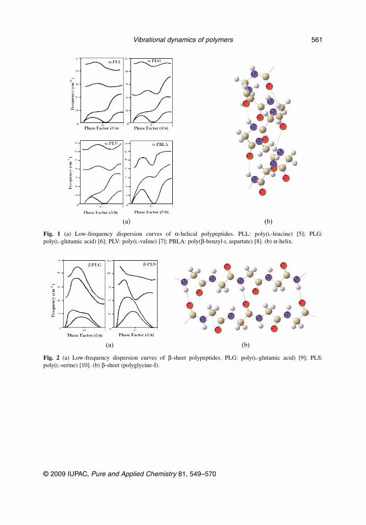
Fig. 1 (a) Low-frequency dispersion curves of α-helical polypeptides. PLL: poly(L-leucine) [5]; PLG:poly(L-glutamic acid) [6]; PLV: poly(L-valine) [7]; PBLA: poly(β-benzyl-L aspartate) [8]. (b) α-helix.
Fig. 2 (a) Low-frequency dispersion curves of β-sheet polypeptides. PLG: poly(L-glutamic acid) [9]; PLS:poly(L-serine) [10]. (b) β-sheet (polyglycine-I).
P. TANDON et al.
© 2009 IUPAC, Pure and Applied Chemistry 81, 549–570
562
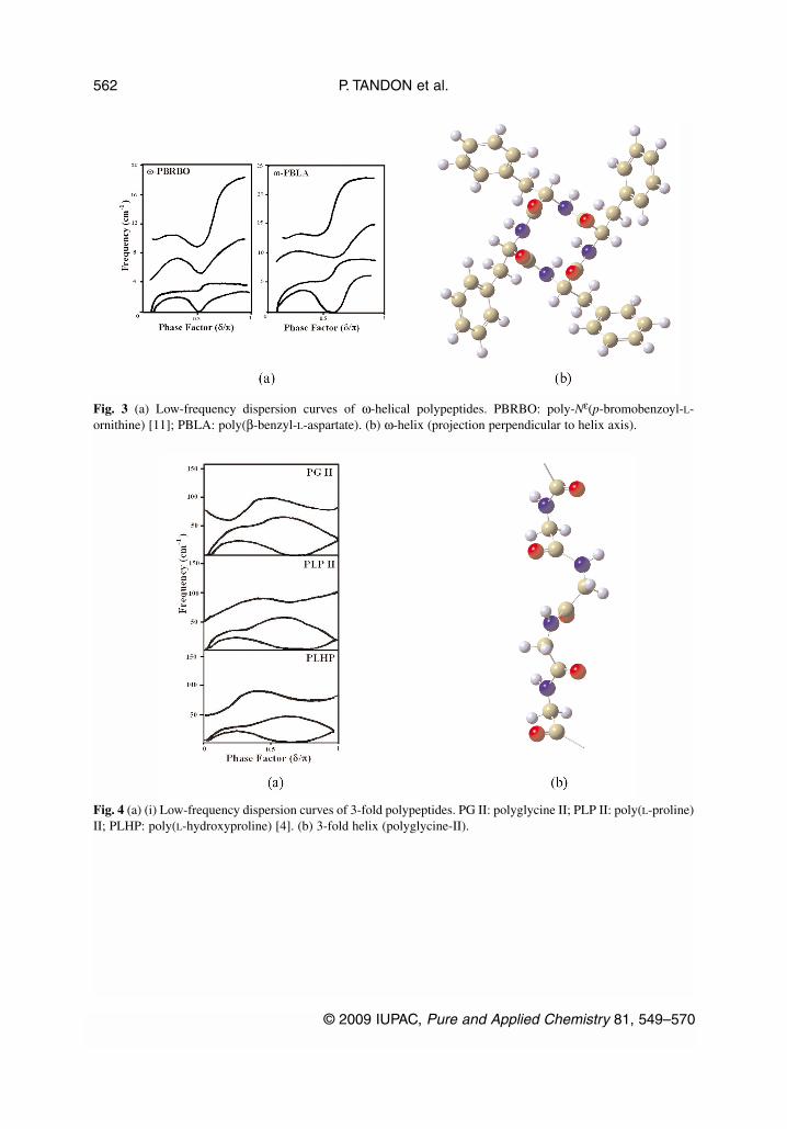
Fig. 3 (a) Low-frequency dispersion curves of ω-helical polypeptides. PBRBO: poly-Nε(p-bromobenzoyl-L-ornithine) [11]; PBLA: poly(β-benzyl-L-aspartate). (b) ω-helix (projection perpendicular to helix axis).
Fig. 4 (a) (i) Low-frequency dispersion curves of 3-fold polypeptides. PG II: polyglycine II; PLP II: poly(L-proline)II; PLHP: poly(L-hydroxyproline) [4]. (b) 3-fold helix (polyglycine-II).
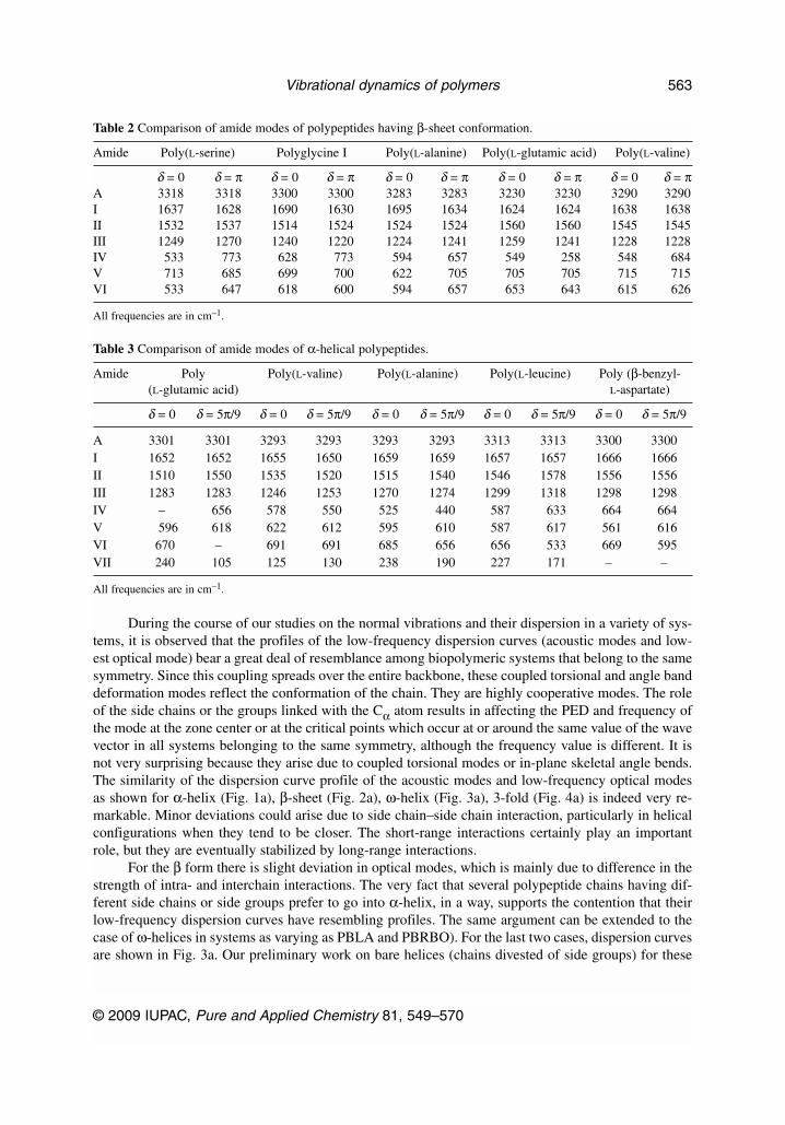
Table 2 Comparison of amide modes of polypeptides having β-sheet conformation.
Amide Poly(L-serine) Polyglycine I Poly(L-alanine) Poly(L-glutamic acid) Poly(L-valine)
δ = 0 δ = π δ = 0 δ = π δ = 0 δ = π δ = 0 δ = π δ = 0 δ = πA 3318 3318 3300 3300 3283 3283 3230 3230 3290 3290I 1637 1628 1690 1630 1695 1634 1624 1624 1638 1638II 1532 1537 1514 1524 1524 1524 1560 1560 1545 1545III 1249 1270 1240 1220 1224 1241 1259 1241 1228 1228IV 533 773 628 773 594 657 549 258 548 684V 713 685 699 700 622 705 705 705 715 715VI 533 647 618 600 594 657 653 643 615 626
All frequencies are in cm–1.
Table 3 Comparison of amide modes of α-helical polypeptides.
Amide Poly Poly(L-valine) Poly(L-alanine) Poly(L-leucine) Poly (β-benzyl-(L-glutamic acid) L-aspartate)
δ = 0 δ = 5π/9 δ = 0 δ = 5π/9 δ = 0 δ = 5π/9 δ = 0 δ = 5π/9 δ = 0 δ = 5π/9
A 3301 3301 3293 3293 3293 3293 3313 3313 3300 3300I 1652 1652 1655 1650 1659 1659 1657 1657 1666 1666II 1510 1550 1535 1520 1515 1540 1546 1578 1556 1556III 1283 1283 1246 1253 1270 1274 1299 1318 1298 1298IV – 656 578 550 525 440 587 633 664 664V 596 618 622 612 595 610 587 617 561 616VI 670 – 691 691 685 656 656 533 669 595VII 240 105 125 130 238 190 227 171 – –
All frequencies are in cm–1.
During the course of our studies on the normal vibrations and their dispersion in a variety of sys-tems, it is observed that the profiles of the low-frequency dispersion curves (acoustic modes and low-est optical mode) bear a great deal of resemblance among biopolymeric systems that belong to the samesymmetry. Since this coupling spreads over the entire backbone, these coupled torsional and angle banddeformation modes reflect the conformation of the chain. They are highly cooperative modes. The roleof the side chains or the groups linked with the Cα atom results in affecting the PED and frequency ofthe mode at the zone center or at the critical points which occur at or around the same value of the wavevector in all systems belonging to the same symmetry, although the frequency value is different. It isnot very surprising because they arise due to coupled torsional modes or in-plane skeletal angle bends.The similarity of the dispersion curve profile of the acoustic modes and low-frequency optical modesas shown for α-helix (Fig. 1a), β-sheet (Fig. 2a), ω-helix (Fig. 3a), 3-fold (Fig. 4a) is indeed very re-markable. Minor deviations could arise due to side chain–side chain interaction, particularly in helicalconfigurations when they tend to be closer. The short-range interactions certainly play an importantrole, but they are eventually stabilized by long-range interactions.
For the β form there is slight deviation in optical modes, which is mainly due to difference in thestrength of intra- and interchain interactions. The very fact that several polypeptide chains having dif-ferent side chains or side groups prefer to go into α-helix, in a way, supports the contention that theirlow-frequency dispersion curves have resembling profiles. The same argument can be extended to thecase of ω-helices in systems as varying as PBLA and PBRBO). For the last two cases, dispersion curvesare shown in Fig. 3a. Our preliminary work on bare helices (chains divested of side groups) for these
© 2009 IUPAC, Pure and Applied Chemistry 81, 549–570
Vibrational dynamics of polymers 563

symmetries also appears to be broadly in support of it. The low-frequency dispersion curve profiles arethus characteristic of their conformation.
Interpretation of spectra of oligomers from dispersion curves
In the absence of INS data, one of the easiest ways to test the meandering part of the dispersion curvesis to look at the spectra of various oligomers. As discussed earlier, the spectra of oligomers are relatedto the dispersion profile of the polymer modes through the phase relationship given by the above ex-pression and provided that both oligo and polymers are conformationally connected. The Raman scat-tering data on oligomers of valine up to the hexamer as reported by Fasman et al. [59] have been used.The observed modes are assigned at appropriate phase values on the corresponding branch of the dis-persion curve from all the possible k values and are plotted in Fig. 5a along with the dispersion curve
P. TANDON et al.
© 2009 IUPAC, Pure and Applied Chemistry 81, 549–570
564
Fig. 5 (a) Dispersion curves of β PLV [13]. (b) Density-of-states of β PLV [13].
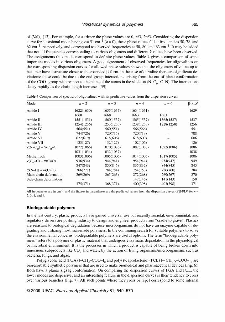
of (Val)n [13]. For example, for a trimer the phase values are 0, π/3, 2π/3. Considering the dispersioncurve for a torsional mode having v = 51 cm–1 (δ = 0), these phase values fall at frequencies 50, 78, and62 cm–1, respectively, and correspond to observed frequencies at 50, 80, and 63 cm–1. It may be addedthat not all frequencies corresponding to various oligomers and different k values have been observed.The assignments thus made correspond to definite phase values. Table 4 gives a comparison of someimportant modes in various oligomers. A good agreement of observed frequencies for oligovalines onthe corresponding dispersion curves for allowed phase values shows that the oligomers of valine up tohexamer have a structure closer to the extended β-form. In the case of di-valine there are significant de-viations: these could be due to the end-group interactions arising from the out-of-plane conformationof the COO– group with respect to the plane of the atoms in the skeleton (N–Cα–C–N). The interactionsdecay rapidly as the chain length increases [59].
Table 4 Comparison of spectra of oligovalines with its predictive values from the dispersion curves.
Mode n = 2 n = 3 n = 4 n = 6 β-PLV
Amide I 1622(1630) 1655(1637) 1634(1631) – 16291660 1668 1663 1663
Amide II 1551(1531) 1560(1537) 1565(1537) 1565(1537) 1537Amide III 1254(1256) 1253(1255) 1238(1253) 1228(1250) 1256Amide IV 564(551) 560(551) 566(566) – 551Amide V 744(726) 720(715) 720(713) – 708Amide VI 622(619) 618(606) 618(609) – 606Amide VII 133(127) 132(127) 102(106) – 126ν(N–Cα) + ν(Cα–C) 1072(1066) 1070(1076) 1087(1080) 1092(1086) 1086
1031(1034) 1032(1037) – – 1038Methyl rock 1003(1006) 1005(1006) 1014(1006) 1017(1005) 1006ν(Cα–C) + ν(C=O) 936(934) 944(941) 954(944) 954(947) 949
847(843) 850(845) 835(832) 844(845) 845ω(N–H) + ω(C=O) 766(771) 784(784) 754(753) 756(760) 784Main-chain deformation 269(269) 265(263) 272(268) 269(267) 270Side-chain deformation – – 147(146) 141(143) 150
375(371) 368(371) 400(398) 403(398) 371
All frequencies are in cm–1, and the figures in parentheses are the predicted values from the dispersion curves of β-PLV for n =2, 3, 4, and 6.
Biodegradable polymers
In the last century, plastic products have gained universal use but recently societal, environmental, andregulatory drivers are pushing industry to design and engineer products from “cradle to grave”. Plasticsare resistant to biological degradation because microorganisms do not have an enzyme capable of de-grading and utilizing most man-made polymers. In the continuing search for suitable polymers to solvethe environmental concerns, biodegradable polymers are useful options. The term “biodegradable poly-mers” refers to a polymer or plastic material that undergoes enzymatic degradation in the physiologicalor microbial environment. It is the processes in which a product is capable of being broken down intoinnocuous subproducts like CO2 and water, by the action of living organisms/microorganisms such asbacteria, fungi, and algae.
Polyglycolic acid (PGA) [–CH2–COO–]n and poly(ε-caprolactone) (PCL) [–(CH2)5–COO–]n arebioresorbable synthetic polymers that are used to make biomedical and pharmaceutical devices (Fig. 6).Both have a planar zigzag conformation. On comparing the dispersion curves of PGA and PCL, thelower modes are dispersive, and an interesting feature in the dispersion curves is their tendency to crossover various branches (Fig. 7). All such points where they cross or repel correspond to some internal
© 2009 IUPAC, Pure and Applied Chemistry 81, 549–570
Vibrational dynamics of polymers 565
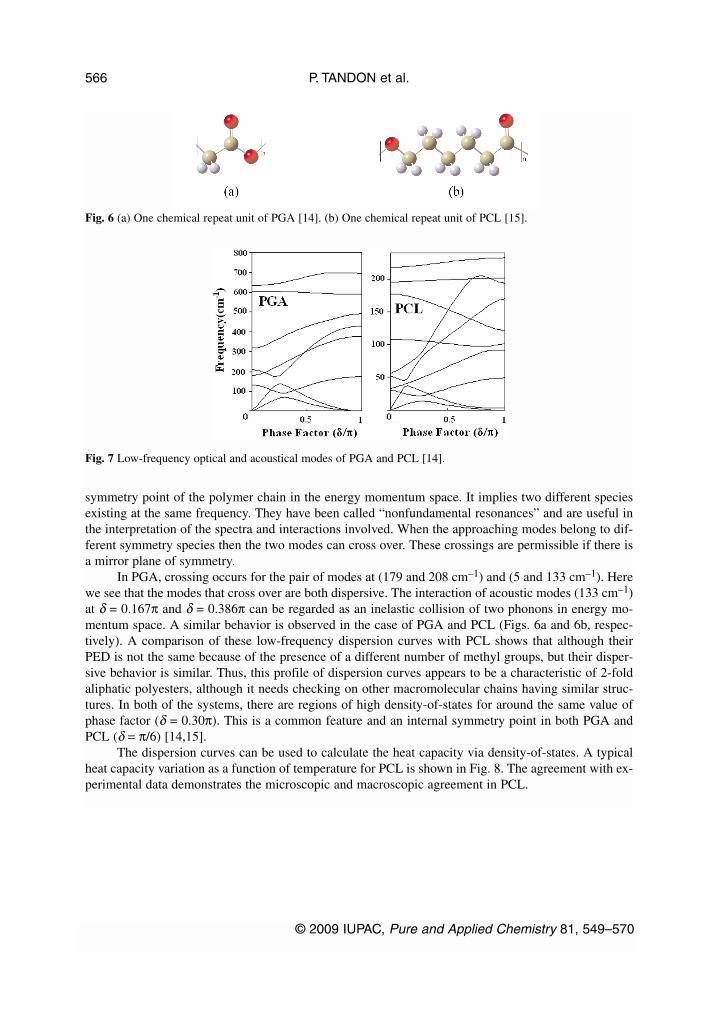
symmetry point of the polymer chain in the energy momentum space. It implies two different speciesexisting at the same frequency. They have been called “nonfundamental resonances” and are useful inthe interpretation of the spectra and interactions involved. When the approaching modes belong to dif-ferent symmetry species then the two modes can cross over. These crossings are permissible if there isa mirror plane of symmetry.
In PGA, crossing occurs for the pair of modes at (179 and 208 cm–1) and (5 and 133 cm–1). Herewe see that the modes that cross over are both dispersive. The interaction of acoustic modes (133 cm–1)at δ = 0.167π and δ = 0.386π can be regarded as an inelastic collision of two phonons in energy mo-mentum space. A similar behavior is observed in the case of PGA and PCL (Figs. 6a and 6b, respec-tively). A comparison of these low-frequency dispersion curves with PCL shows that although theirPED is not the same because of the presence of a different number of methyl groups, but their disper-sive behavior is similar. Thus, this profile of dispersion curves appears to be a characteristic of 2-foldaliphatic polyesters, although it needs checking on other macromolecular chains having similar struc-tures. In both of the systems, there are regions of high density-of-states for around the same value ofphase factor (δ = 0.30π). This is a common feature and an internal symmetry point in both PGA andPCL (δ = π/6) [14,15].
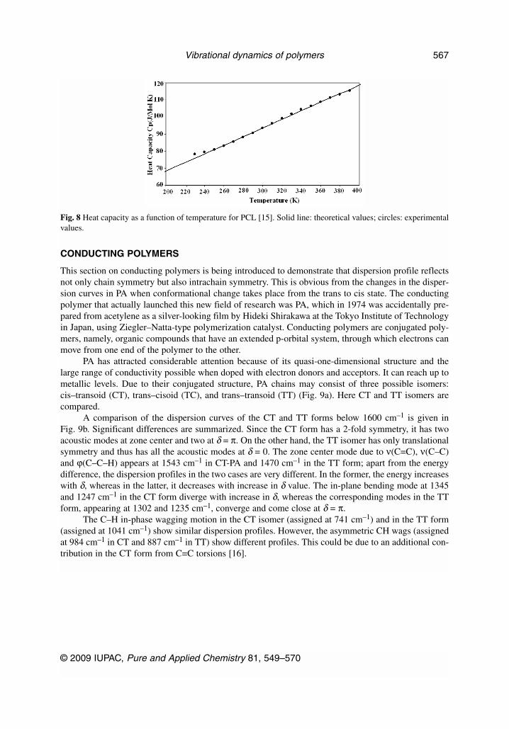
The dispersion curves can be used to calculate the heat capacity via density-of-states. A typicalheat capacity variation as a function of temperature for PCL is shown in Fig. 8. The agreement with ex-perimental data demonstrates the microscopic and macroscopic agreement in PCL.
P. TANDON et al.
© 2009 IUPAC, Pure and Applied Chemistry 81, 549–570
566
Fig. 6 (a) One chemical repeat unit of PGA [14]. (b) One chemical repeat unit of PCL [15].
Fig. 7 Low-frequency optical and acoustical modes of PGA and PCL [14].
CONDUCTING POLYMERS
This section on conducting polymers is being introduced to demonstrate that dispersion profile reflectsnot only chain symmetry but also intrachain symmetry. This is obvious from the changes in the disper-sion curves in PA when conformational change takes place from the trans to cis state. The conductingpolymer that actually launched this new field of research was PA, which in 1974 was accidentally pre-pared from acetylene as a silver-looking film by Hideki Shirakawa at the Tokyo Institute of Technologyin Japan, using Ziegler–Natta-type polymerization catalyst. Conducting polymers are conjugated poly-mers, namely, organic compounds that have an extended p-orbital system, through which electrons canmove from one end of the polymer to the other.
PA has attracted considerable attention because of its quasi-one-dimensional structure and thelarge range of conductivity possible when doped with electron donors and acceptors. It can reach up tometallic levels. Due to their conjugated structure, PA chains may consist of three possible isomers:cis–transoid (CT), trans–cisoid (TC), and trans–transoid (TT) (Fig. 9a). Here CT and TT isomers arecompared.
A comparison of the dispersion curves of the CT and TT forms below 1600 cm–1 is given inFig. 9b. Significant differences are summarized. Since the CT form has a 2-fold symmetry, it has twoacoustic modes at zone center and two at δ = π. On the other hand, the TT isomer has only translationalsymmetry and thus has all the acoustic modes at δ = 0. The zone center mode due to ν(C=C), ν(C–C)and ϕ(C–C–H) appears at 1543 cm–1 in CT-PA and 1470 cm–1 in the TT form; apart from the energydifference, the dispersion profiles in the two cases are very different. In the former, the energy increaseswith δ, whereas in the latter, it decreases with increase in δ value. The in-plane bending mode at 1345and 1247 cm–1 in the CT form diverge with increase in δ, whereas the corresponding modes in the TTform, appearing at 1302 and 1235 cm–1, converge and come close at δ = π.
The C–H in-phase wagging motion in the CT isomer (assigned at 741 cm–1) and in the TT form(assigned at 1041 cm–1) show similar dispersion profiles. However, the asymmetric CH wags (assignedat 984 cm–1 in CT and 887 cm–1 in TT) show different profiles. This could be due to an additional con-tribution in the CT form from C=C torsions [16].
© 2009 IUPAC, Pure and Applied Chemistry 81, 549–570
Vibrational dynamics of polymers 567
Fig. 8 Heat capacity as a function of temperature for PCL [15]. Solid line: theoretical values; circles: experimentalvalues.

CONCLUSION
The symmetry considerations are intimately related to various types of conformations. This is very wellbrought out in the profile of the dispersion curves especially in the low-frequency optical and acousti-cal modes.
ACKNOWLEDGMENT
Financial support to PT and DC from the Council of Science and Technology, Uttar Pradesh, India isgratefully acknowledged.
REFERENCES
1. J. Engel, G. Schwarz. Angew. Chem., Int. Ed. Engl. 9, 389 (1970).2. A. G. Walton, J. Blackwell. Biopolymers, Academic Press, New York (1973).3. G. D. Fasman. Poly-α-Amino Acids, Marcel Dekker, New York (1976).4. V. D. Gupta. Int. J. Quant. Chem. 20, 9 (1981).5. S. Srivastava, P. Tandon, V. D. Gupta, S. Rastogi, L. Burman, S. B. Katti. Pramana 45, 235
(1995).6. S. Srivastava, P. Tandon, V. D. Gupta, S. Rastogi, S. B. Katti, C. Mehrotra. Polym. J. 29, 33
(1997).7. L. Burman, P. Tandon, V. D. Gupta, S. Rastogi. Polym. J. 28, 2389 (1997).8. P. Tandon, V. D. Gupta, O. Prasad, S. Rastogi, B. Katti. J. Polym. Sci., Part B: Polym. Phys. 34,
1213 (1996).9. S. Srivastava, P. Tandon, V. D. Gupta, S. Rastogi, S. B. Katti. Polym. J. 29, 492 (1997).
10. N. K. Misra, D. Kapoor, P. Tandon, V. D. Gupta. Polymer 41, 2095 (2000).
P. TANDON et al.
© 2009 IUPAC, Pure and Applied Chemistry 81, 549–570
568
Fig. 9 (a) Isomers of PA. (b) Comparison of dispersion curves of CT and TT PA [16].
11. A. Gupta, P. Tandon, V. D. Gupta, S. Rastogi, G. P. Gupta. J. Macromol. Sci., Part B: Phys. 34,401 (1995).
12. L. Burman, P. Tandon, V. D. Gupta, S. Srivastava. J. Macromol. Sci., Part B: Phys. 34, 379 (1995).13. L. Burman, P. Tandon, V. D. Gupta, S. Rastogi, S. Srivastava. Biopolymers 38, 53 (1996).14. R. Agarwal, R. M. Misra, P. Tandon, V. D. Gupta. Polymer 45, 5307 (2004).15. R. M. Misra, R. Agarwal, P. Tandon, V. D. Gupta. Eur. Polym. J. 40, 1787 (2004).16. A. Kumar, S. Pande, P. Tandon, V. D. Gupta. J. Macromol. Sci., Part B: Phys. 39, 303 (2000).17. D. Jacquemin, J.-M. Andre, B. Champagne. J. Chem. Phys. 118, 3956 (2003).18. N. J. Ramer, T. Marrone, K. A. Stiso. Polymer 47, 7160 (2006).19. K. Honda, Y. Furukawa, H. Nishide. Vib. Spectrosc. 40, 149 (2006).20. S. Lakard, G. Herlem, B. Lakard, B. Fahys. J. Mol. Struct. (Theochem) 685, 83 (2004).21. M. Kaledin, A. Brown, A. L. Kaledin, J. M. Bowman. J. Chem. Phys. 121, 5646 (2004).22. E. Henssge, D. Dumont, D. Fischer, D. Bougeard. J. Mol. Struct. 482–483, 491 (1999).23. R. H. Boyd, R. H. Gee, J. Han, Y. Jin. J. Chem. Phys. 101, 789 (1994).24. S. Neyertz, D. Brown, J. O. Thomas. J. Chem. Phys. 101, 10064 (1994).25. C. E. Wozny, B. G. Sumpter, D. W. Noid. J. Chem. Phys. 100, 3520 (1994).26. D. Dumont, E. Henssge, D. Ficher, D. Bougeard. Macromol. Theory Simul. 7, 373 (1998).27. D. Dumont, D. Bougeard. Comput. Theo. Polym. Sci. 9, 89 (1999).28. S. Nakamura, M. Ikeguchi, K. Shimizu. Chem. Phys. Lett. 372, 423 (2003).29. V. N. Kabadi, B. M. Rice. J. Phys. Chem. A 108, 532 (2004).30. E. B. Wilson Jr. J. Chem. Phys. 9, 76 (1941).31. E. B. Wilson, J. C. Decius, P. C. Cross. Molecular Vibrations, McGraw-Hill, New York (1955).32. E. P. Wigner. Group Theory and its Application to the Quantum Mechanics of Atomic Spectra,
Academic Press, New York (1959).33. P. W. Higgs. Proc. R. Soc. (London) A220, 472 (1953).34. E. Wodewig. Matrix Calculus, North-Holland, Amsterdam (1959).35. H. Tadokaro. J. Chem. Phys. 33, 1558 (1960).36. C. Y. Liang, S. Krimm, G. B. B. M. Sutherland. J. Chem. Phys. 25, 543 (1953).37. S. Krimm, C. Y. Liang, G. B. B. M. Sutherland. J. Chem. Phys. 25, 549 (1956).38. C. Y. Liang, S. Krimm. J. Chem. Phys. 25, 563 (1956).39. C. Y. Liang, S. Krimm. J. Polym. Sci. 24, 241 (1958).40. C. Y. Liang, S. Krimm. J. Mol. Spectrosc. 3, 554 (1959).41. S. Krimm, C. F. Liang. J. Polym. Sci. 22, 95 (1956).42. S. Krimm. Fortschr. Hochpolymer-Forsch. 2, 51 (1960).43. L. Piseri, G. Zerbi. J. Chem. Phys. 48, 3561 (1968).44. H. C. Urey, H. C. Bradley. Phys. Rev. 38, 1969 (1931). 45. W. Qian, N. G. Mirikin, S. Krimm. Chem. Phys. Lett. 315, 125 (1999). 46. T. H. Benzinger. Nature 229, 100 (1971).47. R. Pan, M. Verma-Nair, B. Wunderlich. J. Therm. Anal. 35, 955 (1989).48. K. A. Roles, A. Xenopoulos, B. Wunderlich. Biopolymers 33, 753 (1993).49. P. W. Atkins, R. S. Friedman. Molecular Quantum Mechanics, 3rd ed., Oxford University Press,
Oxford (2001).50. P. Hohenberg, W. Kohn. Phys. Rev. B 136, 864 (1964).51. W. Kohn, L. J. Sham. Phys. Rev. A 140, 1133 (1965).52. J. C. Slater. Quantum Theory of Molecules and Solids, Vol. 4, McGraw-Hill, New York (1974).53. S. H. Vosko, L. Wilk, M. Nasair. Can. J. Phys. 58, 1200 (1980).54. B. Miehlich, A. Savin, H. Stoll, H. Preuss. Chem. Phys. Lett. 157, 200 (1989).55. C. Lee, W. Yang, R. G. Parr. Phys. Rev. B 37, 785 (1988).56. A. D. Becke. Phys. Rev. A 38, 3098 (1988).57. J. P. Perdew, W. Wang. Phys. Rev. B 45, 13244 (1992).
© 2009 IUPAC, Pure and Applied Chemistry 81, 549–570
Vibrational dynamics of polymers 569

58. R. G. Parr, W. Young. Density Functional Theory of Atoms and Molecules, Oxford UniversityPress, New York (1989).
59. G. D. Fasman, K. Itoh, C. S. Liu, R. C. Lord. Biopolymers 17, 125 (1978).
P. TANDON et al.
© 2009 IUPAC, Pure and Applied Chemistry 81, 549–570
570