congenital nephrogenic diabetes insipidus: the current ... · and therefore a low renal osmolar...
TRANSCRIPT

REVIEW
Congenital nephrogenic diabetes insipidus:the current state of affairs
Daniel Wesche & Peter M. T. Deen & Nine V. A. M. Knoers
Received: 23 November 2011 /Revised: 14 January 2012 /Accepted: 17 January 2012 /Published online: 17 March 2012# IPNA 2012
Abstract The anti-diuretic hormone arginine vasopressin(AVP) is released from the pituitary upon hypovolemia orhypernatremia, and regulates water reabsorption in the renalcollecting duct principal cells. Binding of AVP to the argi-nine vasopressin receptor type 2 (AVPR2) in the baso-lateral membrane leads to translocation of aquaporin 2(AQP2) water channels to the apical membrane of thecollecting duct principal cells, inducing water permeabil-ity of the membrane. This results in water reabsorption fromthe pro-urine into the medullary interstitium following anosmotic gradient. Congenital nephrogenic diabetes insipidus(NDI) is a disorder associated with mutations in either theAVPR2 or AQP2 gene, causing the inability of patients toconcentrate their pro-urine, which leads to a high riskof dehydration. This review focuses on the currentknowledge regarding the cell biological aspects of congenitalX-linked, autosomal-recessive and autosomal-dominantNDI while specifically addressing the latest develop-ments in the field. Based on deepened mechanisticunderstanding, new therapeutic strategies are currentlybeing explored, which we also discuss here.
Keywords Nephrogenic diabetes insipidus . Vasopressintype-2 receptor . Aquaporin-2 water channel .
Pharmacological chaperones
Introduction
Due to the importance of water homeostasis in the humanbody, disorders that interfere with proper urine concentratingability can easily be life-threatening, especially in children.One such disease is nephrogenic diabetes insipidus (NDI).Here, we focus on the inherited form of this syndrome anddescribe the current knowledge concerning clinicalsymptoms, inheritance patterns, and the associated genesand their known disease-causing mutations. We thenaddress the molecular and cellular mechanisms leadingto the disease and finally discuss the latest developmentsaiming to find a cure for this disorder.
Congenital nephrogenic diabetes insipidus
The congenital form of nephrogenic diabetes insipidus(NDI) is a rare inherited disorder, characterized by insensi-tivity of the distal nephron to the antidiuretic effects of theneurohypophyseal hormone arginine vasopressin (AVP). Asa consequence, the kidney loses its ability to concentrateurine, which may lead to severe dehydration and electrolyteimbalance (hypernatremia and hyperchloremia). Patientswith NDI have normal birth weight and pregnancies aresometimes complicated by polyhydramnios. The urine con-centrating defect in NDI is present from birth, and mani-festations of the disorder generally emerge within thefirst weeks of life. With breast milk feedings, infantsusually thrive and do not develop signs of dehydration.This is because human milk has a low salt and protein content,
D. WescheNijmegen Centre for Molecular Life Sciences Graduate School,Radboud University Nijmegen Medical Centre,Nijmegen, The Netherlands
P. M. T. DeenDepartment of Physiology,Nijmegen Centre for Molecular Life Sciences,Radboud University Nijmegen Medical Centre,Nijmegen, The Netherlands
N. V. A. M. Knoers (*)Department of Medical Genetics,University Medical Centre Utrecht,PO Box 85090, 3508 AB Utrecht, The Netherlandse-mail: [email protected]
Pediatr Nephrol (2012) 27:2183–2204DOI 10.1007/s00467-012-2118-8

and therefore a low renal osmolar load. With cows' milkformula feedings, the osmolar load on the kidney increases,resulting in an increased demand for free water. This is notprovided by oral feeding and, therefore, hypernatremic dehy-dration appears. Irritability, poor feeding, and poor weightgain are usually the initial symptoms. Patients are eager tosuck but may vomit during or shortly after the feeding. Dehy-dration is evidenced by dryness of the skin, loss of normal skinturgor, recessed eyeballs, increased periorbital folding, de-pression of the anterior fontanel, and a scaphoid abdomen.Intermittent high fever is a common complication of thedehydrated state, predominantly in very young children. Seiz-ures can occur but are rare and most often seen during therapy,particularly if rehydration proceeds too rapidly. Constipationis a common symptom in children with NDI. Nocturiaand nocturnal enuresis are complaints later in childhood.
Untreated, most patients fail to grow normally. In aretrospective study of 30 male NDI patients, most childrengrew below the 50th percentile, the majority havingstandard deviation (SD) scores lower than −1 [1].Catch-up growth occurs at least in some patients afternormalization of water and electrolyte balance, especially inthose with adherence to treatment. Bone age is generally notdelayed. Weight-for-height SD scores are initially low,followed by global normalization at school age [1].Initial feeding problems and the ingestion of largeamounts of low-caloric fluid resulting in a decreased appetitemay play roles in failure to thrive seen in NDI. Furthermore, itis possible that repeated episodes of dehydration have some asyet undetermined negative effects on growth.
Mental retardation has long been considered an importantcomplication of untreated NDI and assumed to be a sequel ofrecurrent episodes of severe brain dehydration and cerebraledema caused by overzealous attempts at rehydration [2, 3].Additional evidence underscoring the assumption that NDI hasadverse effects on the cerebrum is provided by several reportsdescribing intracranial calcifications in NDI patients [4, 5].Such lesions are generally considered to be the resultof hemorrhage or necrosis. Most of the reported patients withcerebral calcifications were mentally retarded. Nowadays,mental retardation is rare due to earlier recognition and treat-ment of NDI. Reliable estimates of the current frequency ofmental retardation under modern treatment are unknown, but inthe largest psychometric study ever reported, only two of the 17male NDI patients tested (aged 3–30 years) had a total intelli-gence quotient more than 2 SD below the norm. Fourteenpatients had an intelligence score within or above the normalrange and one patient had a general index score between −1and −2 SD [6]. The psychological development of NDI patientsis influenced by a persistent desire for drinking and the need forfrequent voiding, which compete with playing and learning.Therefore, many NDI patients are characterized by hy-peractivity, distractibility, short attention span, and restlessness.
Persistent polyuria can result in the development of mega-cystis, trabeculated bladder wall, hydroureter, and hydroneph-rosis. Large-capacity hypotonic bladder dysfunction mightrequire clean intermittent catheterization [7]. Patients shouldbe trained to void regularly in order to assure that the maximalurinary bladder capacity remains within normal range.
The primary congenital form of NDI has to be differenti-ated from central diabetes insipidus (due to lack of AVP) [8]and from the secondary or acquired forms, which are muchmore common. In our experience, the urinary osmolalityobtained after dDAVP administration in secondary disordersis always higher than in congenital NDI. Several secondarycauses are listed in Table 1 [9–19]. A systematic reviewincluding other less common, mainly drug-induced, second-ary NDI forms is available elsewhere [9, 20].
Physiological significance of water homeostasis
In adults, intra- and extracellular water accounts for approxi-mately 60% of the human body’s total weight. This proportionis higher in infants (70–75%), and falls to 65% in toddlers andyoung children. The maintenance of water and electrolytehomeostasis is vital for a vast number of physiological pro-cesses. Accordingly, the excretion of water is a highly regu-lated process in order to enable the organism to adapt to variedwater uptake and losses and varying body-salt concentrations.In normal circumstances, urinary water loss in children isapproximately 1,000 ml/m2/day. A significant proportion ofthe daily water is lost insensibly, as sweat via the skin and byexhaling (300 ml/m2/day). The feces are usually a minor routeof water output [21, 22].
The average glomerular filtration rate (GFR) equals180 l/day, which is the amount of water the renal glomerularcapillaries convert to pro-urine by passing it on into theBowman's capsule, so the total amount of urine ultimatelyexcreted is less than 1% of the initially filtered volume. Inthe renal proximal tubule and the descending limb ofHenle’s loop, about 90% of the pro-urine is reabsorbedconstitutively via aquaporin 1 water channels (AQP1). Theremaining pro-urine is passed on via the distal convolutedtubule to the collecting duct, where further reuptake isregulated by blood osmolarity and blood volume.
Signaling at organism level
Various sensor systems have evolved to respond to changesin blood composition. Reduced blood volume (hypovolemia) isdetected by endothelial baroreceptors, while increased bloodelectrolyte concentration (hypernatremia) is sensed by hypo-thalamic osmoreceptors. Both trigger the release of arginine-vasopressin (AVP, also: vasopressin, antidiuretic hormone,ADH) from the pituitary gland, where the hormone is storedafter its synthesis in the hypothalamus [23].
2184 Pediatr Nephrol (2012) 27:2183–2204

The receptor for AVP in the kidney, AVPR2, is a G-protein-coupled receptor (GPCR) expressed mainly atthe basolateral site of collecting duct principal cells,where it functions as the key receptor for osmoregulation.AVPR2 has also been found on endothelial cells. There, thereceptor is involved in the secretion of blood clotting andfibrinolytic factors into the bloodstream [24].
Signaling at cellular level
An overview of the AVP-dependent signaling network forthe sustention of water homeostasis is shown in Fig. 1.
After release from the pituitary gland, AVP is transportedvia the bloodstream to the kidneys, where it binds to AVPR2on the principal cells of the collecting duct. Upon binding ofAVP, the receptor is activated and then stimulates GTP-loading of the small GTPase αGS-subunit of its coupledtrimeric G-protein, eventually leading to dissociation ofthe G-protein from the receptor. GTP-αGS then can bindto the membrane-associated enzyme adenylate cyclase (AC),allosterically activating it, which results in an increasedconversion of adenosine triphosphate (ATP) to cyclic aden-osine monophosphate (cAMP). cAMP in turn activates pro-tein kinase A (PKA), leading to phosphorylation of cAMPresponsive element binding protein 1 (CREB-1), Rho-GDPdissociation inhibitor (Rho-GDI) and aquaporin 2 (AQP2).
AQP2 is one of the 13 members of the aquaporin waterchannel family (AQP0-12), of which some are responsiblefor the reabsorption of water in the kidney [25]. Aftertranscription, AQP2 is folded into its native monomericconformation in the endoplasmic reticulum (ER) and homo-tetramerization takes place [26]. The tetramers are thenforwarded to the Golgi apparatus, where two out of fourmonomers are complex N-glycosylated. These functionalwater channels are then stored in endosomal vesicles to betransported to the apical membrane [27]. Upon phosphoryla-tion at Ser256 and Ser269, the transport equilibrium betweenAQP2 shuttling to the apical membrane and re-internalization
by clathrin-mediated endocytosis shifts and the AQP2 con-centration in the apical membrane rises. Thereby, the mem-brane that is otherwise impenetrable for water is renderedpermeable [28]. A mean of three out of four AQP2 monomersof an AQP2 homotetramer is required to be phosphorylated toinduce this change in subcellular localization [28]. However,it has not yet been established whether two phosphorylatedmonomers could also induce translocation on the single-molecule level.
Apical membrane targeting is achieved by specificinteraction between t-(target) and v-(vesicular) SNAREproteins. The apical membrane-specific t-SNARE protein issyntaxin-4 [29], which interacts specifically with thev-SNARE protein VAMP2, located on the cytoplasmic sideof AQP2-containing vesicles [30–34]. The fusion complex isunwound and the v- and t-SNAREs are recycled by the AAA-type ATPase NSF. AQP2-containing vesicles belong to theearly endosomal system [35].
Counterbalancing increased expression on the plasmamembrane, AQP2 is constitutively internalized. Endocytosisis regulated by short-chain ubiquitylation at K270 in theAQP2 C-terminal tail. While the identity of the involvedubiquitin E3 ligase is at present still unknown, first studieshave identified several candidates for further investigation[36].
To be available for recycling, AQP2-containing endo-somes have to be redistributed to the perinuclear region.This process is mediated by minus-end-directed dynein-dependent transport along microtubules [37, 38]. Adaptorproteins in the endosomal membrane are recognized by thedynein-associated dynactin complex [35]. Specificity of theendocytotic AQP2 internalization is mediated by the Rab5protein, an effector-binding factor involved in plasmamembrane-to-early endosome transport [39]. From the endo-somal system – early/late endosomes and/or multivesicularbodies (MVBs) – AQP2 is either recycled by the Rab11-dependent slow recycling pathway or marked for lysosomaldegradation [40]. Prolonged K270 ubiquitylation induces
Table 1 Major secondary causes of nephrogenic diabetes insipidus (NDI)
Cause Mechanism Comments References
Prolonged lithium therapy(treatment of bipolar disorders)
Downregulation of AQP2, ENaC, UT-A1and UT-B; loss of principal cells
Becomes irreversible by unknownmechanisms if left untreated
[9–12]
Hypokalemia Reduction of AQP2 expression, reduction ofmajor sodium transporters (NKCC2, NCC, ENaC)
[13, 14]
Hypercalcemia Reduction of AQP2 and AQP3 expression,reduction of major sodium transporters(NaPi-2, NHE3, Na-K-ATPase, NKCC2)
[15, 16]
Low-protein diet Reduction of AQP2 expression [17]
Urinary tract obstruction Decreased AQP1-4 expression, reduction of majorsodium transporters (NHE3, Na-K-ATPase, NaPi-2,NKCC1, NCC), increased COX-2 levels,upregulated prostaglandin secretion
Reversible after removal of obstruction,slightly reduced urine conc. abilitymay be retained
[18, 19]
AQP, aquaporin; ENaC, epithelial sodium channels; UT, urea transporter
Pediatr Nephrol (2012) 27:2183–2204 2185

MVB trafficking and localization to internal vesicles ofMVBsfollowed by lysosomal degradation, while deubiquitylationincreases localization to early endosomes and the limitingmembrane of MVBs and enables AQP2 recycling [41].
Rho-GDI inhibits dissociation of GDP from Rhosmall monomeric GTPase, thereby downregulating acti-vation of Rho and probably leading to a reorganizationof the actin cytoskeleton that is required for AQP2
ATP
cAMP
AVP
αGS
AC
AVPR2
AQP3
AQP4
AQP2 PKA
P
P
Rho-GDI
P
P P
dynein
dynactin
microtubule
clathrin
Rab-5
Rab-11
MVB
endocytosis
slow recycling pathway
lysosomaldegradation
F-actinre-organization
H2O
GDP
GTP
αGS
AQP2
AQP2
CREB-1
AQP2
AQP2
urinaryexcretion
waterhomeostasis
Syn-4
NSF
VAMP2
QP2
AQP2
exocytosis
P
PP
endosomalvesicle
tightjunction
urine blood
tightjunction
earlyendosomal
vesicle
transcriptiontranslation
RhoGDP
GTP Rho
AQP2monomers
ER exportmaturation
recyclingvesicle
tetramerization
AQP2
collecting duct principal cell
apicalmembrane
basolateralmembrane
UbUb
UbUb
deubiquitylation
Ub Ub
Fig. 1 Intracellular signal transduction pathway initiated by AVP-binding to AVPR2. Via activation of adenylate cyclase (AC) andcAMP-production stimulation, protein kinase A (PKA) is activatedand phosphorylates its target proteins CREB-1, AQP2, and Rho-GDI. The transcription factor CREB-1-p stimulates AQP2 tran-scription, Rho-GDI-p initiates actin reorganization required forAQP2 transport and AQP2-p homotetramers are transported tothe apical membrane. There they render the membrane permeablefor water, which is reabsorbed from the passing pro-urine and
transported back into the bloodstream by AQP3 and AQP4. Rab5-mediated AQP2 endocytosis by clathrin-coated vesicles is triggered byshort-chain ubiquitylation and leads to termination of the response. Inter-nalized AQP2 vesicles are transported to early and late endosomesas well as multivesicular bodies (MVBs) by (-)-end directedtransport along microtubules for storage. From MVBs, they canthen either be lysosomally degraded (prolonged ubiquitylation) orrecycled via the Rab-11-dependent slow recycling pathway(requires deubiquitylation)
2186 Pediatr Nephrol (2012) 27:2183–2204

transport to and accumulation at the apical membrane[42, 43].
Additionally, phosphorylation of the CREB-1 transcriptionfactor stimulates synthesis of AQP2 by binding to the AQP2gene promoter and activating its transcription, which increasescellular AQP2 levels [44]. Since transcriptional activationgenerally leads to a delayed response, this route contributesto longer-lasting AVP sensitization rather than immediatewater permeability.
Driven by the osmotic gradient, which is built up in thekidney hypertonic medullary interstitium, water subsequentlyenters the collecting duct principal cells via the apical mem-brane, and is passed on into the blood stream by constitution-ally expressed aquaporin type 3 and 4 water channels locatedin the basolateral membrane [45].
Activation of AVPR2 by AVP binding therefore leads toincreased water reabsorption in the collecting duct of thekidneys, diluting the blood and restoring osmotic homeostasis.
Besides its function in stimulating water reabsorption viaAQP2 water channels, AVP upregulates reabsorption of sodi-um from the urine passing the collecting duct via activation ofepithelial sodium channels (ENaC), which leads to increasedsodium retention [46]. This may be an important mechanism,although the major contribution to ENaC-mediated regulationof sodium reabsorption is exerted by the aldosterone signalingpathway of the renin-angiotensin-aldosterone system (RAAS)[47]. Additionally, AVP stimulates urea retention by activatingthe urea transporter A1 (UT-A1) [48].
Upon restoration of body water homeostasis, AVPlevels in the blood decrease and consequently the traf-ficking equilibrium shifts to AQP2 re-internalization andredistribution to endosomal vesicles, whereby water reabsorp-tion is reduced [49]. Due to this regulated fluid-absorptionsystem, the body is able to adapt to periods of water shortageand excess water intake.
Genetics and Molecular Mechanisms
Congenital NDI is genetically heterogeneous [50]. In mostcases, the disorder is inherited in an X-linked recessivefashion and is caused by mutations in the vasopressin type-2 receptor gene (AVPR2). About 10% of patients show anautosomal-recessive inheritance as a result of mutations in theaquaporin-2 (AQP2) gene. A few families have been de-scribed with clear autosomal-dominant inheritance of NDI inwhich AQP2 mutations have been identified as well. Thedifferent genetic forms will be discussed in detail below.
X-linked recessive NDI
The X-linked form of NDI (XNDI) is found in about 90% ofNDI patients and is caused by inactivating mutations in the
AVPR2 gene [51–53]. Loss of function of AVPR2 disruptswater reabsorption in the collecting duct due to a loss ofAVP signaling-dependent AQP2 expression and transport tothe apical membrane.
Almost all XNDI patients are male, which is common forX-linked recessive diseases. In women carrying an AVPR2mutation, phenotypic expression of NDI can be either totallyabsent, partially present, or complete. The most likelyexplanation for the existence of different phenotypes inNDI carriers is skewed X-inactivation [54, 55].
Arginine-Vasopressin Receptor Type 2 (AVPR2)
The AVPR2 gene is localized on the X-chromosome at locusXq28. The gene consists of two introns and three exons andtwo isoforms are known that are generated by alternativesplicing (v2a, v2b) [56]. The gene product is a seven-transmembrane domain receptor belonging to the G-protein-coupled receptor (GPCR) superfamily. The core proteinconsists of 371 amino acids with a total weight of40 kDa (SWISS-PROT entry: v2r_human) [57]. TheN-terminus is located on the extracellular side and theC-terminal domain resides in the cytoplasm, renderingAVPR2 a type IV-B transmembrane protein [58, 59].Asn22, located in the N-terminal extracellular domain,is complex glycosylated. After transcription and mRNAprocessing, the primary protein is co-translationallyinserted into the ER membrane with its extracellulardomains located lumenally. Subsequently, folding of theextra- and intracellular domains is assisted by ER and cytosolicchaperones and N-glycosylation starts. Folding also involvesformation of a disulfide bond (C112 and C192) between the firstand second extracellular loops [60–62], which is assisted byprotein disulfide isomerase. Via the exocytotic pathway, thecorrectly folded and glycosylated receptor is transported to theGolgi apparatus, where the high-mannose glycan added inthe ER is further processed into a complex glycan [63].Additionally, it has been suggested that during matura-tion, AVPR2 also becomes O-glycosylated at one ormore serines and/or threonines in the extracellular N-terminal tail, but definite sites remain to be identified[64, 65]. Next, the maturated receptor travels throughthe trans-Golgi network (TGN) to the basolateral cellmembrane and is inserted there. The splice variant v2bhas been implicated with a downregulatory dominant neg-ative effect on v2a trafficking, leading to intracellular retentionby hetero-oligomerization [66, 67]. The natural ligand ofAVPR2 is AVP, but the receptor can also respond to avariety of other natural and synthetic molecules withvarying affinities [68]. The most important alternative naturalagonists are lysovasopressin (LVP) and oxytocin [69], themost widely used synthetic agonist is desmopressin (dDAVP)[70].
Pediatr Nephrol (2012) 27:2183–2204 2187
XNDI mutations
Currently, 221 mutations in the AVPR2 gene that causeXNDI and at least 21 variations that do not lead to diseaseare known (data combined from Spanakis et al. 2008 [71]and the Human Gene Mutation Database (HGMD [72, 73],accessed 07-09-2011). These non-disease-causing mutationsare most likely polymorphisms that can be found in morethan 1% of the population. The G12E mutation has forexample also been found in non-affected individuals,suggesting that it belongs to this class of polymorphisms thatdo not exert a significant effect on proper functioning of thereceptor [74].
The disease-causing mutations where extensively studiedin 2008 by Spanakis et al. in 326 families [71]. They orderedthe 211 mutations that were known by then into 15 differenttypes, namely: missense, nonsense, frameshift caused bydeletions ≤2 nucleotides, between ≥2 and <50 nucleotides,and ≥50 nucleotides, inframe deletions, frameshift due to ≤2nucleotides and >2 nucleotides insertions, inframe insertions,duplications, splice site mutations, double and multiple muta-tions, compound heterozygotes (different mutations on bothalleles) and complex gene rearrangements. Of all mutations,48% aremissensemutations. The secondmost foundmutationin XNDI families was nonsense, with an occurrence of >13%.Small frameshift deletions are the third most common muta-tion found in XNDI families, they are responsible for over10% of the XNDI-causing mutations.
The frequencies of these mutations are still valid sincepresently only ten new mutations have been found (smalldeletion: 222del1 [75], 303del1 [76]; large deletion: 11.2 kbincl. entire gene, described at genomic DNA level [77];nonsense: W156X [78], W200X [79]; missense: L57R[80], Y128D [81], P173L [82], Q174H [83], M311V [84]).
AVPR2 variants and mutations seem to be present in allethnical groups tested with no preference of one mutation/variant for any ethnic group over others.
Mutation classification and XNDI mechanisms
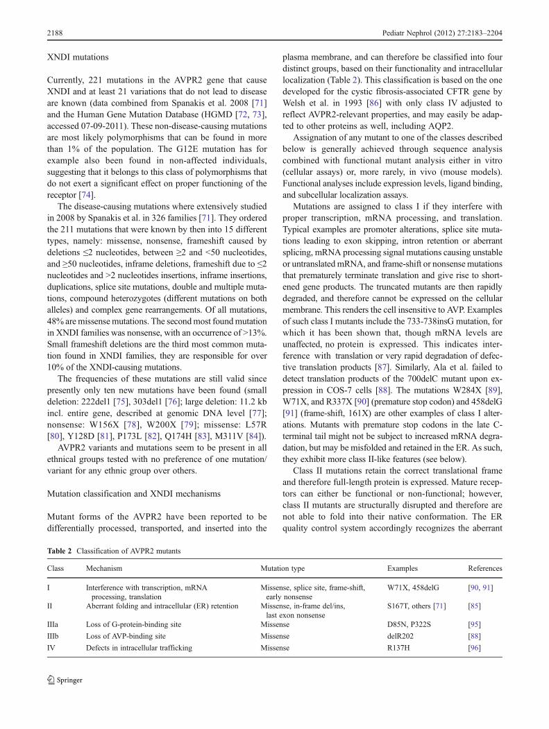
Mutant forms of the AVPR2 have been reported to bedifferentially processed, transported, and inserted into the
plasma membrane, and can therefore be classified into fourdistinct groups, based on their functionality and intracellularlocalization (Table 2). This classification is based on the onedeveloped for the cystic fibrosis-associated CFTR gene byWelsh et al. in 1993 [86] with only class IV adjusted toreflect AVPR2-relevant properties, and may easily be adap-ted to other proteins as well, including AQP2.
Assignation of any mutant to one of the classes describedbelow is generally achieved through sequence analysiscombined with functional mutant analysis either in vitro(cellular assays) or, more rarely, in vivo (mouse models).Functional analyses include expression levels, ligand binding,and subcellular localization assays.
Mutations are assigned to class I if they interfere withproper transcription, mRNA processing, and translation.Typical examples are promoter alterations, splice site muta-tions leading to exon skipping, intron retention or aberrantsplicing, mRNA processing signal mutations causing unstableor untranslated mRNA, and frame-shift or nonsensemutationsthat prematurely terminate translation and give rise to short-ened gene products. The truncated mutants are then rapidlydegraded, and therefore cannot be expressed on the cellularmembrane. This renders the cell insensitive to AVP. Examplesof such class I mutants include the 733-738insG mutation, forwhich it has been shown that, though mRNA levels areunaffected, no protein is expressed. This indicates inter-ference with translation or very rapid degradation of defec-tive translation products [87]. Similarly, Ala et al. failed todetect translation products of the 700delC mutant upon ex-pression in COS-7 cells [88]. The mutations W284X [89],W71X, and R337X [90] (premature stop codon) and 458delG[91] (frame-shift, 161X) are other examples of class I alter-ations. Mutants with premature stop codons in the late C-terminal tail might not be subject to increased mRNA degra-dation, but may be misfolded and retained in the ER. As such,they exhibit more class II-like features (see below).
Class II mutations retain the correct translational frameand therefore full-length protein is expressed. Mature recep-tors can either be functional or non-functional; however,class II mutants are structurally disrupted and therefore arenot able to fold into their native conformation. The ERquality control system accordingly recognizes the aberrant
Table 2 Classification of AVPR2 mutants
Class Mechanism Mutation type Examples References
I Interference with transcription, mRNAprocessing, translation
Missense, splice site, frame-shift,early nonsense
W71X, 458delG [90, 91]
II Aberrant folding and intracellular (ER) retention Missense, in-frame del/ins,last exon nonsense
S167T, others [71] [85]
IIIa Loss of G-protein-binding site Missense D85N, P322S [95]
IIIb Loss of AVP-binding site Missense delR202 [88]
IV Defects in intracellular trafficking Missense R137H [96]
2188 Pediatr Nephrol (2012) 27:2183–2204

folding, and translational products are retained in the ERand incompletely processed. Finally, the quality controlmechanism activates the ER-associated degradation(ERAD) pathway leading to proteasomal degradation ofthe mutant receptors. Prolonged interaction with calnexin,a molecular ER chaperone known to be involved in proteinfolding and ER quality control, has been reported for someAVPR2 mutants [92]. This suggests the involvement ofcalnexin in AVPR2 quality control and that dissociationfrom this chaperone is one of the hurdles for AVPR2 to takebefore continuing its transport to the Golgi complex. Asingle example of retention during receptor maturation inpost-ER compartments such as the Golgi apparatus has alsobeen found [93]. Accumulation of the Y205C mutant wasdetected in spite of low expression levels in the ER-Golgiintermediate compartment (ERGIC) by co-staining withERGIC-53, indicating that ERGIC localization was not anartifact of overloading ER quality control. The amount ofretention and degradation varies within this class, sincedifferent mutations affect protein folding to a different ex-tent, sometimes allowing partial transport of at leastpartially active receptors to the plasma membrane. ClassII mutations are typically insertions or deletions of oneor more nucleotide triplets and missense mutations,slightly altering the amino acid sequence, but retainingthe reading frame. Class II mutations form the mostimportant cause of NDI [71].
Class III mutations do not lead to misfolded receptors,but interfere with proper interaction with their naturalligands, thereby causing reduced/abolished signaling [94].
Class III mutations can be subdivided into two minorgroups. IIIa mutations interfere with binding of or signaltransduction to the coupled trimeric G-protein, especiallythe αGS-subunit, and thereby inhibit or reduce activation ofthe G-protein. Mutations in this class also are missensemutations and in-frame deletions/insertions, which general-ly are located in transmembrane or intracellular domains.Examples are the D85N and P322S mutations [95].
If AVP binding is disrupted or reduced by the mutation,the mutant belongs to class IIIb. Similar to class IIIa muta-tions, these substitutions include missense and small inframeinsertions or deletions and result in full-length expressedprotein. Class IIIb variations are found in extracellulardomains and transmembrane regions. Mutated positions arethought to be located within or close to the AVP binding site,such as delR202 [88].
The last type of XNDI causing defects, class IV, isassigned to all mutations that neither interfere with proteinsynthesis and maturation, nor with ligand binding, but affectother aspects of protein function. We here assign class IV tomutants that differ from wild-type protein in their intracellularlocalization due to altering signal sequences vital for correctintracellular trafficking. The best characterized example is the
R137Hmissense point mutation, which is located in the DRY/H motive, highly conserved in GPCRs. The effect of thismutation is constitutive endocytotic internalization of AVPR2[96], leading to reduced expression of the receptor in theplasma membrane and thereby reduced adenylate cyclase-dependent cAMP signaling upon AVP stimulation.
Some mutants exhibit properties of two or more classesof mutations, indicating that single mutations can affectmultiple features of the receptor. For example, a mutant thatis partially retained in the ER may still be partially trans-ported to the plasma membrane, where it then shows areduced affinity for either AVP or its coupled G-protein,thus having characteristics of class II and IIIa or IIIb,respectively.
Autosomal NDI forms
Autosomal-recessive NDI (ARNDI)
Approximately 10% of NDI patients have the autosomal-recessive form of NDI. Similar to AVPR2 inactivating muta-tions, autosomal-recessive alterations in AQP2 disrupt propersynthesis, functioning, or localization of the gene product,rendering renal collecting duct principal cells irresponsive toAVP stimulation [97].
Aquaporin 2 (AQP2)
The AQP2 gene involved in autosomal NDI is located atchromosome 12q13. It is part of an aquaporin water channelgene cluster, where it maps together with AQP0, AQP5, andAQP6. The gene codes for the 271 amino acid AQP2protein, which consists of six transmembrane domainsconnected by five loops and intracellularly located N- andC- termini (type IV-A TM protein) [98]. Tetramerizationtakes place during and/or after folding of monomers in theER. Subsequently, high-mannose glycans are attached toAsn123 of one or two monomers of a homotetramer [99]and ER export is initiated. During maturation in the Golginetwork, the sugar moieties of the high-mannose glycosylatedmonomers of each AQP2 tetramer are further processed tocomplex glycans [100]. Uponmutation of theN-glycosylationacceptor site (AQP2-N123Q) plasmamembrane expression ofAQP2 was found in X. laevis oocytes and accumulation in theGolgi complex of mammalian cells was shown [26]. Theseresults suggest that glycosylation of AQP2 is not required forpassing ER quality control and ER export, but is essential forpost-Golgi trafficking in mammalian cells.
AQP2 contains a consensus site for PKA phosphorylationat Ser256 [101] and phosphorylation sites controlled byunknown kinases at Ser261, Ser264, and Ser269 [102], alllocated in the cytoplasmic C-terminal tail. Upon phosphory-lation of Ser256, phosphorylation of Ser269 is triggered [103,
Pediatr Nephrol (2012) 27:2183–2204 2189

104] and AQP2 tetramers are translocated to and inserted inthe apical membrane, where they render the membrane waterpermeable. However, recent mass spectrometry data from Xieet al. [105] suggest otherwise: Ser269 was found to be the keyregulator in shifting the equilibrium between apical membranetargeting and re-internalization. High basal Ser256 phos-phorylation levels were found even in the absence ofAVP stimulation and suggested to be required for AVP-dependent induction of Ser269 phosphorylation, which inturn downregulates AQP2 internalization. In addition toregulation of Ser256 and Ser269, Ser261 is dephosphorylated[106] and phosphorylation levels at Ser264 increase [103] uponAVP treatment. However, these last two events seem not to benecessary for translocation of AQP2 to the apical membrane.A review discussing the various, occasionally contradictory,lines of evidence currently present concerning phosphoryla-tion (as well as other post-translational modifications) ofAQP2 has recently been published [107].
The mechanism of selectivity for water of aquaporins ingeneral has been resolved in the homologous AQP1 protein[108] and has further been strengthened by moleculardynamics modeling approaches [109, 110]. The water pore isformed between the first and sixth transmembrane domainsand is lined by the intracellular B-loop and the extracellular E-loop. The residues F56, R195, H186, and C189 located in the ar/R(aromatic/arginine-rich) region determine the water specificityof AQP1 [111, 112], while F24, N76, and N192 are the residuesof the central asparagine/proline/alanine (NPA) region thatensure proton exclusion [113]. In the case of AQP2,the crucial residues correspond to F48, R187, H172, and C181
(ar/R region) and F23, N75, and N184 (NPA region).
ARNDI mutations and mechanisms
There are 40 mutations known to give rise to ARNDI. Theseinclude 32 missense and two nonsense mutations, two 1-bpdeletions, one 2-bp deletion, and three splice site mutations[HGMD [72, 73], accessed 07-09-2011]. In ARNDI, mostalterations are found between the first and the last transmem-brane domain of AQP2. Whereas frameshift and early stopco-don variants lead to shortened translation products that arerapidly degraded, the large group of missense mutations pre-dominantly causes proteins to be folded aberrantly. As forAVPR2, for those mutants this results in ER retention due toextended interaction times with ER chaperones and eventuallyproteasomal degradation. However, some mutants retainintrinsic functionality, and would be able to show at leastpartial activity when expressed in the apical membrane bymeans of forced transport or overexpression [99].
The elucidation of the molecular mechanisms governingwater homeostasis defects in autosomal-recessive NDIpatients has mainly been achieved by extensive research incellular expression systems such as X. laevis oocytes for
water permeability assays [114] and plasma membraneexpression levels, and polarized MDCK cells for sub-cellular localization studies. More recently, mouse modelshave contributed to the confirmation of results obtainedearlier in vitro and their comparability to in vivo situations[115, 116].
Investigation of subcellular localization has revealed ERaccumulation of multiple ARNDI mutants by different tech-niques. Colocalization studies with ER-resident proteins suchas the chaperone Grp94 have been used [117]. Immunocyto-chemistry analysis has revealed dispersed cytosolic staining inunpolarized oocytes and mice kidney cells [99, 114], and ERlocalization has also been demonstrated in polarized MDCKcell lines [116]. Thus, all these findings are consistent with theER retention theory.
For most missense mutants, partial single-channel waterpermeability has been shown in X. laevis oocytes by astandard cell swelling assay [9, 118], indicating that thenative conformation is disturbed only slightly. This suggeststhat the observed disease phenotype is generally due toaberrant subcellular localization of AQP2 rather than lossof function. As we will discuss later, this opens therapeuticoptions directed to restoring mutant trafficking.
Glycosylation of AQP2 is usually assessed by immunoblot-ting, and has revealed the occurrence of four different AQP2forms: unglycosylated (29 kDa), ER-retained high-mannose(32 kDa), ER-exported complex glycosylated (40–45 kDa)and degradation fragments (27 kDa) [99].
Doubt has been cast by Marr et al. [99] on the earlierfinding that some AQP2 mutants in NDI were correctlytransported, but exhibited disrupted functionality [119].While formerly it was found that the AQP2 mutantsT125M and G175R were transported to the plasma membranenormally, but still showed no water permeability increase aftercRNA injection into X. laevis oocytes, this could not be con-firmed by Marr et al. They proposed as a possible explanationfor residual plasma membrane expression the saturating effectof high expression levels of aberrantly folded AQP2 on mutantprotein degradation. Therefore, the main mechanism by whichAQP2 mutants cause ARNDI seems to be ER retardation andthey can thus generally be classified as class II mutants. How-ever, it cannot be excluded that reduced permeability of AQP2mutants at least contributes to NDI phenotypes. Definitiveconfirmation of water channel functionality and transportrequires studies in vivo or at least in mammalian cell lineswithout overexpression of mutant protein [120].
Whatever the case may be regarding the causativeness ofreduced water channel function in NDI, there is no doubtthat different mutants retain different levels of functionalitywhen expressed in the plasma membrane [99, 114]. Thecharacterization of this feature for each mutant is ofhigh therapeutic importance, since it determines whethermolecular chaperones (discussed later) are a valid option.
2190 Pediatr Nephrol (2012) 27:2183–2204

The generation of an in vivo model to study the effect ofthe ARNDI mutations has long been hampered by the factthat mice in contrast to humans seem not to be able tosurvive if AQP2 functionality is completely lost. Miceharboring homozygous T126M mutations died in earlyinfancy [121]. In contrast, the more recent F204Vmouse model [116] was able to survive to adulthood.This has been accounted to a residual transport ofF204V mutant AQP2 homotetramers to the apical mem-brane in collecting duct principal cells, indicating thatspecific mutants may retain some tetramerization andtransporting capability even if excessively ER-retained.A possible explanation is provided by the fact that F204Vresidually matured (complex glycosylated in contrast to high-mannose only) [116], which is required for plasma membraneexpression, but is not essential for tetramerization [26]. It stillneeds to be determined whether residual ER export, glycosyl-ation, tetramerization, and plasma membrane expression alsotake place for AQP2 mutants in NDI patients.
In the F204V mouse model, all common symptomsobserved in humans as well as cellular behavior ofexpression systems (unresponsiveness to dDAVP, ERretention) could be reproduced, indicating its generalsimilarity to a human situation.
In heterozygous mice, Lloyd et al. additionally showedthat AQP2-F204V can homotetramerize as well as formheterotetramers with wt-AQP2 [116]. This is in contrast toprevious findings on the R187C mutant, which could doneither [26, 99].
A single exception to the general mechanism of ER reten-tion of AQP2 mutants in recessive NDI has been found withthe AQP2-P262Lmutant. Due to mechanistic overlap, we willdiscuss the molecular basis of ARNDI caused by this mutantbelow at the end of the section on autosomal-dominant NDI.
Summary autosomal-recessive NDI
Cell expression systems and mouse models have led toseveral theories about pathogenesis in ARNDI, some ofthem generally accepted, others extensively discussed. Theunresponsiveness of kidney collecting duct principal cell toincreased vasopressin levels is caused by the disruption ofAQP2 accumulation in the apical membrane upon AVPstimulation and therefore remaining water impermeability.Except for AQP2-P262L, this is a consequence of the AQP2depletion of endosomal storage vesicles due to failed ERexport. It is commonly accepted that ER retention andsubsequent degradation of AQP2 mutants is the major causa-tive event in ARNDI caused by missense AQP2 proteins [95,117]. While folding and maturation of AQP2 normally isfacilitated by the interaction with ER chaperones, in mutants,most probably, the native conformation is destabilizeddue to the mutation. Followed by aberrant folding, this
induces prolonged interaction with ER quality controlchaperones and eventually targeting for proteasomaldegradation [99, 122].
The role of glycosylation in AQP2 maturation is muchless clear. While non-glycosylated AQP2 appears to be fullyfunctional, compelling evidence has been found for theessentiality of complex glycosylation for translocation tothe apical membrane: AQP2 mutants missing the N123
glycosylation site are trapped in the Golgi network anddo not reach the apical membrane [26]. The underlyingmechanism has not been elucidated yet.
Mouse models now provide tools to study possible thera-peutic options and their safety in a more appropriate settingthan cellular expression systems. However, it has been shownthat mice in part react differently to AQP2 dysfunctionalitythan man does: Homozygous mutant mice are not viable forthe mutants tested to the day, except for F204V [116, 121]. Incontrast, NDI usually is not fatal in infants. Early lethality inmice could be a mere reflection of their high natural urineosmolarity (4,000 mOsm compared to 1,200 mOsm inhumans), the high body surface/volume ratio, thin skin andhigh respiration rate, requiring a high fluid intake/reabsorptionper kilogram body weight needed for pup survival, whilesurvival of human newborns is a consequence of the extensivecare for weak offspring in humans compared to mice. Still, amechanistic difference cannot be excluded. Consequently,conclusions have to be looked upon with caution and criticalevaluation is required.
Autosomal-dominant NDI (ADNDI)
Autosomal-dominant inheritance is the least prominent formof NDI and is responsible for <1% of NDI cases. It alsoinvolves mutations in the AQP2 gene. In contrast toautosomal-recessive NDI AQP2, dominant mutants showperfect functionality, but are not correctly transported tothe apical membrane, resulting in a similar impairment ofurine concentrating ability.
Yet, ADNDI patients show generally higher urineosmolalities and a residual ability to increase the osmolalityof their urine slightly upon dehydration [25]. Thus, waterreabsorption capacity is decreased significantly, but not com-pletely disrupted. This usually leads to a milder phenotypethan that of recessive or X-linked NDI.
ADNDI mutations and mechanisms
Currently, eight mutations causing dominant NDI inheri-tance are known [123–128], each of which was found withina single family. These are three missense mutations, onesingle-nucleotide insertion, and four small deletions (≤10nucleotides). All mutations associated with ADNDI to theday are located in the part of exon 4 of the AQP2 gene that
Pediatr Nephrol (2012) 27:2183–2204 2191

encodes the cytoplasmic C-terminus of the AQP2 protein.This component of AQP2 is not part of the functional waterpore, but contains important sorting signals that governintracellular transport of the protein [95, 125]. AQP2tetramers containing one or more mutant forms areunable to respond properly to signals that normally inducetransport to the apical membrane. Therefore, all autosomal-dominant mutants can be assigned to class IV.
The dominant phenotype of these mutations is due totheir localization in the C-terminal tail of AQP2. Variationsin this part of the protein do not seem to influence correctprotein folding and normal maturation can occur. After ERexport, both homo- and heterotetramers are formed, buttetramers containing one or more mutant AQP2 monomersshow aberrant intracellular localization or insensitivity toAVP signaling-induced re-localization to the apicalmembrane. However, residual trafficking to the apicalmembrane can be detected. This is supposedly due tothe fact that one-sixteenth of all tetramers formed indominant NDI are wt-AQP2-only tetramers [129, 130].
In 2009, Moon et al. [131] reported another heterozygousmutation located in the 6th transmembrane domain of AQP2(S216F), possibly being a novel ADNDI mutation. However,several facts cast doubt on the involvement of S216F in thedominant form of NDI. Firstly, mutation of S216 has beenfound earlier to lead to ARNDI (S216P, [132]). Moreover,ADNDI mutations are usually located in the C-terminaldomain of AQP2 while ARNDI mutations tend to be intransmembrane domains. Lastly, the patient’s parents wereunaffected and could not be genotyped and thus absenceof this mutation could not be proven. Therefore, wepropose functional analyses of the S216F mutant inorder to establish its mechanism and type of inheritancewith sufficient certainty.
The loss of appropriate AQP2 heterotetramer traffickingas a downstream effect of AVP signaling is caused byseveral mechanisms.
As described above, transport of endosomally storedAQP2 tetramers requires phosphorylation of a mean of threeout of the four monomers at S256, which is part of the PKAphosphorylation consensus sequence R253-R254-X255-S256.The first mechanism by which dominant-type mutationscause irresponsiveness of AQP2 tetramers to AVP signalinginvolves disruption of this phosphorylation site. Throughthe G761 (cDNA) to A or T transitions, two of the knownADNDI mutations, the arginine residue at position 254 ischanged into leucine or glutamine [123, 128].
Experimental evidence that these mutations indeedimpair phosphorylation of AQP2 and thereby induce theNDI phenotype, has been brought about by De Mattia et al.[123] for R254L and Savelkoul et al. [128] for R254Q. In X.laevis oocytes and polarized MDCK cells, both mutantsexhibited full functionality of AQP2 tetramers, but decreased
plasma membrane expression and retention in earlyendosomes, even after treatment with the potent PKAstimulating reagent forskolin [123] . The artificial S256Amutant, in which the serine to be phosphorylated was replacedby alanine, showed comparable results, indicating that indeedimpaired phosphorylation of S256 accounts for disruptedtransport to the plasma membrane. Furthermore, co-precipitation of wt- and mutant AQP2 was shown, suggestingthe correctness of the dominant NDI model in which AQP2mutants capture wild-type water channels by heterotetrameri-zation [126, 133]. The importance of phosphorylation atposition 256 as a signal for apical membrane transport hasfurther been strengthened by the fact that phosphorylation ofnearby serines (S261, S264, and S269) could not induce AQP2translocation [101, 134], whereas the artificial AQP2-R254Q-S256D mutant, which is constitutively phosphorylated,showed increased AQP2 levels in the plasma membrane,presumably due to increased AQP2 translocation [103, 123,135, 136]. Conversely, S269 phosphorylation has been impli-cated with AQP2 accumulation in the plasma membrane dueto decreased endocytosis [103]. This example nicely visualizeshow the combination of enduring research with variousexperimental methods and in vitro expression systemshelped to identify complex biochemical matters leading to NDI.
Another mechanism identified is the misrouting of phos-phorylated AQP2 tetramers by the introduction of two baso-lateral sorting signals. An adenosine insertion (c779-780insA) leads to a -1-frameshift that changes the last 12amino acids of the AQP2 C-terminal tail and additionallyshifts the stop codon downstream, thereby lengthening theC-tail by 14 amino acids. Moreover, it introduces two inde-pendent basolateral sorting signals, namely a leucine-basedsequence surrounding the new L261 and an YXXØ-motif atY269QGL (X indicating random and Ø large hydrophobicresidues). Accordingly, tetramers containing this mutanthave been shown to translocate to the basolateral insteadof apical membrane upon phosphorylation [124].
The E258K mutant has been shown to be unresponsive toAVP stimulation because the disruption of the oppositecharges of E258 and R252–254. The repulsion between theresulting positive charges was suggested to disrupt a structuralelement maintained by the naturally opposite charges thatseems to be required for translocation upon phosphorylation.Instead, tetramers containing mutant AQP2 were found to beretained in the Golgi apparatus in X. laevis oocytes [127] andenriched in MVBs in MDCK cells [137, 138].
The mechanism by which the remaining four AQP2mutants found in ADNDI, AQP2-812-818del, AQP2-721delG, AQP2-727delG, and AQP2-763-772del, lead tomisrouting has not yet been fully uncovered. All induce a +1frameshift that shifts the stop codon downstream leading tolengthened AQP2 (34–35 kDa compared to 29 kDa wild-type) with 61 similar C-terminal residues [125]. While the
2192 Pediatr Nephrol (2012) 27:2183–2204

(intra-)cellular localization of 727delG was independent offorskolin treatment [126], the 763-772del mutant has beenfound to translocate to the basolateral membrane uponstimulation with forskolin [133], despite the disappearance ofthe S256 phosphorylation site. The reason for basolateralmisrouting remains to be established, since no experi-mental proof for the suggestions that the elongation ofthe C-terminal tail might introduce new basolateral sort-ing signals or function as such itself [125] has yet beenprovided. The dependence of this sorting on treatmentwith forskolin for 763-772del suggests the involvementof other phosphorylation sites, either upstream of theinsertions or in the lengthened C-tail itself. It remains unclear,why 727delG showed no change in subcellular localizationupon addition of forskolin and how this relates to the observedchanges for the other three deletion mutants.
The subcellular localizations of the two remainingmutants, 727delG and 812-818del, and the effect of stimu-lation of phosphorylation with forskolin on their traffickinghave not yet been investigated. Especially the latter mutantwould be of major interest, since it contains, besides theadded 61 amino acids of the other deletion mutants, theentire wt C-terminal tail as well. Therefore, the naturalS256 PKA phosphorylation site is still present.
Only for c727delG, co-localization with the late endosomal/lysosomal marker Lamp1 has been shown [126]. The absenceof the other mutants in these compartments remains to beconfirmed by co-localization studies with Lamp1 and the earlyendosomal marker EEA1 [133, 139].
ADNDI mouse models
The clinical symptoms caused by the c763-772del mutanthave further been investigated in the first in vivo model, aknock-in mouse harboring this mutation [140]. Typical (AD)NDI symptoms such as polyuria and low-urine osmolality butalso the milder phenotype than that of a recessive NDI mousemodel, could be reproduced, indicating a general applicabilityof this model to research questions in humans. The smallincrease of urine osmolality upon dDAVP administration orfluid deprivation found in human patients was also observedin the mutant mice. Moreover, the presence of residual wt-AQP2 homotetramers in apical membranes and mutant/wtheterotetramers in basolateral membranes of kidney collectingduct cells, as suggested by earlier in vitro-studies, could beconfirmed in vivo.
Summary of autosomal-dominant NDI
Autosomal-dominant NDI is caused by sorting signal-basedretention or basolateral misrouting of mutant-containingAQP2 tetramers. All mutations hitherto known are locatedin the C-terminal tail, indicating the importance of this part
of AQP2 for cellular trafficking [99, 101, 130, 141], whichhas also been shown in other proteins of the AQP family[142]. In wt tetramers, the phosphorylation site at S256serves as an inducible apical sorting signal. It may beinactivated, overruled by basolateral sorting signals, or reprog-rammed to induce basolateral sorting, all causing intracellularmisrouting. Formation of heterotetramers with wt-AQP2 hasbeen shown for all eight mutants known [123–127], providingan explanation for their dominant behavior. Residual 4wt-homotetramers are explanatory of the relatively milder pheno-type when compared to recessive forms of NDI. Additionally,mutant-containing tetramers retain water channel functionalityand thus, if indeed, phosphorylation of three out of the fourmonomers within one tetramer is sufficient for apical shuttling[28], 3 wt/1 mut-heterotetramers also contribute to therelatively milder phenotype.
Mainly studies in X. laevis oocytes and polar MDCK cells,but more recently, also several heterozygous mouse models[143–145] have contributed to the still deepening knowledgeabout the cellular mechanisms involved in ADNDI.
Penetrance of sorting signals in autosomal-dominantand recessive NDI
Interestingly, though located in the AQP2 C-terminal domain,the P262L mutation has been found to cause recessive NDI.The leucine-based basolateral sorting signal introduced byAQP2-P262L seems not to exert any effect following hetero-tetramerizing with wt AQP2, suggesting that it is overruled bythe stronger natural apical sorting signal of wt-AQP2. Onlyupon introduction of another recessive mutation on the otherallele (R187C, A190T), AQP2-P262L fails to form heterote-tramers due to the ER retention of R187C and A190T. Itshomotetramers are mainly retained in intracellular vesicles,while a small fraction is redirected to the basolateral mem-brane upon forskolin-induced phosphorylation [146].
Remarkably, the observations made in vitro regarding thesubcellular localization and inheritance pattern of P262L arecomparable to the results of an accidentally discoveredAQP2-S256L NDI mouse model. This mutant lacks theS256 PKA phosphorylation site, but, in contrast to the humanR254L mutant, which also shows loss of phosphorylation,causes recessive instead of dominant NDI [143]. Altogether,these findings indicate that the destination of AQP2tetramers is determined by the relative strengths of allapical and basolateral sorting signals present [147].
Genetically unresolved NDI cases
For approximately 5% of all NDI patients, it has not yetbeen possible to identify the causative mutations. To theday, it has not been investigated if these mutations mightbe located in promoter regions of the associated genes.
Pediatr Nephrol (2012) 27:2183–2204 2193

Alterations in these spots could result in lower mRNAexpression levels and therefore are viable candidates forclass I mutations in both recessive NDI types. Possibly,searching in AVPR2 and AQP2 promoter regions might behelpful to reveal the causative mutations for the smallpercentage of yet genetically undefined NDI cases.
Development of therapeutic approaches
Conventional therapy
Conventional therapy for NDI consists of replacement ofurinary water losses by adequate fluid supply, in combina-tion with a limited electrolyte diet (low sodium intake) todecrease obligatory water excretion. This is combined withthe administration of the current first-line drug combinationconsisting of hydrochlorothiazide (HTZ, 2–4 mg/kg/24 h) andamiloride (0.3 mg/kg/24 h) [148, 149]. This treatment replacedthe earlier used combination of HTZ and indomethacin, since ithas been reported to have the same efficiency, but without thestrong gastrointestinal and hematopoietic side-effects the lattermedication induces [150]. In young children below the age of 4to 6 years, however, amiloride is less well tolerated because ofpersistent nausea. Therefore we advise the temporary use ofthe combination of indomethacin (2 mg/kg/24 h)-HTZ(2–4 mg/kg/24 h) in these young children. In patientswho cannot tolerate indomethacin, selective inhibitors ofcyclooxygenase-2 (COX-2) might be helpful. Caution inusing indomethacin and selective COX-2 inhibitors inNDI is warranted, as their administration can potentiallylead to the acute deterioration of renal function in dehydratedpatients. Single-drug therapies show lower efficacy and aretherefore not preferred.
The mechanism of action by which HTZ reduces NDIside-effects is somewhat counterintuitive. The drug is com-monly known as a diuretic (increased urine excretion). Thisis achieved by the inhibition of sodium reabsorption in thedistal tubule upon blocking the NaCl co-transporter (NCC).As water follows sodium, this leads to hypovolemia, resultingin a further activation of the renin-angiotensin II- aldosteronesystem. Increased angiotensin II-mediated sodium reabsorp-tion in the proximal tubules is thought to be the main response.In this segment, sodium reabsorption is obligatorily coupled toincreased water reabsorption via AQP1 [151]. Thereby, lesspro-urine reaches the distal tubule and water homeostasis isless dependent on water reabsorption in the collecting duct.
Additionally, HTZ is believed to induceAQP2 upregulation,which might lead to increased water reabsorption even withoutAVP-dependent stimulation [152].
Amiloride acts comparable to HTZ by inhibiting epithelialsodium channels (ENaC), again resulting in lowered sodiumreabsorption and increased hypovolemia [153]. Although
these drugs reduce urine excretion in NDI patients, they areunable to achieve urine volumes produced in healthy individ-uals. Therefore, the general problem remains, although thesymptoms are relieved.
Consequently, current research focuses on methods totreat NDI on a more causative level than solely trying tofight the symptoms. We next discuss the more recentdevelopments in this field based on the hereditary typefor which the approach is suited. In Fig. 2, an overviewof all major mechanisms employed is depicted.
X-linked NDI-directed therapies
Based on the findings of molecular cell biological researchregarding the underlying principles of X-linked NDI, severalnew approaches for treatment have evolved, all morebased on a longer-lasting symptom reduction due totheir more fundamental approach.
Generally, the principles currently investigated can bedivided into three major groups: The first line tries to promotetransport of misfolded, ER retained AVPR2s to the plasmamembrane by assisting with protein folding [154]. Oncerescued, receptors can then fulfill their natural function,since their missense mutations often do not lead to completeloss of function, but rather cause affinities for ligand and/orG-protein to be lowered. The second approach is to directlystimulate the retained receptors intracellularly, while they arestill located in the ER, thereby circumventing the necessity forproper folding [155]. A third approach for NDI treatment hasrecently been provided by promising results that indicated thepossibility to completely circumvent AVPR2 in stimulatingAQP2 membrane translocation [156].
AVPR2 rescue
The extensive retention of misfolded AVPR2 in the ER ledto the suggestion to develop pharmacological chaperonesthat can be administered to the patient and subsequentlyassist with proper folding of the mutant receptors. Thiscould then lead to shielding of the mutants from ERADand recognition by the ER transport system, eventuallyre-establishing expression of (at least partially) activeAVPR2 on the plasma membrane.
The physical principle of small pharmaceutical chaperoneslies in the fact that aggregation of misfolded proteins may leadto a thermodynamically more stable state than folding into thedimeric native conformation. Yet, the energetic barrier, sepa-rating the native state from themisfolded state that can undergooligomerization, usually is too high to be of any biologicalimportance. This is why under normal conditions the kineti-cally favorable product (e.g., the native conformation) ispreferentially formed. However, in an unnatural situation, suchas a mutation that destabilizes the native conformation, as it is
2194 Pediatr Nephrol (2012) 27:2183–2204
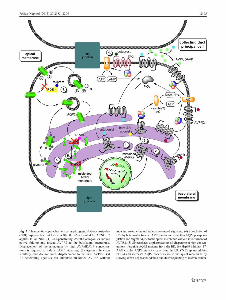
Fig. 2 Therapeutic approaches to treat nephrogenic diabetes insipidus(NDI). Approaches 1–4 focus on XNDI, 5–6 are suited for ARNDI, 7applies to ADNDI. (1) Cell-penetrating AVPR2 antagonists inducenative folding and rescue AVPR2 to the basolateral membrane.Displacement of the antagonist by high AVP/dDAVP concentra-tions is required to induce cAMP signaling. (2) Agonists functionsimilarly, but do not need displacement to activate AVPR2. (3)ER-penetrating agonists can stimulate misfolded AVPR2 without
inducing maturation and induce prolonged signaling. (4) Stimulation ofEP2 by butaprost activates cAMP production as well as AQP2 phosphor-ylation and targets AQP2 to the apical membrane without involvement ofAVPR2. (5) Glycerol acts as pharmacological chaperone in high concen-trations, rescuing AQP2 mutants from the ER. (6) Hsp90-inhibitor 17-AAG enables AQP2 mutant escape from the ER. (7) Rolipram inhibitsPDE-4 and increases AQP2 concentration in the apical membrane byslowing down dephosphorylation and downregulating re-internalization
Pediatr Nephrol (2012) 27:2183–2204 2195

seen in most missense mutations of AVPR2, this equilibriummight change, thereby promoting misfolding and aggregation.
Small-molecule ligands that bind to the native state canin turn either stabilize the native conformation or increasethe energy barrier separating the native state from theaggregation-prone misfolded conformation, enhancingproper folding and promoting transport to the natural cellularlocation by circumventing ER quality control-associatedretention [157].
Proof of principle has already been provided as early asin 1986 [158], but its applicability to X-linked NDI has notbeen tested until the year 2000 by Morello et al. [159]. Theyshowed that, in cell cultures, the cell permeable, non-peptidicAVPR2 antagonists SR121463A and VPA-985 could rescuereceptors by promoting proper folding, increasing cellmembrane receptor load 15-fold and restoring AVP respon-siveness of cells. These drugs are lipophilic small-moleculeAVPR2 antagonists and were therefore anticipated to be cell-permeable and, as they were developed against the normalAVPR2 structure, able to specifically bind the "native" con-formation of the mutants. Since then, in a variety of studies alarge number of AVPR2 mutants have shown the ability ofbeing rescued by molecular chaperones and generally being(at least partially) functional [160–162]. However, two majorproblems prevent progress in this field of research.
First, the ability of the AVPR2 to be stimulated by AVPafter translocation to the plasma membrane has to beensured, which requires dislocation of the pharmacologicalchaperone by AVP after rescuing. Therefore, low-affinityantagonists are believed to have the highest clinical value[163]. However, their efficiency in rescuing is lower than thatof high-affinity ligands and the high concentrations requiredto be administered for sufficient activity by low-affinityantagonists might lead to severe complications in patients.Moreover, Robben et al. have shown that upon pre-treatmentof AVPR2 mutant cells with clinically feasible bloodconcentrations of antagonists, subsequent treatment withdDAVP only led to an increased cAMP response incells pre-treated with high-affinity AVPR2 antagonist[162]. These data indicated that at these antagonistconcentrations, rescue of cell surface expression of theAVPR2 mutants becomes limiting over dislocation ofthe antagonists. These issues have to be addressed inmouse models or clinical studies to be resolved beforeusage of low-affinity ligands as drugs becomes viable.
Secondly, stimulation of AVPR2 on the plasma membraneby AVP leads to increased internalization and rapid degrada-tion of the receptors by the beta-arrestin-MAPK-pathway [65,164], thereby continuously lowering the AVPR2 concentra-tion in the AVP-accessible membrane [155]. This requireshigh-frequency administration of pharmaceuticals to patients,probably also causing unwanted side-effects, again limitingthe clinical value of these compounds. A small-scale clinical
trial conducted in 2006 by Bernier et al. showed that theAVPR1 antagonist SR49059 could induce an increase in urineosmolality in all five patients [161], indicating a generalagreement of in vitro results with a human setting. However,a rapid decline of the drug effect also was observed, probablyassociated with the AVP-induced receptor internalization. Theclinical development of SR49059 was discontinued due tosafety issues as the drug seemed to interfere with thecytochrome P450 metabolic pathway, showing the validity ofthe two major concerns mentioned above. Maybe because ofthe higher potential of other approaches to follow, we are notaware of further clinical studies in which cell-permeableAVPR2 antagonists are used to treat NDI patients.
The use of non-peptide agonists has somewhat circum-vented the first problem, as it has been shown that the com-pounds MCF14, MCF18, and MCF57, all high-affinityagonists of AVPR2, could induce receptor maturation as wellas translocation to the plasma membrane and elicited a cAMPresponse [165]. In contrast to AVP, however, they did notstimulate receptor internalization, but blocked the MAPKpathway responsible for arrestin-induced internalization.
Intracellular receptor activation
Recent studies by Robben et al. have indicated that it ispossible to activate ER-retained, but intrinsically functional,AVPR2 by non-peptide agonists [155]. This activationsurprisingly leads to sufficient cAMP increase to causeAQP2 to be transported to the apical membrane. They usedthe recently for oral administration developed OPC51 and thetwo novel VA88 and VA89 non-peptidic AVPR2 agonists toshow that those are able to stimulate multiple mutants insideof the ER, that then activated their natural signal transductionpathway. In contrast to pharmaceutical chaperone-assistedfolding and rescue of the receptors, the localizationand maturation state of the AVPR2 did not change uponactivation, indicating that these compounds do not actas molecular chaperones.
Additionally, activation of AVPR2 located in the plasmamembrane by AVP leads to rapid degradation of the receptoras a mechanism of adaptation. This limits the efficacy ofreceptor rescue obtained with AVPR2 antagonists or thenon-peptide agonists of the previous paragraph, since afternormal stimulation, a large fraction of the rescued receptors isdegraded and new assisting chaperones have to be adminis-tered. This is not the case for receptor activation within theER; proteasomal degradation of the trapped receptors is notincreased upon intracellular activation by the non-peptideagonists lacking pharmacochaperone function. Anotheradvantage of the use of non-peptidic agonists – as wellas antagonists – for intracellular AVPR2 stimulation isthe high selectivity of non-peptide compounds for the AVPR2.This should prove tominimize the side-effects of administration
2196 Pediatr Nephrol (2012) 27:2183–2204

of those therapeutics, since no other cellular mechanisms areexpected to be activated. Moreover, once a high-affinityantagonist is bound to the receptor in order to restorenative folding, it tends to remain bound. Thereby, itcompetes with the natural agonist AVP, resulting in lessactivation of AVPR2 than predicted from the amountadministered. Therefore, the more strictly dose-dependenteffect of cell-penetrating agonists is expected to offer betterpredictability. Here, the new promising non-peptidic agonistsseem to have a better ability to penetrate cell membranes andshow higher specificity for AVPR2 than the peptide variants invitro. Future in vivo and clinical testing has to confirmwhether this type of drugs has the proposed positiveeffects in patients and meets the safety requirements.
The mode of action by which receptors trapped in the ERcan still activate their coupled G-protein and how this stim-ulates adenylate cyclase is not yet understood. However,most recently, a new model for signaling by internalizedGPCRs has been proposed by Calebiro et al. [166, 167], inwhich GPCRs localized in endosomal membranes can acti-vate their natural pathways (e.g., activation of adenylatecyclase and production of cAMP), if stimulated by lipophil-ic agonists that are able to penetrate the endosomal mem-branes. They also showed earlier that internalized GPCRscan continue signaling from endosomes [168], suggestingthat the classical model, in which internalization solely is adesensitization process, has to be reconsidered. In how farthis model also holds for GPCRs completely trapped in theER/endosomal compartments, remains to be elucidated.Clearly, both AVPR2 rescue and intracellular receptor activa-tion will only work for mutations that affect folding or properintracellular transport and not for truncated receptors, thosethat lost their affinity for AVP or their coupled Gs-protein ormutations that interfere with transcription and translation.Other approaches that can deal with these types of mutantsremain to be found.
Besides the various in vitro expression systems mentioned,a mouse model has recently been constructed, in which theendogenous AVPR2 gene is conditionally knocked out [156].The mice developed extensively increased water intake andurine excretion, and showed dramatically reduced urineosmolality. Additionally, no functional AVPR2 could bedetected in their kidneys. Earlier knockout mice did notsurvive for more than 1 week postnatal, because pupstotally lacking renal water reabsorption are not viable[169]. In contrast, the new mouse model survived nor-mally, since the AVPR2 knockout could be inducedafter reaching adulthood [170]. Therefore, this newmodel might provide a reliable in vivo system for studyingthe effects of drugs on XNDI. We recently developed a mousemodel, in which a mutated AVPR2 can be expressed in aninducible manner. These mice will be used to test the viabilityand safety of rescuing antagonists or intracellularly activating
agonists. As the agonists are still in clinical trials for theircommerciallymore interesting indications, we have towait forthe generosity of the pharmaceutical companies to share theiragonists or for the availability of the drugs on the marketbefore we can test whether these compounds can relieveNDI in patients.
Circumvention of AVPR2
Yet another mechanism that was exploited to alleviateXNDI provides hope for patients with AVPR2 mutants thatcannot be rescued or activated intracellular due to type 1mutations. By stimulation of the E-prostanoid receptor EP4,NDI symptoms were greatly reduced in the conditionalAVPR2-deletion mouse model [156]. This was due to raisedAQP2 levels, most probably a consequence of cAMPproduction caused by EP4 stimulation.
Interestingly, in a very recent study on the effects of EP2and EP4 stimulation on NDI symptoms, Olesen et al. furtherelaborated the underlying mechanisms and showed AVPR2-independent induction of AQP2 phosphorylation and itstranslocation to the apical membrane in MDCK cells[171]. Additionally, they reported relieved NDI symptomsin a rat model following the administration of the EP2agonist butaprost. Due to its resistance to stimulation-induced internalization [172] and its more selective expres-sion [173], the EP2 receptor is the more interesting candi-date for NDI treatment in comparison to the EP4 receptor,also because EP2 agonists have already been tested inclinical studies for other diseases and have shown promisingresults concerning safety issues [174]. However, long-termsafety studies, which are needed when being used in thetreatment of NDI, will need to be performed.
Autosomal-recessive NDI
In general, AQP2 mutants causing autosomal-recessive NDIhave features comparable to X-linked AVPR2 mutants. Forclass I mutants that are strongly truncated due to early stopcodons, early frame shifts, or splice site mutations, itremains extremely difficult to think of therapeutic optionsother than gene therapy. Renal gene therapy has been ham-pered by a low level of accessibility and the absence ofselectivity of gene transfer techniques to and expression inparticular renal cells [95]. Yet, missense mutations are thelargest group of causative alterations in AQP2 for NDI, andgenerally lead to aberrant folding of AQP2 inside the ER.As it is the case for AVPR2, those mutants often retainpartial functionality whenever folding into native conforma-tions is restored. Hence, finding substances that are able to re-establish natural AQP2 folding holds comparable promises forARNDI treatment as it has already been shown for XNDI. Thechallenge is thus to find pharmacological chaperones aimed at
Pediatr Nephrol (2012) 27:2183–2204 2197
rescuing ER-retained AQP2 mutants from ERAD and therebyrestoring the transport of partially functional AQP2 to theplasma membrane. In CHO and MDCK cells, glycerol hasproven the applicability of chemical chaperones to AQP2 byrestoring ER export in high concentrations [117]. Recently,Yang et al. described partial restoration of cellular AQP2processing upon treatment of conditional AQP2-T126Mknock-in mice with an Hsp90 inhibitor, 17-allylamino-demethoxygeldanamycin (17-AAG), eventually resulting inimproved urinary concentrating ability [175]. The authorsproposed several explanations for a possible mechanism.Hsp90 interacts with and promotes ERAD of several aberrant-ly folded proteins, including the mutant cystic fibrosis proteinΔF508-CFTR [176]. By disrupting the interaction of Hsp90with AQP2-T126M, 17-AAG might enable AQP2-T126M toescape from the ER and to be processed further by the Golgicomplex. Another possibility is that 17-AAG directly interactswith heterotetramers containing AQP2-T126M and residualwild-type AQP2 and inhibits their degradation, consequentlyinducing accumulation. There is also some evidence for theinvolvement of Hsp70 in the constitutive recycling pathway ofAQP2 and inhibition of Hsp90 has occasionally been reportedto be associated with induction of Hsp70. Further studies arenecessary to elucidate the mechanism in more detail.
However, inhibition of ER chaperones as common asHsp90 might also have severe side-effects. Hsp90 is anabundant cytosolic chaperone and contributes to variouscellular processes like protein degradation and signal trans-duction. For example, it had been shown that Hsp90 inhibitionwith 17-AAG induced ER stress. This caused increasedexpression of ER chaperones, for example Grp94, andan increase in cytoplasmic calcium levels, eventuallyleading to the disruption of mitochondrial homeostasisand promoting mitochondria-induced apoptosis [177]. Soit is likely that there is a complex mechanistic interplaybetween various factors, that in summary resulted in a partialrestoration of urinary concentration ability in AQP2-T126Mknock-in mice after treatment with 17-AAG. Lengthenedstudies addressing safety issues of Hsp90 or other chaperoneinhibitors have to be conducted in order to elucidate theapplicability of these compounds in NDI therapy.
Autosomal-dominant NDI
For dominant NDI patients, the approaches based onmolecular chaperones that were already suggested forother forms of NDI, are not applicable, since nativefolding occurs. Instead, a search for therapeutics is on-goingthat interfere with misrouting and restore normal apical mem-brane targeting and transport. Since the number of patients isextremely low, and the conventional therapy with diureticscan relieve ADNDI to a certain extent, the amount of researchperformed considering this type of NDI is substantially lower
than for the other subtypes, and in vitro data about possibletherapies for dominant NDI is lacking [25].
The AQP2-Δ763–772 knock-in mouse model generatedby Sohara et al. [140] is the first model for autosomal-dominant NDI, which can be used to test therapeuticapproaches in vivo. After administration of dDAVP to themice, the urine osmolality was increased and administrationof dDAVP was proposed as the therapy of choice. However,high doses of dDAVP and long-term administration arerequired to induce lasting increased AQP2 synthesis, whichcan be beneficial to patients based on the residual wild-typetetramers discussed earlier.
Based on the improvement of AVP-dependent cAMPsignaling of collecting duct cells in a hypercalcemia-inducedNDI mouse model [178], Sohara et al. also tested the phos-phodiesterase 4 inhibitor rolipram in the knock-in dominantNDI mice [140]. Their data indicated that rolipram is able toincrease cAMP levels in the papillae and AQP2 phosphoryla-tion as well as translocation to the apical membrane. If theseresults are reproducible in humans, rolipram might be-come a first therapeutic agent able to improve life quality ofautosomal-dominant NDI patients.
However, as for the Hsp90-inhibition approach, phos-phodiesterase 4 is a common protein that also is involvedin immunosuppressive and anti-inflammatory pathways andits inhibition may have unwanted effects that outweigh theadvantages. Side-effects already discovered in clinical trials,that tested rolipram as antidepressant, were emesis andnausea [179].
Summary and concluding remarks
Besides acquired forms of NDI, three congenital types of thisdisorder exist. X-linked recessive NDI accounts for the vastmajority of patients. They show loss-of-function mutations inthe AVPR2 gene, disrupting the cellular response to AVP. Mostmutations lead to aberrant folding and retention of usuallypartially functional receptors in the ER. For autosomal-recessive NDI, mutations are mainly present in the transmem-brane regions of the AQP2 protein. The major group of themutations consists of missense mutations inducing improperfolding and ER retention, as it is the case in XNDI. Therefore,further research in X-linked as well as autosomal-recessiveNDI focuses on the identification of pharmacologicalchaperones to restore routing of intrinsically (partially)functional proteins to the plasma membrane. In X-linkedNDI, another line of research utilizes membrane-permeableagonists to intracellularly stimulate AVPR2 and thus initiateresponses comparable to normal stimulation by AVP.
For autosomal-dominant NDI, the failure of water transportcan be explained by the importance of the AQP2 C-terminaltail in trafficking of tetrameric protein to the apical membrane.
2198 Pediatr Nephrol (2012) 27:2183–2204

Phosphorylation of S256 in the C-terminus is necessary forAVP-induced accumulation of AQP2 in the apical membrane.In addition to the disruption of this phosphorylation site,AQP2 gene mutations can introduce basolateral or removeapical sorting signals of wt-AQP2, also leading to misrouting.Other approaches than pharmacological chaperones have tobe invented in order to treat ADNDI patients.
Insights into the pathogenesis of the disease have for allNDI subtypes led to the development of in vivo mousemodels, which provide opportunities for clinical safetyassessment of possible therapies. Future research intothe promising approaches mentioned above, combinedwith clinical trials, will show which of those strategiescan improve or replace the current standard therapies forpatients suffering from different NDI types.
References
1. van Lieburg AF, Knoers NV, Monnens LA (1999) Clinicalpresentation and follow-up of 30 patients with congenital nephro-genic diabetes insipidus. J Am Soc Nephrol 10:1958–1964
2. Forssman H (1955) Is hereditary diabetes insipidus of nephro-genic type associated with mental deficiency? Acta PsychiatrNeurol Scand 30:577–587
3. Macaulay D, Watson M (1967) Hypernatraemia in infants as acause of brain damage. Arch Dis Child 42:485–491
4. Kanzaki S, Omura T, Miyake M, Enomoto S, Miyata I, IshimitsuH (1985) Intracranial calcification in nephrogenic diabetes insip-idus. JAMA 254:3349–3350
5. Schofer O, Beetz R, Kruse K, Rascher C, Schutz C, Bohl J (1990)Nephrogenic diabetes insipidus and intracerebral calcification.Arch Dis Child 65:885–887
6. Hoekstra JA, van Lieburg AF, Monnens LA, Hulstijn-DirkmaatGM, Knoers VV (1996) Cognitive and psychosocial functioningof patients with congenital nephrogenic diabetes insipidus. Am JMed Genet 61:81–88
7. Shalev H, Romanovsky I, Knoers NV, Lupa S, Landau D (2004)Bladder function impairment in aquaporin-2 defective nephro-genic diabetes insipidus. Nephrol Dial Transplant 19:608–613
8. Makaryus AN, McFarlane SI (2006) Diabetes insipidus: diagnosisand treatment of a complex disease. Cleve Clin J Med 73:65–71
9. Marples D, Christensen S, Christensen EI, Ottosen PD, Nielsen S(1995) Lithium-induced downregulation of aquaporin-2 waterchannel expression in rat kidney medulla. J Clin Invest 95:1838–1845
10. Klein JD, Gunn RB, Roberts BR, Sands JM (2002) Down-regulation of urea transporters in the renal inner medulla oflithium-fed rats. Kidney Int 61:995–1002
11. Timmer RT, Sands JM (1999) Lithium intoxication. J Am SocNephrol 10:666–674
12. Trepiccione F, Christensen BM (2010) Lithium-induced nephro-genic diabetes insipidus: new clinical and experimental findings.J Nephrol 23(Suppl 16):S43–S48
13. Amlal H, Krane CM, Chen Q, Soleimani M (2000) Early polyuriaand urinary concentrating defect in potassium deprivation. Am JPhysiol Renal Physiol 279:F655–F663
14. Elkjaer M-L, Kwon T-H, Wang W, Nielsen J, Knepper MA,Frøkiaer J, Nielsen S (2002) Altered expression of NHE3, TSC,BSC-1, and ENaC subunits in potassium-depleted rats. Am JPhysiol Renal Physiol 283:F1376–F1388
15. Wang W, Li C, Kwon T-H, Miller RT, Knepper M, Frøkiaer J,Nielsen S (2004) Reduced expression of renal Na+ transporters inrates with PTH-induced hypercalcemia. Am J Physiol RenalPhysiol 286:F535–F545
16. Earm JH, Christensen BM, Frokiaer J, Marples D, Han JS,Knepper MA, Nielsen S (1998) Decreased aquaporin-2 expres-sion and apical plasma membrane delivery in kidney collectingducts of polyuric hypercalcemic rats. J Am Soc Nephrol 9:2181–2193
17. Sands JM, Naruse M, Jacobs JD, Wilcox JN, Klein JD(1996) Changes in aquaporin-2 protein contribute to theurine concentrating defect in rats fed a low-protein diet. J ClinInvest 97:2807–2814
18. Frokiaer J, Li C, Shi Y, Jensen A, Praetorius H, Hansen H, TopcuO, Sardeli C, Wang W, Kwon TH, Nielsen S (2003) Renalaquaporins and sodium transporters with special focus on urinarytract obstruction. APMIS Suppl:71–79
19. Frokiaer J, Marples D, Knepper MA, Nielsen S (1996) Bilateralureteral obstruction downregulates expression of vasopressin-sensitive AQP-2 water channel in rat kidney. Am J Physiol 270:F657–F668
20. Garofeanu CG, Weir M, Rosas-Arellano MP, Henson G, GargAX, Clark WF (2005) Causes of reversible nephrogenic diabetesinsipidus: a systematic review. Am J Kidney Dis 45:626–637
21. Brandis K (2011) Fluid physiology - Section 3.1: Water turnover.URL: http://www.anaesthesiamcq.com/FluidBook/fl3_1.php
22. Trachtman H (2009) Sodium and water. In: Avner ED, HarmonWE, Niaudet P, Yoshikawa N (eds) Pediatr nephrol, 6th edn.Springer, Berlin Heidelberg New York, pp 159–184
23. Sachs H, Takabatake Y (1964) Evidence for a precursor invasopressin biosynthesis. Endocrinol 75:943–948
24. Nossent AY, Robben JH, Deen PM, Vos HL, Rosendaal FR,Doggen CJ, Hansen JL, Sheikh SP, Bertina RM, Eikenboom JC(2010) Functional variation in the arginine vasopressin 2 receptoras a modifier of human plasma von Willebrand factor levels. JThromb Haemost 8:1547–1554
25. Loonen AJ, Knoers NV, van Os CH, Deen PM (2008) Aquaporin2 mutations in nephrogenic diabetes insipidus. Semin Nephrol28:252–265
26. Hendriks G, Koudijs M, van Balkom BW, Oorschot V, KlumpermanJ, Deen PM, van der Sluijs P (2004) Glycosylation is important forcell surface expression of the water channel aquaporin-2 but is notessential for tetramerization in the endoplasmic reticulum. J BiolChem 279:2975–2983
27. Nielsen S, DiGiovanni SR, Christensen EI, Knepper MA, HarrisHW (1993) Cellular and subcellular immunolocalization ofvasopressin-regulated water channel in rat kidney. Proc Natl AcadSci USA 90:11663–11667
28. Kamsteeg EJ, Heijnen I, van Os CH, Deen PM (2000) Thesubcellular localization of an aquaporin-2 tetramer depends onthe stoichiometry of phosphorylated and nonphosphorylatedmonomers. J Cell Biol 151:919–930
29. Mandon B, Chou CL, Nielsen S, Knepper MA (1996) Syntaxin-4is localized to the apical plasma membrane of rat renal collectingduct cells: possible role in aquaporin-2 trafficking. J Clin Invest98:906–913
30. Kanehisa M, Goto S (2000) KEGG: Kyoto encyclopedia of genesand genomes. Nucleic Acids Res 28:27–30
31. Kanehisa M, Goto S, Hattori M, Aoki-Kinoshita KF, Itoh M,Kawashima S, Katayama T, Araki M, Hirakawa M (2006) Fromgenomics to chemical genomics: new developments in KEGG.Nucleic Acids Res 34:D354–D357
32. Kanehisa M, Goto S, Furumichi M, Tanabe M, Hirakawa M(2010) KEGG for representation and analysis of molecularnetworks involving diseases and drugs. Nucleic Acids Res38:D355–D360
Pediatr Nephrol (2012) 27:2183–2204 2199
33. Teng FY, Wang Y, Tang BL (2001) The syntaxins. Genome Biol2:REVIEWS3012
34. Nielsen S, Marples D, Birn H, Mohtashami M, Dalby NO,Trimble M, Knepper M (1995) Expression of VAMP-2-likeprotein in kidney collecting duct intracellular vesicles.Colocalization with Aquaporin-2 water channels. J Clin Invest96:1834–1844
35. Schroer TA (2004) Dynactin. Annu Rev Cell Dev Biol20:759–779
36. Lee YJ, Kwon TH (2009) Ubiquitination of aquaporin-2 in thekidney. Electrolyte Blood Press 7:1–4
37. Vossenkamper A, Nedvetsky PI, Wiesner B, Furkert J, RosenthalW, Klussmann E (2007) Microtubules are needed for theperinuclear positioning of aquaporin-2 after its endocyticretrieval in renal principal cells. Am J Physiol Cell Physiol293:C1129–C1138
38. Marples D, Schroer TA, Ahrens N, Taylor A, Knepper MA,Nielsen S (1998) Dynein and dynactin colocalize with AQP2water channels in intracellular vesicles from kidney collectingduct. Am J Physiol 274:F384–F394
39. Palamidessi A, Frittoli E, Garre M, Faretta M, Mione M, Testa I,Diaspro A, Lanzetti L, Scita G, Di Fiore PP (2008) Endocytictrafficking of Rac is required for the spatial restriction of signalingin cell migration. Cell 134:135–147
40. Stenmark H (2009) Rab GTPases as coordinators of vesicletraffic. Nat Rev Mol Cell Biol 10:513–525
41. Kamsteeg EJ, Hendriks G, Boone M, Konings IB, Oorschot V, vander Sluijs P, Klumperman J, Deen PM (2006) Short-chain ubiquiti-nation mediates the regulated endocytosis of the aquaporin-2 waterchannel. Proc Natl Acad Sci USA 103:18344–18349
42. Klussmann E, Tamma G, Lorenz D, Wiesner B, Maric K,Hofmann F, Aktories K, Valenti G, Rosenthal W (2001) Aninhibitory role of Rho in the vasopressin-mediated translocation ofaquaporin-2 into cell membranes of renal principal cells. J BiolChem 276:20451–20457
43. Tajika Y, Matsuzaki T, Suzuki T, Ablimit A, Aoki T, Hagiwara H,Kuwahara M, Sasaki S, Takata K (2005) Differential regulationof AQP2 trafficking in endosomes by microtubules and actinfilaments. Histochem Cell Biol 124:1–12
44. Yasui M, Zelenin SM, Celsi G, Aperia A (1997) Adenylatecyclase-coupled vasopressin receptor activates AQP2 promotervia a dual effect on CRE and AP1 elements. Am J Physiol 272:F443–F450
45. Nielsen S, Kwon TH, Christensen BM, Promeneur D, Frokiaer J,Marples D (1999) Physiology and pathophysiology of renalaquaporins. J Am Soc Nephrol 10:647–663
46. Blanchard A, Frank M, Wuerzner G, Peyrard S, Bankir L, Jeu-nemaitre X, Azizi M (2011) Antinatriuretic effect of vasopressinin humans is amiloride-sensitive, thus ENaC dependent. Clin JAm Soc Nephrol 6:753–759
47. Stockand JD (2010) Vasopressin regulation of renal sodiumexcretion. Kidney Int 78:849–856
48. Sands JM (2003) Molecular mechanisms of urea transport. JMembr Biol 191:149–163
49. Brown D, Katsura T, Gustafson CE (1998) Cellular mechanismsof aquaporin trafficking. Am J Physiol 275:F328–F331
50. Sands JM, Bichet DG (2006) Nephrogenic diabetes insipidus.Ann Intern Med 144:186–194
51. Birnbaumer M (2001) The V2 vasopressin receptor mutationsand fluid homeostasis. Cardiovasc Res 51:409–415
52. van den Ouweland AM, Dreesen JC, Verdijk M, Knoers NV,Monnens LA, Rocchi M, van Oost BA (1992) Mutations in thevasopressin type 2 receptor gene (AVPR2) associated withnephrogenic diabetes insipidus. Nat Genet 2:99–102
53. Bichet DG (2008) Vasopressin receptor mutations in nephrogenicdiabetes insipidus. Semin Nephrol 28:245–251
54. Nomura Y, Onigata K, Nagashima T, Yutani S, Mochizuki H,Nagashima K, Morikawa A (1997) Detection of skewed X-inactivation in two female carriers of vasopressin type 2 receptorgene mutation. J Clin Endocrinol Metab 82:3434–3437
55. Faerch M, Corydon TJ, Rittig S, Christensen JH, Hertz JM,Jendle J (2010) Skewed X-chromosome inactivation causingdiagnostic misinterpretation in congenital nephrogenic diabetesinsipidus. Scand J Urol Nephrol 44:324–330
56. Firsov D, Mandon B, Morel A, Merot J, Le Maout S, BellangerAC, de Rouffignac C, Elalouf JM, Buhler JM (1994) Molecularanalysis of vasopressin receptors in the rat nephron. Evidence foralternative splicing of the V2 receptor. Pflug Arch 429:79–89
57. Boeckmann B, Bairoch A, Apweiler R, Blatter MC, Estreicher A,Gasteiger E, Martin MJ, Michoud K, O'Donovan C, Phan I,Pilbout S, Schneider M (2003) The SWISS-PROT proteinknowledgebase and its supplement TrEMBL in 2003. NucleicAcids Res 31:365–370
58. Brown CA, Black SD (1989) Membrane topology of mammaliancytochromes P-450 from liver endoplasmic reticulum. Determina-tion by trypsinolysis of phenobarbital-treated microsomes. J BiolChem 264:4442–4449
59. Hartmann E, Rapoport TA, Lodish HF (1989) Predicting theorientation of eukaryotic membrane-spanning proteins. Proc NatlAcad Sci USA 86:5786–5790
60. Conner M, Hawtin SR, Simms J, Wootten D, Lawson Z, ConnerAC, Parslow RA, Wheatley M (2007) Systematic analysis of theentire second extracellular loop of the V(1a) vasopressin receptor:key residues, conserved throughout a G-protein-coupled receptorfamily, identified. J Biol Chem 282:17405–17412
61. Palczewski K, Kumasaka T, Hori T, Behnke CA, Motoshima H,Fox BA, Le Trong I, Teller DC, Okada T, Stenkamp RE,Yamamoto M, Miyano M (2000) Crystal structure of rhodopsin: AG protein-coupled receptor. Science 289:739–745
62. Sangkuhl K, Rompler H, Busch W, Karges B, Schoneberg T(2005) Nephrogenic diabetes insipidus caused by mutation ofTyr205: a key residue of V2 vasopressin receptor function.Hum Mutat 25:505
63. Oksche A, Schulein R, Rutz C, Liebenhoff U, Dickson J, MullerH, Birnbaumer M, Rosenthal W (1996) Vasopressin V2 receptormutants that cause X-linked nephrogenic diabetes insipidus:analysis of expression, processing, and function. Mol Pharmacol50:820–828
64. Sadeghi H, Birnbaumer M (1999) O-Glycosylation of the V2vasopressin receptor. Glycobiology 9:731–737
65. Robben JH, Knoers NV, Deen PM (2004) Regulation of thevasopressin V2 receptor by vasopressin in polarized renal collectingduct cells. Mol Biol Cell 15:5693–5699
66. Sarmiento JM, Anazco CC, Campos DM, Prado GN, Navarro J,Gonzalez CB (2004) Novel down-regulatory mechanism of thesurface expression of the vasopressin V2 receptor by an alternativesplice receptor variant. J Biol Chem 279:47017–47023
67. Gonzalez A, Borquez M, Trigo CA, Brenet M, Sarmiento JM,Figueroa CD, Navarro J, Gonzalez CB (2011) The splice variantof the V2 vasopressin receptor adopts alternative topologies.Biochem 50:4981–4986
68. Bichet DG, Bouvier M, Chini B, Serradeil-Le Gal C, Gimpl G,Guillon G, Kimura T, Knepper MA, Lolait S, Manning M,Mouillac B, Verbalis JG, Wheatley M, Zingg HH (2011)Vasopressin and oxytocin receptors: V2. Last modified on2010-07-01. Accessed on 2011-09-10. IUPHAR database(IUPHAR-DB), http://www.iuphar-db.org/DATABASE/ObjectDisplayForward?objectId0368
69. Thibonnier M, Preston JA, Dulin N, Wilkins PL, Berti-MatteraLN, Mattera R (1997) The human V3 pituitary vasopressinreceptor: ligand binding profile and density-dependent signalingpathways. Endocrinol 138:4109–4122
2200 Pediatr Nephrol (2012) 27:2183–2204

70. Vargas-Poussou R, Forestier L, Dautzenberg MD, Niaudet P,Dechaux M, Antignac C (1997) Mutations in the vasopressinV2 receptor and aquaporin-2 genes in 12 families with congenitalnephrogenic diabetes insipidus. J Am Soc Nephrol 8:1855–1862
71. Spanakis E, Milord E, Gragnoli C (2008) AVPR2 variants andmutations in nephrogenic diabetes insipidus: review and missensemutation significance. J Cell Physiol 217:605–617
72. Krawczak M, Cooper DN (1997) The human gene mutationdatabase. Trends Genet 13:121–122
73. Krawczak M, Ball EV, Cooper DN (1998) Neighboring-nucleotideeffects on the rates of germ-line single-base-pair substitution inhuman genes. Am J Hum Genet 63:474–488
74. Wenkert D, Schoneberg T, Merendino JJ Jr, Rodriguez Pena MS,Vinitsky R, Goldsmith PK, Wess J, Spiegel AM (1996)Functional characterization of five V2 vasopressin receptorgene mutations. Mol Cell Endocrinol 124:43–50
75. Abaci A, Wood K, Demir K, Buyukgebiz A, Bober E, Kopp P(2010) A novel mutation in the AVPR2 gene (222delA) associatedwith X-linked nephrogenic diabetes insipidus in a boy with growthfailure. Endocr Pract 16:231–236
76. Moon SD, Kim JH, Shim JY, Lim DJ, Cha BY, Han JH (2011)Analysis of a novel AVPR2 mutation in a family withnephrogenic diabetes insipidus. Int J Clin Exp Med 4:1–9
77. Fujimoto M, Imai K, Hirata K, Kashiwagi R, Morinishi Y,Kitazawa K, Sasaki S, Arinami T, Nonoyama S, Noguchi E(2008) Immunological profile in a family with nephrogenicdiabetes insipidus with a novel 11 kb deletion in AVPR2 andARHGAP4 genes. BMC Med Genet 9:42
78. Satoh M, Ogikubo S, Yoshizawa-Ogasawara A (2008) Correlationbetween clinical phenotypes and X-inactivation patterns in sixfemale carriers with heterozygote vasopressin type 2 receptor genemutations. Endocr J 55:277–284
79. Sakallioglu O, Tascilar ME, Kalman S, Cheong HI, Atay AA(2009) Nephrogenic diabetes insipidus due to a novel AVPR2mutation. J Pediatr Endocrinol Metab 22:187–189
80. Ranadive SA, Ersoy B, Favre H, Cheung CC, Rosenthal SM,Miller WL, Vaisse C (2009) Identification, characterization andrescue of a novel vasopressin-2 receptor mutation causing nephro-genic diabetes insipidus. Clin Endocrinol (Oxf) 71:388–393
81. Vaisbich MH, Carneiro J, Boson W, Resende B, De ML, HonjoRS, Kim CA, Koch VH (2009) Nephrogenic diabetes insipidus(NDI): clinical, laboratory and genetic characterization of fiveBrazilian patients. Clinics(Sao Paulo) 64:409–414
82. Takatani T, Matsuo K, Kinoshita K, Takatani R, Minagawa M,Kohno Y (2010) A novel missense mutation in the AVPR2 geneof a Japanese infant with nephrogenic diabetes insipidus. JPediatr Endocrinol Metab 23:415–418
83. El-Kares R, Hueber PA, Blumenkrantz M, Iglesias D, Ma K,Jabado N, Bichet DG, Goodyer P (2009) Wilms tumor arisingin a child with X-linked nephrogenic diabetes insipidus. PediatrNephrol 24:1313–1319
84. Sahakitrungruang T, Tee MK, Rattanachartnarong N, ShotelersukV, Suphapeetiporn K,MillerWL (2010) Functional characterizationof vasopressin receptor 2 mutations causing partial and completecongenital nephrogenic diabetes insipidus in Thai families. HormRes Paediatr 73:349–354
85. Oksche A, Dickson J, Schulein R, Seyberth HW, Muller M,Rascher W, Birnbaumer M, Rosenthal W (1994) Two novelmutations in the vasopressin V2 receptor gene in patients withcongenital nephrogenic diabetes insipidus. Biochem Biophys ResComm 205:552–557
86. Welsh MJ, Smith AE (1993) Molecular mechanisms ofCFTR chloride channel dysfunction in cystic fibrosis. Cell73:1251–1254
87. Tsukaguchi H, Matsubara H, Taketani S, Mori Y, Seido T, InadaM (1995) Binding-, intracellular transport-, and biosynthesis-
defective mutants of vasopressin type 2 receptor in patientswith X-linked nephrogenic diabetes insipidus. J Clin Invest 96:2043–2050
88. Ala Y, Morin D, Mouillac B, Sabatier N, Vargas R, Cotte N,Dechaux M, Antignac C, Arthus MF, Lonergan M, Turner MS,Balestre MN, Alonso G, Hibert M, Barberis C, Hendy GN,Bichet DG, Jard S (1998) Functional studies of twelve mutantV2 vasopressin receptors related to nephrogenic diabetesinsipidus: molecular basis of a mild clinical phenotype. JAm Soc Nephrol 9:1861–1872
89. Bichet DG, Birnbaumer M, Lonergan M, Arthus MF, RosenthalW, Goodyer P, Nivet H, Benoit S, Giampietro P, Simonetti S(1994) Nature and recurrence of AVPR2 mutations in X-linkednephrogenic diabetes insipidus. Am J Hum Genet 55:278–286
90. Morello JP, Bichet DG (2001) Nephrogenic diabetes insipidus.Annu Rev Physiol 63:607–630
91. Arthus MF, Lonergan M, Crumley MJ, Naumova AK, Morin D,De Marco LA, Kaplan BS, Robertson GL, Sasaki S, Morgan K,Bichet DG, Fujiwara TM (2000) Report of 33 novel AVPR2mutations and analysis of 117 families with X-linked nephrogenicdiabetes insipidus. J Am Soc Nephrol 11:1044–1054
92. Ellgaard L, Helenius A (2001) ER quality control: towards anunderstanding at the molecular level. Curr Opin Cell Biol 13:431–437
93. Oueslati M, Hermosilla R, Schonenberger E, Oorschot V, BeyermannM, Wiesner B, Schmidt A, Klumperman J, Rosenthal W,Schulein R (2007) Rescue of a nephrogenic diabetes insipidus-causing vasopressin V2 receptor mutant by cell-penetratingpeptides. J Biol Chem 282:20676–20685
94. Pan Y, Wilson P, Gitschier J (1994) The effect of eight V2vasopressin receptor mutations on stimulation of adenylyl cyclaseand binding to vasopressin. J Biol Chem 269:31933–31937
95. Robben JH, Knoers NV, Deen PM (2006) Cell biological aspectsof the vasopressin type-2 receptor and aquaporin 2 water channelin nephrogenic diabetes insipidus. Am J Physiol Renal Physiol291:F257–F270
96. Barak LS, Oakley RH, Laporte SA, Caron MG (2001) Constitutivearrestin-mediated desensitization of a human vasopressin receptormutant associated with nephrogenic diabetes insipidus. Proc NatlAcad Sci USA 98:93–98
97. Knoers N, Monnens LA (1992) Nephrogenic diabetes insipidus:clinical symptoms, pathogenesis, genetics and treatment. PediatrNephrol 6:476–482
98. Bai L, Fushimi K, Sasaki S, Marumo F (1996) Structure ofaquaporin-2 vasopressin water channel. J Biol Chem 271:5171–5176
99. Marr N, Bichet DG, Hoefs S, Savelkoul PJ, Konings IB, De MF,Graat MP, Arthus MF, Lonergan M, Fujiwara TM, Knoers NV,Landau D, Balfe WJ, Oksche A, Rosenthal W, Muller D, van OsCH, Deen PM (2002) Cell-biologic and functional analyses offive new Aquaporin-2 missense mutations that cause recessivenephrogenic diabetes insipidus. J Am Soc Nephrol 13:2267–2277
100. Baumgarten R, Van De Pol MH, Wetzels JF, van Os CH, DeenPM (1998) Glycosylation is not essential for vasopressin-dependent routing of aquaporin-2 in transfected Madin-Darbycanine kidney cells. J Am Soc Nephrol 9:1553–1559
101. Fushimi K, Sasaki S, Marumo F (1997) Phosphorylation of serine256 is required for cAMP-dependent regulatory exocytosis of theaquaporin-2 water channel. J Biol Chem 272:14800–14804
102. Hoffert JD, Pisitkun T, Wang G, Shen RF, Knepper MA (2006)Quantitative phosphoproteomics of vasopressin-sensitive renalcells: regulation of aquaporin-2 phosphorylation at two sites. ProcNatl Acad Sci USA 103:7159–7164
103. Hoffert JD, Fenton RA, Moeller HB, Simons B, Tchapyjnikov D,McDill BW, Yu MJ, Pisitkun T, Chen F, Knepper MA (2008)Vasopressin-stimulated increase in phosphorylation at Ser269
Pediatr Nephrol (2012) 27:2183–2204 2201

potentiates plasma membrane retention of aquaporin-2. J BiolChem 283:24617–24627
104. Moeller HB, MacAulay N, Knepper MA, Fenton RA (2009) Roleof multiple phosphorylation sites in the COOH-terminal tail ofaquaporin-2 for water transport: evidence against channel gating.Am J Physiol Renal Physiol 296:F649–F657
105. Xie L, Hoffert JD, Chou CL, Yu MJ, Pisitkun T, Knepper MA,Fenton RA (2010) Quantitative analysis of aquaporin-2 phos-phorylation. Am J Physiol Renal Physiol 298:F1018–F1023
106. Hoffert JD, Nielsen J, Yu MJ, Pisitkun T, Schleicher SM, NielsenS, Knepper MA (2007) Dynamics of aquaporin-2 serine-261phosphorylation in response to short-term vasopressin treat-ment in collecting duct. Am J physiology Renal Physiol 292:F691–F700
107. Moeller HB, Olesen ET, Fenton RA (2011) Regulation of thewater channel aquaporin-2 by posttranslational modification. AmJ Physiol Renal Physiol 300:F1062–F1073
108. Heymann JB, Engel A (1999) Aquaporins: Phylogeny, Structure,and Physiology of Water Channels. News Physiol Sci 14:187–193
109. Hub JS, Grubmuller H, de Groot BL (2009) Dynamics andenergetics of permeation through aquaporins. What do we learnfrom molecular dynamics simulations? Handb Exp Pharmacol57–76
110. de Groot BL, Grubmuller H (2001) Water permeation acrossbiological membranes: mechanism and dynamics of aquaporin-1and GlpF. Science 294:2353–2357
111. Kozono D, Yasui M, King LS, Agre P (2002) Aquaporin waterchannels: atomic structure molecular dynamics meet clinicalmedicine. J Clin Invest 109:1395–1399
112. Murata K, Mitsuoka K, Hirai T, Walz T, Agre P, Heymann JB,Engel A, Fujiyoshi Y (2000) Structural determinants of waterpermeation through aquaporin-1. Nature 407:599–605
113. de Groot BL, Grubmuller H (2005) The dynamics and energeticsof water permeation and proton exclusion in aquaporins. CurrOpin Struct Biol 15:176–183
114. Lin SH, Bichet DG, Sasaki S, Kuwahara M, Arthus MF, LonerganM, Lin YF (2002) Two novel aquaporin-2 mutations responsible forcongenital nephrogenic diabetes insipidus in Chinese families. JClin Endocrinol Metab 87:2694–2700
115. Boone M, Deen PM (2009) Congenital nephrogenic diabetesinsipidus: what can we learn from mouse models? Exp Physiol94:186–190
116. Lloyd DJ, Hall FW, Tarantino LM, Gekakis N (2005) Diabetesinsipidus in mice with a mutation in aquaporin-2. PLoS Genet 1:e20
117. Tamarappoo BK, Verkman AS (1998) Defective aquaporin-2trafficking in nephrogenic diabetes insipidus and correction bychemical chaperones. J Clin Invest 101:2257–2267
118. Abrami L, Berthonaud V, Deen PM, Rousselet G, Tacnet F,Ripoche P (1996) Glycerol permeability of mutant aquaporin 1and other AQP-MIP proteins: inhibition studies. Pflug Arch431:408–414
119. Goji K, Kuwahara M, Gu Y, Matsuo M, Marumo F, Sasaki S(1998) Novel mutations in aquaporin-2 gene in female siblingswith nephrogenic diabetes insipidus: evidence of disrupted waterchannel function. J Clin Endocrinol Metab 83:3205–3209
120. Kamsteeg EJ, Deen PM (2000) Importance of aquaporin-2expression levels in genotype -phenotype studies in nephro-genic diabetes insipidus. Am J Physiol Renal Physiol 279:F778–F784
121. Yang B, Gillespie A, Carlson EJ, Epstein CJ, Verkman AS (2001)Neonatal mortality in an aquaporin-2 knock-in mouse modelof recessive nephrogenic diabetes insipidus. J Biol Chem 276:2775–2779
122. Deen PM, van Balkom BW, Kamsteeg EJ (2000) Routing of theaquaporin-2 water channel in health and disease. Eur J Cell Biol79:523–530
123. De Mattia F, Savelkoul PJ, Kamsteeg EJ, Konings IB, van derSluijs P, Mallmann R, Oksche A, Deen PM (2005) Lack ofarginine vasopressin-induced phosphorylation of aquaporin-2mutant AQP2-R254L explains dominant nephrogenic diabetesinsipidus. J Am Soc Nephrol 16:2872–2880
124. Kamsteeg EJ, Bichet DG, Konings IB, Nivet H, Lonergan M,Arthus MF, van Os CH, Deen PM (2003) Reversed polarizeddelivery of an aquaporin-2 mutant causes dominant nephrogenicdiabetes insipidus. J Cell Biol 163:1099–1109
125. Kuwahara M, Iwai K, Ooeda T, Igarashi T, Ogawa E, KatsushimaY, Shinbo I, Uchida S, Terada Y, Arthus MF, Lonergan M,Fujiwara TM, Bichet DG, Marumo F, Sasaki S (2001) Threefamilies with autosomal dominant nephrogenic diabetes insipiduscaused by aquaporin-2 mutations in the C-terminus. Am J HumGenet 69:738–748
126. Marr N, Bichet DG, Lonergan M, Arthus MF, Jeck N, SeyberthHW, Rosenthal W, van Os CH, Oksche A, Deen PM (2002)Heteroligomerization of an Aquaporin-2 mutant with wild-typeAquaporin-2 and their misrouting to late endosomes/lysosomesexplains dominant nephrogenic diabetes insipidus. Hum MolGenet 11:779–789
127. Mulders SM, Bichet DG, Rijss JP, Kamsteeg EJ, Arthus MF,Lonergan M, Fujiwara M, Morgan K, Leijendekker R, van derSluijs P, van Os CH, Deen PM (1998) An aquaporin-2 waterchannel mutant which causes autosomal dominant nephrogenicdiabetes insipidus is retained in the Golgi complex. J Clin Invest102:57–66
128. Savelkoul PJ, De MF, Li Y, Kamsteeg EJ, Konings IB, van derSluijs P, Deen PM (2009) p.R254Q mutation in the aquaporin-2water channel causing dominant nephrogenic diabetes insipidusis due to a lack of arginine vasopressin-induced phosphorylation.Hum Mutat 30:E891–E903
129. Shinbo I, Fushimi K, Kasahara M, Yamauchi K, Sasaki S,Marumo F (1999) Functional analysis of aquaporin-2 mutantsassociated with nephrogenic diabetes insipidus by yeast expression.Am J Physiol 277:F734–F741
130. Kamsteeg EJ, Wormhoudt TA, Rijss JP, van Os CH, Deen PM(1999) An impaired routing of wild-type aquaporin-2 aftertetramerization with an aquaporin-2 mutant explains dominantnephrogenic diabetes insipidus. EMBO J 18:2394–2400
131. Moon SS, Kim HJ, Choi YK, Seo HA, Jeon JH, Lee JE, Lee JY,Kwon TH, Kim JG, Kim BW, Lee IK (2009) Novel mutation ofaquaporin-2 gene in a patient with congenital nephrogenic diabetesinsipidus. Endocr J 56:905–910
132. van Lieburg AF, Verdijk MA, Knoers VV, van Essen AJ,Proesmans W, Mallmann R, Monnens LA, van Oost BA, vanOs CH, Deen PM (1994) Patients with autosomal nephrogenicdiabetes insipidus homozygous for mutations in the aquaporin 2water-channel gene. AmJ Hum Genet 55:648–652
133. Asai T, Kuwahara M, Kurihara H, Sakai T, Terada Y, Marumo F,Sasaki S (2003) Pathogenesis of nephrogenic diabetes insipidusby aquaporin-2 C-terminus mutations. Kidney Int 64:2–10
134. van Balkom BW, Savelkoul PJ, Markovich D, Hofman E, NielsenS, van der Sluijs P, Deen PM (2002) The role of putative phos-phorylation sites in the targeting and shuttling of the aquaporin-2water channel. J Biol Chem 277:41473–41479
135. Lu HJ, Matsuzaki T, Bouley R, Hasler U, Qin QH, Brown D(2008) The phosphorylation state of serine 256 is dominant overthat of serine 261 in the regulation of AQP2 trafficking in renalepithelial cells. Am J Physiol Renal Physiol 295:F290–F294
136. Moeller HB, MacAulay N, Knepper MA, Fenton RA (2009) Roleof multiple phosphorylation sites in the COOH-terminal tail ofaquaporin-2 for water transport: evidence against channel gating.Am J Physiol Renal Physiol 296:F649–F657
137. Kamsteeg EJ, Stoffels M, Tamma G, Konings IB, Deen PM (2009)Repulsion between Lys258 and upstream arginines explains the
2202 Pediatr Nephrol (2012) 27:2183–2204

missorting of the AQP2 mutant p.Glu258Lys in nephrogenicdiabetes insipidus. Hum Mutat 30:1387–1396
138. Kamsteeg EJ, Savelkoul PJ, Hendriks G, Konings IB, NivillacNM, Lagendijk AK, van der Sluijs P, Deen PM (2008) Missortingof the Aquaporin-2 mutant E258K to multivesicular bodies/lyso-somes in dominant NDI is associated with its monoubiquitinationand increased phosphorylation by PKC but is due to the loss ofE258. Pflug Arch 455:1041–1054
139. Tajika Y, Matsuzaki T, Suzuki T, Aoki T, Hagiwara H, Tanaka S,Kominami E, Takata K (2002) Immunohistochemical characteriza-tion of the intracellular pool of water channel aquaporin-2 in the ratkidney. Anat Sci Int 77:189–195
140. Sohara E, Rai T, Yang SS, Uchida K, Nitta K, Horita S, Ohno M,Harada A, Sasaki S, Uchida S (2006) Pathogenesis and treatmentof autosomal-dominant nephrogenic diabetes insipidus causedby an aquaporin 2 mutation. Proc Natl Acad Sci USA 103:14217–14222
141. Katsura T, Gustafson CE, Ausiello DA, Brown D (1997) Proteinkinase A phosphorylation is involved in regulated exocytosis ofaquaporin-2 in transfected LLC-PK1 cells. Am J Physiol 272:F817–F822
142. Edemir B, Pavenstadt H, Schlatter E, Weide T (2011) Mechanismsof cell polarity and aquaporin sorting in the nephron. Pflug Arch461:607–621
143. McDill BW, Li SZ, Kovach PA, Ding L, Chen F (2006)Congenital progressive hydronephrosis (cph) is caused by anS256L mutation in aquaporin-2 that affects its phosphorylation andapical membrane accumulation. Proc Natl Acad Sci USA103:6952–6957
144. Rojek A, Fuchtbauer EM, Kwon TH, Frokiaer J, Nielsen S (2006)Severe urinary concentrating defect in renal collecting duct-selective AQP2 conditional-knockout mice. Proc Natl Acad SciUSA 103:6037–6042
145. Yang B, Zhao D, Qian L, Verkman AS (2006) Mouse model ofinducible nephrogenic diabetes insipidus produced by floxedaquaporin-2 gene deletion. Am J Physiol Renal Physiol 291:F465–F472
146. De Mattia F, Savelkoul PJ, Bichet DG, Kamsteeg EJ, Konings IB,Marr N, Arthus MF, Lonergan M, van Os CH, van der Sluijs P,Robertson G, Deen PM (2004) A novel mechanism in recessivenephrogenic diabetes insipidus: wild-type aquaporin-2 rescuesthe apical membrane expression of intracellularly retainedAQP2-P262L. Hum Mol Genet 13:3045–3056
147. Deen PM (2007) Mouse models for congenital nephrogenicdiabetes insipidus: what can we learn from them? NephrolDial Transplant 22:1023–1026
148. Kirchlechner V, Koller DY, Seidl R, Waldhauser F (1999)Treatment of nephrogenic diabetes insipidus with hydrochlorothia-zide and amiloride. Arch Dis Child 80:548–552
149. Alon U, Chan JC (1985) Hydrochlorothiazide-amiloride in thetreatment of congenital nephrogenic diabetes insipidus. Am JNephrol 5:9–13
150. Knoers N, Monnens LA (1990) Amiloride-hydrochlorothiazideversus indomethacin-hydrochlorothiazide in the treatment ofnephrogenic diabetes insipidus. J Pediatr 117:499–502
151. Knoers NV, Deen PM (2001) Molecular and cellular defects innephrogenic diabetes insipidus. Pediatr Nephrol 16:1146–1152
152. Kim GH, Lee JW, Oh YK, Chang HR, Joo KW, Na KY,Earm JH, Knepper MA, Han JS (2004) Antidiuretic effect ofhydrochlorothiazide in lithium-induced nephrogenic diabetesinsipidus is associated with upregulation of aquaporin-2, Na-Cl co-transporter, and epithelial sodium channel. J Am Soc Nephrol15:2836–2843
153. Loffing J, Kaissling B (2003) Sodium and calcium transportpathways along the mammalian distal nephron: from rabbit tohuman. Am J Physiol Renal Physiol 284:F628–F643
154. Los EL, Deen PM, Robben JH (2010) Potential of nonpeptide(ant)agonists to rescue vasopressin V2 receptor mutants for thetreatment of X-linked nephrogenic diabetes insipidus. J Neuro-endocrinol 22:393–399
155. Robben JH, Kortenoeven ML, Sze M, Yae C, Milligan G,Oorschot VM, Klumperman J, Knoers NV, Deen PM (2009)Intracellular activation of vasopressin V2 receptor mutants innephrogenic diabetes insipidus by nonpeptide agonists. ProcNatl Acad Sci USA 106:12195–12200
156. Li JH, Chou CL, Li B, Gavrilova O, Eisner C, Schnermann J,Anderson SA, Deng CX, Knepper MA, Wess J (2009) A selectiveEP4 PGE2 receptor agonist alleviates disease in a new mousemodel of X-linked nephrogenic diabetes insipidus. J Clin Invest119:3115–3126
157. Cohen FE, Kelly JW (2003) Therapeutic approaches to protein-misfolding diseases. Nature 426:905–909
158. Eilers M, Schatz G (1986) Binding of a specific ligand inhibitsimport of a purified precursor protein into mitochondria. Nature322:228–232
159. Morello JP, Salahpour A, Laperriere A, Bernier V, Arthus MF,Lonergan M, Petaja-Repo U, Angers S, Morin D, Bichet DG,Bouvier M (2000) Pharmacological chaperones rescue cell-surface expression and function of misfolded V2 vasopressinreceptor mutants. J Clin Invest 105:887–895
160. Wuller S, Wiesner B, Loffler A, Furkert J, Krause G, HermosillaR, Schaefer M, Schulein R, Rosenthal W, Oksche A (2004)Pharmacochaperones post-translationally enhance cell surfaceexpression by increasing conformational stability of wild-typeand mutant vasopressin V2 receptors. J Biol Chem 279:47254–47263
161. Bernier V, Morello JP, Zarruk A, Debrand N, Salahpour A,Lonergan M, Arthus MF, Laperriere A, Brouard R, BouvierM, Bichet DG (2006) Pharmacologic chaperones as a potentialtreatment for X-linked nephrogenic diabetes insipidus. J Am SocNephrol 17:232–243
162. Robben JH, Sze M, Knoers NV, Deen PM (2007) Functionalrescue of vasopressin V2 receptor mutants in MDCK cells bypharmacochaperones: relevance to therapy of nephrogenic diabetesinsipidus. Am J Physiol Renal Physiol 292:F253–F260
163. Bernier V, Lagace M, Lonergan M, Arthus MF, Bichet DG,Bouvier M (2004) Functional rescue of the constitutivelyinternalized V2 vasopressin receptor mutant R137H by thepharmacological chaperone action of SR49059. Mol Endocrinol18:2074–2084
164. Thibonnier M, Conarty DM, Preston JA, Wilkins PL, Berti-Mattera LN, Mattera R (1998) Molecular pharmacology of humanvasopressin receptors. Adv Exp Med Biol 449:251–276
165. Jean-Alphonse F, Perkovska S, Frantz MC, Durroux T, Mejean C,Morin D, Loison S, Bonnet D, Hibert M, Mouillac B, Mendre C(2009) Biased agonist pharmacochaperones of the AVP V2receptor may treat congenital nephrogenic diabetes insipidus.J Am Soc Nephrol 20:2190–2203
166. Calebiro D, Nikolaev VO, Persani L, Lohse MJ (2010) Signalingby internalized G-protein-coupled receptors. Trends PharmacolSci 31:221–228
167. Calebiro D, Nikolaev VO, Lohse MJ (2010) Imaging of persistentcAMP signaling by internalized G protein-coupled receptors. JMol Endocrinol 45:1–8
168. Calebiro D, Nikolaev VO, Gagliani MC, de FT, Dees C, TacchettiC, Persani L, Lohse MJ (2009) Persistent cAMP-signals triggeredby internalized G-protein-coupled receptors. PLoS Biol 7:e1000172
169. Yun J, Schoneberg T, Liu J, Schulz A, Ecelbarger CA, PromeneurD, Nielsen S, Sheng H, Grinberg A, Deng C, Wess J (2000)Generation and phenotype of mice harboring a nonsense mutationin the V2 vasopressin receptor gene. J Clin Invest 106:1361–1371
Pediatr Nephrol (2012) 27:2183–2204 2203

170. Li Y, Shaw S, Kamsteeg EJ, Vandewalle A, Deen PM (2006)Development of lithium-induced nephrogenic diabetes insipidusis dissociated from adenylyl cyclase activity. J Am Soc Nephrol17:1063–1072
171. Olesen ET, Rutzler MR, Moeller HB, Praetorius HA, Fenton RA(2011) Vasopressin-independent targeting of aquaporin-2 byselective E-prostanoid receptor agonists alleviates nephrogenicdiabetes insipidus. Proc Natl Acad Sci USA 108:12949–12954
172. Desai S, April H, Nwaneshiudu C, Ashby B (2000) Comparisonof agonist-induced internalization of the human EP2 and EP4prostaglandin receptors: role of the carboxyl terminus in EP4receptor sequestration. Mol Pharmacol 58:1279–1286
173. Sugimoto Y, Narumiya S (2007) Prostaglandin E receptors. J BiolChem 282:11613–11617
174. Steinwall M, Akerlund M, Bossmar T, Nishii M, Wright M(2004) ONO-8815Ly, an EP2 agonist that markedly inhibitsuterine contractions in women. BJOG 111:120–124
175. Yang B, Zhao D, Verkman AS (2009) Hsp90 inhibitor partiallycorrects nephrogenic diabetes insipidus in a conditional knock-inmouse model of aquaporin-2 mutation. FASEB J 23:503–512
176. Jiang C, Fang SL, Xiao YF, O'Connor SP, Nadler SG, Lee DW,Jefferson DM, Kaplan JM, Smith AE, Cheng SH (1998) Partialrestoration of cAMP-stimulated CFTR chloride channel activity inDeltaF508 cells by deoxyspergualin. Am J Physiol 275:C171–C178
177. Taiyab A, Sreedhar AS, Rao C (2009) Hsp90 inhibitors, GA and17AAG, lead to ER stress-induced apoptosis in rat histiocytoma.Biochem Pharmacol 78:142–152
178. Wang W, Li C, Kwon TH, Knepper MA, Frokiaer J, Nielsen S(2002) AQP3, p-AQP2, and AQP2 expression is reduced inpolyuric rats with hypercalcemia: prevention by cAMP-PDEinhibitors. Am J Physiol Renal Physiol 283:F1313–F1325
179. Souness JE, Aldous D, Sargent C (2000) Immunosuppressive andanti-inflammatory effects of cyclic AMP phosphodiesterase(PDE) type 4 inhibitors. Immunopharmacol 47:127–162
2204 Pediatr Nephrol (2012) 27:2183–2204