congenitale primaire hypothyroïdie bij een katvdt.ugent.be/sites/default/files/04_2.pdf · vlaams...
TRANSCRIPT

Vlaams Diergeneeskundig Tijdschrift, 2016, 85 349Vlaams Diergeneeskundig Tijdschrift, 2016, 85 Case report 349
BSTRACT
A five and a half-month-old, male domestic shorthair of 1.4 kg was presented with severe constipation. Physical examination showed a dull, small cat with a poor hair coat and excessive scaling, hypothermia and a large amount of feces in the abdomen. Body proportions showed disproportional dwarfism with a large head and a short neck and limbs. Radiographs revealed marked epiphyseal dysgenesis with delayed maturation and ossification. Megacolon was present. Based on an undetectable level of TT4 and an elevated TSH level in serum, congenital primary hypothyroidism was diagnosed. On scintigraphic examination, the diagnosis was confirmed. After several months of levothyroxine therapy, the cat was bright and alert, showed no signs of constipation and developed normally.
SAMENVATTING
Een vijf en een half maanden oude, mannelijke, intacte Europese korthaar van 1,4 kg werd aangeboden met de klacht van erge constipatie. Op het lichamelijk onderzoek viel op dat de kat erg klein en suf was, een slechte vachtkwaliteit met veel schilfers vertoonde, hypothermie en veel ontlasting in het abdomen had. De lichaamsverhoudingen waren duidelijk uit proportie waarbij een groot hoofd, een korte nek en korte ledematen opvallend waren. Op het radiografisch onderzoek waren een vertraagde sluiting van de groeiplaten zichtbaar en een duidelijk megacolon. Gebaseerd op een onmeetbaar laag totaal T4 en een verhoogd serum TSH werd congenitale primaire hypothyroïdie gediagnosticeerd. Het scintigrafisch onderzoek bevestigde deze diagnose. Verscheidene maanden na het opstarten van een levothyroxinetherapie was de kat actief en alert. Hij vertoonde geen tekenen meer van constipatie en ontwikkelde zich verder normaal.
A
INTRODUCTION
Congenital primary hypothyroidism is a rare en-docrine condition in cats. Only a small number of papers describing different etiologies of congenital or spontaneous adult-onset hypothyroidism in cats have been published (Crowe, 2004; Mellanby et al., 2005; Traas et al., 2008; Quante et al., 2010; Galgano et al., 2014; Lim et al., 2014). The congenital form is more common than the naturally acquired form, although both are extremely rare (Bojanic et al., 2011). The incidence of congenital hypothyroidism is unknown because a subset of cases is not diagnosed. Congeni-tal primary hypothyroidism causes disproportion-
Congenital primary hypothyroidism in a cat
Congenitale primaire hypothyroïdie bij een kat
1L. van Bergen, 1I. Bassez, 2G. Junius, 3E. Vandermeulen
1 Dierenartsenpraktijk Malfliet, Martelarenlaan 1, B-9200 Grembergen-Dendermonde 2 Algemeen Medisch Laboratorium, Medvet, Emiel Vloorsstraat 9, B-2020 Antwerpen
3 Vakgroep Medische Beeldvorming van de Huisdieren en Orthopedie van de Kleine Huisdieren,Faculteit Diergeneeskunde, Universiteit Gent, Salisburylaan 133, B-9820 Merelbeke
ate dwarfism, which leads to kittens having a large head and short neck and limbs. (Crowe, 2004; Scott-Moncrieff, 2007). Other common clinical signs are lethargy, mental dullness, delayed dental eruption, constipation, bradycardia and hypothermia (Scott-Moncrieff, 2007; Nelson, 2009; Bojanic et al., 2011; Daminet, 2012).
In this case report, the diagnosis, treatment and outcome of a kitten with congenital primary hypo-thyroidism are described, and its purpose is to make clinicians aware of this condition. It rarely occurs but any clinician should try to recognize this condition as the prognosis improves with fast installment of the treatment.

350 Vlaams Diergeneeskundig Tijdschrift, 2016, 85
CASE REPORT
A five-and-a-half-month-old, intact, male domes-tic shorthair kitten was presented with a two-day his-tory of constipation. The kitten was found in the back-yard of the owner, together with his littermates. The cat was adopted by the owner when he was about two months old. The kitten was clearly smaller than his littermates. He had always been very calm and slept more than the other cats in the household. Since there were no other complaints, the owner did not pay any further attention to it. At the moment of presentation, the owner informed that the kitten had had problems making stool since two days. Examination revealed a dull, small cat with a body weight of 1.4kg. The body temperature was 37.2°C. Furthermore, the cat had a poor hair coat with excessive scaling and all decidu-ous teeth were still present. His body was dispropor-tionate, with a large head, short neck and short limbs (Figures 1A and 1B). The thyroid gland was clearly palpable. The colon was clearly distended on palpa-tion due to severe constipation. Radiographic ex-amination showed epiphyseal dysgenesis and mildly widened vertebral physes (Figures 2A and 2B). Mega-colon was also present containing a large amount of feces. The feces were successfully removed manually under anesthesia and a blood sample was taken. To ameliorate the constipation and suspected colitis, oral lactulose and metronidazole (10 mg/kg q24h) were initiated.
The two main features in this cat were dispropor-tionate dwarfism and megacolon. The differential diagnoses for dwarfism are congenital hypothyroidism hyposomatotropism, chondro-dystrophy, poor qual-ity diet or inadequate caloric intake, gastro-intestinal disorders or parasitism, congenital cardiac anomaly, juvenile diabetes mellitus, portosystemic shunt, hypo-adrenocorticism, renal disorder or lysosomal storage diseases (Nelson, 2009; Lim et. al., 2014). Poor qual-ity diet or inadequate caloric intake was less likely, because the cat had a good appetite and was eating
Figure 1. A. five-and-a-half-month-old, intact, male domestic shorthair. Note the disproportion of his body with a large head and short limbs. B. The hypothyroid cat next to a comparably aged cat to show his small size.
A B
A
B
Figure 2. A. Ventrodorsal and B. right lateral radio-graphs showing delayed ossification and widened growth plates. Note the widened and square vertebral bodies. Megacolon is also present.

Vlaams Diergeneeskundig Tijdschrift, 2016, 85 351
a well-balanced commercial diet. No indications for gastro-intestinal or cardiac disease were present in the history or on physical examination, making gastro-intestinal disorders, parasitism or a congenital cardiac anomaly unlikely.
Hematology and serum biochemistry revealed a slightly lowered hematocrit (20.4%; reference rate (RR) 24.8 – 37.5%), erythrocyte count (4.52 x 10^12/l; RR 5.43 – 10.22 x 10^12/l) and hemoglo-bin (7.1 g/dl; RR 8.0 – 12.9 g/dl), a mild leukocyto-sis (22.44 x 10^9/l; RR 5.50 – 19.50 x 10^9/l) due to an increase in mature neutrophils (17.48 x 10^9/l; RR 2.50 – 12.50 x 10^9/l) and increased alanine aminotransferase (152 U/l; RR 12 – 115 U/l). Together with a reticulocyte count and reticulocyte index within normal limits, the mild anemia was considered to be non-regenerative. The results of the blood analysis excluded renal disorders and juvenile diabetes mel-litus as causes of the dwarfism. Electrolytes (sodium, potassium, calcium and phosphorus) and cortisol were within reference ranges, so hypoadrenocorti-cism was very unlikely. Vitamin B12 turned out to be normal and this result excluded dwarfism as a result of decreased serum cobalamin levels. The total serum thyroxine (TT4) level was undetectable (< 0.5 µg/dl; RR 1.1 – 3.5 µg/dl). Marked elevation of the thyroid stimulating hormone (TSH) level was detected, us-ing a canine-specific chemiluminescent immunoassay (ECLIA) (6.70 ng/ml; RR 0 – 0.6 ng/ml). Insulin-like growth factor 1 (IGF-1) was measured to investigate for hyposomatotropism and was found to be normal (450.3 ng/ml; RR 48.4 – 544.0 ng/ml). Liver function was further investigated by measuring blood ammo-nia concentration and pre- and postprandial bile acids, but since they were within normal limits, a portosys-temic shunt could be excluded. Based on the low se-rum TT4, the elevated cTSH and the normal IGF-1, primary hypothyroidism was diagnosed in this cat.
To confirm the diagnosis of congenital primary hypothyroidism, the cat was referred for a diagnostic thyroid scintigraphic scan. For this purpose, 117 MBq of sodium pertechnetate (Na99mTcO4) was injected intravenously through an indwelling catheter in the cephalic vein. A planar static scan was performed 20 minutes after administration of the radiofarmaceuti-cal, with the cat in sternal position above the gamma-camera. Both thyroid glands were severely enlarged and demonstrated an increased uptake of Na99mTcO4 (Figures 3A and 3B). Thyroid function is often ex-pressed as the ratio of uptake in the thyroid gland in comparison to the salivary gland uptake (normal ra-tio is approximately 1/1). However, in this patient, the salivary glands could not be reliably delineated. Alternatively, the percentage of the injected dose of Na99mTcO4 that is accumulated in the thyroid gland can be calculated (%TcU), although there is a wide range of normal values reported in the literature, from 0.25 to 3.9% (Mooney et al., 1992; Nap et al., 1994; Daniel et al., 2002; Daniel and Brawner, 2006; Lee et al., 2010). The patient's uptake was markedly in-
creased with 7.38 % TcU in the left and 6.15 % TcU in the right thyroid gland.
Treatment with levothyroxine was started at an ini-tial dose of 50µg once daily. After four weeks, blood analysis was repeated: the non-regenerative anemia and leukocytosis had been normalized, and the pre-viously reported increased alanine aminotransferase was within normal ranges. Total T4 had increased to the low normal range but TSH was still too high (1.2 ng/ml), despite its marked decrease. Therefore, the levothyroxine dosage was increased to 50µg twice a day. The cat was reevaluated several times during the following months and two more adjustments of the
Figure 3. A. Ventral static acquisition of the patient. Both thyroid glands are markedly enlarged and have an increased pertechnetate uptake. Pertechnetate activity in the stomach is physiologic. Salivary glands are non-discernable in this patient. B. Similar scan of a cat with normal thyroid function.
A
B
352 Vlaams Diergeneeskundig Tijdschrift, 2016, 85
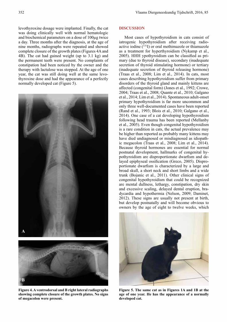
levothyroxine dosage were implanted. Finally, the cat was doing clinically well with normal hematologic and biochemical parameters on a dose of 100µg twice a day. Three months after the diagnosis, at the age of nine months, radiographs were repeated and showed complete closure of the growth plates (Figures 4A and 4B). The cat had gained weight (up to 3.1 kg) and the permanent teeth were present. No complaints of constipation had been noticed by the owner and the therapy with lactulose was stopped. At the age of one year, the cat was still doing well at the same levo-thyroxine dose and had the appearance of a perfectly normally developed cat (Figure 5).
DISCUSSION
Most cases of hypothyroidism in cats consist of iatrogenic hypothyroidism after receiving radio- active iodine (131I) or oral methimazole or thiamazole as a treatment for hyperthyroidism (Nykamp et al., 2005). HHH ypothyroidism can be classified as pri-mary (due to thyroid disease), secondary (inadequate secretion of thyroid stimulating hormone) or tertiary (inadequate secretion of thyroid releasing hormone) (Traas et al., 2008; Lim et al., 2014). In cats, most cases describing hypothyroidism suffer from primary disorders of the thyroid gland and mainly kittens are affected (congenital form) (Jones et al., 1992; Crowe, 2004; Traas et al., 2008; Quante et al., 2010; Galgano et al., 2014; Lim et al., 2014). Spontaneous adult-onset primary hypothyroidism is far more uncommon and only three well-documented cases have been reported (Rand et al., 1993; Blois et al., 2010; Galgano et al., 2014). One case of a cat developing hypothyroidism following head trauma has been reported (Mellanby et al., 2005). Even though congenital hypothyroidism is a rare condition in cats, the actual prevalence may be higher than reported as probably many kittens may have died undiagnosed or misdiagnosed as idiopath-ic megacolon (Traas et al., 2008; Lim et al., 2014). Because thyroid hormones are essential for normal postnatal development, hallmarks of congenital hy-pothyroidism are disproportionate dwarfism and de-layed epiphyseal ossification (Greco, 2005). Dispro-portionate dwarfism is characterized by a large and broad skull, a short neck and short limbs and a wide trunk (Bojanic et al., 2011). Other clinical signs of congenital hypothyroidism that could be recognized are mental dullness, lethargy, constipation, dry skin and excessive scaling, delayed dental eruption, bra-dycardia and hypothermia (Nelson, 2009; Daminet, 2012). These signs are usually not present at birth, but develop postnatally and will become obvious to owners by the age of eight to twelve weeks, which
Figure 5. The same cat as in Figures 1A and 1B at the age of one year. He has the appearance of a normally developed cat.
A
B
Figure 4. A ventrodorsal and B right lateral radiographs showing complete closure of the growth plates. No signs of megacolon were present.

Vlaams Diergeneeskundig Tijdschrift, 2016, 85 353
was also the case in this cat (Bojanic et al., 2011). When congenital hypothyroidism is suspected based on clinical signs, a diagnosis can be made by measur-ing total T4 and endogenous TSH concentrations. Af-fected cats are expected to have low total T4 and high TSH concentrations if the cause is thyroid-dependent. Since there is no feline-specific TSH assay available, a chemiluminescent immunoassay for canine TSH is used to measure feline TSH concentrations (Greco, 2006; Galgano et al., 2014).
Primary congenital hypothyroidism can be divided into two main categories: thyroid dyshormonogenesis (goitrous) and dysmorphogenesis (non-goitrous). The goitre in the thyroid dyshormonogensis category is the result of an increased TSH concentration in res-ponse to low thyroid hormone concentrations and subsequent thyroid hyperplasia. In case of thyroid dysmorphogenesis, there are defects to the TSH re-ceptor, which lead to development defects and aplasia of the thyroid gland (Quante et al., 2010; Bojanic et al., 2011). In the cat of the present case, the thyroid glands were palpable and scintigraphy showed an in-creased uptake of Na99mTcO4, so thyroid dyshormono-genesis was suspected. Increased uptake of Na99mTcO4 indicates a functional NaI-symporter transport mech-anism (Quante, 2010). Thyroid dysmorphogenesis is less likely as aplastic thyroid glands would not be visible on these scans. Na99mTcO4 is routinely used in thyroid scintigraphy, as it mimics the biologic behav-ior of iodine to a certain extent. The uptake mecha-nism of both Na99mTcO4 and iodine into the thyrocytes uses the NaI-symporters. However, Na99mTcO4 will not be incorporated into thyroid hormones (no fur-ther organification). 123I on the other hand undergoes organification and would have been the best tool to describe the mechanism of congenital hypothyroid-ism in this case. Despite the less accurate reflection of the thyroid function compared to 123I, Na99mTcO4 is considered appropriate to evaluate thyroid function. Further, the cost of radioactive iodine isotopes (123I and 131I) are higher than the readily available Na99mT-cO4. Lastly, even though 131I has reportedly been used in low doses, it holds a radiotoxic component (beta-particle decay) that is useful for therapeutic purposes but also contributes to a higher radiation burden for the patient.
Defects during the synthesis of thyroid hormones may occur at several levels, such as impaired uptake of iodine by the thyroid gland (through the NaI-sym-porter) or deficient organification (by thyroid peroxi-dase and thyroid oxidase-2 enzymes) and transport (by pendrin) of iodine (Bojanic et al., 2011). Jones et al. (1992) reported an organification defect in a family of Abyssian cats with an autosomal recessive mode of inheritance. IV administration of sodium perchlorate as an active competitor for Na99mTcO4 (or radioactive iodine isotopes) in the thyroid glands is known as a technique to pinpoint the pathology more exactly, but was not pursued in this case.
The diagnosis of hypothyroidism was made and le-vothyroxine therapy was started. Several months and a few dosage adjustments later, signs of hypothyroid-ism resolved and the cat was doing well. Cats that suf-fer from congenital hypothyroidism and receive levo-thyroxine therapy for this condition may have a good prognosis, as in the present case. The long-term out-come however is unknown, but depends on the etio- logy and the age when treatment is initiated, since thyroid hormone is necessary for the normal develop-ment of bones, joints and the central nervous system (Bojanic et al., 2011).
ACKNOWLEDGEMENT
The authors want to thank family Verschueren. Without their support and love for their cat, this ar-ticle would not have been possible.
REFERENCES
Blois S.L., Abrams-Ogg A.C.G., Mitchell C., Yu A., Stoewen D., Lillie B.N., Kiupel M. (2010). Use of the thyroid scintigraphy and pituitary immunohistochemistry in the diagnosis of spontaneous hypothyroidism in a mature cat. Journal of Feline Medicine and Surgery 12, 156-160.
Bojanic K., Acke E., Jones B.R. (2011). Congenital hypo- thyroidism of dogs and cats: A review. New Zealand Journal 59, 115-122.
Crowe A. (2004). Congenital hypothyroidism in a cat. The Canadian Veterinary Journal 45, 168-170.
Daminet S. Feline hypothyroidism. (2012). In: Mooney C.T., Peterson M.E. (editors). BSAVA Manuel of Canine and Feline Endocrinology. Fourth edition, British Small Animal Veterinary Association, Gloucester, p. 111-115.
Galgano M., Spalla I., Callegari C., Auriemma E., Zanna G., Ferro S., Zini E. (2014). Primary hypothyroidism and thyroid goiter in an adult cat. Journal of Veterinary Inter-nal Medicine 28, 682-686.
Greco D.S. (2005). Diagnosis of congenital and adult-onset hypothyroidism in cats. Clinical techniques in Small Ani-mal Practice 21, 40-44.
Mellanby R.J., Jeffery N.D., Gopal M.S., Herrtage M.E. (2005). Secondary hypothyroidism following head trauma in a cat. Journal of Feline Medicine and Surgery 7, 135-139.
Nelson R.W. Disorders of the hypothalamus and pituitary gland. (2009). In: Nelson R.W., C. Guillermo Couto (edi-tors). Small Animal Internal Medicine. Fourth edition, Mosby Elsevier, St. Louis, Missouri, p. 709-714.
Nykamp S.G., Dykes N.L., Zarfoss M.K., Scarlett J.M. (2005). Association of the risk of development of hypo-thyroidism after iodine 131 treatment with the pretreat-ment pattern of sodium pertechnetate Tc 99m uptake in the thyroid gland in cats with hyperthyroidism: 165 cases (1990-2002). Journal of the American Veterinary Asso-ciation 226, 1671-1675.
Lim C.K., Rosa C.T., de Witt Y., Schoeman J.P. (2014). Congenital hypothyroidism and concurrent renal insuf-ficiency in a kitten. Journal of the South African Veteri-nary Association 85, 1-6.

354 Vlaams Diergeneeskundig Tijdschrift, 2016, 85
Quante S., Fracassi F., Gorgas D., Kircher P.R., Boretti F.S., Ohlerth S., Reusch C.E. (2010). Congenital hypo-thyroidism in a kitten resulting in decreased IGF-I con-centration and abnormal liver function tests. Journal of Feline Medicine and Surgery 12, 487-490.
Scott-Moncrieff J.C. (2007). Clinical signs and concurrent diseases of hypothyroidism in dogs and cats. Veterinary Clinics of North America: Small Animal Practice 37, 709-722.
Traas A.M., Abbot B.L., French A., Giger U. (2008). Con-genital thyroid hypoplasia and seizures in 2 littermate kittens. Journal of Veterinary Internal Medicine 22, 1427-1431.
World Veterinary Poultry Associati onWVPA - Belgian Branch
WVPA België organiseert jaarlijks verschillende studienamiddagen omtrent recente
problemati ek bij industrieel gehouden pluimvee. De thema’s van de studienamiddagen
spelen in op de huidige problemati ek in de pluimveesector en proberen tegemoet te
komen aan de verwachti ngen van de pluimveedierenarts.
WVPA België heeft een acti viteiten programma uitgewerkt voor het academiejaar 2016-
2017.
De onderwerpen zijn als volgt:
- Vectorvaccin: voorzien voor januari-februari 2017
- In ovo feeding: voorzien voor maart- april 2017
- Persisterende Salmonella en E.coli infecti es: voorzien voor mei - juni 2017
Deelname aan het programma is grati s voor leden.
Alle informati e over het lidgeld alsook het rekeningnummer waarop het lidgeld kan gestort
worden, kan u vinden op de website www.wvpa.be.
Met vriendelijke groeten,
WVPA België
126588M103064
126588M103064.indd 1 30/11/16 15:39