connecting transcriptional control to chromosome …symposium.cshlp.org/content/75/227.full.pdf ·...
TRANSCRIPT
Embryonic stem cells (ESCs) provide an attractivemodel system for studying the transcriptional control ofcell state. Their two key propertiesÑ self-renewal andpluripotencyÑ allow them to be propagated almost indef-initely in culture, yet differentiate into any cell type in thebody (Fig. 1). Although the regulatory networks that giverise to the various cell states are highly complex, a numberof simple regulatory concepts have emerged from studiesof ESC transcriptional control and cellular reprogram-ming. These concepts, which involve the roles of tran-scription factors, signal transduction pathways, andregulators of chromatin structure in the control of cellstate, are not necessarily limited to ESCs and may applyto all cell types.
TRANSCRIPTION FACTORS AND CONTROLOF ESC STATE
DNA-binding transcriptional regulators are key to spe-cific gene control. Early studies into the transcriptionalcontrol of the Escherichia coli lac operon created a frame-work for understanding gene control in all of biology(Jacob and Monod 1961). In the absence of lactose, the lacoperon is repressed by the lac repressor, a DNA-bindingprotein that binds specifically to a DNA element justdownstream from the transcription start site called the lacoperator. When the repressor is bound to the lac operator,transcription initiation by RNA polymerase is inhibited. Iflactose is present, it is metabolized into allolactose, whichbinds the lac repressor protein and alters its conformation,thus preventing it from binding to the lac operator, and al-lowing transcription to occur. A second control mecha-nism, that involves sensing of the nutrient environment anda second DNA-binding transcriptional regulator, can con-tribute to further activation of the lac operon in the absenceof glucose. Cyclic adenosine monophosphate (cAMP) is asignaling molecule whose prevalence is inversely propor-
tional to that of glucose. It binds to catabolite activator pro-tein (CAP), which undergoes a conformational change thatallows it to bind a DNA element called the CAP-bindingsite, located upstream of the transcription start site, fromwhich it recruits RNA polymerase, thus activating tran-scription. The fundamental concept that emerged fromthese studiesÑ that gene control relies on specific DNA-binding repressors and activators and the specific sequenceelements that they recognize in the genomeÑ continues tounderpin our understanding of the control of gene expres-sion in all organisms.
DNA-binding transcription factors comprise the largestsingle class of proteins encoded in the human genome,representing ~10% of all protein-coding genes (Lander etal. 2001; Levine and Tjian 2003; Babu et al. 2004). Mosteukaryotic DNA-binding transcription factors appear tofunction as transcriptional activators but can be bound byother proteins that cause the multiprotein complex to actas a repressor. Transcription factors bind to both promoter-proximal DNA elements and to distal regions 1—100 kbaway from the promoter (D’Alessio et al. 2009; Narlikarand Ovcharenko 2009; Pan et al. 2010). The distal ele-ments that are involved in positive gene regulation aregenerally called enhancers, and these elements are typi-cally bound by multiple transcription factors. The bestcharacterized of these enhancers is that of the INF-β pro-moter, where eight transcription factor molecules occupya 50-bp segment of DNA (Maniatis et al. 1998; Agaliotiet al. 2000; Panne 2008).
In ESCs, the transcription factors Oct4, Sox2, andNanog are key in determining and maintaining cell state.The critical role of the transcription factors Oct4 andNanog was initially established based on their unique ex-pression in ESCs and the impact of their loss of expressionon cell state (Scholer et al. 1990; Nichols et al. 1998;Chambers et al. 2003; Mitsui et al. 2003; Chambers andSmith 2004; Hart et al. 2004). The high-mobility-group
Connecting Transcriptional Control to ChromosomeStructure and Human Disease
J.J. NEWMAN AND R.A. YOUNGWhitehead Institute for Biomedical Research and Department of Biology,Massachusetts Institute of Technology, Cambridge, Massachusetts 02142
Correspondence: [email protected]
We review key insights into transcriptional regulation of cell state that have emerged from the study of embryonic stem cells.These insights are described in the context of historical studies of the roles of transcription factors, signal transduction path-ways, and regulators of chromatin structure. We highlight recent studies that have led to the model that mediator and cohesinphysically and functionally connect the enhancers and core promoters of a key subset of active genes in cells, thus generatingcell-type-specific DNA loops linked to the gene-expression program of each cell. Mutations in the genes encoding mediatorand cohesin components can cause an array of human developmental syndromes and diseases, and we discuss the implicationsof these findings for the mechanisms involved in these diseases.
Cold Spring Harbor Symposia on Quantitative Biology,Volume LXXV. ©2010 Cold Spring Harbor Laboratory Press 978-1-936113-07-1 227
016_227-236_Newman_99_S75.qxd_Symposium 75 5/13/11 10:13 AM Page 227

protein Sox2 was added to the list of key regulators ofESC state when it was discovered that Sox2 forms a het-erodimer with Oct4 (Ambrosetti et al. 1997, 2000; Avilionet al. 2003; Masui et al. 2007).
The study of genes occupied and regulated by Oct4,Sox2, and Nanog has led to three key concepts in tran-scriptional control of ESC state. First, the master regula-tors collaborate to regulate each of their own promoters,forming an interconnected autoregulatory loop (Fig. 2A)(Boyer et al. 2005). This regulatory feature probably con-tributes to the ability to jump-start the ESC gene-expres-sion program during reprogramming by forced expressionof exogenous reprogramming factors (Jaenisch and Young2008). It is also thought to generate a bistable state forESCs: a stable positive-feedback-controlled gene-expres-sion program when the master transcription factors are ad-equately expressed and a stimulus to enter a differentiation
program when any one of the master transcription factorsis no longer fully functional. A second concept is that themaster regulators collaborate to regulate their target genes(Boyer et al. 2005). A third concept is that these regulatorsfunction to activate expression of genes necessary tomaintain ESC state, while contributing to silencing ofgenes whose repression is essential to maintaining thatstate. These silenced genes encode developmental regula-tors whose expression is required for the appropriate dif-ferentiation of ES cells and whose repression is critical formaintaining the self-renewing and pluripotent propertiesof ESCs (Fig. 2B) (Boyer et al. 2005; Lee et al. 2006; Lohet al. 2006).
Additional transcription factors contribute to the tran-scriptional control of ES cells. Sall4 and Tcf3 have re-cently been shown to occupy most of the same genesbound by Oct4, Sox2, and Nanog (Wu et al. 2006; Zhanget al. 2006; Chen et al. 2008a; Cole et al. 2008). Transcrip-tion factors c-Myc, Esrrb, Trim28, and Ronin are also im-portant for the control of proliferation and maintenance ofESC state (Chen et al. 2008b; van den Berg et al. 2008;Zhang et al. 2008; Hu et al. 2009; Dejosez et al. 2010). Itis likely that additional transcription factors among thehundreds that are expressed in ESCs will emerge as im-portant contributors to the larger gene-expression programof these cells.
The power of a small set of master transcription factorsto establish and maintain ESC state has been elegantlydemonstrated by cellular reprogramming with definedtranscription factors. Takahashi and Yamanaka (2006) firstshowed that the forced expression of Oct4, Sox2, Klf4,and c-Myc can reprogram fibroblasts into induced plurip-otent stem cells (iPSCs). These findings have been repro-duced by many other laboratories using combinations ofOct4 and other transcription factors in various somaticcells (Maherali et al. 2007; Wernig et al. 2007, 2008; Ma-herali and Hochedlinger 2008; Park et al. 2008a,b; Silvaet al. 2008). Reprogramming is now being used to gener-ate patient-specific iPSCs that should be useful for regen-
228 NEWMAN AND YOUNG
Oct4
Sox2
Nanog
Pou5f1
Sox2
Nanog
Active
Repressed
Oct4
Sox2
Nanog
Polycomb
SetDB1
DOT1Trithorax
Genes to maintain pluripotency Oct4, Sox2, Nanog, Tcf3, Id1
Developmental regulators Olig2, Pax6, Neurog1, Hoxb1
Pol2
Pol2
SET2
A
B
Figure 2. Transcription factors and chromatin-modify-ing enzymes contribute to control of ESC gene expres-sion. (A) Oct4, Sox2, and Nanog form an interconnectedautoregulatory loop, where all three transcription factorsregulate their own genes. (B) Oct4, Sox2, and Nanogoccupy enhancers associated with actively transcribedgenes, where there is evidence for RNA polymerase II(Pol2) and chromatin-modifying complexes associatedwith transcriptional activity, including Trithorax, DOT1,and SET2. These active genes encode proteins requiredfor the maintenance of ESC state, including key ESC-specific transcription factors and inhibitors of differen-tiation (Id1). Oct4, Sox2, and Nanog also occupy silentgenes with a nonproductive form of Pol2 and chro-matin-modifying complexes associated with transcrip-tional repression, including Polycomb and SetDB1.These repressed genes encode proteins required forearly differentiation and lineage commitment and arepoised for transcription during differentiation.
Skin cells
Neurons
Muscle cells
Red blood cells
Lung cells
Pancreatic cells
Pluripotent Multipotent Unipotent
Blastocyst
Ectoderm
Mesoderm
EndodermEmbryonic stem cells
Skin cells
Neurons
MMuscle cells
d blood cells
ung cells
Pancreatic cells
Red
unL
RR
L
Ectoderm
Mesoderm
Endoderm
Blastocyst
Embryonic stem cells
Figure 1. ESCs are derived during the blastocyst stage of embryo-genesis. At this stage, cells are pluripotent and can proliferate thisway in culture indefinitely under the right conditions. As ESCsdifferentiate during development, they are fated for particular lin-eages and take on a multipotent cell state. Within each lineage, acell can continue to differentiate into any of the more than 200defined cell types of an adult mammal. Examples of these fullydifferentiated unipotent cell states are depicted here.
016_227-236_Newman_99_S75.qxd_Symposium 75 5/13/11 10:13 AM Page 228

erative medicine and the study of disease mechanisms(Dimos et al. 2008; Ebert et al. 2009; Soldner et al. 2009;Trounson 2009; Kiskinis and Eggan 2010).
SIGNAL TRANSDUCTION AND CONTROLOF ESC STATE
The culture of ESCs initially required a layer of irradi-ated fibroblasts in order to obtain the factors necessary forproliferation and pluripotency. These fibroblasts producecytokines and growth factors necessary for ESC pluripo-tency and self-renewal (Smith and Hooper 1983). The keyfactors secreted by fibroblasts have been identified, andESCs can now be grown in chemically defined mediumin the absence of irradiated fibroblasts. Leukemia in-hibitory factor (LIF), Wnt, and bone morphogenetic pro-tein 4 (BMP4) were among the factors supplied by thefibroblasts and found to impact murine ESC state(Williams et al. 1988; Smith et al. 1998; Ying and Smith2003; Sato et al. 2004; Okita and Yamanaka 2006). Thus,LIF, Wnt, and BMP4 signaling pathways can contributeto maintenance of ESC state (Fig. 3).
LIF, which was originally identified through its abilityto inhibit growth of myeloid leukemia cells, has been im-plicated in growth promotion and differentiation of vari-ous cell types (Hilton 1992). A member of the IL-6 familyof cytokines, it is expressed in the trophectoderm of thedeveloping embryo, with its receptor (LIFR) expressedthroughout the inner cell mass (ICM). ESCs are derivedfrom the ICM of blastocysts, so removing them from blas-tocysts also removes their source of this cytokine. LIFbinding to LIFR leads to phosphorylation and activationof the Janus kinase/signal transducers and activators oftranscription (JAK/STAT) and mitogen-activated proteinkinase (MAPK) cascades. In ESCs, activated STAT3 istranslocated to the nucleus where it acts as a transcriptionfactor (Stahl et al. 1995; Auernhammer and Melmed2000). STAT3 interacts with the core transcription factorsOct4, Sox2, and Nanog, explaining how LIF signalingconnects to the core regulatory circuitry and thus con-tributes to maintenance of the murine ESC state (Chen etal. 2008b). LIF has been demonstrated to activate STAT3
in human ESCs but is unable to maintain their pluripo-tency (Daheron et al. 2004; Humphrey et al. 2004). Inmouse embryogenesis, LIF is produced to allow a preg-nant female to maintain her embryos in an ESC state untilher environment is appropriate for development and birth(Nichols et al. 2001). This is likely the reason why LIF hasa profound impact on the mouse ESC state but has no ef-fect on the maintenance of human ESCs.
Wnt signaling contributes to the maintenance of ESCstate (Yoshikawa et al. 1997; Aubert et al. 2002; Kielmanet al. 2002; Sato et al. 2004) and is mediated by the intra-cellular signaling protein β-catenin. In the absence of Wntsignaling, β-catenin is phosphorylated and subsequently de-graded by the axin/GSK-3/APC complex. When Wnt pro-teins bind to cell-surface receptors of the Frizzled family,the receptors activate Dishevelled proteins, which inhibitthe axin/GSK-3/APC complex. Thus, when the Wnt signal-ing pathway is activated, unphosphorylated forms of β-catenin accumulate in the cytoplasm and shuttle into thenucleus (Reya and Clevers 2005). Within the nucleus, β-catenin interacts with DNA-bound cofactors of the lym-phoic enhancer factor (LEF)/T-cell factor (TCF) family ofproteins. The key LEF/TCF component expressed in ESCsis TCF3, which co-occupies promoters with Oct4, Sox2,and Nanog. In the absence of β-catenin, TCF3 acts as a tran-scriptional repressor, whereas when bound by β-catenin, itacts as an activator. Thus, Wnt signaling connects directlyto the core regulatory circuitry of ESCs by converting TCF3from a repressor to an activator (Cole et al. 2008).
The BMP4 signaling pathway is a member of the TGF-β superfamily of signaling pathways (Shi and Massagué2003). Activation of the BMP4 pathway stimulates thephosphorylation of Smad proteins 1, 5, and 8. Phosphor-ylation of the Smads leads to an interaction with the co-Smad Smad4. Once together in a complex with Smad4,the Smad proteins shuttle to the nucleus where they act toregulate gene expression. BMP4 has been shown to main-tain pluripotency of ESCs by activating genes encodinginhibitors of differentiation proteins and can function to-gether with LIF in the absence of serum to maintain ESCstate (Ying and Smith 2003). As with the transcription fac-tors targeted by the LIF and Wnt signaling pathways,
TRANSCRIPTIONAL CONTROL AND CHROMOSOME STRUCTURE 229
Figure 3. The Wnt, LIF, and BMP4 signaling pathways connect to the transcriptional regulatory circuitry of ESCs. The terminal com-ponents of each of these signaling pathways are transcription factors that often co-occupy enhancers with Oct4, Sox2, and Nanog.
Oct4
Sox2
Nanog
BMP4
Wnt3a
LIF
Tcf3
Stat3
Smad1/5/8
Transcriptionfactors
Signalingpathways
Chromatin modifiers/
geneexpression
Polycomb
SetDB1
DOT1Trithorax
Genes to maintain pluripotency Oct4, Sox2, Nanog, Tcf3, Id1
Developmental regulators Olig2, Pax6, Neurog1, Hoxb1
Pol2
Pol2
SET2
016_227-236_Newman_99_S75.qxd_Symposium 75 5/13/11 10:13 AM Page 229
Smad1 often occupies genes with ESC master transcrip-tion factors Oct4, Sox2, and Nanog (Chen et al. 2008b).
New knowledge of the contributions of signaling path-ways to the control of ESC state has led to advances in cel-lular reprogramming. A role for Wnt signaling in the coreregulatory circuitry of ESCs (Cole et al. 2008) led Marsonet al. (2008) to show that Wnt3a conditioned media dra-matically improve the efficiency of cellular reprogrammingin the absence of the proto-oncogene c-Myc. Similarly, byinhibiting TGF-β signaling using an inhibitor specific forpart of the Activin/Nodal signaling pathway (a pathwayknown to promote differentiation of murine ESCs), repro-gramming efficiency was improved in the absence of c-Myc (Maherali and Hochedlinger 2009). The inhibition ofthis pathway likely blocked other signals of differentiationand so lowered the threshold for the expression of genesnecessary to regain the pluripotent state. These results areimportant because of the interest in eliminating exogenousproto-oncogenes from reprogramming protocols.
CHROMATIN STRUCTURE AND CONTROLOF CELL STATE
Nucleosomes represent the fundamental unit of chro-matin and are made up of an octamer of four core histoneproteins around which 147 bp of DNA are wrapped (Risand Kubai 1970; Olins and Olins 1974; Finch and Klug1976; Kornberg and Klug 1981; Luger et al. 1997; Daveyet al. 2002). Nucleosomes compact the mammaliangenome by ~100,000-fold (Goetze et al. 2007). Genomicregions that are densely populated by nucleosomes andhighly compacted are generally more transcriptionallysilent than those that have lower nucleosome density. Nu-cleosomes are distributed throughout the genome but aredepleted from active promoter regions (Bernstein et al.2004; Gilbert et al. 2004; Yuan et al. 2005; Segal et al.2006; Mavrich et al. 2008; Schones et al. 2008; Tirosh andBarkai 2008; Jiang and Pugh 2009). The presence of tran-scription factors and the transcription initiation apparatusat active promoters are thought to influence the local den-sity and positioning of nucleosomes.
Two classes of nucleosome regulators have been de-scribed. ATP-dependent chromatin remodeling complexesare able to mobilize nucleosomes, which can enhance orreduce access to DNA sequences by transcription factorswith consequent effects on gene activity (Tsukiyama andWu 1995; Tsukiyama et al. 1995; Kingston and Narlikar1999; Narlikar et al. 2002; Sif 2004; de la Serna et al.2006a,b; Ho and Crabtree 2010; Saladi and de la Serna2010). There are also a large number of histone-modifyingenzymes whose activities contribute to local gene activity(Kouzarides 2007). Transcription factors and componentsof the transcription apparatus can bind and recruit ATP-dependent chromatin remodeling complexes and histone-modifying enzymes to specific sites in the genome(Roeder 2005; Segal et al. 2006; Panne 2008; Cairns2009). In this manner, most nucleosome regulators, whichhave no sequence-specific binding properties of their own,are brought to specific sites to facilitate gene activity orrepression.
Histone-modifying enzymes contribute to gene controlby chemically modifying lysine, arginine, serine, and otherresidues in the amino-terminal “tails” of histone proteinsthat then form binding sites for other proteins that con-tribute to positive or negative regulation of gene expres-sion. For example, histone acetyltransferases (HATs) canacetylate specific lysine residues that form sites for bind-ing by regulatory proteins that contain bromodomains(Phillips 1963; Kouzarides 2000; Roth et al. 2001; Yangand Seto 2007). Histone methyltransferases (HMTs) canmethylate specific lysine and arginine residues, formingsites for binding by regulatory proteins that contain chro-modomains (Berger 2002; Yeates 2002; Gerber and Shi-latifard 2003; Trievel 2004). There are also enzymes thatcan remove these modifications from histones, thus con-ferring the opposite effect on gene control (Hassig andSchreiber 1997; Thiagalingam et al. 2003; Verdin et al.2003; Agger et al. 2008; Kim et al. 2009).
Polycomb group (PcG) proteins and SetDB1 are amongthe chromatin regulators that have roles in the maintenanceof embryonic cell states. Genetic studies established thatPcG genes are key regulators of early development inmetazoans (Faust et al. 1998; O’Carroll et al. 2001; Pasiniet al. 2004; Breiling et al. 2007). Later studies revealed thatPcG proteins repress gene expression, in part by methylat-ing and ubiquitylating histone tails (Pirrotta 1998; Orlando2003; Ringrose and Paro 2004; Schuettengruber et al.2007; Schwartz and Pirrotta 2007). In ESCs, PcG proteinsoccupy and silence genes encoding developmental regula-tors (Bernstein et al. 2006; Boyer et al. 2006; Lee et al.2006; Pan et al. 2007; Zhao et al. 2007; Yeap et al. 2009).
SetDB1 knockdown causes loss of ESC state (Bilodeauet al. 2009; Yuan et al. 2009; Lohmann et al. 2010).SetDB1 is among the histone H3 lysine 9 methytrans-ferases, which have roles in gene repression (Schultz et al.2002; Ayyanathan et al. 2003; Wang et al. 2003). SetDB1occupies and methylates nucleosomes at many of the samesilent developmental genes that are occupied by PcG pro-teins (Bilodeau et al. 2009; Yuan et al. 2009). Thus, it ap-pears that both contribute to repression of this key set ofdevelopmental regulators, whose expression leads to lossof ESC state (Fig. 2B).
MEDIATOR AND COHESIN CONNECT GENEEXPRESSION AND CHROMATIN
ARCHITECTURE
In eukaryotes, enhancer elements are typically locatedat substantial distances from the core promoter, where thetranscription initiation apparatus is bound (Banerji et al.1981; Benoist and Chambon 1981; Wasylyk et al. 1983;Maniatis et al. 1987; Levine and Tjian 2003; Visel et al.2009). Transcription factors bind cofactors that can bindRNA Pol2 and are thus thought to bridge the enhancers towhich they are bound and the transcriptional machinerylocated at the promoter (Hampsey and Reinberg 1999;Naar et al. 2001; McKenna and O’Malley 2002; Spiegel-man and Heinrich 2004; Thomas and Chiang 2006; Fudaet al. 2009). Among the cofactors are mediator and p300,which have been shown to bind both transcription factors
230 NEWMAN AND YOUNG
016_227-236_Newman_99_S75.qxd_Symposium 75 5/13/11 10:13 AM Page 230
and the transcription initiation apparatus (Roeder 1998;Taatjes and Tjian 2004; Conaway et al. 2005; Kornberg2005; Malik and Roeder 2005, 2008; Visel et al. 2009). Itthus seems likely that specific DNA loop formation is anatural consequence of the mechanism of transcriptionalactivation in eukaryotes.
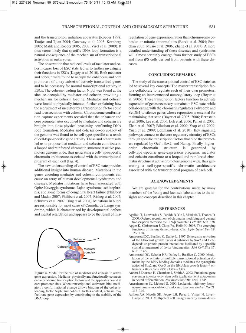
The observation that reduced levels of mediator and co-hesin cause loss of ESC state led us to further investigatetheir functions in ESCs (Kagey et al. 2010). Both mediatorand cohesin were found to occupy the enhancers and corepromoters of a key subset of actively transcribed genesand to be necessary for normal transcriptional activity inESCs. The cohesin-loading factor Nipbl was found at thesites co-occupied by mediator and cohesin, providing amechanism for cohesin loading. Mediator and cohesinwere found to physically interact, further explaining howthe recruitment of mediator by a transcription factor couldlead to association with cohesin. Chromosome conforma-tion capture experiments revealed that the enhancer andcore promoter sites occupied by mediator and cohesin arebrought into close physical proximity, confirming DNAloop formation. Mediator and cohesin co-occupancy ofthe genome was found to be cell-type specific as a resultof cell-type-specific gene activity. These and other resultsled us to propose that mediator and cohesin contribute toa looped and reinforced chromatin structure at active pro-moters genome wide, thus generating a cell-type-specificchromatin architecture associated with the transcriptionalprogram of each cell (Fig. 4).
The new understanding of control of ESC state providesadditional insight into human disease. Mutations in thegenes encoding mediator and cohesin components cancause an array of human developmental syndromes anddiseases. Mediator mutations have been associated withOpitz-Kaveggia syndrome, Lujan syndrome, schizophre-nia, and some forms of congenital heart failure (Philibertand Madan 2007; Philibert et al. 2007; Risheg et al. 2007;Schwartz et al. 2007; Ding et al. 2008). Mutations in Nipblare responsible for most cases of Cornelia de Lange syn-drome, which is characterized by developmental defectsand mental retardation and appears to be the result of mis-
regulation of gene expression rather than chromosome co-hesion or mitotic abnormalities (Borck et al. 2004; Stra-chan 2005; Musio et al. 2006; Zhang et al. 2007). A moredetailed understanding of these diseases and syndromeswill almost certainly emerge from further study of ESCsand from iPS cells derived from patients with these dis-eases.
CONCLUDING REMARKS
The study of the transcriptional control of ESC state hasled to several key concepts. The master transcription fac-tors collaborate to regulate each of their own promoters,forming an interconnected autoregulatory loop (Boyer etal. 2005). These transcription factors function to activateexpression of genes necessary to maintain ESC state, whilecollaborating with the chromatin regulators Polycomb andSetDB1 to silence genes whose repression is essential formaintaining that state (Boyer et al. 2005, 2006; Bernsteinet al. 2006; Lee et al. 2006; Loh et al. 2006; Pan et al. 2007;Zhao et al. 2007; Bilodeau et al. 2009; Yeap et al. 2009;Yuan et al. 2009; Lohmann et al. 2010). Key signalingpathways connect to the core regulatory circuitry of ESCsthrough specific transcription factors that bind the promot-ers regulated by Oct4, Sox2, and Nanog. Finally, higher-order chromatin structure is generated bycell-type-specific gene-expression programs; mediatorand cohesin contribute to a looped and reinforced chro-matin structure at active promoters genome wide, thus gen-erating a cell-type-specific chromatin architectureassociated with the transcriptional program of each cell.
ACKNOWLEDGMENTS
We are grateful for the contributions made by many members of the Young and Jaenisch laboratories to the in-sights and concepts described in this chapter.
REFERENCES
Agalioti T, Lomvardas S, Parekh B, Yie J, Maniatis T, Thanos D.2000. Ordered recruitment of chromatin modifying and generaltranscription factors to the IFN-β promoter. Cell 103: 667—678.
Agger K, Christensen J, Cloos PA, Helin K. 2008. The emergingfunctions of histone demethylases. Curr Opin Genet Dev 18:159—168.
Ambrosetti DC, Basilico C, Dailey L. 1997. Synergistic activationof the fibroblast growth factor 4 enhancer by Sox2 and Oct-3depends on protein-protein interactions facilitated by a specificspatial arrangement of factor binding sites. Mol Cell Biol 17:6321—6329.
Ambrosetti DC, Scholer HR, Dailey L, Basilico C. 2000. Modu-lation of the activity of multiple transcriptional activation do-mains by the DNA binding domains mediates the synergisticaction of Sox2 and Oct-3 on the fibroblast growth factor-4 en-hancer. J Biol Chem 275: 23387—23397.
Aubert J, Dunstan H, Chambers I, Smith A. 2002. Functional genescreening in embryonic stem cells implicates Wnt antagonismin neural differentiation. Nat Biotechnol 20: 1240—1245.
Auernhammer CJ, Melmed S. 2000. Leukemia-inhibitory factor-neuroimmune modulator of endocrine function. Endocr Rev 21:313—345.
Avilion AA, Nicolis SK, Pevny LH, Perez L, Vivian N, Lovell-Badge R. 2003. Multipotent cell lineages in early mouse devel-
TRANSCRIPTIONAL CONTROL AND CHROMOSOME STRUCTURE 231
Pol IITBP
Mediatorcomplex
Enhancer
Cohesin
Pol IIIIIIIIIIIIIIIIIPTBPP
edMeomco
Enhancer
ohesin
IIIIII
Figure 4. Model for the role of mediator and cohesin in activegene expression. Mediator physically and functionally connectsenhancer-bound transcription factors and the apparatus bound atcore promoter sites. When transcriptional activators bind medi-ator, a conformational change allows binding of the cohesin-loading factor Nipbl and cohesin. In this context, cohesin mayfacilitate gene expression by contributing to the stability of theDNA loop.
016_227-236_Newman_99_S75.qxd_Symposium 75 5/13/11 10:13 AM Page 231

opment depend on SOX2 function. Genes Dev 17: 126—140.Ayyanathan K, Lechner MS, Bell P, Maul GG, Schultz DC, Ya-
mada Y, Tanaka K, Torigoe K, Rauscher FJ III. 2003. Regulatedrecruitment of HP1 to a euchromatic gene induces mitoticallyheritable, epigenetic gene silencing: A mammalian cell culturemodel of gene variegation. Genes Dev 17: 1855—1869.
Babu MM, Luscombe NM, Aravind L, Gerstein M, TeichmannSA. 2004. Structure and evolution of transcriptional regulatorynetworks. Curr Opin Struct Biol 14: 283—291.
Banerji J, Rusconi S, Schaffner W. 1981. Expression of a β-globingene is enhanced by remote SV40 DNA sequences. Cell 27:299—308.
Benoist C, Chambon P. 1981. In vivo sequence requirements ofthe SV40 early promotor region. Nature 290: 304—310.
Berger SL. 2002. Histone modifications in transcriptional regula-tion. Curr Opin Genet Dev 12: 142–148.
Bernstein BE, Liu CL, Humphrey EL, Perlstein EO, Schreiber SL.2004. Global nucleosome occupancy in yeast. Genome Biol 5:R62.
Bernstein BE, Mikkelsen TS, Xie X, Kamal M, Huebert DJ, CuffJ, Fry B, Meissner A, Wernig M, Plath K, et al. 2006. A bivalentchromatin structure marks key developmental genes in embry-onic stem cells. Cell 125: 315—326.
Bilodeau S, Kagey MH, Frampton GM, Rahl PB, Young RA. 2009.SetDB1 contributes to repression of genes encoding develop-mental regulators and maintenance of ES cell state. Genes Dev23: 2484—2489.
Borck G, Redon R, Sanlaville D, Rio M, Prieur M, Lyonnet S,Vekemans M, Carter NP, Munnich A, Colleaux L, et al. 2004.NIPBL mutations and genetic heterogeneity in Cornelia deLange syndrome. J Med Genet 41: e128.
Boyer LA, Lee TI, Cole MF, Johnstone SE, Levine SS, Zucker JP,Guenther MG, Kumar RM, Murray HL, Jenner RG, et al. 2005.Core transcriptional regulatory circuitry in human embryonicstem cells. Cell 122: 947—956.
Boyer LA, Plath K, Zeitlinger J, Brambrink T, Medeiros LA, LeeTI, Levine SS, Wernig M, Tajonar A, Ray MK, et al. 2006. Poly-comb complexes repress developmental regulators in murineembryonic stem cells. Nature 441: 349—353.
Breiling A, Sessa L, Orlando V. 2007. Biology of polycomb andtrithorax group proteins. Int Rev Cytol 258: 83—136.
Cairns BR. 2009. The logic of chromatin architecture and remod-elling at promoters. Nature 461: 193—198.
Chambers I, Smith A. 2004. Self-renewal of teratocarcinoma andembryonic stem cells. Oncogene 23: 7150—7160.
Chambers I, Colby D, Robertson M, Nichols J, Lee S, Tweedie S,Smith A. 2003. Functional expression cloning of Nanog, apluripotency sustaining factor in embryonic stem cells. Cell113: 643—655.
Chen X, Vega VB, Ng HH. 2008a. Transcriptional regulatory net-works in embryonic stem cells. Cold Spring Harb Symp QuantBiol 73: 203—209.
Chen X, Xu H, Yuan P, Fang F, Huss M, Vega VB, Wong E, OrlovYL, Zhang W, Jiang J, et al. 2008b. Integration of external sig-naling pathways with the core transcriptional network in em-bryonic stem cells. Cell 133: 1106—1117.
Cole MF, Johnstone SE, Newman JJ, Kagey MH, Young RA. 2008.Tcf3 is an integral component of the core regulatory circuitryof embryonic stem cells. Genes Dev 22: 746—755.
Conaway RC, Sato S, Tomomori-Sato C, Yao T, Conaway JW.2005. The mammalian Mediator complex and its role in tran-scriptional regulation. Trends Biochem Sci 30: 250—255.
Daheron L, Opitz SL, Zaehres H, Lensch MW, Andrews PW, Its-kovitz-Eldor J, Daley GQ. 2004. LIF/STAT3 signaling fails tomaintain self-renewal of human embryonic stem cells. StemCells 22: 770—778.
D’Alessio JA, Wright KJ, Tjian R. 2009. Shifting players and par-adigms in cell-specific transcription. Mol Cell 36: 924—931.
Davey CA, Sargent DF, Luger K, Maeder AW, Richmond TJ. 2002.Solvent mediated interactions in the structure of the nucleosomecore particle at 1.9 Å resolution. J Mol Biol 319: 1097—1113.
Dejosez M, Levine SS, Frampton GM, Whyte WA, Stratton SA,Barton MC, Gunaratne PH, Young RA, Zwaka TP. 2010.
Ronin/Hcf-1 binds to a hyperconserved enhancer element andregulates genes involved in the growth of embryonic stem cells.Genes Dev 24: 1479—1484.
de la Serna IL, Ohkawa Y, Higashi C, Dutta C, Osias J, Komma-josyula N, Tachibana T, Imbalzano AN. 2006a. The microph-thalmia-associated transcription factor requires SWI/SNFenzymes to activate melanocyte-specific genes. J Biol Chem281: 20233—20241.
de la Serna IL, Ohkawa Y, Imbalzano AN. 2006b. Chromatin re-modelling in mammalian differentiation: Lessons from ATP-dependent remodellers. Nat Rev Genet 7: 461—473.
Dimos JT, Rodolfa KT, Niakan KK, Weisenthal LM, MitsumotoH, Chung W, Croft GF, Saphier G, Leibel R, Goland R, et al.2008. Induced pluripotent stem cells generated from patientswith ALS can be differentiated into motor neurons. Science 321:1218—1221.
Ding N, Zhou H, Esteve PO, Chin HG, Kim S, Xu X, Joseph SM,Friez MJ, Schwartz CE, Pradhan S, et al. 2008. Mediator linksepigenetic silencing of neuronal gene expression with x-linkedmental retardation. Mol Cell 31: 347—359.
Ebert AD, Yu J, Rose FF Jr, Mattis VB, Lorson CL, Thomson JA,Svendsen CN. 2009. Induced pluripotent stem cells from aspinal muscular atrophy patient. Nature 457: 277—280.
Faust C, Lawson KA, Schork NJ, Thiel B, Magnuson T. 1998. ThePolycomb-group gene eed is required for normal morphogeneticmovements during gastrulation in the mouse embryo. Develop-ment 125: 4495—4506.
Finch JT, Klug A. 1976. Solenoidal model for superstructure inchromatin. Proc Natl Acad Sci 73: 1897—1901.
Fuda NJ, Ardehali MB, Lis JT. 2009. Defining mechanisms thatregulate RNA polymerase II transcription in vivo. Nature 461:186—192.
Gerber M, Shilatifard A. 2003. Transcriptional elongation by RNApolymerase II and histone methylation. J Biol Chem 278:26303—26306.
Gilbert N, Boyle S, Fiegler H, Woodfine K, Carter NP, BickmoreWA. 2004. Chromatin architecture of the human genome: Gene-rich domains are enriched in open chromatin fibers. Cell 118:555—566.
Goetze S, Mateos-Langerak J, van Driel R. 2007. Three-dimen-sional genome organization in interphase and its relation togenome function. Semin Cell Dev Biol 18: 707—714.
Hampsey M, Reinberg D. 1999. RNA polymerase II as a controlpanel for multiple coactivator complexes. Curr Opin Genet Dev9: 132—139.
Hart AH, Hartley L, Ibrahim M, Robb L. 2004. Identification,cloning and expression analysis of the pluripotency promotingNanog genes in mouse and human. Dev Dyn 230: 187—198.
Hassig CA, Schreiber SL. 1997. Nuclear histone acetylases anddeacetylases and transcriptional regulation: HATs off toHDACs. Curr Opin Chem Biol 1: 300—308.
Hilton DJ. 1992. LIF: Lots of interesting functions. TrendsBiochem Sci 17: 72—76.
Ho L, Crabtree G.R. 2010. Chromatin remodelling during devel-opment. Nature 463: 474—484.
Hu G, Kim J, Xu Q, Leng Y, Orkin SH, Elledge SJ. 2009. Agenome-wide RNAi screen identifies a new transcriptionalmodule required for self-renewal. Genes Dev 23: 837—848.
Humphrey RK, Beattie GM, Lopez AD, Bucay N, King CC, FirpoMT, Rose-John S, and Hayek A. 2004. Maintenance of pluripo-tency in human embryonic stem cells is STAT3 independent.Stem Cells 22: 522—530.
Jacob F, Monod J. 1961. Genetic regulatory mechanisms in thesynthesis of proteins. J Mol Biol 3: 318—356.
Jaenisch R, Young R. 2008. Stem cells, the molecular circuitry ofpluripotency and nuclear reprogramming. Cell 132: 567—582.
Jiang C, Pugh BF. 2009. Nucleosome positioning and gene regu-lation: Advances through genomics. Nat Rev Genet 10: 161—172.
Kagey MH, Newman JJ, Bilodeau S, Zhan Y, van Berkum NL, Or-lando DA, Ebmeier CC, Goossens J, Rahl P, Levine SS, et al.2010. Mediator and cohesin connect gene expression and chro-matin architecture. Nature 467: 430–435.
232 NEWMAN AND YOUNG
016_227-236_Newman_99_S75.qxd_Symposium 75 5/13/11 10:13 AM Page 232

Kielman MF, Rindapaa M, Gaspar C, van Poppel N, Breukel C,van Leeuwen S, Taketo MM, Roberts S, Smits R, Fodde R.2002. Apc modulates embryonic stem-cell differentiation bycontrolling the dosage of β-catenin signaling. Nat Genet 32:594—605.
Kim JK, Samaranayake M, Pradhan S. 2009. Epigenetic mecha-nisms in mammals. Cell Mol Life Sci 66: 596—612.
Kingston RE, Narlikar GJ. 1999. ATP-dependent remodeling andacetylation as regulators of chromatin fluidity. Genes Dev 13:2339—2352.
Kiskinis E, Eggan K. 2010. Progress toward the clinical applicationof patient-specific pluripotent stem cells. J Clin Invest 120: 51—59.
Kornberg RD. 2005. Mediator and the mechanism of transcrip-tional activation. Trends Biochem Sci 30: 235—239.
Kornberg RD, Klug A. 1981. The nucleosome. Sci Am 244: 52—64.Kouzarides T. 2000. Acetylation: A regulatory modification to rival
phosphorylation? EMBO J 19: 1176—1179.Kouzarides T. 2007. Chromatin modifications and their function.
Cell 128: 693—705.Lander ES, Linton LM, Birren B, Nusbaum C, Zody MC, Baldwin
J, Devon K, Dewar K, Doyle M, FitzHugh W, et al. 2001. Initialsequencing and analysis of the human genome. Nature 409:860—921.
Lee TI, Jenner RG, Boyer LA, Guenther MG, Levine SS, KumarRM, Chevalier B, Johnstone SE, Cole MF, Isono K, et al. 2006.Control of developmental regulators by Polycomb in human em-bryonic stem cells. Cell 125: 301—313.
Levine M, Tjian R. 2003. Transcription regulation and animal di-versity. Nature 424: 147—151.
Loh YH, Wu Q, Chew JL, Vega VB, Zhang W, Chen X, BourqueG, George J, Leong B, Liu J, et al. 2006. The Oct4 and Nanogtranscription network regulates pluripotency in mouse embry-onic stem cells. Nat Genet 38: 431—440.
Lohmann F, Loureiro J, Su H, Fang Q, Lei H, Lewis T, Yang Y,Labow M, Li E, Chen T, et al. 2010. KMT1E mediated H3K9methylation is required for the maintenance of embryonic stemcells by repressing trophectoderm differentiation. Stem Cells28: 201—212.
Luger K, Mader AW, Richmond RK, Sargent DF, Richmond TJ.1997. Crystal structure of the nucleosome core particle at 2.8Å resolution. Nature 389: 251—260.
Maherali N, Hochedlinger K. 2008. Guidelines and techniques forthe generation of induced pluripotent stem cells. Cell Stem Cell3: 595—605.
Maherali N, Hochedlinger K. 2009. Tgfβ signal inhibition coop-erates in the induction of iPSCs and replaces Sox2 and cMyc.Curr Biol 19: 1718—1723.
Maherali N, Sridharan R, Xie W, Utikal J, Eminli S, Arnold K,Stadtfeld M, Yachechko R, Tchieu J, Jaenisch R, et al. 2007. Di-rectly reprogrammed fibroblasts show global epigenetic remod-eling and widespread tissue contribution. Cell Stem Cell 1:55—70.
Malik S, Roeder RG. 2005. Dynamic regulation of pol II transcrip-tion by the mammalian mediator complex. Trends Biochem Sci30: 256—263.
Malik S, Roeder RG. 2008. Epigenetics? Mediator does that too!Mol Cell 31: 305—306.
Maniatis T, Goodbourn S, Fischer JA. 1987. Regulation of inducibleand tissue-specific gene expression. Science 236: 1237—1245.
Maniatis T, Falvo JV, Kim TH, Kim TK, Lin CH, Parekh BS,Wathelet MG. 1998. Structure and function of the interferon-βenhanceosome. Cold Spring Harb Symp Quant Biol 63: 609—620.
Marson A, Foreman R, Chevalier B, Bilodeau S, Kahn M, YoungRA, Jaenisch R. 2008. Wnt signaling promotes reprogrammingof somatic cells to pluripotency. Cell Stem Cell 3: 132—135.
Masui S, Nakatake Y, Toyooka Y, Shimosato D, Yagi R, TakahashiK, Okochi H, Okuda A, Matoba R, Sharov AA, et al. 2007.Pluripotency governed by Sox2 via regulation of Oct3/4 expres-sion in mouse embryonic stem cells. Nat Cell Biol 9: 625—635.
Mavrich TN, Jiang C, Ioshikhes IP, Li X, Venters BJ, Zanton SJ,Tomsho LP, Qi J, Glaser RL, Schuster SC, et al. 2008. Nucleo-
some organization in the Drosophila genome. Nature 453: 358—362.
McKenna NJ, O’Malley BW. 2002. Combinatorial control of geneexpression by nuclear receptors and coregulators. Cell 108:465—474.
Mitsui K, Tokuzawa Y, Itoh H, Segawa K, Murakami M, TakahashiK, Maruyama M, Maeda M, Yamanaka S. 2003. The homeo-protein Nanog is required for maintenance of pluripotency inmouse epiblast and ES cells. Cell 113: 631—642.
Musio A, Selicorni A, Focarelli ML, Gervasini C, Milani D, RussoS, Vezzoni P, Larizza L. 2006. X-linked Cornelia de Lange syn-drome owing to SMC1L1 mutations. Nat Genet 38: 528—530.
Naar AM, Lemon BD, Tjian R. 2001. Transcriptional coactivatorcomplexes. Annu Rev Biochem 70: 475—501.
Narlikar L, Ovcharenko I. 2009. Identifying regulatory elementsin eukaryotic genomes. Brief Funct Genomic Proteomic 8: 215—230.
Narlikar GJ, Fan HY, Kingston RE. 2002. Cooperation betweencomplexes that regulate chromatin structure and transcription.Cell 108: 475—487.
Nichols J, Zevnik B, Anastassiadis K, Niwa H, Klewe-NebeniusD, Chambers I, Scholer H, Smith A. 1998. Formation of pluripo-tent stem cells in the mammalian embryo depends on the POUtranscription factor Oct4. Cell 95: 379—391.
Nichols J, Chambers I, Taga T, Smith A. 2001. Physiological ra-tionale for responsiveness of mouse embryonic stem cells togp130 cytokines. Development 128: 2333—2339.
O’Carroll D, Erhardt S, Pagani M, Barton SC, Surani MA,Jenuwein T. 2001. The Polycomb-group gene Ezh2 is requiredfor early mouse development. Mol Cell Biol 21: 4330—4336.
Okita K, Yamanaka S. 2006. Intracellular signaling pathways reg-ulating pluripotency of embryonic stem cells. Curr Stem CellRes Ther 1: 103—111.
Olins AL, Olins DE. 1974. Spheroid chromatin units (v bodies).Science 183: 330—332.
Orlando V. 2003. Polycomb, epigenomes, and control of cell iden-tity. Cell 112: 599–606.
Pan G, Tian S, Nie J, Yang C, Ruotti V, Wei H, Jonsdottir GA, Stew-art R, Thomson JA. 2007. Whole-genome analysis of histoneH3 lysine 4 and lysine 27 methylation in human embryonic stemcells. Cell Stem Cell 1: 299—312.
Pan Y, Tsai CJ, Ma B, Nussinov R. 2010. Mechanisms of transcrip-tion factor selectivity. Trends Genet 26: 75—83.
Panne D. 2008. The enhanceosome. Curr Opin Struct Biol 18:236—242.
Park IH, Lerou PH, Zhao R, Huo H, Daley GQ. 2008a. Generationof human-induced pluripotent stem cells. Nat Protoc 3: 1180—1186.
Park IH, Zhao R, West JA, Yabuuchi A, Huo H, Ince TA, LerouPH, Lensch MW, Daley GQ. 2008b. Reprogramming of humansomatic cells to pluripotency with defined factors. Nature 451:141—146.
Pasini D, Bracken AP, Jensen MR, Lazzerini Denchi E, Helin K.2004. Suz12 is essential for mouse development and for EZH2histone methyltransferase activity. EMBO J 23: 4061—4071.
Philibert RA, Madan A. 2007. Role of MED12 in transcription andhuman behavior. Pharmacogenomics 8: 909—916.
Philibert RA, Bohle P, Secrest D, Deaderick J, Sandhu H, CroweR, Black DW. 2007. The association of the HOPA(12bp) poly-morphism with schizophrenia in the NIMH genetics initiativefor schizophrenia sample. Am J Med Genet B NeuropsychiatrGenet 144B: 743—747.
Phillips DM. 1963. The presence of acetyl groups of histones.Biochem J 87: 258—263.
Pirrotta V. 1998. Polycombing the genome: PcG, trxG, and chro-matin silencing. Cell 93: 333—336.
Reya T, Clevers H. 2005. Wnt signalling in stem cells and cancer.Nature 434: 843—850.
Ringrose L, Paro R. 2004. Epigenetic regulation of cellular mem-ory by the Polycomb and Trithorax group proteins. Annu RevGenet 38: 413—443.
Ris H, Kubai DF. 1970. Chromosome structure. Annu Rev Genet4: 263—294.
TRANSCRIPTIONAL CONTROL AND CHROMOSOME STRUCTURE 233
016_227-236_Newman_99_S75.qxd_Symposium 75 5/13/11 10:13 AM Page 233

Risheg H, Graham JM Jr, Clark RD, Rogers RC, Opitz JM,Moeschler JB, Peiffer AP, May M, Joseph SM, Jones JR, et al.2007. A recurrent mutation in MED12 leading to R961Wcauses Opitz-Kaveggia syndrome. Nat Genet 39: 451—453.
Roeder RG. 1998. Role of general and gene-specific cofactors inthe regulation of eukaryotic transcription. Cold Spring HarbSymp Quant Biol 63: 201—218.
Roeder RG. 2005. Transcriptional regulation and the role of diversecoactivators in animal cells. FEBS Lett 579: 909—915.
Roth SY, Denu JM, Allis CD. 2001. Histone acetyltransferases.Annu Rev Biochem 70: 81—120.
Saladi SV, de la Serna IL. 2010. ATP dependent chromatin remod-eling enzymes in embryonic stem cells. Stem Cell Rev 6: 62—73.
Sato N, Meijer L, Skaltsounis L, Greengard P, Brivanlou AH. 2004.Maintenance of pluripotency in human and mouse embryonicstem cells through activation of Wnt signaling by a pharmaco-logical GSK-3-specific inhibitor. Nat Med 10: 55—63.
Scholer HR, Ruppert S, Suzuki N, Chowdhury K, Gruss P. 1990.New type of POU domain in germ line-specific protein Oct-4.Nature 344: 435—439.
Schones DE, Cui K, Cuddapah S, Roh TY, Barski A, Wang Z, WeiG, Zhao K. 2008. Dynamic regulation of nucleosome position-ing in the human genome. Cell 132: 887—898.
Schuettengruber B, Chourrout D, Vervoort M, Leblanc B, CavalliG. 2007. Genome regulation by polycomb and trithorax pro-teins. Cell 128: 735—745.
Schultz DC, Ayyanathan K, Negorev D, Maul GG, Rauscher FJIII. 2002. SETDB1: A novel KAP-1-associated histone H3, ly-sine 9-specific methyltransferase that contributes to HP1-me-diated silencing of euchromatic genes by KRAB zinc-fingerproteins. Genes Dev 16: 919—932.
Schwartz YB, Pirrotta V. 2007. Polycomb silencing mechanismsand the management of genomic programmes. Nat Rev Genet8: 9—22.
Schwartz CE, Tarpey PS, Lubs HA, Verloes A, May MM, RishegH, Friez MJ, Futreal PA, Edkins S, Teague J, et al. 2007. Theoriginal Lujan syndrome family has a novel missense mutation(p.N1007S) in the MED12 gene. J Med Genet 44: 472—477.
Segal E, Fondufe-Mittendorf Y, Chen L, Thastrom A, Field Y,Moore IK, Wang JP, Widom J. 2006. A genomic code for nu-cleosome positioning. Nature 442: 772—778.
Shi Y, Massagué J. 2003. Mechanisms of TGF-β signaling fromcell membrane to the nucleus. Cell 113: 685—700.
Sif S. 2004. ATP-dependent nucleosome remodeling complexes:Enzymes tailored to deal with chromatin. J Cell Biochem 91:1087—1098.
Silva J, Barrandon O, Nichols J, Kawaguchi J, Theunissen TW,Smith A. 2008. Promotion of reprogramming to ground statepluripotency by signal inhibition. PLoS Biol 6: e253.
Smith TA, Hooper ML. 1983. Medium conditioned by feeder cellsinhibits the differentiation of embryonal carcinoma cultures.Exp Cell Res 145: 458—462.
Smith SK, Charnock-Jones DS, Sharkey AM. 1998. The role ofleukemia inhibitory factor and interleukin-6 in human reproduc-tion. Hum Reprod (suppl 3) 13: 237—243 (discussion 244—246).
Soldner F, Hockemeyer D, Beard C, Gao Q, Bell GW, Cook EG,Hargus G, Blak A, Cooper O, Mitalipova M, et al. 2009. Parkin-son’s disease patient-derived induced pluripotent stem cells freeof viral reprogramming factors. Cell 136: 964—977.
Spiegelman BM, Heinrich R. 2004. Biological control through reg-ulated transcriptional coactivators. Cell 119: 157—167.
Stahl N, Farruggella TJ, Boulton TG, Zhong Z, Darnell JE Jr, Yan-copoulos GD. 1995. Choice of STATs and other substrates spec-ified by modular tyrosine-based motifs in cytokine receptors.Science 267: 1349—1353.
Strachan T. 2005. Cornelia de Lange syndrome and the link be-tween chromosomal function, DNA repair and developmentalgene regulation. Curr Opin Genet Dev 15: 258—264.
Taatjes DJ, Tjian R. 2004. Structure and function of CRSP/Med2;a promoter-selective transcriptional coactivator complex. MolCell 14: 675—683.
Takahashi K, Yamanaka S. 2006. Induction of pluripotent stemcells from mouse embryonic and adult fibroblast cultures by de-
fined factors. Cell 126: 663—676.Thiagalingam S, Cheng KH, Lee HJ, Mineva N, Thiagalingam A,
Ponte JF. 2003. Histone deacetylases: Unique players in shapingthe epigenetic histone code. Ann NY Acad Sci 983: 84—100.
Thomas MC, Chiang CM. 2006. The general transcription machin-ery and general cofactors. Crit Rev Biochem Mol Biol 41: 105—178.
Tirosh I, Barkai N. 2008. Two strategies for gene regulation by pro-moter nucleosomes. Genome Res 18: 1084—1091.
Trievel RC. 2004. Structure and function of histone methyltrans-ferases. Crit Rev Eukaryot Gene Expr 14: 147—169.
Trounson A. 2009. New perspectives in human stem cell therapeu-tic research. BMC Med 7: 29.
Tsukiyama T, Wu C. 1995. Purification and properties of an ATP-dependent nucleosome remodeling factor. Cell 83: 1011—1020.
Tsukiyama T, Daniel C, Tamkun J, Wu C. 1995. ISWI, a memberof the SWI2/SNF2 ATPase family, encodes the 140 kDa subunitof the nucleosome remodeling factor. Cell 83: 1021—1026.
van den Berg DL, Zhang W, Yates A, Engelen E, Takacs K, Bezs-tarosti K, Demmers J, Chambers I, Poot RA. 2008. Estrogen-related receptor β interacts with Oct4 to positively regulateNanog gene expression. Mol Cell Biol 28: 5986—5995.
Verdin E, Dequiedt F, Kasler HG. 2003. Class II histone deacety-lases: Versatile regulators. Trends Genet 19: 286—293.
Visel A, Rubin EM, Pennacchio LA. 2009. Genomic views of dis-tant-acting enhancers. Nature 461: 199—205.
Wang H, An W, Cao R, Xia L, Erdjument-Bromage H, Chatton B,Tempst P, Roeder RG, Zhang Y. 2003. mAM facilitates conver-sion by ESET of dimethyl to trimethyl lysine 9 of histone H3 tocause transcriptional repression. Mol Cell 12: 475—487.
Wasylyk B, Wasylyk C, Augereau P, Chambon P. 1983. The SV4072 bp repeat preferentially potentiates transcription startingfrom proximal natural or substitute promoter elements. Cell 32:503—514.
Wernig M, Meissner A, Foreman R, Brambrink T, Ku M, Hoched-linger K, Bernstein BE, Jaenisch R. 2007. In vitro reprogram-ming of fibroblasts into a pluripotent ES-cell-like state. Nature448: 318—324.
Wernig M, Zhao JP, Pruszak J, Hedlund E, Fu D, Soldner F, Broc-coli V, Constantine-Paton M, Isacson O, Jaenisch R. 2008. Neu-rons derived from reprogrammed fibroblasts functionallyintegrate into the fetal brain and improve symptoms of rats withParkinson’s disease. Proc Natl Acad Sci 105: 5856—5861.
Williams RL, Hilton DJ, Pease S, Willson TA, Stewart CL, GearingDP, Wagner EF, Metcalf D, Nicola NA, Gough NM. 1988.Myeloid leukaemia inhibitory factor maintains the developmen-tal potential of embryonic stem cells. Nature 336: 684—687.
Wu Q, Chen X, Zhang J, Loh YH, Low TY, Zhang W, Sze SK, LimB, Ng HH. 2006. Sall4 interacts with Nanog and co-occupiesNanog genomic sites in embryonic stem cells. J Biol Chem 281:24090—24094.
Yang XJ, Seto E. 2007. HATs and HDACs: From structure, func-tion and regulation to novel strategies for therapy and preven-tion. Oncogene 26: 5310—5318.
Yeap LS, Hayashi K, Surani MA. 2009. ERG-associated proteinwith SET domain (ESET)-Oct4 interaction regulates pluripo-tency and represses the trophectoderm lineage. EpigeneticsChromatin 2: 12.
Yeates TO. 2002. Structures of SET domain proteins: Protein lysinemethyltransferases make their mark. Cell 111: 5—7.
Ying QL, Smith AG. 2003. Defined conditions for neural commit-ment and differentiation. Methods Enzymol 365: 327—341.
Yoshikawa Y, Fujimori T, McMahon AP, Takada S. 1997. Evidencethat absence of Wnt-3a signaling promotes neuralization insteadof paraxial mesoderm development in the mouse. Dev Biol 183:234—242.
Yuan GC, Liu YJ, Dion MF, Slack MD, Wu LF, Altschuler SJ,Rando OJ. 2005. Genome-scale identification of nucleosomepositions in S. cerevisiae. Science 309: 626—630.
Yuan P, Han J, Guo G, Orlov YL, Huss M, Loh YH, Yaw LP, Rob-son P, Lim B, Ng HH. 2009. Eset partners with Oct4 to restrictextraembryonic trophoblast lineage potential in embryonic stemcells. Genes Dev 23: 2507—2520.
234 NEWMAN AND YOUNG
016_227-236_Newman_99_S75.qxd_Symposium 75 5/13/11 10:13 AM Page 234

Zhang J, Tam WL, Tong GQ, Wu Q, Chan HY, Soh BS, Lou Y, YangJ, Ma Y, Chai L, et al. 2006. Sall4 modulates embryonic stemcell pluripotency and early embryonic development by the tran-scriptional regulation of Pou5f1. Nat Cell Biol 8: 1114—1123.
Zhang B, Jain S, Song H, Fu M, Heuckeroth RO, Erlich JM, JayPY, Milbrandt J. 2007. Mice lacking sister chromatid cohesionprotein PDS5B exhibit developmental abnormalities reminiscentof Cornelia de Lange syndrome. Development 134: 3191—3201.
Zhang X, Zhang J, Wang T, Esteban MA, Pei D. 2008. Esrrb acti-vates Oct4 transcription and sustains self-renewal and pluripo-tency in embryonic stem cells. J Biol Chem 283: 35825—35833.
Zhao XD, Han X, Chew JL, Liu J, Chiu KP, Choo A, Orlov YL,Sung WK, Shahab A, Kuznetsov VA, et al. 2007. Whole-genome mapping of histone H3 Lys4 and 27 trimethylations re-veals distinct genomic compartments in human embryonic stemcells. Cell Stem Cell 1: 286—298.
TRANSCRIPTIONAL CONTROL AND CHROMOSOME STRUCTURE 235
016_227-236_Newman_99_S75.qxd_Symposium 75 5/13/11 10:13 AM Page 235