consensus statement on negative pressure wound therapy for the management of diabetic wounds
TRANSCRIPT

8/13/2019 Consensus Statement on Negative Pressure Wound Therapy for the Management of Diabetic Wounds
http://slidepdf.com/reader/full/consensus-statement-on-negative-pressure-wound-therapy-for-the-management-of 1/33

8/13/2019 Consensus Statement on Negative Pressure Wound Therapy for the Management of Diabetic Wounds
http://slidepdf.com/reader/full/consensus-statement-on-negative-pressure-wound-therapy-for-the-management-of 2/33
Evidence-Based MedicineIncluded in this manuscript is a review of current liter-
ature on diabetic foot wound s, which was examined based
on the classification of evidence-based medicine as
described by The O xford Cent re for Eviden ce-Based
Medicine.
1
By classifying the evidence, critical decisionscan be made when determining patient care. The classifi-
cation of evidence ranges from highest (level 1) to lowest
(level 5) and is subcategorized by letters. According to
this system, level 1a evidence includes the systematic
review of rand omized, con trolled tria ls; levels 2 through 4
are cohort studies of varying degrees of quality; and level
5 is expert opinion without explicit critical appraisal.
Rand omized, co ntro lled trials (RCTs) or th e systematic
review of severa l RCTs is much more likely to present con-
sistent data and will help clinicians determine whether a
treatment is effective or inappropriate. However, per-
forming these rigorous studies in wound care is compli-
uators, and the complexity in standardizing the control
arm or general medical care are all contributing factors to
this challenge. If no RCT data is available for a specific
patient situation, clinicians turn to the published litera-
ture or rely on clinical judgment.
More th an 300 articles have been published o n V.A.C.
®
Therapy (Figure 1), including the first large RCT2 pub-
Consensus Statement on Negative Pressure
Wound Therapy (V.A.C. ® Therapy) for the
Management of Diabetic Foot Wounds
Abstract: In 2004, a multidisciplina ry expert pan el convened a t th e Tucson Expert Consensus Con ference ( TECC)
to determine appropriate use of negative pressure wound therapy as delivered by a Vacuum Assisted Closure ® device
(V.A.C.® Therapy, KCI, San Antonio, Tex) in th e treatment of diab etic foot woun ds. These guidelines were upda ted by
a second multidisciplina ry expert pan el at a consensus conference on the use of V.A.C.® Therapy, held in February
2006, in Miami, Florida. This updated version of the guidelines summarizes current clinical evidence, provides prac-
tical guidance, offers best practices to clinicians treating diabetic foot wounds, and helps direct future research.The Miami consensus panel discussed the following 12 key questions regarding V.A.C.® Thera py: 1) H ow long shou ld
V.A.C.® Thera py be used in th e treatment of a diabetic foot woun d? 2) Sho uld V.A.C.® Therapy be applied without
debr iding th e wound? 3) Ho w sho uld th e patient using V.A.C.® Thera py be evaluat ed o n a n o utpat ient ba sis? 4) When
should V.A.C.® Therapy be applied fo llowing revascularizat ion ? 5) When should V.A.C. ® Therapy be applied after inci-
sion, d rain age, an d deb ridemen t of infection? 6) Shou ld V.A.C.® Therapy be applied over an active soft tissue infec-
tion ? 7) H ow should V.A.C. ® Thera py be used in patien ts with o steomyelitis? 8) H ow should no ncom pliance to V.A.C.®
Therapy be de fined? 9) How sho uld V.A.C. ® Thera py be used in com bina tion with ot her mo da lities? 10) Shou ld small,
superf icial wound s be con sidered for V.A.C.® Therapy? 11) How should success in the use of V.A.C. ® Therapy be
defined ? 12) H ow can on e combin e effective offloading a nd V.A.C.® Therapy?
4 00
3 50
3 00
2 50
2 00
1 50
1 00
50
01 9 9 5 1 9 9 6 1 9 9 7 1 9 9 8 1 9 9 9 2 0 0 0 2 0 0 1 2 0 0 2 2 0 0 3 2 0 0 4 2 0 0 5 2 0 0 6
Ar t ic lesAbstrac tsText BooksCase StudiesGuidelines
N u m b e r o f P u b l i c a t i o n s
Year
George Andros, M D; David G. Armstrong, DPM, PhD; Christopher E. Attinger, M D; Andrew J.M. Boulton, MD, FRCP;
Robert G. Frykberg, DPM, M PH; Warren S. Joseph, DPM; Lawrence A. Lavery, DPM, MPH; Stephan M orbach, M D;
Jeffrey A. Niezgoda, M D, FACHM, FACEP; Boulos Toursarkissian , M D

8/13/2019 Consensus Statement on Negative Pressure Wound Therapy for the Management of Diabetic Wounds
http://slidepdf.com/reader/full/consensus-statement-on-negative-pressure-wound-therapy-for-the-management-of 3/33
lished in The Lancet in November 2005 and several small
RCTs3–14 published previously that demonstrate the effica-
cy of V.A.C.® Therapy for various wound types (Table 1).
In addition to this level 1 evi-
dence, many case studies,
including some large case
series, demonstrate the clin-
ical benefits of V.A.C.®
Therapy, and the outcomes
in these cases are consistent
w ith the RCT data. In add i-
tion to specific patient data,
several guide lines and con-
sensus conferences have been held, involving key opin ion
leaders w ith multidisciplinary experience in their associ-
ated fields. These conferences have covered several
wound types including pressure ulcers, d iabetic foot
wounds, open abdom inal wounds, and complex chest
and open sternotomy wounds. Several treatment
algorithms have been developed ba sed on the experience
of these multidisciplinar y panels of experts and are being
adopted and implemented by clin icians. Although
gu idelines or consensus publica tions are considered level
5 evidence, the clin ical evidence included in th is consen-
sus documen t is based o n level 1 and level 2 evidence that
supports the recommendations from the multidiscipli-
nary expert panel.
Foot Ulceration Among People with DiabetesThe world is facing a major epide mic of d iabetes.
Abo ut 194 million people worldw ide, or 5.1%, in the age
group 20–79 were estimated to have d iabetes15in 2003.
This estimate is expected to increase to some 333 m il-
lion, or 6.3% of the adult populat ion ,15 by 2025. In 2003,
the number of Americans w ith d iabetes was 18.2 m illion.
This number has increased since 2003; d iabetes now
aff ects 20.8 million Americans.16 In the Un ited States, d ia-
betes is expected to increase 60% over the next 22 years,
wh ile in Europe d iabetes is expected to increase 16%.
D iabetes is expected to increase in Australia by 59%, in
South America by 88%, and in Africa, Middle East, and
Asia by a tremendous 98%. India is the world capital of
known diabetes. There are currently more than 30 mil-
lion people living w ith diabetes in India. There is also a n
increasing number of young people and ch ildren w ith
type 2 d iabetes, especially amon g ethn ic m inor ity groups.
This increase in diabetes is m ainly attributed to modern-
iza tion or western iza tion of the world’s societ ies.17
The incidence of foot ulceration is extraord inarily
h igh among people w ith diabetes. Those at greatest risk
of developing foot ulcers
include those who have a
past history of foot ulcers,
those who have undergone
amputations, or those w ith
m icrovascular complica -
tions. Foot ulcers develop in
about 15% of patients w ith
d iabetes, and foot disorders
are a leading cause of ho spi-
ta liza tion for patients w ith d iabetes.18–20 The life time risk
of a person w ith diabetes developing a foot ulcer could
be as h igh as 25%.21 Up to 70% of all leg amputations in
the Un ited States are performed on people w ith d ia-
betes, 22 and approximately 85% of lower limb amputa-
tions in patients w ith d iabetes are preceded by foot ulcer-
at ion ,19 h igh ligh ting the importance of prevention and
appropriate management of foot lesions.
All people w ith d iabetes are at risk for developing foot
ulcers, regardless of sympto ms, race, or age. The best way
for a clin ician to determ ine ulceration risk is to remove
pa tients’ shoes and socks and look at the ir feet.
Professor JA Lindsay of Belfast once sa id, “For one
mistake made for not know ing, 10 mistakes are made
2
CONSENSUS STATEMENT ON NEGATIVE PRESSURE WOUND THERAPY (V.A.C. ® THERAPY) FOR THE MANAGEMENT OF DIABETIC FOOT WOUNDS
Table 1. Published randomized, controlled trialswith V.A.C.®Therapy
THE BEST WAY FOR A CLINICIAN TO
DETERMINE ULCERATION RISK IS TO
REMOVE PATIENTS’ SHOES AND SOCKS
AND LOOK AT THEIR FEET.
Autho r and Year Topic of Study # of Pat ients
Ar m st r on g2 2 0 0 5 Diab et ic foo t am puta tions 1 6 2
T immer s-Jukema 3 2 0 0 5 Skin b lood flow 1 0
Jones-Banwell4 2 0 0 5 Inter face layer s 4 0
Jeschke5 2 0 0 4 V.A.C.® with In tegr a 1 2
Moisidis6 200 4 S k in gr afts 22
M oues7,8 2 0 0 4 * B a c t er ia l load 54
Eginton 9 20 03 D iabet ic foot wounds 10
Wanne r 10 2 0 0 3 Pr essur e ulcer s 2 2
For d 11 2 0 0 2 Pr essur e ulcer s 2 8
Joseph12 2 0 0 0 C hr on ic wound s 2 4
McCallon 13 20 00 D iabet ic foot wounds 10
Genecov 14 1 9 9 8 Skin gr aft donor r e-ep itheliza tion 1 0
* Pub l ished a second ar t icle fr om same RCT p r esent ing economic da ta .

8/13/2019 Consensus Statement on Negative Pressure Wound Therapy for the Management of Diabetic Wounds
http://slidepdf.com/reader/full/consensus-statement-on-negative-pressure-wound-therapy-for-the-management-of 4/33
for not looking.” The key to preventing
d iabet ic foot ulcers is to always consider
pa tients’ foot health.
Structures of Diabetic Foot CareThe increasing global inc idence of d iabetes
comes w ith an increase in disabling complica-
tions, includ ing the d iabetic foot. Greater
awareness of the problem among people w ith
the disease, healthcare providers, and health-
care decision makers is needed in order to
reduce lower-extremity amputations that result
from the diabetic foot. Other integral parts of
the solution include structured screening tools
to identify those at risk and the implementation of stan-
dardized prevention and treatment protocols. The 2005
Year o f th e D iabetic Foot cam paign was an important step
in increasing the awareness of d iabetic foot issues,
addressing the human and economic burden of foot com-
plica tions in people w ith diabetes through press confer-
ences and other worldw ide events.23
Stud ies have shown th at amp utation rates can be sign if-
icantly reduced in people w ith diabetes by implementing
the follow ing strategies:
• Inspection of feet and foo twear during patients’
regular visits
• Use of p reventive foot and shoe care in high-risk
feet (eg, pod iatry, protective shoes, education )
• Implementation of a multifactorial and
multidisciplinary approach to care for
established foot ulcers
• Early d iagnosis of peripheral vascular d isease and
vascular intervention if required
• Con tinuou s follow-up o f pat ients w ith
previous foot ulcers
• R eg istration of amputations and foot ulcers.24,25
The International D iabetes Federation (IDF) global
gu ideline for type 2 diabetes declares that d efined control
intervals and preventive actions should be taken for
pa tients at d ifferent risk levels. The multid isciplinary foot
care team is considered the most effective approach for
the management o f the ulcerated diabetic foot, foot infec-
tion, and other foot care emergencies.26 D ifferent coun-
tr ies and healthca re systems have implemented such mul-
tifactorial approaches to d iabetic foot care,27,28 some
reporting success29,30 and some failures (Table 2).31,32
There are remarkable d ifferences among healthcare
systems across Europe. No com mon structure of diabetic
foot care exists between coun tries. As a consequence, the
EUR OD IALE consortium was founded to describe differ-
ences in individual disease specific factors, ma nagement
strategies, and healthcare organ izational aspects of d ia-
betic foot d isease across Europe. Furthermore, the con-
sortium loo ks at d ifferences in outcomes in terms of clin-
ical endpoints, quality of life, and healthcare consump-
tion. Final results are expected later in 2006. The plan is
to use these data to develop a European consensus on
best mana gement of diabetic foot disease w ith a focus on
op timal organiza tion of care and resource ut ilization .33
The stat us of d iabetic foot care varies around the globe.
In Ch ina, the number of scien tific publications on the
to pic increased from 6 in 1996 to 176 in 2003. H owever,
no podiat rists w ith professional train ing and few diabetes
educators are ava ilable in Ch ina. R ecently, the
International Consensus on the D iabetic Foot was trans-
lated and published in Ch inese, and multid isciplinary
foot care teams are beginn ing to work in some larger ho s-
pitals (Prof. Zhangrong Xu, personal commun ica tion ,
April 2006).
Although population-based data are no t available,
rough estimates from India ind icate that approximately
40,000 legs are amputated every year. Almost 75% of
amputations are performed in patients having neuro-
pathic feet w ith secondary infection, which is potentially
preventable. The urgent need to train clin icians in India
in diabetic foot care based on these astound ing statistics
resulted in a concept called the “Step-by-Step Project.” 34
The pro ject, fund ed by the World D iabetes Foundation ,
3
Table 2. Status of diabetic foot care around the globe
Count r y Populat ionDiabetes Amputat ions in pat ients
pr evalence with diabetes
United Sta tes 2 9 5 ,7 3 4 ,1 3 4 7 % (2 0 0 5 ) 8 2 ,0 0 0
Ch ina 1 .2 5 b il lion 2 .7 % (2 0 0 2 ) 7 0 0 ,0 0 0 *
Ind ia 1 .0 7 b il lion 2 % r u r a l / 1 2 % ur ban (2 0 0 0 ) 4 0 ,0 0 0
Tanzania 3 5 m ill ion 1 % r u r a l / 4 –1 2 % ur ban No da ta
Ger m any 8 2 .5 m ill ion 7 % (2 0 0 1 ) 2 9 ,0 0 0
Fr ance 6 2 m ill ion 3 .2 % (1 9 9 9 ) 1 7 ,0 0 0
* Sou r ce: Wo r ld D iabetes Foundat ion Annua l Rev iew, 200 4.
8/13/2019 Consensus Statement on Negative Pressure Wound Therapy for the Management of Diabetic Wounds
http://slidepdf.com/reader/full/consensus-statement-on-negative-pressure-wound-therapy-for-the-management-of 5/33
4
involved 115 teams of physicians and nurses from In dia,
Tan zan ia, and several ne ighboring countries.
Healthcare providers received structured d iabetic foot
care education and tra in ing in 2 phases: basic co urses in
2004 and advanced courses in 2005. Goals of the Step-
by-Step Project were to create awareness of d iabetic foot
problems in the participa ting countries; provide tra in-
ing in d iabetic foot management to clin icians; facilitate
the d isseminat ion of information among healthcare
providers; reduce the risk of complica tions associated
w ith d iabetes; and empower patients w ith d iabetes to
take better care of their feet. The pro ject’s strategies to
reduce amputation rates included foot inspection at
every patient visit; early detection of neuropathy and
ischemia; continual follow-up of h igh-risk patients; and
preventive foot care and early warn ing sign education.35
Lon g-term n etworks are h elping to ensure percolation
of knowledge throughout the countries. 34 If successful,
th is project could become a mo del for the implementa-
tion of d iabetic foot care education and tra in ing pro-
grams in oth er developing countries.
A structured exchange program between d iabetic foot
centers of excellence in German y and Ind ian centers par-
ticipa ting in the Step-by-Step Project is planned to take
place in 2006 as an a dd-on to th is project.
Alarm ing amputation incidence data was recently pub-
lished in Germany. The researchers used hospital per-
formance and expenditure statistics to obta in a compre-
hensive count of lower limb amputations and calculated
the number of amputations in patients w ith d iabetes as
well as the number of d iabetes-related amputa tions by
using routine data from the Local Health Insurance
Funds (AOK) and previous analyses from w ith in
Germany. According to the data, surgeons performed
alm ost 44,000 lower limb amputations and 4,000 amputa -
tion revisions in Germany in 2001. Nearly 29,000 of tho se
lower limb amputations were performed on patients w ith
d iabetes.36 The actual number of amputations may be
even h igher, according to the latest data.37 Disease-man-
agement programs have been implemented for people
w ith type 2 (2003) and type 1 (2005) d iabetes to improve
care quality.38 Even though patients’ participa tion in these
programs is volunta ry, 1.5 m illion people w ith type 2 d ia-
betes registered by July 1, 2005. These programs are
designed to affect the quality of care of patients suffering
from chronic d iseases by defining the con tents and devel-
op ing timeframes for the treatment of diabetes and its
complica tions, as well as providing interfaces among the
d ifferent levels of care. Fam ily physicians deliver basic
care for people w ith type 2 d iabetes, wh ile diabetologists
provide basic care for those w ith type 1 d iabetes. These
programs include foot screen ing and inspection at
defined intervals. Pro viders are obliged to refer h igh-risk
feet, ulceration, and suspicion of diabetic osteoarthropa-
thy at predefined interfaces to special ized diabetic foot
clin ics. According to the German Diabetes Associat ion
quality criter ia from the group working on the diabetic
foot, 130 outpatient d iabetic foot clin ics and approxi-
ma tely 70 special ized hospital departments using a multi-
d isciplinar y approach h ave been approved to date. Yet,
despite clearly defined interfaces, less than 20% of
pa tients w ith diabetes and foo t problems are referred to a
special ized diabetic foot clinic, according to an initial
evaluation of the d isease management program for peo-
ple w ith type 2 d iabetes.38
In France, physician s per for med 17,000 lower-extrem i-
ty amputations on people w ith diab etes. While surgeons
amputated above the ankle in approximately 40% of
these amputations, only 38% of amputees had experi-
enced a vascular assessment before amputation .39
Pat ients in France are una ble to contact special ists d irect-
ly. General practitioners serve as care managers for
pa tients, including patients w ith diabetes. Fifteen foot
care clin ics (prima rily in associat ion w ith un iversity hos-
pitals) o ffer a multidiscipl inary approach, but the overall
organiza tion of d iabetic foot management in France is
not clearly delineat ed. To da te, pod iat ric care is poorly
re imbursed, and only 20% of pat ients w ith d iabetes are
screened using a 10-g mo no filament. A program tha t w ill
screen and treat patients w ith pre-ulcera tive conditions is
be ing developed. A special health network w ill provide
free care 5 times a year to those at increased risk for d ia-
be tic foot lesions (Dr. Jean Lou is Richard, personal com-
mu nica tion, April 2006).
Stud ies in the Un ited Kingdom40 reported an increase
in amputation despite the St. Vincent Declaration to
reduce amputations by 50%. Sweden, however, has been
successful in reducing the number of amputations. All
Swed ish citizens carry cards that conta in personal med-
ical data. The cards facilitate accurate databases and,
together w ith well-org an ized diabetes care, have probably
resulted in a fall in the amputation rate.41
CONSENSUS STATEMENT ON NEGATIVE PRESSURE WOUND THERAPY (V.A.C. ® THERAPY) FOR THE MANAGEMENT OF DIABETIC FOOT WOUNDS
8/13/2019 Consensus Statement on Negative Pressure Wound Therapy for the Management of Diabetic Wounds
http://slidepdf.com/reader/full/consensus-statement-on-negative-pressure-wound-therapy-for-the-management-of 6/33
5
The prevalence of foot ulceration in various stud ies
worldw ide is important to consider. For example, in a
Swed ish study conducted in 1990, study subjects had a
foot ulcer prevalence < 1% in a study population of
pa tients w ith t ype 1 diabe tes aged 15 to 50 years. H owever,
in a study from the U nited Kingdo m, 1.4% of th e patien t
population in the study had active ulcers, and th is study
comprised patients w ith active ulcers and th ose w ith a h is-
tory of ulcers ( ie, 4.8% of the population had ulcers
before or during th e study). In th e develop ing world, like
southern Africa, especially in Algeria, 12% of the patien t
population had active ulcers and 6.7% were amputees.
The United States also has a h igh rate of amputation ,
wh ich is 8.1 per 1,000 persons w ith diabetes. More recent
data from the population in San Antonio, Texas, repo rted
the incidence of ulceration to be about 68.4 per 1,000
persons w ith d iabetes per
year.42,43
Worldw ide, particularly in
develop ing countries, d ia-
betes is increasingly com-
mon . As discussed at the Pan
American Health Organ i-
za tion conference, wh ich
took place in 2003, ther e is a h igh prevalence o f type 2 dia-
betes and neuropathy in the Car ibbean and Central
America. More than 20% of some Car ibbean island popu-
lations have d iabetes. In Bra zil, it is estimated that 7.6% of
the population has diabetes.44,45 Amputation ra tes are h igh ,
and few d iabetes foot services are available in th ese areas.
A retrospective study from Trinida d 46 investigated 187
major amputations and found the vast major ity (> 80%)
were due to diabetic foot problems. Most amputation s
were a bove-the-knee a mputa tions (63%). Peripheral vas-
cular disease was rare compa red to n europa thy at 27% ver-
sus 92%, respectively.
A multidisciplinary approach to d iabetic foot recon-
struction is necessary to achieve salvage rates of 95% or
greater. The reconstruction should be biomechanically
sound to prevent recurrence of foot ulceration. There is
no formula for successful d iabetic foot reconstruction ,
thus it is critical to initially salvage all potentially viable tis-
sue and use it creatively to rebuild a functional foot.
Mayfield et al47 have shown that th e more of the foo t one
man ages to salvage, the longer the pat ient’s life expectan-
cy w ill be. That may be, in part, because the longer the
foot, the less the energy required for ambulation and,
hence, th e less stress on the h eart.
The evidence continues to mount that multidisciplinary
foot-care teams should trea t active d iabetic foot problems
to reduce the number of amputations. The a im should
also be to properly organ ize preventive care for peo ple at
h igh risk and continuo usly follow-up w ith patients having
previous foo t ulcers. The a vailability of such structures for
all patients w ith diabetes at risk world w ide should be con-
sidered a m ajor future goal.
Putting Feet FirstAs previously mentioned, the International Diabetes
Federation designa ted 2005 the Year o f th e Diabetic Foot,
and since th is designation, progress has been made in
bu ild ing awareness among clin icians and the public that
diabetic foot problems are a
major worldw ide concern.
However, challenges remain
in stressing several impor-
tant messages:
• Prevention is the first
step toward solving d iabetic
foot problems—up to 85%
of amputations can be avoide d 48
• R eduction in the number of amputations can be
ach ieved through education and identifica tion of the
h igh-risk foot
• Strategies aimed at foot ulcer prevention are cost
effective and can be co st saving.
Each year, in a year-long campaign, the International
Diabetes Federation high lights a diabetes-related top ic
that its members believe is particularly important. Last
year’s campaign, which focused on “putting feet first,”
looked at preventing amputation, screening for ulceration ,
and treating the diabetic foot. Culm ina ting the year, The
Lancet launched an issue almost exclusively dedicated to
the diabetic foot to co incide w ith World Diabetes Day
(November 14th), a date that marks the birth date of
Frederick Ban ting, who discovered insulin w ith Best,
Collip, and McLeod in Toro nto in 1922. The publication
of this special issue sign ifies the first time any major non-
special ist journal had focused on the d iabetic foot, which
illustrates the importance of diabetic foot problems—not
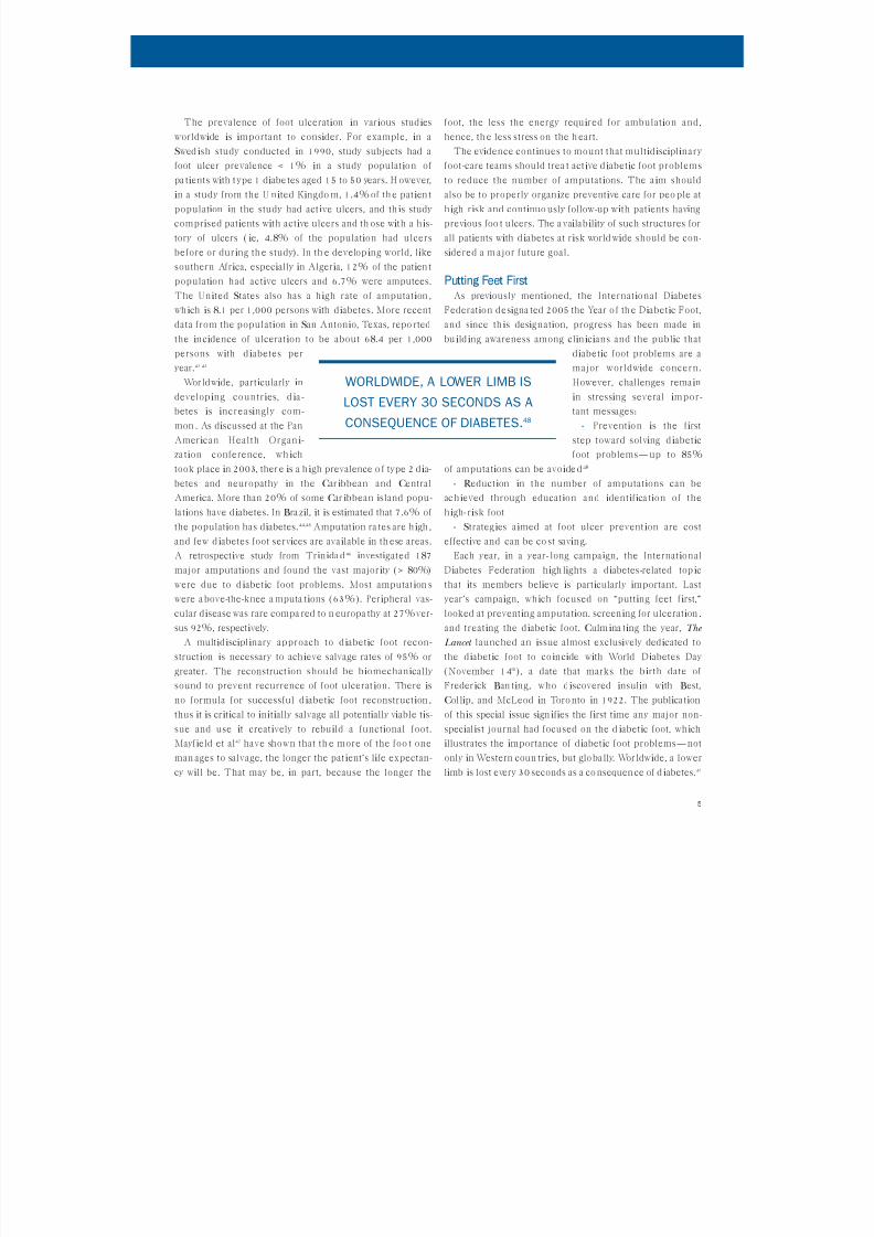
only in Western coun tries, but glo ba lly. Wor ldw ide, a lower
limb is lost every 30 seconds as a co nsequen ce of d iabetes.49
WORLDWIDE, A LOWER LIMB IS
LOST EVERY 30 SECONDS AS A
CONSEQUENCE OF DIABETES.48
8/13/2019 Consensus Statement on Negative Pressure Wound Therapy for the Management of Diabetic Wounds
http://slidepdf.com/reader/full/consensus-statement-on-negative-pressure-wound-therapy-for-the-management-of 7/33
Diabetic Foot Ulceration: Causal PathwaysNeuropathy. Clin icians must screen for neuropathy,
wh ich is the compon ent cause in a reported 78% of fo ot
ulceration cases.50 Wh ile the annual risk of foot ulcera-
tion is slightly more than 2.0% among all patients w ith
d iabetes,51it is between 5.0% and 7.5% among pat ients
w ith d iabetes and neuropathy.52 In a UK population -
based study of type 2 diabetes published in 1994, 42% of
the 811 subjects included in the study had clinical evi-
dence of neuropathy and 11% had vascular d isease.53
The investigators, therefore, conservatively estimated
that m ore than 50% of older pat ients w ith type 2 d iabetes
are at risk for foot problems. Half of these pat ients w ill
be asymptomatic.53 Thus, d iabetic neuropath y is a para-
dox because some ind ividuals experience severe pa in
w ith preserved sensation, wh ile others experience much
less pain and loss of sensation, an d o thers have no symp-
toms at all.
Largely a “forgotten complica tion,” diabetic neuropa-
thy often goes und iagnosed. The American Diabetes
Associat ion (ADA) commissioned a survey in 2005 and
found that only 1 in 4 survey respondents who experi-
enced symptoms of neuropathy had been d iagnosed w ith
the condition . Th is survey found that 56% of respondents
who had experienced symptoms were not fam iliar w ith
the term “diabetic neuropathy,” and wh ile 62% believed
th eir symptoms were associated w ith their diabetes, only
42% had been told by their physicians that d iabetes was
the cause. In the Un ited Kingdom Prospective Diabetes
Study (UKPDS), 11% of subjects had neuropathy at the
time of diagnosis of d iabetes, indica ting that patients may
present d iabetic foot prob lems to surgeons, pod iat rists, or
pr imary care physicians as d iagnostic features of d ia-
betes.54
Neuropath ic ulcers are frequently complicated by
infection. In a study by R eiber,50investigators reviewed
several cases to determ ine key component causes that
resulted in diabetic foot ulceration. Investigators found
that wh ile a single compon ent cause may be important in
the development of ulceration, it would no t cause ulcera-
tion on its own; however, ulcers would develop when com-
bined w ith other component causes. This study showed
that the most important component cause of diabetic
ulceration was neuropath y, which was present in 4 out of
5 subjects (78%). Other causative factors include infec-
tion and ischemia. It is man dato ry that ph ysicians treating
pa tients w ith diabetes and foot problems determ ine
wh ich components of this et iologic tr iad (neuropathy,
infection, ischemia) are contribu ting to the foot ulcer in
each patient.
Foot ulcers rarely result from a single pathology but
rather from multiple contributory causes, wh ich lead to
the breakdown of the h igh-risk foot.55 In add ition to the
et iologic tr iad noted by R eiber, the combina tion of neu-
ropathy, deformity, and trauma has been shown to cause
foot ulceration in 63% of cases.50Several ad d itional stud-
ies found a ca usal relationsh ip between pressure an d d ia-
be tic foot ulcer formation. The results from several d ia-
be tic neuropathy stud ies56–58 suggest that h igh foot pres-
sures are associated w ith first and recurrent plantar neu-
ropathic ulcers; foot pressure abnormalities precede the
appearance of neuropathy; h igh foot pressure pred icts
ulcers; and presence of a plantar callus is associated w ith
h igh pressure and pred icts ulcer formation. G iven these
and other predictors of ulceration, it is estimated that at
least 80% of ulcers are preventable. 53
Tests for neuropathy detection. On examina tion, the
symptoms of neuropathy are usually bilateral, but they
may be more severe on one side. Most often, however,
symptoms are symmetrical. Often, when d iabetic neu-
ropathy rapidly progresses, the physician may attribute
the symptoms to another cause. Several simple, inexpen-
sive tests, such as the neuropathy d isab ility score (NDS)
and monofilaments, are effective in detecting d iabetic
neuropathy. A neurologic examinat ion of the lower
extremities involves the use of a 10-g monof ilament or a
composite score, such as a mod ified NDS, to test sensa-
tion .42
In a prospective study,59investigators showed that d ia-
be tic neuropathy leads to foot ulcerat ion. Th is observa-
tional study consisted of 469 patients who were screened
when a n ew diabetes center open ed in 1988. The subjects
were assessed by vibration perception using a Bio-
Thesiometer (Bio-Medical Instrument Company,
Newbury, Ohio), wh ich is a han d-held d evice that sem i-
quantita tively measures vibration perception. Subjects
also received foot care education. Investigators followed
the patients to determ ine who developed foot ulcers. The
results of th is study showed that those patients w ith no
neuropathy (vibration perception threshold [VPT] < 15)
had an annual risk of developing an ulcer below 1%.
Those subjects w ith defin itive neur opathy (VPTs > 25)
6
CONSENSUS STATEMENT ON NEGATIVE PRESSURE WOUND THERAPY (V.A.C. ® THERAPY) FOR THE MANAGEMENT OF DIABETIC FOOT WOUNDS

8/13/2019 Consensus Statement on Negative Pressure Wound Therapy for the Management of Diabetic Wounds
http://slidepdf.com/reader/full/consensus-statement-on-negative-pressure-wound-therapy-for-the-management-of 8/33
7
had a 7-fold increase risk or a 5% annual r isk of develop-
ing foot ulcers. This study was later repeated and includ-
ed multiple centers in North America and Europe w ith
more than 1,000 subjects w ith diabetes and defin ite neu-
ropathy but no past h istory of ulcers and no evidence of
peripheral vascular d isease. The subjects were seen every
3 months by the investiga tive pod iat rist or a special ist
nurse. The annual risk of first ulcers in th is group51 was
greater tha n 7%. The da ta from this study can be used to
power calculations for further stud ies. Investigators in t h is
study also showed that electrophysiology was the best pre-
dictor of foot ulcers. In more soph isticated studies where
nerve function is measured, electrophysiology is a good
surrogate marker for risk factors of neuro pathy.60
R esults from a study by Booth a nd Young 61 indicated
that not all 10-g mo nof ilaments buckle at 10 g of force.
Differences in manufacturer and cycles of applied stress
may make these devices inac-
curate and possibly hyper-
sensitive to identifying lo ss of
protect ive sensation. The
authors concluded that
Ba iley Instruments
(Lancashire, UK) and Owen
Mumford (Oxford, UK) fila-
ments were the most accu-
rate among 160 monofila-
ments tested. Any clinic eval-
ua ting multiple patients
should, if possible, have multiple 10-g monofilaments
ava ilable to a void over-d iagnosing loss of protective sensa-
tion .62
Abbott et al51 studied 9,710 patients w ith d iabetes who
underwent foot screen ing in 6 districts of Northwest
England to d etermine the inc idence of and clin ically rel-
evant risk factors for new foot ulcerat ion in the commu-
n ity healthcare setting. Investigators used the NDS,
encompassing sensory modalities of vibration, pinp rick,
and hot a nd co ld rods. The researchers reported th at 291
ulcers developed in the 2-year study period and recom-
mended the NDS, 10-g monofilament, and palpation of
foot pulses as screen ing tools. The best pred ictor of risk
of ulcers in the study was the NDS. Patients scoring 6
had an ann ual risk of ulceration of 6.0%, wh ile tho se scor-
ing < 6 had a 1.0% ann ual r isk of ulceration .
A recent study by Mirand a-Palma et al63 compared dif-
ferent screen ing m ethod s for at-risk feet and suggested
the Bio-Thesiometer and the NDS had h igher sensitivities
than the monofilament.
Peripheral Vascular Disease and Diabetic Foot UlcersPeripheral vascular disease. When treating a d iabetic
foot ulcer, the clinician’s first priority should be to treat
and drain any invasive infection that is present and per-
form debridement if necrosis is present. However, follow-
ing the dra inage of infection and prior to elective
debridement, clin icians must determine vascular supply
adequacy. For ischem ic wounds, clinicians should delay
aggressive debridement beyond what is needed to control
infection until after proper revasculariza tion.
Diagnosing ischemia in the diabetic foot. Atherosclerosis in
pa tients w ith d iabetes is h istologically identical to that
seen in those w ithout diabetes. The major difference is
the distribution of disease.
People w ith d iabetes tend to
have tibioperoneal d isease
w ith long segment occlu-
sions and calcifica tion pre-
do mina ting. When femoral
d isease is also present, it
tends to be d iffuse w ithout
any single focal dominant
lesion. Another d ifference is
the presence of abnormally
th ick capillary basement
membranes in patients w ith diabetes. Functional differ-
ences in the m icrovasculature may also exist. The con-
cept, however, of un ique anatomic abnormalities in the
microcirculation of the patient w ith diabetes, precluding
any revascularization success, is incorrect.
The pulse exam may show a palpable femoral and
popliteal pulse in the absence of palpable pedal
pulses. Wh ile reassuring, the presence of palpable pedal
pulses does not mean normal perfusion exists.64
Pulsat ion may be transm itted and felt d istal to an
occluded vessel due to the calc ifica tion seen in people
w ith diabetes. Further vasculariza tion is warranted if
the d iabetic foot ulcer fa ils to progress well. Therefore,
for many patients, clin icians should perform a non inva-
sive arterial evaluation w ith segmental pressures,
ankle-brachial index (ABI), toe-brach ial index (TBI) ,
and pulse volume recording.
THE CONCEPT OF UNIQUE
ANATOMIC ABNORMALITIES IN THE
MICROCIRCULATION OF THE
PATIENT WITH DIABETES,
PRECLUDING ANY REVASCULARIZATION
SUCCESS, IS INCORRECT.
8/13/2019 Consensus Statement on Negative Pressure Wound Therapy for the Management of Diabetic Wounds
http://slidepdf.com/reader/full/consensus-statement-on-negative-pressure-wound-therapy-for-the-management-of 9/33
8
Vascular diagnostic studies. When faced w ith abnormal
vascular lab studies, the treating clin ician must determine
whether the amount of blood flow present is sufficient to
heal the foot wound. Controversy persists as to what con-
stitutes adequate perfusion .65 The Soc iety for Vascular
Surgery defines critical limb ischemia as the presence of
ulceration or gangrene w ith an ankle systolic pressure <
60 mmHg, a m etatarsal pressure < 40 mmH g, or toe pulse
volume recordings (PVR s) that are n on-pulsatile. 66 In
practice, however, many clinicians prefer to have a toe
pressure > 60 mmH g. The ABI is notoriously unreliable in
pa tients w ith d iabetes because the medial calcifica tion
present in th e vessel tends to art ificially elevate a nkle pres-
sure and ABI. Alternatively, many clinicians use the tran-
scutane ous pressure of oxygen ( TcpO 2). A TcpO 2 over 30
mmHg is desirable for adequate healing. In general,
wh ile low values (either toe p ressure o r TcpO 2) can be
predictive of nonhealing or poor healing, higher values
do not necessarily guarantee healing success. Thus, wh en
faced w ith a problematic or refractory d iabetic wound,
the clinician must consider revascularization, whenever
feasible or possible.
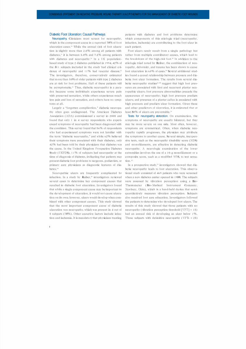
Reconstructive Surgery of the Diabetic FootProper debridement, infection control, adequate blood
supply, an d use of grafts or flaps when n ecessary are key fac-
tors for successful foo t recon struction. U se of negative pres-
sure wound therapy via the V.A.C.® Therapy System (KCI,
San Anton io, Tex) h elps prepare the woun d to either heal
by secondary intention or to be closed by simple recon-
structive means. If the wound is to be skin g raf ted , V.A.C.®
Therapy provides the ideal dressing to assist in obtaining
the highest possible take rate. Use of V.A.C.® Therapy in
foot reconstruction ha s enabled clinician s to solve complex
wound problems (eg, exposed bone, jo ints, and tendons)67
solved in the past w ith microsurgery but now routinely
treated w ith more simple solutions (Figure 2).
Adequate blood flow. Opt imal blood flow must be
ach ieved prior to perform ing reconstructive surgery. The
clin ician should not in itiate reconstruction until new
granulating tissue, neo-epitheliza tion at the wound edge,
and wrinkled skin at the wound borders are present. If
the patient has been revascular ized, it takes 4 to 10 days
follow ing bypass surgery and up to 28 days follow ing
endovascular intervention for the new blood flow to have
ma ximal effect at the wound’s edge.68 Caselli et al69 stud-
ied maximal TcpO 2 and suggested that a longer wa it may
be needed . In gen eral, however, clinicians should exped i-
tiously carry out pod iat ric procedures, w ith the goal of
ach ieving wound closure in the foot as soon as possible.
Endovascular revasculariza tion ha s a high sho rt-term fa il-
ure rate, while bypasses suffer fa ilures at a much lower
rate. The timing of debridement and revascularization in
the dysvascular patient is complicated, because only dead
CONSENSUS STATEMENT ON NEGATIVE PRESSURE WOUND THERAPY (V.A.C. ® THERAPY) FOR THE MANAGEMENT OF DIABETIC FOOT WOUNDS
FIGURE 2: Complex diabetic foot wound with exposed tendon and bone. Wound bed prepared for skin graft with V.A.C.®Therapy.

8/13/2019 Consensus Statement on Negative Pressure Wound Therapy for the Management of Diabetic Wounds
http://slidepdf.com/reader/full/consensus-statement-on-negative-pressure-wound-therapy-for-the-management-of 10/33
9
tissue, which can be hard to identify under ischemic con-
ditions, should be removed. If wet gangrene is present,
debridement should precede revascularization. If the
wound is relatively stable, debridement should be in itiat -
ed after revascularization, when there are signs that the
new blood flow is affecting the wound (eg, presence of
new granulation tissue). If dry gangrene is present, the
gangrenous edges w ill have to be monitored closely for
the development of wet gangrene so that further necrosis
does not occur.
Revascularization options. For further workup, clinician s
must obtain an angiogram (eg, conventional, magnetic
resonance, or computed tomography angiography) fol-
low ing vascular lab stud ies. Iodinated contrast exposure
may affect the choice of revascularization techn ique in
pa tients w ith diabetes and renal insufficiency. Current
revascularization options in the patient w ith diabetes
include conventional open surgery and endovascular
interventions. The 2 options are not mutually exclusive
and can be combined (eg, iliac stenting combined w ith
femorodistal bypass grafting). The cho ice of revasculariza-
tion techn ique w ill depend on the n ature of the d isease,
local expertise, extent of tissue loss, patient’s medical con-
dition, and conduit availab ility. Open surgical techniques
include endarterectomy for local lesions and bypass for
long occlusions. People w ith diabetes frequently have spar-
ing of some pedal vessel, such as the dorsal is pedis artery,
which is a good target for bypass graft ing .70 A single seg-
ment grea ter saphenous vein is the best conduit for use in
such reconstructions. If a heel lesion is present, experi-
enced clinicians may prefer a posterior tibial bypass.71 If
the clin ician is planning a transmetatarsal amputation
(TMA), either an anterior (AT/DP) or posterior (PT)
revascularization is acceptable.72
Endovascular options include angiopla sty, w ith or w ith -
out stenting, and atherectomy (ie, atherectomy w ith
exc imer laser or a plaque excision device). Wh ile subint i-
mal angioplasty has gained w idespread popularity in
Europe,73it has not become as popular in the Un ited
States. It is important in endovascular intervention to
avo id confusing angiographic w ith hemodynamic success.
Clinicians should repeat vascular lab studies follow ing
endovascular intervention to ensure sufficient hemody-
nam ic improvement. If feasible, poor risk patients w ith no
autogenous conduit may fare better if treated w ith
endovascular options. Similarly, clin icians may approach
focal stenotic lesions, as opposed to long calcified occlu-
sions, w ith endovascular techn iques.
For the pod iat ric procedure, clin icians can safely stop
an ticoagulation used in the period after the vascular
operation. No such guide lines, however, are available fol-
low ing endo vascular revascularization.
If revasculariza tion fails, the likelihood of limb salvage
is much higher w ith a closed and healed foot wound.
Toursarkissian et al74 from the University of Texa s, San
Anton io, examined outcomes follow ing d istal bypass graft
occlusions in patients w ith diabetes. The researchers
found that the presence of a foot wound at the t ime of
bypass failure was associated w ith a much h igher rate of
limb loss—67% versus 32% for cases w ith no foot wounds
at the time of bypass failure. V.A.C.® Therapy is extremely
useful in these cases, as it has been shown to help
decrease the time required for wound healing of d iabetic
foot ulcers.13
Revascularization implications. It is important to realize
that the decision for revascularization is a sign ificant com-
mitment for clin ician and patient. A recent review of 318
bypass patients conducted by Gosh ima et al75 from the
U n iversity of Arizona found that wh ile the perioperative
mortality was < 1%, 50% of the pat ients required at least
one additional surgery w ith in 3 months of their index
procedure. R enal failure patients were also mo re likely to
require r e-ad mission to th e hospital, an d 54% of pa tients
took longer than 3 months to heal their d iabetic foot
ulcers. Again, th is is a potential area where the use of
V.A.C.® Therapy could b e benef icial .
Clin icians can use V.A.C.® Therapy to manage compli-
ca tions follow ing b ypass surgery as well. Woun d pro blems
frequently follow bypass surgery, espec ially in the
saphenectomy site, and V.A.C.® Therapy can h elp achieve
closure of these wounds. However, clin icians should exer-
cise caution if the graft has been left in situ. It is prefer-
able, in these cases, for clinicians to a pply local flaps or a
bioengineered tissue graft to co ver the vascular graft after
debridement. If this is not possible, and a direct V.A.C.®
Therapy applica tion is needed, it is preferable that clini-
cians apply a non adh erent layer over the vascular graft to
avoid d irect con tact o f th e V.A.C.® Therapy foam.
Clinician s can app ly V.A.C.® Therapy to defects created by
flap donor sites as well.
V.A.C. ® Therapy application and the ischemic diabetic
foot ulcer. It is generally preferred tha t clinicians revascu-

8/13/2019 Consensus Statement on Negative Pressure Wound Therapy for the Management of Diabetic Wounds
http://slidepdf.com/reader/full/consensus-statement-on-negative-pressure-wound-therapy-for-the-management-of 11/33
1 0
la rize before applying V.A.C.® Therapy on an ischemic
diabetic foot ulcer. Most patients had n ormal ABIs in the
large clin ical trial on V.A.C.® Therapy published in The
Lancet .2 In gen eral, a TcpO 2 > 40 mmHg is desirable, and
several case series reported fa ilure in patients w ith inade-
quate flow.76,77 However, clinicians occasionally ach ieve
success in th is area w ith the use of V.A.C.® Thera py. Man y
reports3–11 on the use of V.A.C.® Therapy after failed revas-
culariza tion have found increased chances of success.
Clinicians should consider V.A.C.® Therapy as an adjunct
to other modalities in an effort to avoid amputation.
Debridement. The first step to successful foot recon-
struct ion, assum ing adequate blood flow has been
ach ieved, is debridement (see the Peripheral Vascular
Disease and Diabetic Foot Ulcers section in these guide lines).
The debrided wound should be free of all necrotic t issue
and debris and should have at its base clean, healthy,
bleed ing tissue. During debridement, the clin ician
should on ly remove dead tissue while preserving all other
tissue. The clin ician should be aggressive enough to
ensure that all necrotic tissue is removed but gentle
enough to a void damaging the remain ing viable tissue. I f
dissection is required, the clin ician should use a surgica l
blade and skin ho oks, rather than pickups and cautery, to
avo id damaging the normal tissue. Since the peripheral
tissue is the future source of new tissue growth, th e more
intact it is after the debridement, the better it w ill be able
to promote future healing. An alternative debrid ing tool
is a hydrosurgical water kn ife (Versajet® , Smith &Nephew,
Cambridge, U K). The Versajet forces a narro w stream of
water across a small gap (8 mm–5 mm) at pressure that
can reach 15,000 psi. This creates a vacuum around the
stream (Venturi effect) that draws in the surrounding tis-
sue and pulverizes it. One advantage of using the water
knife is the depth of cut can be altered by adjust ing the
pressure setting. Th is m inimizes damage to or accidental
removal of normal tissue, which sometimes occurs w ith
normal surgical debrid ing techn iques. The softer the tis-sue, the lower the pressure setting needs to be. It is par-
ticularly useful when debriding large areas or when
preparing a wound for skin grafting. 78
As mentioned previously, the clin ician should be
aggressive when debriding necrotic tissue. Future recon-
struction plans should not affect the amount of t issue that
needs to be debrided. The process should consist of tak-
ing serial thin slices of tissue until normal tissue appears.
The presence of clotted veins in the skin, fat, or muscle
ind icates that the local circulation to that area is obstruct-
ed and the tissue is most likely no t viable. The presence of
stringy fascia or tendon indicates no n-viab ility, and the t is-
sue should be shaved to shiny hard tendon or fascia. The
presence of soft grey bone indicates dead bone, and the
bone should be sawed, burred, or rongeured back to
clean, hard bone w ith punctuate bleeding at the surface
(Figure 3). Odor is an excellent indicator of whether a
wound has been adequately debrided. If there is a per-
sistent od or post-debr idement, further debridement is
needed. When the od or is no longer present, the clin ician
can feel comfortable that the wound ha s been ad equately
debrided.
Deep tissue cultures should be obta ined during
debridemen t, and broa d-spectrum antibiot ics should be
started after the procedure. If cellulitis is present, the
cutaneous border of erythema should be delineated w ith
a magic marker and the time noted. The wound should
be checked w ith in the next 6 hours for resolution of thecellulitis. If the infection h as spread beyond the outlined
border, then either wound debridement was inadequate
or the antibiot ics are inappropriate, and further debride-
ment and/or antibiot ic adjustment is needed.
The wet-to-dry dressing provides an alternative option
to surgical debridement. In th is method, saline-mois-
tened gauze is placed upon the wound a nd a llowed to dry.
Upon removing the dressing, the necrotic tissue that has
CONSENSUS STATEMENT ON NEGATIVE PRESSURE WOUND THERAPY (V.A.C. ® THERAPY) FOR THE MANAGEMENT OF DIABETIC FOOT WOUNDS
FIGURE 3: Diabetic foot wound with osteomyelitisfollowing debridement.
8/13/2019 Consensus Statement on Negative Pressure Wound Therapy for the Management of Diabetic Wounds
http://slidepdf.com/reader/full/consensus-statement-on-negative-pressure-wound-therapy-for-the-management-of 12/33
1 1
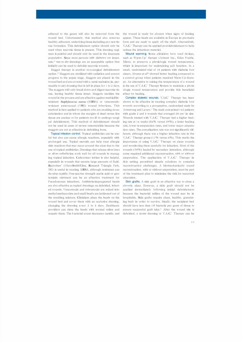
adhered to the gauze w ill also be removed from the
wound bed. Unfortunately, th is method also removes
healthy, adh erent, underlying tissue including an y new tis-
sue formation. Th is debridement option should only be
used when necrotic tissue is present. Th is dressing regi-
men is p a inful and should only be used in the insensate
population. Since most persons w ith diabetes are insen-
sate,53 wet-to-dry dressings are an acceptable option that
initially can be used to debride necrotic wounds.
Maggot therapy is anoth er no n-surgical debridement
op tion .79 Maggots are sterilized w ith radiat ion and cannot
progress to the pupae stage. Maggots are placed in the
wound bed an d are covered w ith a semi-occlusive ( ie, per-
meable to air) dr essing that is left in place fo r 1 to 2 da ys.
The m aggots w ill only break down an d d igest necrotic t is-
sue, leaving healthy tissue intact. Maggots sterilize the
wound in the pro cess and are effective against methicillin-
resistant Staphylococcus aureus (MRSA) or vancomycin-
re sistant enterococci (VR E) wound infections. Th is
method is best applied to patients who are awa iting revas-
culariza tion and in whom the margins of dead versus live
tissue are unclear or for patients too ill to undergo surgi-
cal debridement. This method of debridement should
not be used in cases of severe osteomyelitis because the
maggots are not as effect ive in debriding bone.
Topical infection control. Topical antibiotics can be use-
ful but also can cause allergic reactions, especially w ith
pro lon ged use. Top ical steroids can help treat allerg ic
skin reactions that may occur around the ulcer due to the
use of topical antibiot ics. Dressings that release silver ion s
or silver-sulfad iaz ine work well for all wounds in manag-
ing topical infection. Cadexomer iod ine is also helpful,
especially in wounds that secrete large amounts of fluid.
Bactroban® (GlaxoSmithKline, R esearch Triangle Park,
NC) is useful in treating MRSA, although resistance can
develop ra pidly. O ne-qua rter strength acetic ac id or gen-
ta micin o intment can be an effective treatment for
Pseudomonas infections. Antibiot ic-impregnated beadsare also effective as topical dressings on debrided, infect-
ed wounds. Vancomycin and tobramycin are m ixed into
methyl-metha crylate an d small bead s are fashioned out of
the resulting mixture. Clin icians place the beads on the
wound bed and cover them w ith an occlusive dressing ,
changing the dressing every 2 to 3 days. Healthcare
providers can rinse the beads w ith normal saline and
reapply them. The b acterial count decreases rap idly, and
the wound is ready for closure when signs of healing
appear. These beads are ava ilable in Europe in pre-mad e
form and are ready to apply off the shelf. Alternat ively,
V.A.C.® Therapy can be appl ied po st-deb ridement to help
reduce the infectious material.
Wound warming. Some clin icians have used devices,
such as Warm-Up® therapy (Arizant Inc., Eden Pra irie,
Minn), to preserve a physiologic wound temperature,
wh ich is important for mainta ining cell function. In a
small, randomized trial of 10 patients w ith d iabetic foot
ulcers, Alvarez et al80 showed better healing compared to
a control group when patients received Warm -U p th era-
py. An alternative to raising the temperature of a wound
is the use of V.A.C.® Therapy System to mainta in a ph ysi-
ologic wound temperature and provide th is beneficial
effect for healing .
Complex diabetic wounds. V.A.C.® Therapy has been
shown to be effective in treating complex diabetic foot
wounds according to a prospective, randomized study by
Armstrong and Lavery.2 The study com prised 162 patients
w ith grade 2 and 3 wounds that averaged 20 cm 2 in size.
Woun ds treated w ith V.A.C.® Therapy had a higher heal-
ing rate at 16 weeks (56% versus 39%), a faster heal ing
rate, lo wer re-amputa tion rates, and lower major amputa-
tion rates. The com plica tion rate was not sign ificantly dif-
ferent, although there was a h igher infection rate in the
V.A.C.® Therapy group (11% versus 6%). Th is marks the
importance of using V.A.C.® Therapy on clean wounds
and monito ring them carefully for infection. Most of the
wounds (70%) healed by secondary intention, although
some required additional reconstruction, w ith or w ithout
amputation. The applica tion of V.A.C.® Therapy in
th is setting perm it ted simple solutions to complex
reconstructive challenges. A biomechan ically sound
reconstruction, w ith or w ithout amputation, must be part
of the treatment plan to min imize the risk for recurrent
ulceration .
Skin grafts. A skin graft is an effective way to close achron ic ulcer. However, a skin graft should not be
applied immediately follow in g in itial debridement
because the bacterial milieu of the wound may be in
hospitable. Skin grafts require clean, healthy, granulat-
ing beds in order to survive. Ideally, the recipient bed
should have less than 105 bacteria per gram of tissue to
ensure successful graft take.81 After the wound site is
debrided, a moist dressing or V.A.C.® Therapy can be

8/13/2019 Consensus Statement on Negative Pressure Wound Therapy for the Management of Diabetic Wounds
http://slidepdf.com/reader/full/consensus-statement-on-negative-pressure-wound-therapy-for-the-management-of 13/33
1 2
applied until the wound has developed a healthy,
well-vascularized granulation bed. If there is no response,
wound healing a djuncts, such as growth fa ctors, cultured
skin, or h yperbaric oxygen (HBO), might b e necessary to
fa cilitate a healthy granulating bed.
The follow ing factors are critical to ensure a good take
when applying a skin graft: avoiding infection; en suring
adherence of the graft to the underlying bed; avo id ing
seroma or hematoma development between the skin
graft and the wound bed; and avo id ing shearing forces
that might detach the skin graft from the underlying
wound bed. To en sure a h igher skin graft ta ke rate, clini-
cians should curette or shave down the ex isting granula-
tion tissue to rem ove any bacteria tha t may still l ie w ith in
interstices. The skin graft should be meshed (1:1 or
1.5:1) to prevent a seroma or hematoma from build ing
up between the skin graft and the underlying wound
bed. The graft should be placed on the wound bed and
secured into position by a few strategically placed stitch-
es or staples. Follow ing graft applica tion, a w ide-meshed
nonadherent dressing should be placed (petrolatum-
impregnated gauze or silicone mesh) on the skin graft
and then V.A.C.® GranuFoam® should be placed on top
of the mesh w ith continuous suction for the next 3 to 5
days. The V.A.C.® Therapy Dressing conforms to the
underlying wound bed and, thus, ensures good contact
of the skin graft to the underlying wound bed, regardless
of wound bed con tour or d epth. V.A.C.® Thera py cont in-
uously removes any fluid that may appear, preventingflu id bu ild up that could d isrupt the contact of the graft
to th e wound bed. V.A.C.® Therapy ensures good contact
between the skin graft and the underlying bed, making it
d iff icult for shear forces to d isrupt the graft. V.A.C.®
Therap y also h elps remove infectious materials.
Clin icians can expect up to a 95% skin graft take w ith
adequate debridement, proper wound preparation, and
use of V.A.C.® Therapy as a topical dressing. A study com-
pa ring the effectiveness of V.A.C.® Therapy to bolsterdressings for fresh skin grafts82 demo nstrated a 97% com-
plete skin graft ta ke using V.A.C.® Therapy versus 81% for
the gro up dressed w ith b olster dressings.
Skin gra fts can a lso be used in inhospitable wound beds
(eg, those w ith exposed bone or tendon), provided the
wound bed has been adequately prepared. The applica-
tion of a collagen latt ice framework covered w ith a th in
silicone sheet (Integra® , Integra Life Sciences, Plainsboro,
NJ) creates a vascularized neodermis that can be skin
grafted. The exposed bone or tendon is debrided and
pulse irr igated. The sheet of collagen lattice framework is
meshed and placed on the debrided wound and secured
w ith a few strategically placed sutures or staples. It is cov-
ered w ith the V.A.C.® Thera py Dressing and con nected to
the V.A.C.® device, which is placed on continuous nega-
tive pressure and changed every 2 to 3 days unt il the col-
lagen lattice turns pink, as it develops a vascular network
(7 to 14 days). The silicone sheet is then removed and
covered w ith a th in skin g raft ( 10/1000" ). The use of the
collagen lattice framework to create a hospitable wound
bed fo r eventual skin grafting has allowed wounds to heal
w ith a simple skin graft that in the past required complex
flap recon struction .5,83,84
The Ilizarov frame has proved to be effective in sal-
vag ing infected Charcot joints in the presence of open
wounds, underlying joints, bone, and/or exposed ten-
don. In the past, th is would have required a free flap to
adequately cover the wound, wh ich was a formidable
undertaking because it had to be performed w ith in the
confines of a metal frame. Now, small local fasciocuta-
neous flaps can be ro tated to cover the exposed bone an d
tendon and a skin graft can be used to cover the rema in-
der of the wound. V.A.C.® Therapy can be applied over
the entire skin-graf ted a rea fo r 3 to 5 days. This provide s
a simple solution to wound problems that in the past
either required microsurgery or led to a below-the-knee
amputation .V.A.C.® Therapy, when used after adequate debr ide-
ment in a well-vascularized bed, prepares the wound for
closure by secondar y intention, delayed primary closure,
skin graft, or flap coverage (Figure 4). V.A.C.® Therapy
draws the wound edges together, reduces bacter ial colo-
CONSENSUS STATEMENT ON NEGATIVE PRESSURE WOUND THERAPY (V.A.C. ® THERAPY) FOR THE MANAGEMENT OF DIABETIC FOOT WOUNDS
FIGURE 4: V.A.C.®Therapy incombination with the V.A.C.® GranuFoam®Heel Dressingallows placement of negativepressure tubing on top of thefoot for patient comfort.
8/13/2019 Consensus Statement on Negative Pressure Wound Therapy for the Management of Diabetic Wounds
http://slidepdf.com/reader/full/consensus-statement-on-negative-pressure-wound-therapy-for-the-management-of 14/33
1 3
n iza tion by removing infectious material, and assists in
healthy granulation tissue form ation. V.A.C.® Thera py can
then be used a s a bolster-type dressing over skin grafts to
help ensure a h igher rate of skin-graft take. V.A.C.®
Therapy has revolution ized soft-tissue reconstruction of
the foot and ankle because it has enabled closure of
wounds by simple techniques that in the past would have
required complex pedicled or m icrosurgical free flaps.
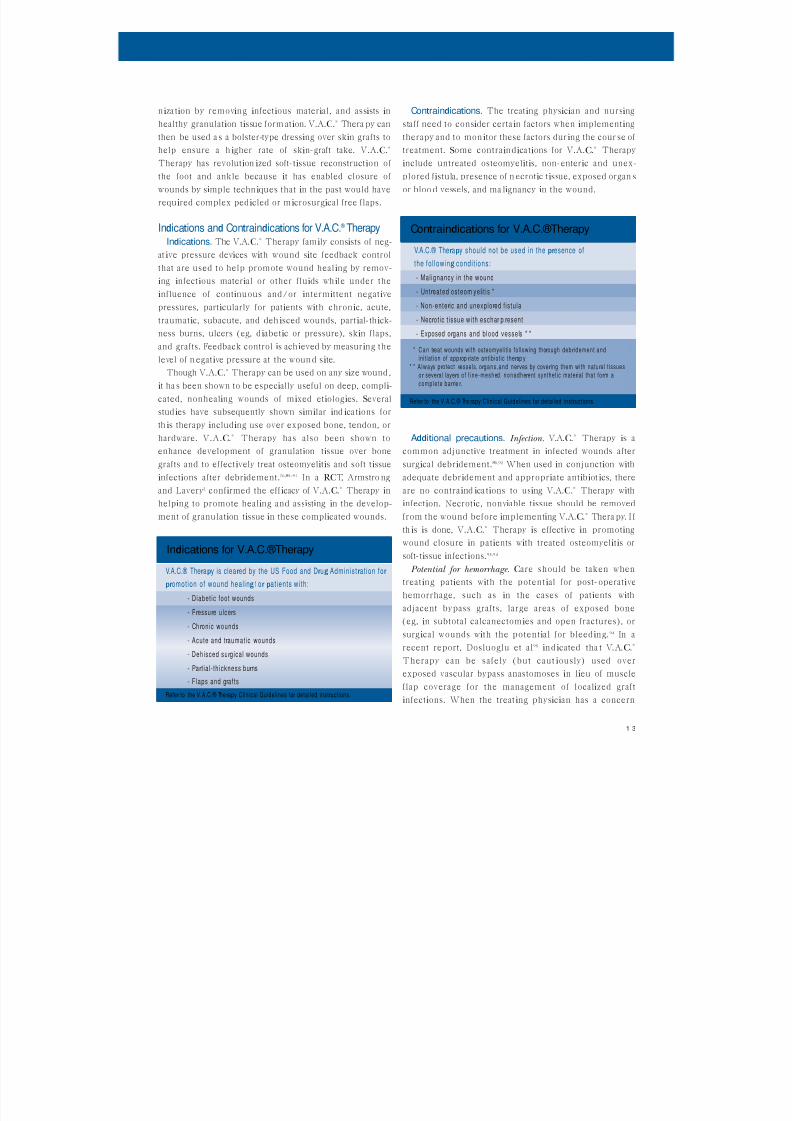
Indications and Contraindications for V.A.C. ® Therapy
Indications. The V.A.C.® Therapy family consists of neg-
at ive pressure devices w ith wound site feedback control
that are used to help promote wound healing by remov-
ing infectious material or other fluids wh ile under the
influence of continuous and/or intermittent negative
pressures, particularly for patients w ith chronic, acute,
traumatic, subacute, and deh isced wounds, partial-thick-
ness burns, ulcers (eg, diabetic or pressure), skin flaps,
and grafts. Feedback control is ach ieved by measuring the
level of n egative pressure at the woun d site.Though V.A.C.® Therapy can be used on any size wound ,
it ha s been shown to be especially useful on deep, compli-
cated, nonhealing wounds of mixed etiologies. Several
stud ies have subsequently shown similar ind ica tions for
th is therapy including use over exposed bone, tendon, or
hardware. V.A.C.® Therapy has also been shown to
enhance development of granulation tissue over bone
grafts and to effectively treat osteomyelitis and soft tissue
infections after debridement.76,85–91 In a RCT, Armstro ngand Lavery2 confirmed the efficacy of V.A.C.® Therapy in
helping to promote healing and assisting in the develop-
ment of granulation tissue in these complicated wounds.
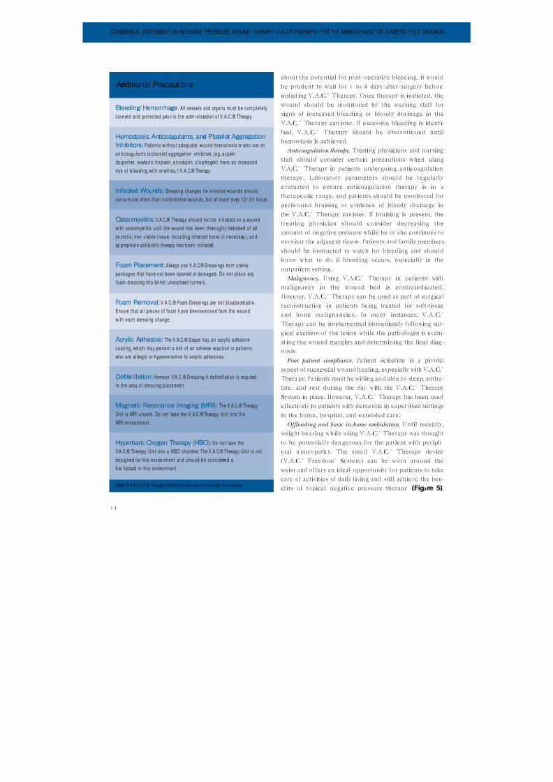
Contraindications. The treating physician and nursing
staff need to consider certain factors when implementing
therapy and to mon itor these factors during the cour se of
treatment. Some contraindications for V.A.C.® Therapy
include untreated osteomyelitis, non-enteric and unex-
plored fistula, presence of n ecrotic tissue, exposed organ s
or bloo d vessels, and ma lignancy in the wound.
Additional precautions. Infection. V.A.C.® Therapy is a
common adjunctive treatment in infected wounds after
surgical debridement.85,92 When used in conjunction w ith
adequate debridement and appropriate antibiot ics, thereare no contraind ica tions to using V.A.C.® Therapy w ith
infection. Necrotic, nonviable tissue should be removed
from the wound before implementing V.A.C.® Thera py. I f
th is is done, V.A.C.® Therapy is effective in promoting
wound closure in patients w ith treated osteomyelitis or
soft-tissue infections.93,94
Potential for hemorrhage. Care should be taken when
treating patients w ith the potential for post-operative
hemorrhage, such as in the cases of patients w ithadjacent bypass grafts, large areas of exposed bone
(eg, in subtotal calcanectom ies and open fractures), or
surgical wounds w ith the potential for bleed ing. 94 In a
recent report, Dosluoglu et al95ind icated tha t V.A.C.®
Therapy can be safely (but caut iously) used over
exposed vascular bypass anastomoses in lieu of muscle
flap coverage for the management of localized graft
infections. When the treat ing physician has a concern
V.A.C.® Ther apy is clear ed by the US Food and Dr ug Administr at ion for
pr omotion of wound heal ing f or pat ients wi th:
- D iabet ic foot wounds
- Pr essu r e ulcer s
- Ch r onic wounds
- Acute and tr aumat ic wounds
- Dehisced su r gical wounds
- Par t ia l - thickness bur ns
- F laps and gr afts
Refer to the V.A.C.® Ther apy Clinical Guidelines for detailed instr uctions.
Indications for V.A.C.®Therapy
V.A.C.® Ther apy should not be used in the pr esence of
the fol lowing condi t ions:
- Mal ignancy in the wound
- Untr eated osteom yeli t is *
- Non-enter ic and unexplo r ed f istula
- Necr ot ic t issue w i th escha r p r esent
- Exposed o r gans and blood vessels * *
* C a n tr eat wounds with osteomyelit is fol lowing thor ough deb r idement and
init iat ion of app r op r iate antibiot ic ther ap y* * A lways pr otect vessels, o r gans,and ner ves by cove r ing them with natur al t issues
o r sever al layer s o f f ine-meshed, nonadher ent synthetic mater ial that fo r m acomple te barr ie r .
Refer to the V.A.C.® Ther apy Clinical Guidelines for detailed instr uctions.
Contraindications for V.A.C.®Therapy
8/13/2019 Consensus Statement on Negative Pressure Wound Therapy for the Management of Diabetic Wounds
http://slidepdf.com/reader/full/consensus-statement-on-negative-pressure-wound-therapy-for-the-management-of 15/33
1 4
about the potential for post-operative bleed ing, it would
be prudent to wa it for 1 to 3 days after surgery before
in itiat ing V.A.C.® Therapy. Once therapy is in itiated, the
wound should be mon itored by the nursing staff for
signs of increased bleed ing or bloody dra inage in the
V.A.C.® Thera py can ister. If excessive bleed ing is identi-
fied, V.A.C.® Therapy should be d iscontinued until
hemostasis is ach ieved.
Anticoagulation therapy. Treating physicians and nursing
staff should consider certain precautions when using
V.A.C.® Therapy in patients undergoing anticoagulationtherapy. Laboratory parameters should be regularly
evaluated to ensure anticoagulation therapy is in a
therapeutic range, and pat ients should be monitored for
periwound bruising or evidence of bloody drainage in
the V.A.C.® Therapy can ister. If bruising is present, the
treating physician should consider decreasing the
amount of negative pressure wh ile he or she continues to
mo nitor the adjacent tissue. Patients and family members
should be instructed to watch for bleed ing and shouldknow what to do if bleeding occurs, especially in the
outpatient setting .
Malignancy. U sing V.A.C.® Therapy in patients w ith
malignancy in the wound bed is contra ind icated.
However, V.A.C.® Therapy can be used as part of surgical
reconstruction in patients being treated for soft-tissue
and bone malignancies. In many instances, V.A.C.®
Therapy can be implemented immediately follow ing sur-
gical excision of the lesion while the pathologist is evalu-at ing the wound margins and determin ing the final d iag -
nosis.
Poor patient compliance. Pa tient selection is a pivotal
aspect of successful wound h ealing, especially w ith V.A.C.®
Thera py. Pa tients must be w illing an d able to sleep, ambu-
late, and rest during the day w ith the V.A.C.® Therapy
System in place. However, V.A.C.® Therapy has been used
effectively in patients w ith dementia in supervised settings
in the home, ho spital, and extended care.Offloading and basic in-home ambulation. U ntil recently,
weight bearing wh ile using V.A.C.® Therapy was thought
to be potentially dan gerous for the pat ient w ith periph -
eral n euro path y. The sma ll V.A.C.® Therapy device
(V.A.C.® Freedom®System) can be worn around the
wa ist and offers an ideal opportun ity for patients to take
care of activities of daily l iving and still ach ieve the ben-
ef its of topical negative pressure therapy (Figure 5).
CONSENSUS STATEMENT ON NEGATIVE PRESSURE WOUND THERAPY (V.A.C. ® THERAPY) FOR THE MANAGEMENT OF DIABETIC FOOT WOUNDS
Bleeding / Hemorrhage:All vessels and or gans must be com pletely
cove r ed and p r otected p r io r to the adm inistr ation of V.A.C.® Ther apy.
Hemostasis, Anticoagulants, and Platelet Aggregation
Inhibitors: Patients w i thout adequate wound hemostasis o r who a r e on
ant icoagulants o r p latelet aggr egation inhibitor s (eg, asp ir in ,
ibup r ofen , w ar fa r i n , h e p ar i n , enoxapr i n , c lop idogr el) have an inc r eased
r isk of bleeding,w i th o r wit hou t V.A.C.® Ther apy.
Infected Wounds: Dr essing changes fo r infected wounds should
occur m or e of ten than noninfected wounds, but at least ever y 12–24 hou r s.
Osteomyelitis:V.A.C.® Ther apy should not be ini t iated on a wound
with osteomyelitis until the wound has been tho r oughly deb r ided of all
ne cr ot ic, non-viable t issue, including infected bone ( i f necessar y ), a n d
ap p r op r iate ant ibiot ic ther apy has been ini t iated.
Foam Placement: Always u se V.A.C.® D r essings fr om ster i le
packages that have not been opened o r damaged. Do not place any
foam d r essing into bl ind/ unexplor ed tunnels.
Foam Removal: V.A.C.® Foam Dr essings ar e not bioabsor bable.
Ensur e that al l p ieces of foam h ave been r emoved fr om the wound
with each d r essing change.
Acrylic Adhesive: The V.A.C.® Dr ape has an ac r ylic adhesive
coat ing, which may p r esent a r isk of an adver se r eact ion in pat ients
who a r e aller gic o r hyper sensitive to ac r ylic adhesives.
Defibrillation: Remove V.A.C.® D r essing if defib r i l lation is r equir ed
in the a r ea of d r essing placement.
Magnetic Resonance Imaging (MRI): The V.A.C.® Ther apy
Unit is MRI unsafe. Do not take the V.A.C.® Ther apy Unit into theMRI envir onment.
Hyperbaric Oxygen Therapy (HBO): Do not take the
V.A.C.® Ther apy Unit into a HBO chambe r . The V.A.C.® Ther apy Unit is not
designed for this envir onment and should be conside r ed a
fi r e hazar d in this envir onment.
Refer to the V.A.C.® Ther apy Clinical Guidelines for detailed instr uctions.
Additional Precautions

8/13/2019 Consensus Statement on Negative Pressure Wound Therapy for the Management of Diabetic Wounds
http://slidepdf.com/reader/full/consensus-statement-on-negative-pressure-wound-therapy-for-the-management-of 16/33
1 5
There are dressing applica tion techn iques to help
ensure that no add itional pressure is applied as a conse-
quence of tubing placement. Th is involves using V.A.C.®
GranuFoam® to a llow placement o f the T.R .A.C. Pad ® or
tubing to the dorsum of the foot. 96 The tubing can then
ex it the proximal or anterior aspect of a removable cast
walker (Active Offload ing Walker, R oyce Med ical,Camarillo, Cal if). Th is entire construct can then be
wrapped in a cohesive bandage, allow ing the patient to
walk in a protected fash ion w ith V.A.C.® Therapy in place
wh ile ensuring adherence to pressure offload ing. Th is
device has been dubbed an “ instant total contact cast”
(Figure 6).97,98 R ecent data 99 suggest that it does not
impart a clin ically sign ificant amount of increased pres-
sure to the plan tar aspect of the foot provided tha t V.A.C.®
Therapy is applied w ith in the removable cast walker as
described. Without allow ing some degree of activity, most
pa tients would be relegated to bedrest or prolonged hos-
pita lization, and patient compliance could become an
issue.
Basic Science and Mechanisms of Action for
V.A.C.®TherapyThe V.A.C.® Therapy System consists of a special ized
dressing that includes an adhesive drape and a resilient,
sterile, open -cell foam d ressing that is cut to fill a wound
defect and can transmit pressure equally throughout the
foam . An evacuation tube is applied to the d ressing and is
atta ched t o th e V.A.C.® Therapy device, which delivers
regulated negative pressure to t he wound site (Figure 7).
In theory, applied negative pressure w ill assist in devel-
opment of granulation tissue in a previously nonhealing
wound, leading to wound con tracture and n eo-epitheliza-
tion. Applying controlled negative pressure to the wound
edge removes interstitial fluid and infectious materials
and provides a closed moist wound-healing environment.
Thus, given the action of V.A.C.® Therapy, it is possible
that the follow ing mechanisms occur: provision of a mo istwound healing environment; improved management of
exudate; removal of infectious materials w ith in the
wound; mainta ined wound temperature; and physical
stimulation of cells.
Moist wound healing. The V.A.C.® Therapy System cre-
ates a moist wound-healing environmen t. Advantages of a
mo ist wound bed include promotion of granulation tissue
formation in acute and chron ic wounds, reduced pain,
and reduced exposure to infection .100 The simplest out-come of this is that moisture in the wound bed prevents
the formation o f eschar tha t would delay epithelial migra-
tion. In the moist wound bed, the epithelium has a
smooth er path way to re-epithelize the wound surface.
Additionally, in th is more aqueous milieu, growth factors
are more active, more available, and more easily synthe-
sized than in a desiccated environment. Matrix materials
may be more a vailable as well, and m oist wounds maint ain
FIGURE 5: V.A.C.®Freedom®is a lightweight, portable systemwith an extended battery life and carrying case that enablepatients to return to work and daily activities.
FIGURE 6:The “instant totalcontact cast” withV.A.C.®Therapy(“instant TCC”).
FIGURE 7:V.A.C.®Therapyapplication.
8/13/2019 Consensus Statement on Negative Pressure Wound Therapy for the Management of Diabetic Wounds
http://slidepdf.com/reader/full/consensus-statement-on-negative-pressure-wound-therapy-for-the-management-of 17/33
1 6
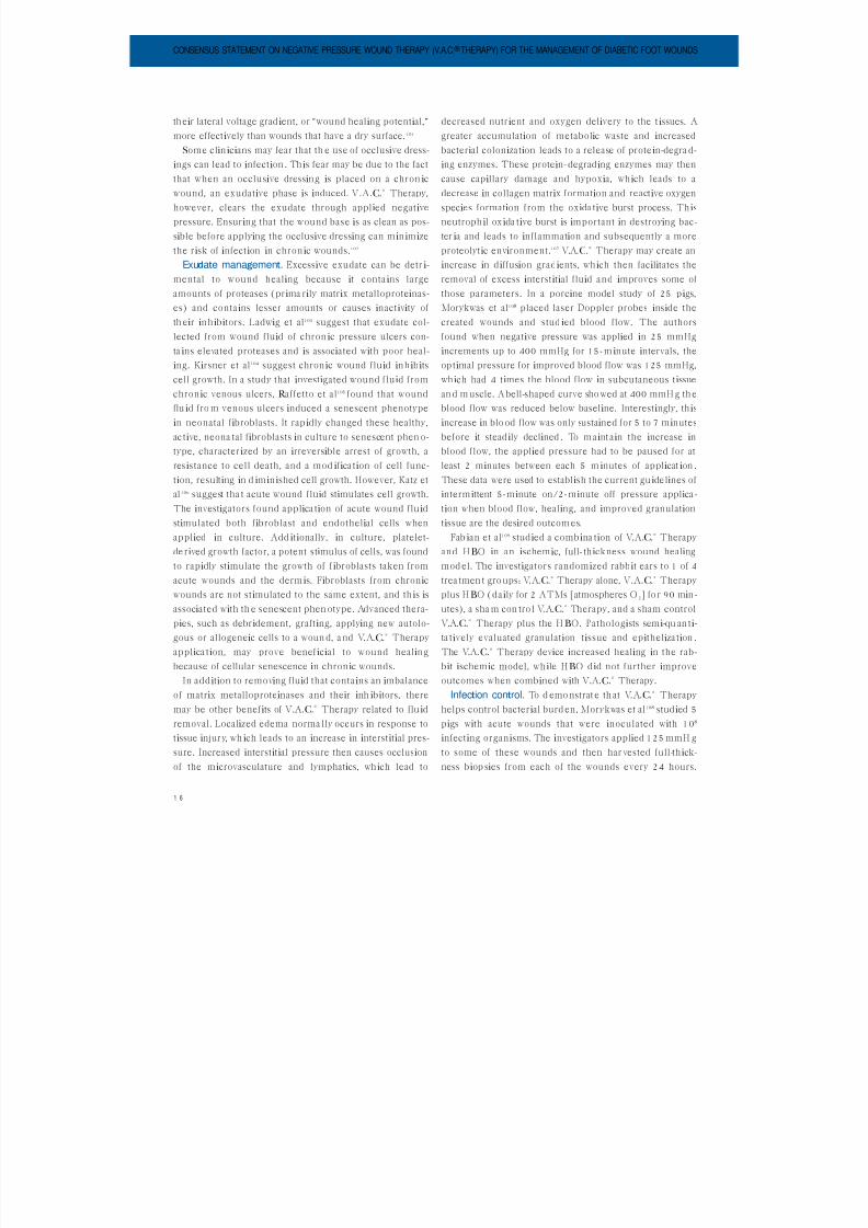
their lateral voltage grad ient, or “wound healing potential,”
more effectively than wounds that have a dry surface. 101
Some clin icians may fear that th e use of occlusive dress-
ings can lead to infection . Th is fear may be due to the fact
that when an occlusive dressing is placed on a chron ic
wound, an exudative phase is induced. V.A.C.® Therapy,
however, clears the exudate through applied negative
pressure. Ensuring that the wound base is as clean as pos-
sible before applying the occlusive dressing can min imize
the risk of infection in chron ic wounds.102
Exudate management. Excessive exudate can be detr i-mental to wound healing because it conta ins large
amounts of proteases (prima rily matrix metalloproteinas-
es) and conta ins lesser amounts or causes inactivity of
th eir inh ibitors. Ladw ig et al103 suggest that exudate col-
lected from wound fluid of chron ic pressure ulcers con-
ta ins elevated proteases and is associated w ith poor heal-
ing. Kirsner et al104 suggest chronic wound fluid inhibits
cell growth. In a study that investigated wound fluid from
chronic venous ulcers, R affetto et al105 found that woundflu id fro m venous ulcers induced a senescent phenotype
in neonatal fibroblasts. It rapidly changed these healthy,
ac tive, neona tal fibroblasts in culture to senescent phen o-
type, characterized by an irreversible arrest of growth, a
resistance to cell death, and a mod ifica tion of cell func-
tion, resulting in d iminished cell growth. However, Katz et
al 106 suggest tha t acute wound fluid stimulates cell growth.
The investigators found applica tion of acute wound flu id
stimulated both fibroblast and endothelial cells whenapplied in culture. Add itionally, in culture, platelet-
derived growth factor, a potent stimulus of cells, was found
to rapidly stimulate the growth of fibroblasts taken from
acute wounds and the dermis. Fibroblasts from chronic
wounds are not stimulated to the same extent, and this is
associated w ith th e senescent phen otype. Advanced thera-
pies, such as debridement, grafting, applying new autolo-
gous or allogeneic cells to a woun d, a nd V.A.C.® Therapy
applica tion, may prove beneficial to wound healingbecause of cellular senescence in chronic wounds.
In addition to removing fluid that contains an imbalance
of matrix metalloproteinases and their inh ibitors, there
may be other benefits of V.A.C.® Therapy related to fluid
removal. Localized edema norma lly occurs in response to
tissue injury, wh ich leads to an increase in interstitial pres-
sure. Increased interstitial pressure then causes occlusion
of the microvasculature and lymphatics, which lead to
decreased nutrient and oxygen delivery to the tissues. A
greater accumulation of metabolic waste and increasedbacterial colonization leads to a release of prote in-degra d-
ing enzymes. These protein-degrading enzymes may then
cause capillary damage and hypoxia, which leads to a
decrease in collagen matrix formation and reactive oxygen
species formation from the oxida tive burst process. This
neutroph il ox ida tive burst is important in destroying bac-
ter ia and leads to inflammation and subsequently a more
proteolytic environment.107 V.A.C.® Therapy may create an
increase in diffusion gradients, which then facilitates theremoval of excess interstitial fluid and improves some of
those parameters. In a porcine model study of 25 pigs,
Morykwas et al108 placed laser Doppler probes inside the
created wounds and stud ied blood flow. The authors
found when negative pressure was applied in 25 mmHg
increments up to 400 mmHg for 15-m inute intervals, the
optimal pressure for improved blood flow was 125 mmHg,
which had 4 times the blood flow in subcutaneous tissue
an d m uscle. A bell-shaped curve sho wed at 400 mmH g th eblood flow was reduced below baseline. Interestingly, this
increase in blo od flow was only sustained for 5 to 7 minutes
before it steadily declined . To m aint ain the increase in
blood flow, the applied pressure had to be paused for at
least 2 minutes between each 5 m inutes of applicat ion .
These data were used to establish the current guidelines of
intermittent 5-minute on/2-minute off pressure applica-
tion when blood flow, healing, and improved granulation
tissue are the desired outcom es.Fab ian et al109 studied a combina tion of V.A.C.® Therapy
and HBO in an ischemic, full-thickness wound healing
mod el. The investigators randomized rabbit ears to 1 of 4
trea tmen t gro ups: V.A.C.® Therapy alone, V.A.C.® Therapy
plus HBO (daily for 2 ATMs [atmospheres O 2] fo r 90 min-
utes), a sha m con tro l V.A.C.® Therapy, and a sham control
V.A.C.® Therapy plus the HBO. Pathologists semi-qu an ti-
ta tively evaluated granulation tissue and epitheliza tion .
The V.A.C.® Therapy device increased healing in the rab-bit ischemic model, while HBO did not further improve
outcomes when combined w ith V.A.C.® Therapy.
Infection control. To d emo nstrat e th at V.A.C.® Therapy
helps control bacterial burd en, Morykwas et al108 studied 5
pigs w ith acute wounds that were inoculated w ith 108
infecting organisms. The investigators applied 125 mmH g
to some of these wounds and then har vested full-thick-
ness biop sies from each of the wounds every 24 hours.
CONSENSUS STATEMENT ON NEGATIVE PRESSURE WOUND THERAPY (V.A.C. ® THERAPY) FOR THE MANAGEMENT OF DIABETIC FOOT WOUNDS

8/13/2019 Consensus Statement on Negative Pressure Wound Therapy for the Management of Diabetic Wounds
http://slidepdf.com/reader/full/consensus-statement-on-negative-pressure-wound-therapy-for-the-management-of 18/33
1 7
They found that between Day 4 and Day 5, wounds treat-
ed w ith V.A.C.® Therapy had a decrease in the bacterialload (105), while the control wounds not receiving V.A.C.®
Therapy co nt inued to h ave elevated levels of bacteria. In
a subsequent pro spective, randomized study, Moues et al7
showed that V.A.C.® Therapy d id not sign ificantly
decrease the bacterial load more than conventional moist
dressing therapy. Interestingly, they found that the non-
fermentative gram-negative bacilli showed a sign ificant
decrease in wounds treated w ith V.A.C.® Therapy, where-
as Staphylococcus aureus showed a sign ificant increase inwounds treated w ith V.A.C.® Therapy. Despite the
increase in Staphylococcus aureus, V.A.C.® Therapy sign ifi-
cantly decreased the wound surface area when compared
to conventional moist dressing therapy. The authors
noted that factors other than reduction of the bacterial
bioburden were responsible for successful healing .
Looking bill et al110suggested th at the presence of h igher
bacterial load w ith in woun ds delays healing, wh ile Krizek
and R obson111 demonstrated that decreasing the woundbacterial count to 105 or less was sufficient to ensure good
skin graft take. In a retrospective study, Weed et a l112 fa iled
to show that the bacterial count dro pped between 104–106
w ith pro long ed use of th e V.A.C.® Therapy System, but the
wounds still responded positively to the V.A.C.® Therapy
System. In other stud ies,66 V.A.C.® Therapy was shown to
reduce the bacterial count to the 104 and 106 range w ith in
4 to 5 days. Th is suggests that bacterial burden reduction
may occur w ith use o f V.A.C.® Therapy w ith in the first 4 to5 days, making subsequent therapy effective in decreasing
wound size. The ro le of instilling antibacterial irrigants via
the V.A.C.® Instill®System to h elp further control the b ac-
ter ial bioburden is ano ther applica tion of V.A.C.® Therapy
(Figure 8). There are 2 reports in the literature by
Bernstein an d Tam 92 an d Wolvos113 show ing clinical effica-
cy of the V.A.C.® Instill® System for treatment of diabeticfoot wound s.
Silver as an irr igant or as a dressing is becoming more
commonly used in wound care for managing infected
wounds or wounds at r isk for infection. The new V.A.C.®
GranuFoam ®Silver™ Dressing (Figure 9) is an option for
cases of complex, colon ized, or infected wounds post-
debridement, for reduction of bacterial bioburden, andto help reduce the risk of recurren t infection in patients
who have severe comorbidities and a h istory of chron ic
wound colonization. The V.A.C.® GranuFoam ®Silver™
Dressing can also be placed over split-thickness skin grafts
in combina tion w ith an appropriate nonadherent layer, as
the dressing has the necessary antimicrobial coverage in
ad d ition to its bolstering effect.
Physical stimulation of cells. In 1892, Julius Wolfe no ted
that bone changed shape in response to physical stress.Subsequently, a German histologist, Richard Thoma,
extra pola ted Wolfe’s observation to soft tissues and found
that the development of blood vessels is governed by
dynam ic forces acting on their walls as follows: an
increase in velocity of blood flow causes d ila tion of the
lumen; an increase in lateral pressure of the vessel walls
causes it to thicken; and an increase in en d pressure caus-
es formation of n ew capillaries. This is the law of tension
FIGURE 8: The V.A.C.®Instill® System allows automated delivery of topical wound solutions to wound sites inaddition to providing benefitsof V.A.C.®Therapy.
FIGURE 9: Ionic silver is micro-bonded to the dressing in sucha way that the foam retains the same porosity and structure of
traditional V.A.C.®GranuFoam®Dressing to simulate the samemicromechanical effects.
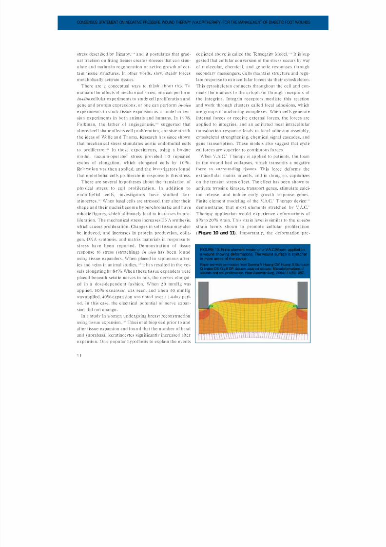
8/13/2019 Consensus Statement on Negative Pressure Wound Therapy for the Management of Diabetic Wounds
http://slidepdf.com/reader/full/consensus-statement-on-negative-pressure-wound-therapy-for-the-management-of 19/33
1 8
stress described by Ilizarov,114 and it postulates that grad-
ual traction on living tissues create s stresses that ca n stim-ulate and ma inta in regeneration or active growth of cer-
ta in tissue structures. In other words, slow, steady forces
metabolically activate tissues.
There are 2 conceptual ways to th ink about th is. To
evaluate the effects of mecha nical stress, one can per form
in-vitro cellular experiments to study cell proliferation and
gene and protein expressions, or one can perform in-vivo
experiments to study tissue expansion as a model or ten-
sion experiments in both an imals and humans. In 1978,Folkman, the father of angiogenesis,115 suggested that
altered cell shape affects cell proliferation, consistent w ith
the ideas of Wolfe an d Thoma. R esearch h as since shown
that mechanical stress stimulates aortic endothelial cells
to proliferate.116 In these experiments, using a bovine
mo del, vacuum-oper ated stress provided 10 repeated
cycles of elongation, wh ich elongated cells by 10%.
R elaxation was then applied, and the investigators found
that endothelial cells proliferate in response to th is stress.There are several hypotheses about the translat ion of
physical stress to cell proliferation. In add ition to
endothel ial cells, investigators have stud ied ker-
at inocytes.117 When basal cells are stressed, they alter their
shape and their nuclei becom e h yper-chroma tic and h ave
mito tic figures, which ultimately lead to increases in pro-
liferation. The mechanical stress increa ses DNA synthesis,
wh ich causes proliferation. Changes in soft tissue m ay also
be induced, and increases in protein production, colla-gen, DNA synthesis, and matrix materials in response to
stress have been reported. Demonstration of tissue
response to stress (stretching) in vivo has been found
using tissue expanders. When placed in saphenous arter-
ies and veins in an imal studies,118 it ha s resulted in th e ves-
sels elongating by 84%. When t hese tissue expanders were
placed beneath sciat ic nerves in rats, the nerves elongat-
ed in a dose-depend ent fa shion. When 20 mmHg was
applied, 30% expansion was seen, and when 40 mmHgwas applied, 40% expan sion was noted over a 14-da y peri-
od. In th is case, the electrical potential of nerve expan-
sion did not change.
In a study in women undergoing breast reconstruction
using t issue expansion ,119 Takei et al biop sied prior to and
after tissue expansion and foun d that the number of basal
and suprabasal keratinocytes sign ificantly increased after
expansion. On e popular hypothesis to expla in the events
de picted above is called t he Tensegrity Model.120 It is sug-
gested that cellular con version of the stress occurs by wayof molecular, chemical, and genetic responses through
secondary messengers. Cells mainta in structure and regu-
late response to extracellular fo rces via their cytoskeleton.
Th is cytoskeleton connects throughout the cell and con-
nects the nucleus to the cytoplasm through receptors of
the integrins. Integrin receptors med iate this reaction
and work through clusters called focal adhesions, which
are groups of anchoring complexes. When cells generate
internal forces or receive external forces, the forces areapplied to integrins, and an activated local intracellular
transduction response leads to focal adhesion assembly,
cytoskeletal strengthen ing, chemical signal cascades, and
gene transcript ion. These models also suggest that cycli-
cal forces are superior to continuous fo rces.
When V.A.C.® Therapy is applied to patients, the foam
in the wound bed collapses, wh ich transmits a negative
force to surround ing tissues. This force deforms the
extracellular matrix in cells, and in doing so, capita lizeson the tension stress effect. The effect has been shown to
ac tivate tyrosine kinases, transport genes, stimulate calci-
um release, and induce early growth response genes.
Fin ite element modeling of the V.A.C.® Therapy device121
demo nstrated th at m ost elements stretched by V.A.C.®
Therapy applica tion would experience deformations of
5% to 20% stra in. This stra in level is similar to the in-vitro
strain levels shown to promote cellular proliferation
(Figure 10 and 11). Importantly, the deformation pre-
CONSENSUS STATEMENT ON NEGATIVE PRESSURE WOUND THERAPY (V.A.C. ® THERAPY) FOR THE MANAGEMENT OF DIABETIC FOOT WOUNDS
FIGURE 10: Finite element model of a V.A.C.®foam applied toa wound showing deformations. The wound surface is stretchedin most areas of the device.
Reprinted with permission f rom Saxena V, Hwang CW,Huang S, EichbaumQ, Ingber DE, Orgil l DP. Vacuum-assisted closure: Microdeformations ofwounds and cell proliferation. Plast Reconstr Surg . 2004;114(5):1087.

8/13/2019 Consensus Statement on Negative Pressure Wound Therapy for the Management of Diabetic Wounds
http://slidepdf.com/reader/full/consensus-statement-on-negative-pressure-wound-therapy-for-the-management-of 20/33
1 9
dicted by the mo del also was similar in morpho logy to the
surface undulations observed in h istological cross-sec-
tions of the wounds. The authors hypothesized that th is
tissue deform ation stretches individual cells, thereby pro-
mo ting proliferation in the wound m icroenvironment.
The authors also found evidence that in chron ic woundsthe applica tion of V.A.C.® Therapy increased the concen-
tration of n ew microvasculature and d ecreased the ma trix
metalloprote inase concentration. They suggested th at th e
microdeformation caused by the V.A.C.® Therapy System
contributed to the change.122
Through the aforementioned mechanisms, the above
cited studies suggest t ha t V.A.C.® Therapy may improve
healing by providing a moist wound healing environ-
ment, improve management of exudate and bacterialburden w ith in the wound, ma inta in wound temperature,
and physically stimulate cells to proliferate.
Benefits. Improvement of skin graft take. In many
clin ical applica tions, V.A.C.® Therapy w ill be used not to
affect primary closure but to enhance the progression of
wounds in preparation for early surgery and to decrease
the severity of the procedure on the reconstructive
ladder. The modality can also help stab ilize flaps,
grafts, and traumatic wounds in preparation for eventual
closure.93,123In itially, some feared that suction from V.A.C.®
Therapy might disturb a graft from its recipient bed, but
clin icians have found that the negative pressure actually
enhances the take of the skin graft by provid ing a bolster
and preventing fluid accumulation beneath the graft.
Schneider et al124 used a nonadherent layer between the
skin graft and the dressing to provide an interfa ce. Th is
method applied continuous bolster of the graft to the
rec ipient bed while min imizing tissue in-growth into the
foam . The investigators left the d ressing in place for 3 or4 days until the graft had taken successfully.
Wound bed granulation. In conjunction w ith the
ba sic tenets of wound healing and appropriate wound
care ( including debridem ent) , V.A.C.® Therapy has been
reported to promote local perfusion in wounds.125–128 By
applying negative pressure un iformly to all points of a
wound, V.A.C.® Therapy w ill also assist in wound contrac-
tion and n ew tissue d evelopmen t. This occurs not only in
pa tients w ith diabetic foot u lcers but also in patients whoare nonoperative (eg, tho se who a re too ill or unstable to
return to the operating room for closure). Although con-
cern was reported regarding use of V.A.C.® Thera py over
exposed bone, tendon, or hardware, current reports in
the literature now support its use in such situation s
(Figure 12).86,88,89,128–130 V.A.C.® Therapy may promote per-
FIGURE 11: Application of V.A.C.®Therapy in
duces surfaceundulations stretching cells in the wound. Wound with
V.A.C.® device at 7 days (lower right) has a higher surface area(blue line) and more blood vessels than a wound not exposed to the foam (lower left).
Reprinted with permission f rom Saxena V, Hwang CW, Huang S,Eichbaum Q, Ingber DE, Orgil l DP. Vacuum-assisted closure:Microdeformations of wounds and cell proliferation.Plast Reconstr Surg . 2004;114(5):1087.
FIGURE 12: Complex diabetic foot wound with
exposed bone and tendon.

8/13/2019 Consensus Statement on Negative Pressure Wound Therapy for the Management of Diabetic Wounds
http://slidepdf.com/reader/full/consensus-statement-on-negative-pressure-wound-therapy-for-the-management-of 21/33
2 0
fu sion and granulat ion tissue formation in these
wounds to the po int that the exposed structures arecovered, and the wound may be successfully grafted or
closed. Some clin icians w ill apply V.A.C.® Therapy b efore
leaving the operating room; however, it is necessary to
wait until hemostasis is ach ieved before applying th is
ac tive thera py.
In a study on both an imals and hum ans, Genecov et al14
covered half the skin of graft donor sites w ith V.A.C.®
Therapy dressing and half w ith a th in-film adhesive dress-
ing. Biopsies were taken every other da y from th e an ima lmodel and on Day 4 and Day 7 from the human model.
The a utho rs found that the V.A.C.® Therapy donor site
wounds had epithelized sign ificantly faster in both an i-
mal and human models. No d ifference in the degree of
pa in in the huma n studies was reported .
Saxena et al121 designed a fin ite element model of the
wound in contact w ith the foam , which showed microde-
formation of tissue between contact po ints of the foam,
resulting in increased surface area of the wound. Thesepred icted deformations are sim ilar to m icroscop ic
woun d cross-sections of V.A.C.® Thera py applica tion. The
investigators hypothesized that stretch ing the wound sur-
face by inducing wound undulation s would stimulate cell
d ivision, proliferation, and angiogenesis through mecha-
n isms described by Ingber.131 Th is was later con firmed in
an an imal study by Chen et al.132
Ease of dressing changes. An add itional benefit is the
ease of applying these dressings. V.A.C.®
Therapy allowsfor reduced ha nd s-on care, mean ing dressing changes
can be performed every 48 hours rather than multiple
times daily, unless the wound is infected (12–24 hours
is recommended for infected wounds).93,123 The device
can be applied at home or in alternative care settings,
and dressings can be changed more frequently if there is
concern about what is taking place under the d ressing in
the early stages of treatment. Mult iple wounds can be
treated at a single time by using a Y-conn ector o rbr idg ing techn ique.
As adjunctive therapy. V.A.C.® Therapy is most often used
adjunctively w ith other agents or modalities. In a brief
review, Espensen et al133 described multimodal therapy
using a tissue substitute in concert w ith V.A.C.® Therapy.
The use of V.A.C.® Therapy as a means to prepare a
wound to receive a skin substitute as well as to promote
take of th e skin substitutes has become increasingly com-
mon. Usually, some type of nonadherent dressing is
applied between th e graft a nd th e V.A.C.® GranuFoam®
to prevent adherence of the graft to the foam. Other
recent reviews have also documented the benefits of
using V.A.C.® Therapy in conjunction w ith various wound
healing a gents or plastic procedures to augment wound
healing. 93,94,123,128,134
Clinical Evidence for V.A.C.®TherapyIn 1997, Morykwas an d Argenta 108,129 reported the orig-
inal an imal and clin ical stud ies on the V.A.C.® TherapySystem a nd showed th at use of V.A.C.® Thera py resulted
in enhanced granulation tissue formation and improved
bacterial clearance compared to control dressings.
There also appeared to be increased flap survival. Some
of the therapeutic benefits of V.A.C.® Therapy that were
postulated by Morykwas and Argenta included aggres-
sive reduction in local interstitial edema, increases in
local blood flow, evacuation of excessive dra inage,
decreased bacterial colon iza tion, and conversion of anopen wound into a controlled closed wound, wh ich facil-
itates less frequent dressing changes and protects the
wound from the patient and h is or her surround ings.
Therapeutic benefits also included provid ing a moist
wound -hea ling environment, which has become the
standard of care. These f ind ings, including those on
enhan ced cutaneous blood flow during V.A.C.® Therapy,
have recently been reviewed and corroborated in both
an imal and h uman studies.3,93,135
One large a nd 2 smaller RCTs demo nstra ted level 1 evi-
dence for V.A.C.® Therapy’s effectiveness in treating d ia-
be tic foot wounds (Figure 13). In a large RCT of 162
pa tients w ith d iabetes and partial foot amputation
CONSENSUS STATEMENT ON NEGATIVE PRESSURE WOUND THERAPY (V.A.C. ® THERAPY) FOR THE MANAGEMENT OF DIABETIC FOOT WOUNDS
FIGURE 13: Diabetic foot amputation treated withV.A.C.®Therapy.
8/13/2019 Consensus Statement on Negative Pressure Wound Therapy for the Management of Diabetic Wounds
http://slidepdf.com/reader/full/consensus-statement-on-negative-pressure-wound-therapy-for-the-management-of 22/33
2 1
wound s, Armstrong a nd La very2 determined that patients
treated w ith V.A.C.® Therapy had better outcomes com-pared to control in proportion, rate of healing and fewer
re-am putat ions. In th is 16-week, 18-site trial, the
researchers’ a im was to determine if V.A.C.® Therapy
improved the proportion and rate of wound healing in
these complex wounds. Patients were random ized to
receive either V.A.C.® Therapy (n = 77) w ith dressing
changes every 48 hours or moist wound therapy (n = 85).
Pa tients in the control arm were treated w ith alginates,
hydrocolloids, foams, or hydrogels, accord ing to stan-dardized consensus guidelines, and these dressings were
changed daily, unless the treating physician recommend-
ed otherw ise. All study part icipants received appropriat e
offloading th erap y (a pressure-relief walker or sandal).
Pa tients were treated until ach ievement of complete
wound closure or completion of the 112-day act ive treat-
ment phase. Complete wound closure was defined as
100% re-epitheliza tion w ithout drainage. Wh ile the pri-
mary endpoint was wound closure, researchers alsoreported on rates of wound closure, granulation tissue
formation, and amputations.
The rates of complete wound closure, t ime to closure,
granulation tissue formation, a nd re-amputation favored
V.A.C.® Therapy. Forty-three V.A.C.® Therapy patients
(56%) ach ieved complete wound closure compa red to 33
(39%) in the control group (P = 0.040). The rate of
wound healing was stat istically sign ificant in favor of the
V.A.C.®
Thera py group based on a shorter time to closure(P = 0.005) as was the time to granulation tissue forma-
tion (76%–100%, P = 0.002). Comparison of the number
of second amputations also favored V.A.C.® Therapy
pa tients (2) compared to control patients (9), although
the d ifference was not sign ificant. The complica tion rate
was not sign ificantly different between the groups, and
infections were the most commonly reported adverse
event.
This RCT differs from previous stud ies of open footwounds in patients w ith diabetes because it included larg-
er wounds. A literature search for RCTs on diabetic foot
wounds3,75,85–88,93,136 reveals a concentration on superficial
neuropathic foot wounds (2.4 cm2 to 2.9 cm 2). The
wounds in the current study, however, were 7 to 8 times
that size (~20.7 cm2). The authors consider that it is
“somewhat compelling” that the proportion of healed
wounds was similar—and, in fact, better—than in other
non-V.A.C.® Therapy stud ies w ith more superficial
wounds and the same endpoints.The au tho rs conclud ed th at V.A.C.® Therapy seems to
be a safe and effect ive treatment for complex diabetic
foot wounds. Most wounds (70%) healed by secondary
intention, although some required add itional reconstruc-
tion, w ith or w ithout amputation. Thus, the applica tion of
V.A.C.® Therapy in th is setting permitted simple solution s
to complicated reconstructive challeng es. The f ind ings in
th is large RCT are consistent w ith outcomes noted in 2
smaller V.A.C.® Therapy RCTs.9,13
In a random ized, controlled, crossover study, Eginton
et al9 com pare d V.A.C.® Therapy to mo ist gauze dressings
in the treatment of large d iabetic foot wounds. Patients
were rando mized to one treatment for 2 weeks and then
received the alternative treatment for 2 weeks. Seven
wounds in 6 patients provided the data for statistica l
analysis. V.A.C.® Therapy decreased wound volume and
depth sign ificantly more than moist gauze dressing s (59%
versus 0% and 49% versus 8%, respectively). G iven thesmall sample size and limited duration of each 2-week
treatment phase, the auth ors concluded th at V.A.C.®
Therapy is effective over a 2-week per iod but stated that it
was not possible to determine the appropriate length of
V.A.C.® Therapy based on the results of th is tr ial.
However, the ability of V.A.C.® Therapy to decrease
wound depth and volume shown in t his study is consistent
w ith the granulation tissue formation find ings of
Armstrong and Lavery.2
In another small RCT, McCallon et al13 sought to
determ ine whether V.A.C.® Therapy increased wound res-
olution when compared to saline-mo istened gauze
in the treatment of postoperative diabetic foot wound s. In
th is pilot study of 10 pat ients (V.A.C.® Therapy, n = 5; con-
trol, n = 5), outcome measures were t ime to
sat isfactory healing (wh ich was ach ieved either b y delayed
pr imary intention o r by seconda ry intention) and change
in woun d surfa ce area . The V.A.C.®
Therapy group ha d anaverage time to satisfactory healing of 22.8 (± 17.4) days,
compared to the control group average of 42.8 ( ± 32.5)
days. The authors considered that the small sample size
(n = 10) precluded statistical sign ificance. Average
changes in woun d surface a rea also fa vored the V.A.C.®
Therapy group, which showed a 28.4% (± 24.3) average
decrease compared to th e control group (9.5%, ± 16.9)
average increase in surfa ce area. O n averag e, V.A.C.®
8/13/2019 Consensus Statement on Negative Pressure Wound Therapy for the Management of Diabetic Wounds
http://slidepdf.com/reader/full/consensus-statement-on-negative-pressure-wound-therapy-for-the-management-of 23/33
2 2
Therapy patients achieved satisfactory healing via defini-
tive closure approximately 3 weeks sooner than controlpa tients. The results of th is pilot study are also con sistent
w ith the find ings of the large RCT in wh ich V.A.C.®
Therapy wounds ach ieved complete wound closure more
favorably than control wounds.
Though V.A.C.® Therapy can be used on any size
wound, it has been shown to be especially useful on deep,
complicated, nonhealing wounds of m ixed etiologies. In
2002, Armstrong et al136 presented level 2 evidence in a
retrospective review of 31 consecutive patients w ith dia-betes at 2 wound center s. V.A.C.® Therapy was applied to
large wounds follow ing surgical debridement and contin-
ued until 100% gra nula tion tissue co verage was ach ieved.
The wound size was 28 cm 2 w ith a mean duration of the
wounds before therapy of 25 weeks. The investigators’
outcomes were time to closure, proportion of healing,
healing at the same level of initial debridement w ithout
requiring a more proximal amputation, and incidence of
complica tions. About 90% of th ese wound s healed a t theinitial level at a mean of 8 weeks, and wh ile the mean
duration of V.A.C.® Therapy was approximately 5 weeks,
most often it was used for only 2 weeks. Mult imodal ther-
ap ies, includ ing split-thickness skin gra fts, were then used
to facilitate closure after the wound had been prepared
w ith V.A.C.® Therapy. The few complications were 6
reports of wound maceration under and around the
dressing, 1 occasion of periwound cellulitis, and 1 deep
space infection. Three of 31 patients received higher levelamputation s (1 below-the-knee and 2 tran smetata rsal
amputations), wh ich is consistent w ith what can be
expected w ith in th is patient population. The auth ors con-
cluded tha t “th e use of V.A.C.® Therapy to assist in the
development of a granular base in diabetic foot wounds
may show promise in th is patient population that is at a
h igh risk for amputations.”
In a report of level 2 evidence from a Veterans Affairs
Med ical Center, Page et al91
retrospect ively reviewed 22pa tients w ith 23 d iabetic foot ulcers on whom V.A.C.®
Therapy had been applied and compared these patients
to 24 patients treated w ith standard therapy. This was a
h istorical review rather than a comparative trial. V.A.C.®
Therapy was continued until the wounds were filled w ith
granulation tissue or prepared for eventual closure.
Pa tients were followed da ily as inpatients; once they left
the hospital, they were followed weekly. Patients were fol-
lowed either until healed or for 1 year after in itial surge ry.
With the patients on standard therapy (moist saline dress-ings), the wounds took longer to fill as well as to heal;
there were sign ificant differences between groups for
both parameters. Page et al also stud ied complica tions,
but they did not specifically enumerate which complica-
tions they were studying. Such complica tions, however,
would include infections or having to return to surgery
for further revisions and/or amputations. Again, there
were sign ificantly fewer complica tions in the V.A.C.®
Therapy-treated gro up compa red to th e control gro upand sign ificantly fewer additional surgeries necessary in
the actively treated group. The investigators stud ied the
readmission days as well as the number of readmissions.
G enera lly, they foun d th at V.A.C.® Therapy patients
required fewer adm issions and fewer days in the ho spita l
because they were able to be treated as outpat ients or in
a skilled nursing care center much earlier than the con-
trol patients.91
As seen in th is retrospective review by Page et a l, a skingraft was used in some instances to facilitate closure
after preparing the wound bed w ith V.A.C.® Therapy.
However, it is important to assess whether a nonadher-
ent layer is necessary to avo id tissue in-growth into the
foam. I n a study by Schneider et al,124 investigators used
a nonadherent layer interface between the skin graft
and the dressing. Th is method applied continuous bol-
ster of the graft to the recipient bed wh ile m in im izing
tissue in-growth into th e foam. The investigators left thedressing in place for 3 or 4 days until the graft had taken
successfully.
Mo isid is et al6 also assessed whether split-thickness skin
graft take is improved qualita tively or quantita tively in a
small, prospective, blinded, RCT comparing V.A.C.®
Therapy to standard bolster dressings. Th is level 1 evi-
dence study evaluated 22 adult inpatients w ith wounds
requiring skin grafting between July 2001 and July 2002.
After grafting, each wound half was random ized toreceive either a standard bolster dressing or V.A.C.®
Therapy. Skin gr aft a ssessment was perform ed a t 2 weeks
by a single blinded observer. There were 20 patients in
the study group w ith a median age o f 64 years. The m ean
wound size was 128 cm 2. At 2 weeks, woun ds tha t received
V.A.C.® Therapy had a greater degree of epithelization in
6 cases (30%), the same degree of ep ithelization in 9
cases (45%), and less ep ithelization in 5 cases (25%),
CONSENSUS STATEMENT ON NEGATIVE PRESSURE WOUND THERAPY (V.A.C. ® THERAPY) FOR THE MANAGEMENT OF DIABETIC FOOT WOUNDS

8/13/2019 Consensus Statement on Negative Pressure Wound Therapy for the Management of Diabetic Wounds
http://slidepdf.com/reader/full/consensus-statement-on-negative-pressure-wound-therapy-for-the-management-of 24/33
compared w ith their respective control wounds. Graft
quality follow ing V.A.C.® Therapy was subjectively deter-mined to be better in 10 cases (50%), equivalent in 7
cases (35%), and worse in 3 cases (15%). Although the
quantita tive graft take was not sign ificant, the qualita tive
graft take was found to be sign ificantly better w ith the use
of V.A.C.® Thera py (P < 0.05).
Aside from the aforementioned studies, there are mo re
than 300 manuscripts ranging between level 1 and level 5
evidence that demonstrate the safety and eff icacy of
V.A.C.®
Therapy and show how th is dynamic therapy canbe an important part of the management strategy for
treating diabetic foot wounds. Figure 14 illustrates a clin-
ical treatment pathway for d iabetic foot ulcers including
the appropriate use of V.A.C.® Therapy.
Economic Impact of V.A.C. ® Therapy
D iabetic foot problems are a major worldw ide concernand can result in sign ificant healthcare costs. Peripheral
neuropa thy and vascular disease are ma jor risk factors for
pa tients w ith diabetes and predispose these patients to
foot ulceration and lower-extremity amputation. It is also
important to consider when assessing the economic
impact of diabetic foot disease that the lifet ime risk of
developing a foot ulcer can be as h igh as 25%, and the
rate of the fo ot ulcer recurrence is greater than 50% after
3 years.48
The costs associated w ith major amputation arestaggering, not only from an economic perspective, but
from a psychosocial and emotional perspective as well.
Wh ile traditiona l method s may offer the mo st expeditiou s
solution to a chron ic problem, the lon g-term econo mic
WOUND
UT Grade 1
> UT Grade 1
Medical assessment
Appropriate dressing
Offloading
Not progressing
toward healing
at 30 days
Revascularization,
as appropriate
Infection control,
as appropriate
Re-evaluate weekly
Reassess therapy
Infected wound
No infected wound
Progressing toward
healing at 30 days
Continue current
treatment regimen
Dressing changes
every 12–24 hours
Continue V.A.C.®
Therapy
Consider V.A.C.®
Therapy
No excessive
maceration/infection
Excessive
maceration/infection
Patient with
complex DFU*
Re-evaluate wound
after 24 hours
Dressing changes
every 48 hours
Re-evaluate every
2–4 weeks
WOUND
HEALED
* complex = > UT Grade 1; may also include
Grade 1 if patient has failed appropriate therapy
as defined in recommendations
FIGURE 14: Treatment of Diabetic Foot Ulcer (DFU) with V.A.C. ® Therapy
2 3
CONSENSUS STATEMENT ON NEGATIVE PRESSURE WOUND THERAPY (V A C ® THERAPY) FOR THE MANAGEMENT OF DIABETIC FOOT WOUNDS

8/13/2019 Consensus Statement on Negative Pressure Wound Therapy for the Management of Diabetic Wounds
http://slidepdf.com/reader/full/consensus-statement-on-negative-pressure-wound-therapy-for-the-management-of 25/33
2 4
outlook is one of escalating medical and emotional bur-
den. It is, therefore, in the best interest of both thepa tient and wound care special ist to utilize alternative
management strategies to ach ieve healing of d iff icult
wounds and provide limb salvage not based on da ily cost
but on th e overall econom ic impact to hea l these wounds.
Gordois et al137 reported that the total annual cost of dia-
betic peripheral neuropathy and its complica tions in the
U nited States was estimated to b e between $4.6 and $13.7
billion ( data from 2001). Up to 27% of the d irect medica l
cost of d iabetes may be attributed to diabetic peripheralneuropathy.137
The use of the V.A.C.® Therapy System (Figure 15) can
be an important addition to wound management strate-
gies, help decrease overall healthcare expend itures, and
improve quality of life for patients. In addition to the
tremendous physical toll of foo t ulceration and amputa-
tion, there is an enormous economic impact. The esti-
mated cost of treating a single foot ulceration42
dur ing a2-year period is $28,000, and foot problems may account
for 40% of healthcare resources in developing coun-
tr ies.48
Shearer et al138 confirmed that patients w ith diabetes
and neuropath ic risk factors incur 5 times more direct
me dical costs for ulcers and amputations and live for 2
months less than patients w ithout neuropathy. Swed ish
researchers reported tha t an intensified prevention strat-
egy involving education, foot care, and footwear would
be cost effective and even cost saving if applied topa tients w ith risk factors.139
Armstrong et al140 presented a poster at the 2006
American Diabetes Associat ion meeting that compared
successful healing endpo ints when treating diabetic foot
ulcers w ith V.A.C.® Therapy versus convention al wet-to-
mo ist therapy to a ssess the o verall treatm ent cost of the 2
therapies in community-based ho me hea lthcare pa tients.
The data was based on a retrospect ive database analysis
of Med icare Part B d iabetic foot ulcer outpatient data forpa tients treated w ith V.A.C.® Therapy between 1996 and
2004. The results were compared to published analyses
of diabetic foot ulcers treated using wet-to-moist treat-
ment. More than 1,100 patients in the V.A.C.® Therapy
group were matched to the wet-to-mo ist comparison
group of 586 patients. All data were equally matched
except for wound size, wh ich was larger w ith in the
V.A.C.® Therapy group. The data showed that after 12
weeks of trea tmen t, 39.5% of the V.A.C.®
Therapy-treatedpa tients ach ieved successful healing compared to 23.9%
of the wet-to-moist group (P < 0.001). After 20 weeks of
treatment, 46.3% of the V.A.C.® Therapy group ach ieved
successful healing endpoints compared to 32.8% of the
wet-to-moist group (P < 0.001). In add ition, the 20-week
expected cost o f treat ment for V.A.C.® Therapy p at ients
in the home care setting was reduced by more than 40%
when co mpared to th e wet-to-moist treatment option
($16,733 versus $28,691). Thus, the V.A.C.®
Therapypa tients were associated w ith greater healing at 12 weeks
an d 20 weeks, which resulted in successful treatment o ut-
comes at an overall lower cost of care when compared to
wet-to-mo ist treatment of patients w ith diabetic foot
ulcers in the outpatient hom e care setting.
Frykberg et al141 presented a poster at the 2006
International Society for Pharmacoeconom ics and
Outcomes R esearch meeting that assessed the benef its
of V.A.C.®
Therapy in reducing the incidence of lower-extrem ity amputations in patients w ith d iabetic foot
ulcers compared to trad itional wound therapies. A ret-
rospective da tabase ana lysis was conducted using adm in-
istrative data from 2 d ifferent sources. The commercial
dataset yielded more than 3,500 patients meeting the
study crite ria, wh ile the Med icare data set included more
than 12,700 patients. Th is retrospective analysis demon-
strated that the proportion of patients w ith an amputa-
CONSENSUS STATEMENT ON NEGATIVE PRESSURE WOUND THERAPY (V.A.C. ® THERAPY) FOR THE MANAGEMENT OF DIABETIC FOOT WOUNDS
FIGURE 15: The V.A.C.®Therapy Family of devices.

8/13/2019 Consensus Statement on Negative Pressure Wound Therapy for the Management of Diabetic Wounds
http://slidepdf.com/reader/full/consensus-statement-on-negative-pressure-wound-therapy-for-the-management-of 26/33
2 5
tion was lower follow ing treatment w ith V.A.C.® Therapy
compared to the control gro up. This find ing was consis-tent for the commercial and Med icare datasets. From
the commercial dataset population, V.A.C.® Thera py was
associated w ith up to a 34% lower amputation occur-
rence compared to the control group ( P = 0.09). In the
Med icare population, the d iabetic foot ulcer risk-adjust-
ed amputation percentages were 16.6% for control and
10.8% for V.A.C.® Therapy groups (P = 0.007). The
results from 2 distinct patient samples suggest that
V.A.C.®
Thera py lowers the incidence of amputations byup to 35% in comparison w ith trad itional wound thera-
pies. V.A.C.® Therapy, therefore, has the potent ial to
substantially improve treatment outcomes for patients
w ith d iabetic foot ulcers and to reduce the econom ic
burden of lower-extrem ity amputations on the health-
care system.
Niezgoda et al142 investi-
gated the cost effect iveness
of V.A.C.®
Therapy as anadjunctive modality in the
overall management of
pa tients at risk for limb loss
due to complex wounds.
Evaluation o f cost efficacy of
V.A.C.® Therapy was accomplished in a group of 10
pa tients who presented to th e authors’ wound ca re cen-
ter for evaluation and second opin ion regard ing poten-
tial limb salvage. All of these patients had previouslyreceived the recommenda tion for b elow-the-knee am pu-
ta tions at other centers. All 10 pat ients had sign ificant
wound healing compromise, including 1 patient w ith an
exposed Ach illes tendon overlying a necrotic calcaneal
wound complicated by osteomyelitis. Several patients
developed extensive soft tissue loss due to necrot izing
fasciitis; some included exposed bone, fascia, and ten-
don. Despite prior below-the-knee a mputa tion recom-
mendations, these patients were h ighly motivated toach ieve limb salvage. All pat ients were mana ged w ith an
aggressive limb salvage effort, wh ich included arterial
vascular evaluation and intervention, if ind icated; plastic
and orthoped ic surgical con sultation for tissue stabiliza -
tion and closure, when appropriate; HBO; and aggres-
sive wound care utilizing V.A.C.® Therapy. All patients
were d ischarged after successfully ach ieving bipedal
ambulation .
The po sitive clinical outcomes in th is group of pat ients
led to a retrospect ive analysis to assess whether incorpo-ra tion of V.A.C.® Therapy provided economic benefit.
The investigators d iscovered that these 10 pat ients
averaged 17 days of ho spita liza tion, including both acute
and in-hosp ital rehabilita tion days. In comparison, the
hospital d iagnosis-related gr oup d ata base revealed
that patients who un derwent b elow-the-knee ampu tation
for similar cond itions had an average hospita liza tion of
31 da ys.
Analysis of data specific to Med icare patients143
hasdetermined that the national daily hospital cost per
pa tient averages $1,426. Simple calculation suggests that
when V.A.C.® Therapy is incorporated into a comprehen-
sive man agemen t strategy, as in t h is group of limb salvage
pa tients, a cost avoidan ce of $19,964 can be ap preciated
by decreasing hospital stay
($1,426 x 14 d ays).
In add ition, ach ieving
limb salvage and ma int ain-ing bipedal ambulation also
avo ided the cost of prosthe-
sis fabrica tion, fitt ing, and
rehabilita tion. It has been
reported 137 that these costs
can range from $20,000 to $40,000. Thus, a strategy that
atta ins limb salvage can ach ieve cost avoidance of more
than $73,000 per patient.
This information suggests that these cost conclusion sare optim istic and that shortened hospital stays,
decreased overall medical costs, and limb salvage can be
atta ined when V.A.C.® Therapy is incorporated into the
care plan of patients at risk for limb loss.142
Moues et al8 showed in a prospective RCT of level 1 evi-
dence tha t th e costs of V.A.C.® Therapy were simila r
when compared to conventional therapy (moist gauze)
in th e man agement o f full-thickness wounds that
required surgical clo sure. To a ch ieve th is, the d irectme dical costs of the to tal num ber of resources needed to
ac h ieve a healthy, granulating wound bed th at was ready
for surgical closure were calculated. Fifty-four patients
ad mitted to a department of plast ic and reconstructive
surgery were recruited into the trial. Cost analysis sho wed
sign ificantly h igher mean material expenses for wounds
treated w ith V.A.C.® Therapy (Euro 414 ± 229 [SD]; US
$520.65) compared w ith con ventional th erapy (Euro 15
V.A.C.®THERAPY AS PART OF AWOUND MANAGEMENT STRATEGY
COULD LEAD TO COST AVOIDANCE.
CONSENSUS STATEMENT ON NEGATIVE PRESSURE WOUND THERAPY (V.A.C. ® THERAPY) FOR THE MANAGEMENT OF DIABETIC FOOT WOUNDS

8/13/2019 Consensus Statement on Negative Pressure Wound Therapy for the Management of Diabetic Wounds
http://slidepdf.com/reader/full/consensus-statement-on-negative-pressure-wound-therapy-for-the-management-of 27/33
2 6
± 11; P < 0.0001; US $18.86) but sign ificantly lower mean
nursing expenses (Euro 33 ± 31 and Euro 83 ± 58; US
$41.50 and US $104.38) for V.A.C.® Therapy and conven-
tional therapy, respectively (P < 0.0001). Hospita liza tion
costs were lower in the V.A.C.® Therapy group (Euro 1788
± 1060; US $2,248.59) than in th e conventional treatment
group (Euro 2467 ± 1336; P < 0.043; US $3,102.50) due to
an average shorter duration until they were ready for sur-
gical closure. There was no sign ificant difference in total
costs per patient between the 2 therap ies (Euro 2235 ±
1301 for V.A.C.®
Therapy [US $2,810.74] versus Euro2565 ± 1384 for conventional therapy [US $3,225.74]).
The authors concluded that the lower number of t ime-
consuming dressing changes and the shorter durat ion
un til they were ready for surgery compensated for the
h igher material costs of V.A.C.® Therapy. As a result,
V.A.C.® Therapy was equally as expensive as conventional
mo ist gauze.
The large RCT by Armstrong and Lavery2is also being
assessed f or V.A.C.®
Therapy cost effectiveness in treatingcomplex diabetic foot wounds versus standard mo ist
wound therapy. The preliminary data presented here
appear to suggest that ad d ing V.A.C.® Therapy as part of a
wound m ana gement strategy could lead to cost avoidance
when managing diabetic foot problems and can result in
sign ificant co st savings.
Conclusion
G uide lines fo r th e use of V.A.C.®
Therapy in the treat-ment of the diabetic foot were first introduced in 2004 by
a panel of experts who convened in Tucson, Ariz. Since
2004, the clin ical evidence in support of V.A.C.® Therapy
has increased, including the publica tion of a large RCT in
The Lancet in November 2005, as well as several small
RCTs, retro spect ive stud ies, and large case series.
Clinicians must choose the most appropriate treatment
for their patients using eviden ce-based med icine, and
although this consensus document
is cons
idered level 5
evidence, the findings are based on level 1 and level 2 evi-
dence that supports the recommendations from the mul-
tidisciplinary expert panel. Wh ile proper debridement,
infection control, and adequate blood supply are
required for successful limb salvage or foot reconstruc-
tion, the use of the V.A.C.® Therapy System has enabled
clin icians to solve complex wound problems w ith more
simple solutions.
Important Questions and Answers on Appropriate
V.A.C. ®
Therapy Use1. How long should V.A.C.® Therapy be used in treatment of a
diabetic foot wound? In most cases, V.A.C.® Therapy may be
used to ach ieve a healthy granular bed prior to possible
consideration of other modalities or surgical procedures.
V.A.C.® Therapy may be used occasionally until closure.
For example, its continued use may be considered in a
subset of patients if they are too ill to undergo add itional
procedures or refuse add itional surgical intervention .
Wound heal ing progress is important to justify continuedV.A.C.® Thera py use.
2. Should V.A.C.® Therapy be applied without debriding the
wound? No. Surgical debridement is critical to healing the
diabetic foot wound in the presence of devita lized tissue
and peri-woun d hyperkerato sis; therefore, the wound
must be debrided prior to applica tion of V.A.C.® Therapy.
If surgical debridement is not an option, mechanical or
enzymatic debridement should be considered prior to
placem ent of V.A.C.®
Therapy in order to ach ieve the fullbenefit of therapy. Cau tion should be taken when using
V.A.C.® Therapy after surgically debriding patients on
an ticoagulants.
3. How should the patient using V.A.C.® Therapy be evaluated
on an outpatient basis? R esponsible clin icians should evalu-
ate patients on an outpatient basis pr ior to in itiat ing
V.A.C.® Therapy, then regularly evaluate the wound’s
progress in the follow ing areas: reduction in depth,
reduction in surface area, and increased granulation tis-sue. A wound that fails to improve during a period o f 2–4
weeks should prompt the clin ician to consider alternative
and/or adjunctive therapies, assuming the clin ician has
revisited the possibilities of inadequate debridement,
infection, ischemia, inadequate offload ing, nutritional
status, and V.A.C.® Therapy compliance.
4. When should V.A.C.® Therapy be applied following revas-
cularization? Advanced wound healing m oda lities,
includ
ing V.A.
C.
®
Therapy, are not substitutes for revas-
culariza tion. V.A.C.® Therapy should not be applied to
an ischemic wound if revasculariza tion has not been
considered. Clin icians may apply V.A.C.® Therapy fol-
lo w ing revasculariza tion if: 1) infection is being treated;
2) there is no active bleed ing (remember to exercise
caution in patients taking anticoagulants); and 3) there
is m in imal necrotic t issue remain ing a nd no eschar. The
goal is to close the wound as soon as is medically possi-
( )

8/13/2019 Consensus Statement on Negative Pressure Wound Therapy for the Management of Diabetic Wounds
http://slidepdf.com/reader/full/consensus-statement-on-negative-pressure-wound-therapy-for-the-management-of 28/33
2 7
ble, but if there are concerns of potential bleed ing, it is
recommended to wait at least 24 hours before plac ingV.A.C.® Therapy.
5. When should V.A.C.® Therapy be applied after
incision, drainage, and debridement of infection? Generally,
V.A.C.® Therapy should not be applied immediately
follow ing infection incision, dra inage, and debride-
ment. R ather, the clin ician should evaluate the wound
for at lea st 24 ho urs before V.A.C.® Thera py applica tion ,
keeping in mind that residual necrotic tissue is an
imped iment to healing and debridement at the time ofinfection dra inage is essential at th is stage. R epeated
debridement ma y be required in order for the pat ient to
receive the full benefit of V.A.C. Therapy.® If the clin i-
cian decides to apply V.A.C.® Therapy immediately fol-
lo w ing an incision and dra inage procedure, the wound
should be evaluated every 12 hours for the f irst 2 to 3
days, and if there is still active infection, the dressing
should be changed every 12–24 hours until the clin ica l
signs of infection resolve.6. Should V.A.C.® Therapy be applied over an active soft tis-
sue infection? V.A.C.® Therapy can be a useful adjunct in
the management of diabetic foot infections; however,
V.A.C.® Therapy should not be applied to an untreated
d iabetic foot infection. The clin ical importance of
assessing colon iza tion and quantita tive bacteriologic
assessment of th e wound during V.A.C.® Thera py has not
yet been elucidated. The d iagnosis of the genesis and
resolut ion o f infect ion is a clin ical one (see theInfectious D iseases Society of America [ISDA] guide-
lines).144
7. How should V.A.C.® Therapy be used in patients with
osteomyelitis? V.A.C.® Therapy should not be used in cases
of untreated osteomyelitis. Prior to applica tion of V.A.C.®
Therapy, clinicians should surgically resect and/or med-
ically suppress osteomyelitis. V.A.C.® Therapy is useful in
obtaining granulation tissue over exposed bo ne in these
pa tients. Wh
ile wounds that probe deeply are clearly a
risk factor for infection, one should keep in mind that
exposed bone is not necessarily infected bo ne.66,145
8. How should noncompliance to V.A.C.® Therapy be defined?
In general, clin icians should not use V.A.C.® Therapy on
pa tients who historically demonstrate an ina bility or
un w illingness to participate in their own care. R epeated
events of noncompliance are indica tions that a clin ician
should choose an alternate technology or approach.
Adequate compliance means that patients are using the
de vice appropriately and consistently and are receivingdressing ch anges as prescribed.
9. How should V.A.C.® Therapy be used in combination with
other modalities? Published literature has shown that
V.A.C.® Therapy can be used in combina tion w ith a variety
of modalities includ ing bioengineered tissues, antimicro-
bial-contain ing dressings, cytokines, HBO therapy, and
other advanced mod alities. R ecent nonrandomized stud-
ies suggest that V.A.C.® Therapy can be used successfully
in conjunction w ith acellular matrix scaffolds.83
Clin ician sshould employ the criteria in gu ide line #3 to mon ito r
progress.
10. Should small, superficial wounds be considered for
V.A.C.® Therapy? Generally, if small, superficial wounds
are respond ing well to adequate debr idement and
offload ing, V.A.C.® Therapy may not be necessary.
Clin icians should not determ ine the appropr iateness
of V.A.C.® Therapy based only on wound size or depth
but rather should ta ke into account anatom ic location ,involvement of underlying structures, and future func-
tional concerns.
11. How should success in the use of V.A.C.® Therapy be
defined? The primary defin itions of success are reduced
time to complete healing; increased prevalence of heal-
ing at a po int in time; and a dequate preparation fo r sur-
gical closure. Secondary defin itions of success for
V.A.C.® Therapy use include increased rate of healing;
decreased time to 100% granular bed; improved qualityof life; decreased requirement for further procedures;
pa in reduction; d ecreased cost of care; red uced level of
nursing care required; fewer peri-therapeutic complica -
tions; lower proportion of amputation at given time
po ints; impro ved long -term healing; and increased dura-
bility of closure.
12. Can one combine effective offloading with V.A.C.®
Therapy? It may be feasible to combine V.A.C.® Therapy
bridg
ing techn
iques w
ith effect
ive offload
ing modal
ities
to allow limited outpatient weight-bearing. ■
References
1. Centre for Evidence-Based Med icine. Levels of evidence and
grades of recommendation. Available at: http://www.cebm.net/
levels_of_eviden ce.asp. Accessed September 28, 2005.
2. Armst ron g D , Lave ry L; D iabetic Foot Study Consortium. Negative
CONSENSUS STATEMENT ON NEGATIVE PRESSURE WOUND THERAPY (V.A.C. ® THERAPY) FOR THE MANAGEMENT OF DIABETIC FOOT WOUNDS

8/13/2019 Consensus Statement on Negative Pressure Wound Therapy for the Management of Diabetic Wounds
http://slidepdf.com/reader/full/consensus-statement-on-negative-pressure-wound-therapy-for-the-management-of 29/33
2 8
pressure wound therapy after partial d iabetic foot amputation: a
multicentre, random ised controlled tr ial. Lancet.2005;366(9498):1704–1710.
3. Timmers MS, Le Cessie S, Banwell P, Jukema GN. The effects of
varying degrees of pressure delivered by negative-pressure wound
therapy on skin perfusion. Ann Plast Surg. 2005;55(6):665–671.
4. Jones SM, Banwell PE, Shakespeare PG . Interface d ressings influ-
ence the delivery of topical negative-pressure therapy. Plast Reconstr
Surg. 2005;116(4):1023–1028.
5. Jesch ke MG , R ose C, Angele P, Fuchtm eier B, Nerlich MN, Bolder
U. D evelopment of new reconstructive techn iques: use of Integrain combina tion w ith fibr in glue and negative-pressure th erapy for
reconstruction of acute and chron ic wounds. Plast Reconstr Surg.
2004;113(2):525–530.
6. Moisid is E, Hea th T, Boorer C, Ho K, Deva AK. A prospect ive,
blinded, randomized, controlled clin ical trial of top ical negative
pressure use in skin grafting. Plast Reconstr Surg .
2004;114(4):917–922.
7. Moues CM, Vos MC, van den Bemd GJ, Stijnen T, H ovius SER .
Bacterial load in relation t o vacuum-assisted closure wound thera-py: a prospective random ized trial. Wound Repair Regen.
2004;12(1):11–17.
8. Moues CM, van den Bemd GJ, Meerd ing WJ, Hovius SE. An eco-
no mic evaluation o f the u se of TNP on full-th ickness wounds. J
Wound Care. 2005;14(5):224–227.
9. Eginto n MT, Brown KR , Seabrook GR , Towne JB, Cambria R A. A
prospect ive randomized evaluation of negative-pressure wound
dressings for d iabetic foot wounds. Ann Vasc Surg.
2003;17(6):645–649.10. Wanner MB, Schwarzl F, Strub B, Zaech GA, P ierer G.
Vacuum-assisted wound closure for cheaper and mo re comfortable
healing of pressure sores: a prospect ive study. Scand
J Plast Reconstr Surg Hand Surg . 2003;37(1):28–33.
11. Ford CN, R einhard ER , Yeh D, et al. In ter im ana lysis of a prospec-
tive, random ized trial of vacuu m-assisted closure versus the
Healthpo int system in the management of pressure ulcers. Ann
Plast Surg. 2002;49(1):55–61.
12. Joseph E, Ha mori C
A,B
ergmanS
,R
oaf E,S
wann NF, Anastasi G W.
A prospective, randomized trial of vacuum-assisted closure versus
standard therapy of chron ic nonhealing wounds. WOUNDS.
2000;12(3):60–67.
13. McCallon SK, Kn ight CA, Valiulus JP, Cun n ingha m MW,
McCulloch JM, Farinas LP. Vacuum-assisted closure versus saline -
mo istened gauze in the healing of postoperative d iabetic foot
wounds. Ostomy Wound Manage. 2000;46(8): 28–34.
14. Gen ecov DG , Schneider AM, Morykwas MJ, Parker D, Wh ite WL,
Argenta LC
. A controlled subatmospheri
c pressure dressi
ng
increases the rate of skin graft donor site reepithelial iza tion. Ann
Plast Surg. 1998;40(3):219–225.15. International D iabetes Federation. D iabetes Atlas. Ava ilable at:
http://www.eatlas.idf.org/Prevalence/All_diabetes. Accessed May
17, 2006.
16. Centers for D isease Control and Prevention. National D iabetes
Fact Sheet 2005. Ava ilable at: http://www.cdc.gov/d iabetes/
pubs/estimates05.htm#prev. Accessed May 17, 2006.
17. Wild S, R og lic G, G reen A, Sicree R , K ing H. Global prevalence of
d iabetes: estimates for the year 2000 and projections for 2030.
Diabetes Care. 2004;27(5):1047–1053.18. Mayfield JA, R eiber GE, Sanders LJ, Jan isse D, Pogach LM.
Prevent ive foot care in people w ith d iabetes. Diabetes Care.
1998;21(12):2161–2177.
19. Palumbo PJ , Melton LJ I II . Peripheral vascular d isease and
d iabetes. In: Diabetes in America: Diabetes Data Compiled 1984 .
Wash ington , DC: Government Print ing Of f ice; 1985:
XV–1–XV–21.
20. Consensus Development Conference on D iabetic Foot Wound
Care: 7–8 April 1999, Boston, Massachusetts. American D iabetesAssociat ion . Diabetes Care. 1999;22(8):1354–1360.
21. Singh N, Armstrong DG, Lipsky BA. Preventing foot ulcers in
pa tients w ith d iabetes. JAMA. 2005;293(2):217–228.
22. R eiber GE, Ledoux WR . Ep ide miology of d iabetic foot ulcers and
amputations: evidence for prevention. In: Williams R , Herm an W,
Kinmo nth AL, Wareha m NJ, eds. The Evidence Base for Diabetes Care .
Hoboken, NJ: J Wiley &Sons; 2002:642–665.
23. Bakker K. The year of the d iabetic foot and beyond. Diabetes Voice.
2005;50(4):41–43.24. I nt er na tional Consensus on the D iabetic Foot. Amsterdam, The
Netherlands: Internationa l Working Group on the D iabetic Foot;
1999.
25. Apelqvist J, Larsson J. What is the most effect ive way to red uce inc i-
dence of amputation in the d iabetic foot? Diabetes Metab Res Rev.
2000;16(Suppl 1):S75–S83.
26. International D iabetes Federation. Global guide line fo r Type 2 dia-
betes. Brussels, Belg ium: IDF Clin ical Guide lines Task Force; 2005.
27.Si
bbaldR
G, Kensholme A,C
arter L, Knowles A, Tyrrell W.S
pecial
foot clin ics for pat ients w ith d iabetes. J Wound Care.
1996;5(5):238–243.
28. Faglia E, Favales F, Aldegh i A, et al. Change in major amputation
rate in a center ded icated to d iabetic foot care during the 1980s:
prognostic determ inants for major amputation. J Diabetes
Complications. 1998;12(2):96–102
29. Pedr osa HC, Leme LAP, Novaes C, et al. The d iabetic foot in South
America: progress w ith the Bra zilian Save the D iabetic Foot
Project. Int Diabetes Monitor. 2004;16:10–16.
8/13/2019 Consensus Statement on Negative Pressure Wound Therapy for the Management of Diabetic Wounds
http://slidepdf.com/reader/full/consensus-statement-on-negative-pressure-wound-therapy-for-the-management-of 30/33
2 9
30. van Ho utum WH, R auwerda JA, R uwaard D, Schaper NC, Bakker
K. R eduction in d iabetes-related lower-extrem ity amputations inThe Netherlands: 1991–2000. Diabetes Care. 2004;27(5):1042–1046.
31. Anon . An aud it of amputation in a rural health d istr ict. Pract Diabet
Int. 1997;14:175–178.
32. Tra utn er C, Haastert B, Spraul M, G ian i G , Berger M. Unchanged
inc idence of lower-limb amputations in a German City, 1990–1998.
Diabetes Care. 2001;24(5):855–859.
33. EUR ODIALE. General information . Available at: http://www.euro-
d iale.org. Accessed May 18, 2006.
34. Pendsey S. Step-by-step project on d iabetic foot to help reduce legamputations by 50%. Asian J Diabetol. 2005;7(3):48–50.
35. Cal iforn ia Pod iat ric Med ical Associat ion. Step-by-Step: WDF-fund-
ed project to set up 100 d iabetic foot clin ics in Ind ia. Available at:
http://www.pod iat rists.org/enewsroom/news/news2005/diabetic-
clin icsin ind ia/. Accessed May 25, 2006.
36. Heller G, Gun ster C, Schellschmidt H. How frequent are d iabetes-
related amputations of the lower limbs in Germany? An analysis on
the basis of rout ine data . Dtsch Med Wochenschr .
2004;129(9):429–433.37. Heller G, Gunster C, Swart E. The frequency of lower limb amputa-
tions in Germa ny. Dtsch Med Wochenschr . 2005;130(28–29):1689–1690.
38. Weber I, Brenner G, Altenhofen L, Brech W, Ha nsen L. D ie ambu-
lante Versorgung von D iabetikern in Deutschland. Available at:
http://www.zi-berlin.de/news/downloads/110705Diabetikerver
sorgung.p df. Accessed June 13, 2006.
39. Institut National de Veille San ita ire. Bulletin Ep ide miologiq ue
Hebdomada ire. Ava ilable at: http://www.invs.sante.fr/beh/
2006/10/beh_10_2006.pdf . Accessed June8
, 2006.40. Man i R , Krentz AJ, Shearman CP. D iabetic foot amputation: the
need for an object ive assessment tool. WOUNDS .
2003;15(7):241–245.
41. Larsson J, Apelqvist J, Agardh CD, Stenstrom A. Decreasing inc i-
dence of major amputation in d iabetic patients: a consequence of
a multid iscipl inary foot care team approach? Diabet Med.
1995;12(9):770–776.
42. R amsey SD, Newton K, Blough D, et al. Incidence, outcomes, and
cost of foot ulcersin pat
ients w
ith d
iabetes. Diabetes Care.
1999;22(3):382–387.
43. Lavery LA, Armstron g DG, Wund erlich R P, et al. D iabetic foot syn-
drome: evaluating the prevalence and inc idence of foot pathology
in Mexican Americans and n on-H ispan ic wh ites from a d iabetes
d isease management cohort. Diabetes Care. 2003;26(5):1435–1438.
44. Boulton AJ, Vileikyte L. The d iabetic foot: the scope of the prob-
lem. J Fam Pract. 2000;49(11 Suppl):S3–S8.
45. Boulton AJM, Vileikyte L. D iabetic foot problems and their man-
agement around the world. In: Bowker JH, Pfeiffer MA, eds.Levin
and O’Neal’s The Diabetic Foot . 6th ed. St. Louis, Mo: Mosby;
2001:261–271.46. Na ra yn singh V, Singh M, R amdass MJ, et al. Major lower limb
amputations in Trin idad: a retrospective analysis—amputations.
The Diabetic Foot . 2002;5(2).
47. Mayfield JA, R eiber GE, Maynard C, Czern iecki JM, Caps MT,
Sangeorzan BJ. Sur vival follow ing lower-limb amputation in a vet-
eran population . J Rehabil Res Dev. 2001;38(3):341–345.
48. Boulton AJ, Vileikyte L, R agna rson-Tennvall G , Apelqvist J. The
global burden of d iabetic foot d isease. Lancet.
2005;366(9498):1721–1726.49. International D iabetes Federation. Time to act: d iabetes and foot
care. Brussels, Belg ium: International D iabetes Federation; 2005.
50. R eiber GE, Vileikyte L, Boyko EJ, et al. Causal pathways for inc i-
dent lower-extrem ity ulcers in patients w ith d iabetes from two set-
tings. Diabetes Care. 1999;22(1):157–162.
51. Abbott CA, Car ringt on AL, Ashe H, et a l. The No rth -West D iabetes
Foot Care Study: inc idence of, and r isk factors for, new d iabetic
foot ulceration in a commun ity-based pat ient cohort. Diabet Med.
2002;19(5):377–38
4.52. Abbott CA, Vileikyte L, Williamson S, Car rington AL, Boulton AJ.
Multicenter study of the inc idence of and pred ict ive risk factors for
d iabetic neuropath ic foot ulceration . Diabetes Care.
1998;21(7):1071–1075.
53. Kumar S, Ashe H, Parnell LN, et al. The prevalence of foot ulcera-
tion and its correlates in type 2 d iabetic patients: a population -
based stud y. Diabet Med. 1994;11(5):480–484.
54. UK Prospect ive D iabetes Study (UKPDS) Group. Intensive blood-
glucose control w ith sulfonylureas or
insul
in compared w
ith con-
ventional treatment and risk of complica tions in patients w ith type
2 d iabetes (UKPDS 33). Lancet . 1998;352(9131):837–853. Erratum
in: Lancet. 1999;354(9178):602.
55. Boulton AJM. The pathway to ulceration: aetiopathogenesis. In:
Boulton AJM, Cavanagh PR , R ayman G, eds. The Foot in Diabetes . 4th
ed. Hobo ken, NJ: Wiley &Sons; 2006:51–67.
56. Boulton AJ, Hard isty CA, Betts R P, et al. Dynam ic foot pressure and
other stud ies as d iagnostic and management a ids in d iabetic neu-
ropathy. Diabetes Care. 198
3;6(1):26–33.57. Veves A, Murra y HJ, Youn g MJ, Boulton AJ. The risk of foot ulcer-
at ion in d iabetic patients w ith h igh foot pressure: a prospect ive
study. Diabetologia . 1992;35(7):660–663.
58. Murray HJ, Young MJ, Hollis S, Boulton AJ. The associat ion
between callus formation, h igh pressures and neuropathy in d ia-
bet ic foot ulceration. Diabet Med. 1996;13(11):979–982.
59. You ng MJ, Breddy JL, Veves A, Boulton AJ. The pred ict ion of d ia-
be tic neuropath ic foot ulceration using vibration perception
thresholds. A prospective study. Diabetes Care
. 1994;17(6):557–560.
CONSENSUS STATEMENT ON NEGATIVE PRESSURE WOUND THERAPY (V.A.C. ® THERAPY) FOR THE MANAGEMENT OF DIABETIC FOOT WOUNDS
8/13/2019 Consensus Statement on Negative Pressure Wound Therapy for the Management of Diabetic Wounds
http://slidepdf.com/reader/full/consensus-statement-on-negative-pressure-wound-therapy-for-the-management-of 31/33
3 0
60. Car rington AL, Shaw JE, Van Sch ie CH, et al. Can motor nerve con-
duction velocity pred ict foot pro blems in d iabetic subjects over a 6-year outcome period? Diabetes Care. 2002;25(11):2010–2015.
61. Booth J, Young MJ. Differences in performance of commercially
ava ilable 10–g monofilaments. Diabetes Care. 2000;23(7):984–998.
62. Armstron g DG. The 10-g mono filament: the d iagnostic d ivin ing
rod for the d iabetic foot? Diabetes Care . 2000;23(7):887.
63. Miranda-Palma B, Sosenko JM, Bowker JH, Mizel MS,
Boulton AJ. A compar ison of the monofilament w ith other testing
modalities for foot ulcer suscept ibility. Diabetes Res Clin Pract.
2005;70(1):8
–12.64. Andr os G, Ha rris R W, Dula wa LB, Oblath R W, Salles-Cunha SX.
The need for arter iography in d iabetic patients w ith gangrene and
palpable foot pulses. Arch Surg. 1984;119(11):1260–1263.
65. Apelqvist J, Castenfors J, Larsson J, Stenstrom A, Agardh CD.
Prognostic value of systolic ankle and toe blood pressure levels in
outcomes of d iabetic foot ulcers. Diabetes Care. 1989;12(6):373–378.
66. R utherford RB, Baker JD, Ernst C, John ston KW, Port er JM, Jones
DN. R ecommended standards for reports dealing w ith lower
extremity
ischem
ia.
R evised vers
ion. J Vasc Surg.
1997;26(3):517–538.
67. DeFranzo AJ, Argenta LC, Marks MW, et a l. The use of vacu um-
assisted closure therapy for the treatment of lower extrem ity
wounds w ith exposed bone. Plast Reconstr Surg.
2001;108(5):1184–1191.
68. R hodes GR , King TA. Delayed skin oxygenation follow ing d istal tib-
ial revascularizat ion. Implica tions for wound healing in late ampu-
ta tions. Am Surg. 1986;52(10):519–525.
69.C
aselli A, Lat
ini V, Lapen na A, et al. Transcuta neou s oxygen
tension mon ito ring after successful revascular iza tion in
d iabetic patients w ith ischem ic foot ulcers. Diabet Med.
2005;22(4):460–465.
70. Pomposelli FB, Kansal M, Hamdan AD, et al. A decade of exper i-
ence w ith dorsalis ped is artery bypass: analysis of outcome in more
than 1000 cases. J Vasc Surg. 2003;37(2):307–315.
71. Gentile AT, Berman SS, R einke KR , et a l. A regional pedal ischemia
scoring system for d ecision ana lysis in patients w ith heel ulceration .
Am J Surg. 1998
;176(2):109–114.72. Tou rsark issian B, Hagino R T, Kha n K, Schoolfield J, Sh ireman PK,
Harkless L. Healing of transmetatarsal amputat ions in d iabetic
pa tients: is angiography pred ict ive? Ann Vasc Surg.
2005;19(6):769–773.
73. Lazaris AM, Tsiam is AC, Fishw ick G, Bol ia A, Bell PR . Clin ical out-
come of primary infraing uinal subint imal angioplasty in d iabetic
pa tients w ith critical lower limb ischemia. J Endovasc Ther.
2004;11(4):447–453.
74. Tou rsa rkissian B, Sh ireman PK, Schoolfield J, Blumoff R L.
Outcomes follow ing d istal bypass graft occlusion in d iabetics. Ann
Vasc Surg. 2003;17(6):670–675.75. Gosh ima KR , M ills JL Sr, Hughes JD. A new look at outcomes after
infraing uinal bypass surgery: trad itional reporting standards sys-
tematically underestimate the expend iture or effort requ ired to
atta in limb salvage. J Vasc Surg. 2004;39(2):330–335.
76. Mendon ca DA, Cosker T, Makwan a NK. Vacu um a ssisted closure to
aid wound healing in foot and ankle surgery. Foot Ankle Int .
2005;26(9):761–766.
77. Clare MP, Fitzg ibbons TC, McMullen ST, Stice RC, Hayes DF,
Henkel L. Experience w
ith the vacuum ass
isted closure negat
ive
pressure techn ique in the treatment o f non-healing d iabetic and
dysvascular woun ds. Foot Ankle Int. 2002;23(10):896–901.
78. Klein MB, Hunter S, H eimbach DM, et al. The Versajet water d is-
sector: a new tool for tangential excision. J Burn Care Rehab.
2005;26(6):483–487.
79. Sherman R A, Sherman J, G ilead L, Lipo M, Mumcuo glu KY.
Maggot therapy in outpatients. Arch Phys Med Rehabil.
2001;82(9):1226–1229.
80. Alvare z OM,
R ogers
RS,B
ooker JG, Patel M. Effect of nonconta ctnormotherm ic wound therapy on the healing of neuropath ic ( d ia-
bet ic) foot ulcers: an interim analysis of 20 patients. J Foot Ankle
Surg. 2003;42(1):30–35.
81. R obson MC, Heggers JP. Delayed wound closure based on bacteri-
al counts. J Surg Oncol. 1970;2(4):379–383.
82. Scherer LA, Sh iver S, Chang M, Meredith JW, Ow ings JT. The vac-
uum assisted closure device: a method of secur ing skin grafts and
improving graft survival. Arch Surg. 2002;137(8):930–934.
83. Mo
iemen N
S,
Staiano JJ, Ojeh NO , Thway Y, Frame JD.
R econstruct ive surgery w ith a dermal regeneration template: clin i-
cal and h istological stud y. Plast Reconstr Surg. 2001;108(1):93–103.
84. Molnar JA, Defranzo AJ, Hada egh A, Morykwas MJ, Shen P,
Argenta LC. Acceleration of Integra incorporation in complex tis-
sue defects w ith subatmospheric pressure. Plast Reconstr Surg.
2004;113(5):1339–1346.
85. Wongwor awat MD, Schnall SB, Holtom PD, Moon C, Sch iller F.
Negative pressure dressings as an alternative techn ique for the
treatment ofi
nfected wounds. Clin Orthop Relat Res.2003;(414):45–48.
86. Archde aco n MT, Messerschm itt P. Modern pap ineau techn iq ue
w ith vacuum-assisted closure. J Orthop Trauma. 2006;20(2):134–137.
87. Geller SM, Longton JA. Ulceration of pyoderma gangrenosum
treated w ith negative pressure wound therapy. J Am Podiatr Med
Assoc. 2005;95(2):171–174.
88. Datiashvili R O, Knox KR . Negative pressure dressings: an alterna-
tive to free tissue t ransfer? WOUNDS. 2005;17(8):206–212.
89. Pelham FR , Kubiak EN, Sathappan SS, D i Cesare P E. Top ical neg-
8/13/2019 Consensus Statement on Negative Pressure Wound Therapy for the Management of Diabetic Wounds
http://slidepdf.com/reader/full/consensus-statement-on-negative-pressure-wound-therapy-for-the-management-of 32/33
3 1
at ive pressure in the treatment of infected wounds w ith exposed
orthopaed ic implants. J Wound Care. 2006;15(3):111–116.90. R epta R , Ford R , Hoberman L, R echner B. The use of n egat ive-pres-
sure therapy and skin grafting in the tr eatment of soft-tissue d efects
over the Ach illes tendon. Ann Plast Surg. 2005;55(4):367–370.
91. Page JC, Newswander B, Schwenke DC, Hansen M, Ferguson J.
R etrospect ive analysis of negative pressure wound therapy in open
foot wounds w ith sign ificant soft tissue defects. Adv Skin Wound
Care. 2004;17(7):354–364.
92. Bernstein BH, Tam H . Com bina tion of subatmospheric pressure
dressing and grav
ity feed ant
ibioticinst
illat
ion
in the treatment of
post-surgical d iabetic foot wounds: a case ser ies. WOUNDS.
2005;17(2):37–48.
93. Venturi ML, Att inger CE, Mesbah i AN, Hess CL, Graw KS.
Mechan isms and clin ical applica tions of the vacuum -assisted clo-
sure (VAC) device: a review. Am J Clin Dermatol. 2005;6(3):185–194.
94. Petrie N, Potter M, Banwell P. The management of lower extrem i-
ty wounds using topical negative pressure. Int J Low Extrem Wounds.
2003;2(4):198–206.
95. Dosluoglu HH ,S
chimpf DK,
Schultz
R ,C
herr GS
. Preservation of
infected and exposed vascular grafts using vacuum assisted closure
w ithout muscle flap coverage. J Vasc Surg. 2005;42(5):989–992.
96. Greer SE, Duth ie E, Cartolano B, Koehler KM, Mayd ick-Youn gber g
D, Lo nga ker MT. Techn iques for applying subatmospheric pres-
sure dressing to wounds in d iff icult regions of anatomy. J Wound
Ostomy Continence Nurs. 1999;26(5):250–253.
97. Armstrong DG, Short B, Espensen EH, Abu-R umman PL, Nixon
BP, Bou lton AJ. Tech n ique for fabrica tion of an “ instant tota l-con-
tact cast” for treatment of neuropathi
c di
abeti
c foot ulcers. J Am Podiatr Med Assoc. 2002;92(7):405–408.
98. Armstron g DG, Lavery LA, Wu S, Boulton AJ. Evaluation of remo v-
able and irremovable cast walkers in the healing of d iabetic foot
wounds: a random ized controlled trial. Diabet es Care.
2005;28(3):551–554.
99. Armstron g DG, Kunze K, Mart in BR , Kimb riel HR , Nixon BP,
Boulton AJ. Plantar pressure changes using a novel negat ive
pressure wound therapy techn ique. J Am Podiatr Med Assoc.
2004;94(5):456–460.
100. Haimo w itz JE, Margolis DJ. Moist wound healing. In: Krasner D,
Kane D, eds. Chronic Wound Care: A Clinical Source Book for Healthcare
Professionals . 2nd ed. Wayne, Pa: Health Management Publica tions;
1997:49–55.
101. Weiss DS, Kirsner R , Eaglstein WH. Electrical stimulation and
wound healing. Arch Dermatol . 1990;126(2):222–225.
102. R osenfeldt FL, Negri J, Hold away D, et al. Occlusive wrap dressing
reduces infection rate in saphenous vein harvest site. Ann Thorac
Surg. 2003;75(1):101–105.
103. Ladw ig GP, R obson MC, Liu R , Kuhn MA, Mu ir DF, Schultz GS.
R at ios of activated matrix metalloproteina se-9 to t issue inh ibitor ofmatrix metalloproteinase-1 in wound fluids are inversely correlat ed
w ith healing of pressure ulcers. Wound Repair Regen.
2002;10(1):26–37.
104. Kirsner RS, Katz MH, Eaglstein WH, Falan ga V. The b iology of
wound fluid. WOUNDS. 1993;5(3):122–128.
105. R affetto JD, Mendez MV, Marien BJ, et al. Changes in cellular
mo tility and cytoskeletal act in in fibroblasts from patients w ith
chron ic venous insufficiency and in neonatal fibroblasts in the
presence of chronic wound flu
id . J Vasc Surg.
2001;33(6):1233–1241.
106. Katz MH , Alvare z AF, Kirsner RS, Eaglstein WH, Falan ga V. Hum an
wound fluid from a cute wounds stimulates fibroblast and endothe-
lial cell growth. J Am Acad Dermatol. 1991;25(6 Pt 1):1054–1058.
107. Kan azawa H, Kur ihara N, H irata K, Fujimoto S, Taked a T. The ro le
of free rad icals and neutroph il elastase in development of pul-
monary emphysema. Intern Med. 1992;31(7):857–860.
108. Morykwas MJ, Argent a LC, Shelton-Brown EI, McGuirt W. Vacu um-
assisted closure: a new method for wound control and t reatment—
an imal stud ies and basic foundation. Ann Plast Surg.
1997;38(6):553–562.
109. Fabian TS, Kaufman HJ, Lett ED, et al. The evaluat ion of
subatmospheric pressure and hyperbar ic oxygen in ischem ic
full-th ickness wound healing. Am Surg. 2000;66(12):1136–1143.
110. Looking bill DP, Miller SH, Kno wlesRC. Bacteriology of chron ic leg
ulcers. Arch Dermatol. 1978;114(12):1765–1768.
111. Krizek TJ, R obson MC. The evolution of quantita tive bacteriology
i
n wound management. Am J Surg. 1975;130(5):579–58
4.112. Weed T, R at liff C, Drake DB. Quantifying bacterial bioburden
dur ing negative pressure wound therapy: does the wound
VAC enhance bacterial clearance? Ann Plast Surg.
2004;52(3):276–280.
113. Wolvos T. Wound inst illation—the next step in negative pressure
wound therapy. Lessons learned from in itial experiences. Ostomy
Wound Manage. 2004;50(11):56–66.
114. Ilizarov G A. The tension-stress effect on the gen esis and growth of
tissues. Part I. The influence of stability of fixa tion an d soft-tissue
preservation. Clin Orthop Relat Res. 1989;(238):249–281.
115. Folkman J, Moscona A.R ole of cell shape in growth control. Nature.
1978;273(5661):345–349.
116. Sum pio BE, Banes AJ, Levin LG, Johnson G Jr. Mechan ical stress
stimulates aortic endothelial cells to proliferate. J Vasc Surg.
1987;6(3):252–256.
117. Bere iter-Ha hn J, Luers H. Subcellular ten sion fields and mechan i-
cal resistance of the lamella front related to the d irection of loco-
mo tion . Cell Biochem Biophys. 1998;29(3):243–262.
CONSENSUS STATEMENT ON NEGATIVE PRESSURE WOUND THERAPY (V.A.C. ® THERAPY) FOR THE MANAGEMENT OF DIABETIC FOOT WOUNDS

8/13/2019 Consensus Statement on Negative Pressure Wound Therapy for the Management of Diabetic Wounds
http://slidepdf.com/reader/full/consensus-statement-on-negative-pressure-wound-therapy-for-the-management-of 33/33
3 2
118. Fu G, Zen g Y, Xia Z, Lee J. Biorheological features of some soft tis-
sues under a surgical t
issue expans
ion procedure. Biorheology.
1997;34(4–5):281–293.
119. Takei T, Mills I, Arai K, Sum pio BE. Molecular basis for tissue
expansion: clin ical imp lica tions for the surgeon. Plast Reconstr Surg.
1998;102(1):247–258.
120. Ingber DE. Mechanosensation through integrins: cells act locally
but th ink glob ally. Proc Natl Acad Sci USA. 2003;100(4):1472–1474.
121. Saxena V, Hwan g CW, Hua ng S, Eichbaum Q, Ingber D,
O rg ill DP. Vacuum-assisted closure: m icrodeformat ions of
wounds and cell proliferat
ion. Plast Reconstr Surg. 2004;
114(5):1086–1098.
122. Greene AK, Puder M, R oy R , et al. Microdeformat ional wound
therapy: effects on a ngiogenesis and matrix metalloproteinases in
chron ic wounds of 3 deb ilita ted patients. Ann Plast Surg.
2006;56(4):418–422.
123. Lam bert KV, H ayes P, McCarthy M. Vacuum assisted closure: a
review of development and current a pplica tions. Eur J Vasc Endovasc
Surg. 2005;29(3):219–226.
124.S
chnei
der AM, Morykwas MJ, Argenta LC
. A new and reli
ablemethod o f securing skin grafts to the d iff icult recipient bed. Plast
Reconstr Surg. 1998;102(4):1195–1198.
125. Morykwas MJ, Argenta LC. Nonsurgical modalities to enhance
healing and care of soft t issue wounds. J South Orthop Assoc.
1997;6(4):279–288.
126. Morykwas MJ, Faler BJ, Pearce DJ, Argenta LC. Effects of varying
levels of subatmo spheric pressure on th e rate of gra nulation tissue
formation in experimental wounds in sw ine. Ann Plast Surg.
2001;47(5):547–551.
127. Vo inchet V, Magalo n G . Vacuum -assisted closure. Woun d
healing by negat ive pressure. Ann Chir Plast Esthet.
1996;41(5):583–589.
128. Webb LX . New techn iques in wound management: vacuum-assist-
ed wound closure. J Am Acad Orthop Surg. 2002;10(5):303–311.
129. Argenta LC, Morykwas MJ. Vacuum-assisted closure: a n ew method
for wound control and treatment—clin ical experience. Ann Plast
Surg. 1997;38(6):563–577.
130. Wu SH, Zecha PJ, Feitz R , Hovius SER . Vacuum thera py as an inter-
me d iate phase in wound closure: a clin ical experience. Euro J Plast
Surg. 2000;23(4):174–177.
131. Ingber D. In search of cellular control: signal transduction in con-
text. J Cell Biochem Suppl. 1998;30–31:232–237.
132. Chen SZ, Li J, Li XY, Xu LS. Effects o f vacuum -assisted closure on
wound microcirculation: an experimental study. Asian J Surg.
2005;28(3):211–217.
133. Espensen EH, Nixon BP, Lavery LA, Armstrong DG. Use of subat-
mospheric (VAC) therapy to improve bioengineered tissue gra fting
in d iabetic foot wounds. J Am Podiatr Med Assoc.
2002;92(7):395–397.134. Carson SN, Overall K, Lee-Jahsha n S, Tra vis E. Vacuum -assisted clo-
sure used for healing chron ic wounds and skin grafts in the lower
extrem ities. Ostomy Wound Manage. 2004;50(3):52–58.
135. Wackenfors A, Sjogren J, Gustafsson R , Algotsson L, Ingemansson
R , Malmsjo M. Effects of vacuum-assisted closure therapy on
ing uinal wound edge microvascular blood flow. Wound Repair Regen.
2004;12(6):600–606.
136. Armstron g DG , Lavery LA, Abu-R umman P, et al. Outcomes of sub-
atmospheric pressure dress
ing therapy on wounds of the d
iabet
ic
foot. Ostomy Wound Manage. 2002;48(4):64–68.
137. Gordois A, Scuffham P, Shea rer A, Og lesby A, Tob ian JA. The
health care costs of d iabetic peripheral neuropathy in the US.
Diabetes Care. 2003;26(6):1790–1795.
138. Shearer A, Scuffham P, Gordo is A, Ogleshy A. Pred icted costs and
outcomes from reduced vibration detection in people w ith d ia-
betes in the US. Diabetes Care . 2003;26(8):2305–2310.
139. R agna rson-Tennvall G , Apelqvist J. Prevention of d iabetes-related
foot ulcers and am putati
ons: a cost-uti
li
ty analysi
s based on Markovmodel simulation. Diabetologia. 2001;44(11):2077–2087.
140. Armstrong DG, Boulton A, Lavery L, N iezgoda J, Sheehan P.
Negative pressure wound therapy and d iabetic foot ulcer healing
success: a retrospective study of a commun ity-based t herap y for
hom e-health car e patients. Presented at the American D iabetes
Associat ion 66th Annual Scient ific Sessions in Wash ington, DC, June
9–13, 2006.
141. Frykberg R , Hayden CT, Williams D, Keith MS. Negative pressure
wound therapy associat ion w ith reduced occurrence of d iabetic
foot ulcer amputations: a retrospective study of payor cla ims data.
Presented at the ISPO R 11th Annual Internat ional Meeting in
Ph iladelphia, Pa, May 20–24, 2006.
142. Niezgoda JA, Page JC, Kaplan M. The econom ic value of negative
pressure wound therapy. Ostomy Wound Manage.
2005;51(2A):44S–47S.
143. American Hospital Associat ion Annual Survey 2002
144. Lipsky BA, Berendt AR , Deery HG, et al. D iagnosis and treatment
of d iabetic foot infections.Clin Infect Dis.
2004;39(7):885–910.
145. Lavery LA, Armstrong D G, Wunderlich R P, Mohler MJ, Wend el CS,
Lipsky BA. Risk factors for foot infections in ind ividuals w ith d ia-
betes. Diabetes Care. 2006;29(6):1288–1293.