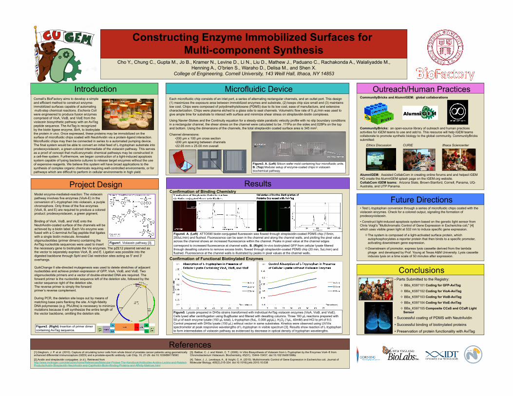
constructing enzyme immobilized surfaces for multi

1
CommunityBricks and AlumniGEM: global collaborations CommunityBricks: an open-source library of outreach and human practices activities for iGEM teams to use and add to. This resource will help iGEM teams collaborate to promote synthetic biology to the global community. CommunityBricks submitted: Ethics Discussion CURIE Ithaca Sciencenter Parts Submitted to the Registry: BBa_K597101 Coding for GFP-AviTag BBa_K597102 Coding for VioA-AviTag BBa_K597103 Coding for VioB-AviTag BBa_K597104 Coding for VioE-AviTag BBa_K597105 Composite CCaS and CCaR Light Sensor • Successful coating of PDMS with NeutrAvidin • Successful binding of biotinylated proteins • Preservation of protein functionality with AviTag Conclusions Model enzyme-mediated-reaction: The violacein pathway involves five enzymes (VioA-E) in the conversion of L-tryptophan into violacein, a purple chromophore. Only three of the five enzymes (VioA, B, and E) are required to produce a colored product: prodeoxyviolacein, a green pigment. Binding of VioA, VioB, and VioE onto the NeutrAvidin-coated surface of the channels will be achieved by a biotin label. Each Vio enzyme was fused with a C-terminal AviTag peptide that ligates with a single biotin molecule. Annealed oligonucleotides (primer dimers) containing the AviTag nucleotide sequences were used to insert the necessary gene to biotinylate the Vio enzymes. The pZE12 plasmid served as the vector to separately express VioA, B, and E. Ligation was possible into the digested backbone through SphI and ClaI restriction sites acting as 5' and 3' overhangs. QuikChange II site-directed mutagenesis was used to delete stretches of adjacent nucleotides and achieve protein expression of GFP, VioA, VioB, and VioE. Two oligonucleotide primers and a vector of double-stranded DNA are required. The forward primer is the nucleotide sequence left of the deletion site, followed by the vector sequence right of the deletion site. The reverse primer is simply the forward primer’s reverse complement. During PCR, the deletion site loops out by means of matching base pairs flanking the site. A high-fidelity DNA polymerase (e.g. PfuUltra) is necessary to minimize mutations because it will synthesize the entire length of the vector backbone, omitting the deletion site. Project Design Each microfluidic chip consists of an inlet port, a series of alternating rectangular channels, and an outlet port. This design (1) maximizes the exposure area between immobilized enzymes and substrate, (2) keeps chip size small and (3) maintains low cost. Chips were composed of polydimethylsiloxane (PDMS) due to its low cost, ease of manufacture, and extensive characterization. Chips were plasma etched to a glass side to seal channels. Volumetric flow rate of 5 µL/min was used to give ample time for substrate to interact with surface and minimize shear stress on streptavidin-biotin complexes. Using Navier-Stokes and the Continuity equation for a steady-state parabolic velocity profile with no slip boundary conditions in a rectangular channel, the shear stress on the channel were calculated to be .111Pa on the sides and.028Pa on the top and bottom. Using the dimensions of the channels, the total streptavidin coated surface area is 345 mm 2 . Channel dimensions: •200 µm x 100 µm cross section •200 µm spacing between channels •22.05 mm x 25.00 mm overall Confirmation of Binding Chemistry Confirmation of Functional Biotinylated Enzymes Results Cornell’s BioFactory aims to develop a simple and efficient method to construct enzyme- immobilized surfaces capable of automating multi-step chemical reactions. Escheria Coli were engineered to produce fusion enzymes comprised of VioA, VioB, and VioE from the violacein biosynthetic pathway with an AviTag peptide sequence. The AviTag is recognized by the biotin ligase enzyme, BirA, to biotinylate the protein in vivo. Once expressed, these proteins may be immobilized on the surface of microfluidic chips coated with NeutrAvidin via a protein-ligand interaction. Microfluidic chips may then be connected in series to a automated pumping device. The final system would be able to convert an initial feed of L-tryptophan substrate into prodeoxyviolacein, a green-colored intermediate of the violacein pathway. This serves as a proof of concept that multi-enzymatic chemical pathways may be constructed in a cell-free system. Furthermore, we began construction of a light-induced apoptosis system capable of lysing bacteria cultures to release target enzymes without the use of expensive reagents. We believe this system will have broad applications to the synthesis of complex organic chemicals requiring well-controlled environments, or for pathways which are difficult to perform in cellular environments in high yield. Introduction • Test L-tryptophan conversion through a series of microfluidic chips coated with the violacein enzymes. Check for a colored output, signaling the formation of prodeoxyviolacein. • Construct light-induced apoptosis system based on the genetic light sensor from Chris Voigt’s "Multichromatic Control of Gene Expression in Escherichia coli," [4] which uses visible green light at 532 nm to induce specific gene expression. The system is composed of a light-activated surface protein, which autophosphorylates a reporter protein that then binds to a specific promoter, activating downstream gene expression. Downstream of promotor, express lysis cassette derived from the lambda phage and developed by Prof. Young at Texas A&M University. Lysis cassette induces lysis on a time scale of 50 minutes after expression. Future Directions [1].Gleghorn, J. P. et al. (2010). Capture of circulating tumor cells from whole blood of prostate cancer patients using geometrically enhanced differential immunocapture (GEDI) and a prostate-specific antibody. Lab Chip, 10, 27-29. doi:10.1039/B917959C [2].Avidin and streptavidin conjugates. (n.d.). Retrieved from http://www.invitrogen.com/site/us/en/home/References/Molecular-Probes-The-Handbook/Antibodies-Avidins-Lectins-and-Related- Products/Avidin-Streptavidin-NeutrAvidin-and-CaptAvidin-Biotin-Binding-Proteins-and-Affinity-Matrices.html [3]. Balibar, C. J. and Walsh, C. T. (2006). In Vitro Biosynthesis of Violacein from L-Tryptophan by the Enzymes VioAE from Chromobacterium Violaceum. Biochemistry, 45(51), 15444-15457. doi:10.1021/bi061998z [4]. Tabor, J. J., Levskaya, A., & Voight, C. A. (2010). Multichromatic Control of Gene Expression in Escherichia coli. Journal of Molecular Biology, 405(2),315-324. doi:10.1016/j.jmb.2010.10.038 References Figure4. A. (Left) ATTO590 biotin-conjugated fluorescein was flowed through streptavidin-coated PDMS chip (15min, 200uL/min) and flushed. Fluorescence can be seen in the channel and along the channel walls, and plotting the pixel value across the channel shows an increased fluorescence within the channel. Peaks in pixel value at the channel edges correspond to increased fluorescence at channel walls. B. (Right) In-vivo biotinylated GFP from cellular lysate filtered through desalting columns to remove excess biotin, flowed through streptavidin-coated PDMS chip (20 min, 5uL/min) and flushed. Fluorescence at the channel walls is illustrated by peaks in pixel values at the channel walls. Cho Y., Chung C., Gupta M., Jo B., Kramer N., Levine D., Li N., Liu D., Mathew J., Paduano C., Rachakonda A., Walaliyadde M., Henning A., O’brien S., Waraho D., Delisa M., and Shen X. College of Engineering, Cornell University, 143 Weill Hall, Ithaca, NY 14853 Constructing Enzyme Immobilized Surfaces for Multi-component Synthesis Figure1. Violacein pathway [3]. Microfluidic Device Figure3. A. (Left) Silicon wafer mold containing four microfluidic units. B. (Top) Modular setup of enzyme-coated chips in violacein biochemical pathway. Figure5. Lysate prepared in DH5α strains transformed with individual AviTag violacein enzymes (VioA, VioB, and VioE). Cells lysed after centrifugation using BugBuster and filtered with desalting columns. Three 160 μL reactions prepared with 50 μl of each enzyme lysate (150 μL total), L-tryptophan (9uL, 0.008 μg/μL), H 2 O 2 (1μL, 40mM) and HCl to pH of 9.0. Control prepared with DH5α lysate (150 μL) without vector in same substrates. Kinetics were observed using UV/Vis spectrometer at peak responsive wavelengths of L-tryptophan in visible spectrum [3]. Results show reaction of L-tryptophan to form intermediates of violacein pathway as evidenced by decrease in optical density of tryptophan wavelengths. Figure2. (Right) Insertion of primer dimer containing AviTag sequence. Outreach/Human Practices AlumniGEM: Assisted CollabCom in creating online forums and and helped iGEM HQ create the AlumniGEM splash page on the iGEM.org website. CollabCom iGEM teams: Arizona State, Brown-Stanford, Cornell, Panama, UQ- Australia, and UTP Panama.

Upload: others

Post on 21-Mar-2022

3 views

Category:

Documents


0 download

TRANSCRIPT

CommunityBricks and AlumniGEM: global collaborations

CommunityBricks: an open-source library of outreach and human practices activities for iGEM teams to use and add to. This resource will help iGEM teams collaborate to promote synthetic biology to the global community. CommunityBricks submitted:

Ethics Discussion CURIE Ithaca Sciencenter

•  Parts Submitted to the Registry:   BBa_K597101 Coding for GFP-AviTag

  BBa_K597102 Coding for VioA-AviTag

  BBa_K597103 Coding for VioB-AviTag

  BBa_K597104 Coding for VioE-AviTag

  BBa_K597105 Composite CCaS and CCaR Light Sensor

•  Successful coating of PDMS with NeutrAvidin

•  Successful binding of biotinylated proteins

•  Preservation of protein functionality with AviTag

Conclusions

Model enzyme-mediated-reaction: The violacein pathway involves five enzymes (VioA-E) in the conversion of L-tryptophan into violacein, a purple chromophore. Only three of the five enzymes (VioA, B, and E) are required to produce a colored product: prodeoxyviolacein, a green pigment.

Binding of VioA, VioB, and VioE onto the NeutrAvidin-coated surface of the channels will be achieved by a biotin label. Each Vio enzyme was fused with a C-terminal AviTag peptide that ligates with a single biotin molecule. Annealed oligonucleotides (primer dimers) containing the AviTag nucleotide sequences were used to insert the necessary gene to biotinylate the Vio enzymes. The pZE12 plasmid served as the vector to separately express VioA, B, and E. Ligation was possible into the digested backbone through SphI and ClaI restriction sites acting as 5' and 3' overhangs.

QuikChange II site-directed mutagenesis was used to delete stretches of adjacent nucleotides and achieve protein expression of GFP, VioA, VioB, and VioE. Two oligonucleotide primers and a vector of double-stranded DNA are required. The forward primer is the nucleotide sequence left of the deletion site, followed by the vector sequence right of the deletion site. The reverse primer is simply the forward primer’s reverse complement.

During PCR, the deletion site loops out by means of matching base pairs flanking the site. A high-fidelity DNA polymerase (e.g. PfuUltra) is necessary to minimize mutations because it will synthesize the entire length of the vector backbone, omitting the deletion site.

Project Design

Each microfluidic chip consists of an inlet port, a series of alternating rectangular channels, and an outlet port. This design (1) maximizes the exposure area between immobilized enzymes and substrate, (2) keeps chip size small and (3) maintains low cost. Chips were composed of polydimethylsiloxane (PDMS) due to its low cost, ease of manufacture, and extensive characterization. Chips were plasma etched to a glass side to seal channels. Volumetric flow rate of 5 µL/min was used to give ample time for substrate to interact with surface and minimize shear stress on streptavidin-biotin complexes.

Using Navier-Stokes and the Continuity equation for a steady-state parabolic velocity profile with no slip boundary conditions in a rectangular channel, the shear stress on the channel were calculated to be .111Pa on the sides and.028Pa on the top and bottom. Using the dimensions of the channels, the total streptavidin coated surface area is 345 mm2.

Channel dimensions: • 200 µm x 100 µm cross section • 200 µm spacing between channels • 22.05 mm x 25.00 mm overall

Confirmation of Binding Chemistry

Confirmation of Functional Biotinylated Enzymes

Results

Cornell’s BioFactory aims to develop a simple and efficient method to construct enzyme- immobilized surfaces capable of automating multi-step chemical reactions. Escheria Coli were engineered to produce fusion enzymes comprised of VioA, VioB, and VioE from the violacein biosynthetic pathway with an AviTag peptide sequence. The AviTag is recognized by the biotin ligase enzyme, BirA, to biotinylate the protein in vivo. Once expressed, these proteins may be immobilized on the surface of microfluidic chips coated with NeutrAvidin via a protein-ligand interaction. Microfluidic chips may then be connected in series to a automated pumping device. The final system would be able to convert an initial feed of L-tryptophan substrate into prodeoxyviolacein, a green-colored intermediate of the violacein pathway. This serves as a proof of concept that multi-enzymatic chemical pathways may be constructed in a cell-free system. Furthermore, we began construction of a light-induced apoptosis system capable of lysing bacteria cultures to release target enzymes without the use of expensive reagents. We believe this system will have broad applications to the synthesis of complex organic chemicals requiring well-controlled environments, or for pathways which are difficult to perform in cellular environments in high yield.

Introduction

•  Test L-tryptophan conversion through a series of microfluidic chips coated with the violacein enzymes. Check for a colored output, signaling the formation of prodeoxyviolacein.

•  Construct light-induced apoptosis system based on the genetic light sensor from Chris Voigt’s "Multichromatic Control of Gene Expression in Escherichia coli," [4] which uses visible green light at 532 nm to induce specific gene expression.

 The system is composed of a light-activated surface protein, which autophosphorylates a reporter protein that then binds to a specific promoter, activating downstream gene expression.

 Downstream of promotor, express lysis cassette derived from the lambda phage and developed by Prof. Young at Texas A&M University. Lysis cassette induces lysis on a time scale of 50 minutes after expression.

Future Directions

[1].Gleghorn, J. P. et al. (2010). Capture of circulating tumor cells from whole blood of prostate cancer patients using geometrically enhanced differential immunocapture (GEDI) and a prostate-specific antibody. Lab Chip, 10, 27-29. doi:10.1039/B917959C [2].Avidin and streptavidin conjugates. (n.d.). Retrieved from http://www.invitrogen.com/site/us/en/home/References/Molecular-Probes-The-Handbook/Antibodies-Avidins-Lectins-and-Related-Products/Avidin-Streptavidin-NeutrAvidin-and-CaptAvidin-Biotin-Binding-Proteins-and-Affinity-Matrices.html

[3]. Balibar, C. J. and Walsh, C. T. (2006). In Vitro Biosynthesis of Violacein from L-Tryptophan by the Enzymes VioA−E from Chromobacterium Violaceum. Biochemistry, 45(51), 15444-15457. doi:10.1021/bi061998z [4]. Tabor, J. J., Levskaya, A., & Voight, C. A. (2010). Multichromatic Control of Gene Expression in Escherichia coli. Journal of Molecular Biology, 405(2),315-324. doi:10.1016/j.jmb.2010.10.038

References

Figure4. A. (Left) ATTO590 biotin-conjugated fluorescein was flowed through streptavidin-coated PDMS chip (15min, 200uL/min) and flushed. Fluorescence can be seen in the channel and along the channel walls, and plotting the pixel value across the channel shows an increased fluorescence within the channel. Peaks in pixel value at the channel edges correspond to increased fluorescence at channel walls. B. (Right) In-vivo biotinylated GFP from cellular lysate filtered through desalting columns to remove excess biotin, flowed through streptavidin-coated PDMS chip (20 min, 5uL/min) and flushed. Fluorescence at the channel walls is illustrated by peaks in pixel values at the channel walls.

Cho Y., Chung C., Gupta M., Jo B., Kramer N., Levine D., Li N., Liu D., Mathew J., Paduano C., Rachakonda A., Walaliyadde M., Henning A., O’brien S., Waraho D., Delisa M., and Shen X.

College of Engineering, Cornell University, 143 Weill Hall, Ithaca, NY 14853

Constructing Enzyme Immobilized Surfaces for Multi-component Synthesis

Figure1. Violacein pathway [3].

Microfluidic Device

Figure3. A. (Left) Silicon wafer mold containing four microfluidic units. B. (Top) Modular setup of enzyme-coated chips in violacein biochemical pathway.

Figure5. Lysate prepared in DH5α strains transformed with individual AviTag violacein enzymes (VioA, VioB, and VioE). Cells lysed after centrifugation using BugBuster and filtered with desalting columns. Three 160 µL reactions prepared with 50 µl of each enzyme lysate (150 µL total), L-tryptophan (9uL, 0.008 µg/µL), H2O2 (1µL, 40mM) and HCl to pH of 9.0. Control prepared with DH5α lysate (150 µL) without vector in same substrates. Kinetics were observed using UV/Vis spectrometer at peak responsive wavelengths of L-tryptophan in visible spectrum [3]. Results show reaction of L-tryptophan to form intermediates of violacein pathway as evidenced by decrease in optical density of tryptophan wavelengths.

Figure2. (Right) Insertion of primer dimer containing AviTag sequence.

Outreach/Human Practices

AlumniGEM: Assisted CollabCom in creating online forums and and helped iGEM HQ create the AlumniGEM splash page on the iGEM.org website. CollabCom iGEM teams: Arizona State, Brown-Stanford, Cornell, Panama, UQ-Australia, and UTP Panama.