construction, analysis, and transcription of model nucleosomal templates
TRANSCRIPT
Methods 33 (2004) 18–24
www.elsevier.com/locate/ymeth
Construction, analysis, and transcription of modelnucleosomal templates
Wendy Walter and Vasily M. Studitsky*,1
Department of Biochemistry and Center for Molecular Medicine and Genetics, Wayne State University School of Medicine, Detroit, MI 48201, USA
Accepted 30 October 2003
Abstract
Transcription through the nucleosome by Saccharomyces cerevisiae RNA polymerase II (Pol II) is characterized by an almost
absolute block to transcription at physiological ionic strength and displacement of one H2A/H2B dimer to form a hexasome [Mol.
Cell 9 (2002) 541]. In previous studies of Pol II transcription through chromatin, templates containing nucleosomes in multiple
positions were used. These templates do not allow detailed analysis of the mechanism of transcription through chromatin. Here, we
describe the development of a new template that is only long enough to accommodate a single nucleosome position along the DNA
so that all of the templates are identical and allow for more in-depth analysis. After ligation of the nucleosome to promoter DNA or
assembled elongation complexes, the mechanism of transcription through this uniquely positioned nucleosome by various RNA
polymerases can be analyzed.
� 2003 Elsevier Inc. All rights reserved.
Keywords: Chromatin; Nucleosome positioning; Transcription; Elongation; RNA polymerase II
1. Introduction
In eukaryotic cells, transcription occurs on DNA that
is packaged into chromatin. Chromatin fibers are madeup of discrete repeating units called nucleosomes. Each
nucleosome core is made up of 146 bp of DNA wrapped
around a histone octamer containing two molecules
each of histones H2A, H2B, H3, and H4 [2]. Linker
histone H1 binds where DNA enters and exits the nu-
cleosome.
Genes that are actively transcribed by RNA poly-
merase II (Pol II) still retain their nucleosomal structure,indicating that even if the nucleosome is disrupted
during transcription, recovery of this structure occurs
almost immediately after passage of the enzyme (see
[3,4] for reviews). Thus, Pol II encounters a nucleosomal
barrier to transcription approximately every 200 bp.
Exactly how Pol II negotiates this obstacle has been the
* Corresponding author.
E-mail address: [email protected] (V.M. Studitsky).1 Present address: Department of Pharmacology, University of
Medicine and Dentistry of New Jersey, 675 Hoes Lane, Room 405,
Piscataway, NJ 08854, USA.
1046-2023/$ - see front matter � 2003 Elsevier Inc. All rights reserved.
doi:10.1016/j.ymeth.2003.10.016
subject of intense research. However, the mechanism has
remained elusive due to the lack of suitable model in
vitro systems. Promoter-dependent transcription initia-
tion in crude extracts [5] or with highly purified proteins[6] results in low efficiency of template utilization. This
makes analysis of the fate of nucleosomes after tran-
scription nearly impossible due to a high background of
non-transcribed templates. In contrast, efficient end-
initiation is supported by DNA templates containing a
single-stranded, 30-extending oligo(dC) ‘‘tail’’ [7]. How-
ever, in this system, determination of the fate of the
nucleosome during transcription is complicated by theformation of extremely stable DNA-Pol II complexes at
the end of DNA [8]. Moreover, end-initiated and pro-
moter-initiated RNA polymerases differ in the way they
progress through the nucleosome [8].
We have recently developed a novel approach to
study the mechanism of transcription through the nu-
cleosome by Pol II. This approach involved the use of
ECs that were assembled from purified oligonucleotidesand RNA polymerase and ligation of the ECs to tem-
plates with defined nucleosome positions ([1,9], Fig. 1A).
Pol II uses a very different mechanism for transcription
through the nucleosome than Pol III and bacteriophage
W. Walter, V.M. Studitsky / Methods 33 (2004) 18–24 19
SP6 RNA polymerase [1,10,11]. The nucleosomal bar-rier is much stronger for Pol II. Moreover, where tran-
scription through the nucleosome results in transfer of
the complete histone octamer upstream of the tran-
scribing enzyme for SP6 RNA polymerase and Pol III,
transcription through the nucleosome by Pol II results in
the loss of an H2A/H2B dimer [1]. Remarkably, it has
recently been shown that Escherichia coli RNA poly-
merase (RNAP) also uses this Pol II-type mechanism[12]. Thus, the bacterial enzyme provides a suitable
model system for analysis of the general aspects of the
mechanism.
Our previous studies on the mechanism of transcrip-tion through the nucleosome with Pol II and RNAP
were performed using a mononucleosomal template that
allowed for different nucleosome positions on the DNA
[1]. Therefore, the template used for transcription was a
mixture of differently positioned mononucleosomes. The
heterogeneity of templates makes detailed analysis of the
mechanism of transcription through the nucleosomes
(such as analysis of nucleosome-specific pausing pattern[13,14] or arrest of RNA polymerase at unique position
within the nucleosome [10]) impossible. Here, we
describe construction, analysis, and transcription of a
shorter nucleosomal template that allows for only one
position of the histone octamer on the DNA.
2. Materials and methods
2.1. Design of the DNA fragment for the generation of
uniquely positioned nucleosomes
In our previous studies, a 204 bp fragment of DNA
was used for nucleosome reconstitution and transcrip-
tion by Pol II and RNAP [1,12]. While this template is
only long enough to accommodate one nucleosome perDNA molecule, reconstitution results in a mixture of
mononucleosomes with different positions along the
DNA. This made a detailed analysis of the mechanism
of transcription through the nucleosome difficult.
Therefore, a template containing a uniquely positioned
nucleosome was constructed.
Uniquely positioned nucleosomes can be obtained
using different strategies. First, differently positionedisomers can be separated in and extracted from a native
gel [15]. However, the isomers can be separated well only
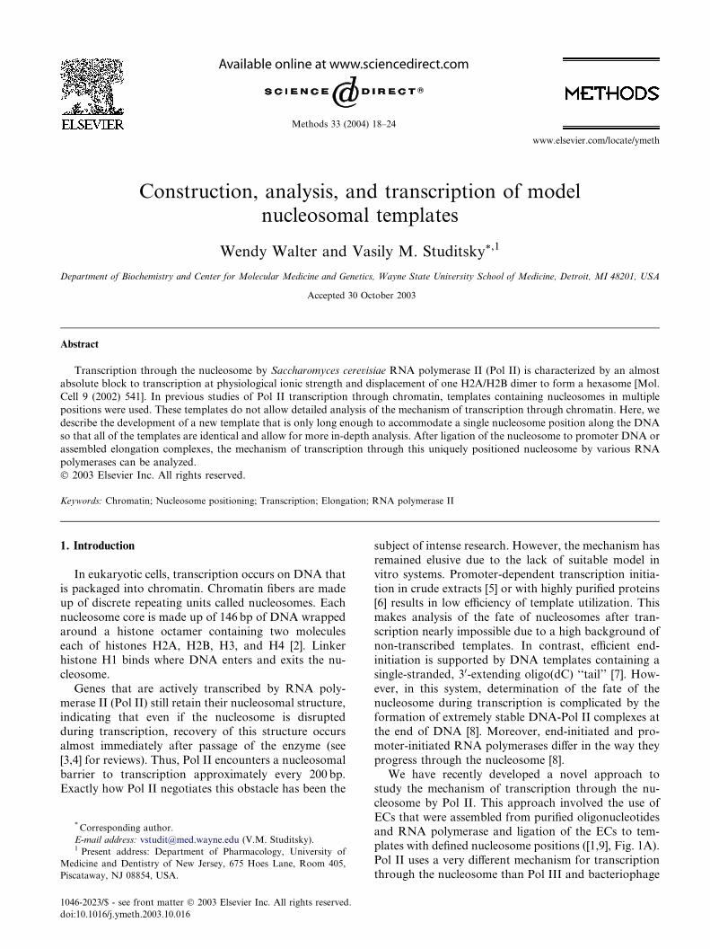
ig. 1. Experimental system using assembled ECs to study transcrip-
on through the nucleosome by E. coli RNAP. (A) Experimental ap-
roach for transcription through the nucleosome using immobilized
Cs. EC9 assembled on 50 bp DNA is immobilized on Ni2þ–NTA
garose and washed. The 150bp nucleosome (or DNA) is ligated to the
Cs and the complexes are washed. Transcription is resumed after the
ddition of NTPs. For analysis of the templates, the promoter DNA
agment is labeled on the 50 end of the 50R oligo (indicated by a star).
or transcript analysis, RNA can be labeled at the 50 end of the RNA
ligo or radioactive nucleotides can be added at any position by
alking the RNAP (see B). (B) Walking of RNA polymerase along the
mplate. Immobilized EC9 is ligated to the nucleosome and washed.
NAP is walked to the +45 position by the addition of ATP, CTP, and
TP. In some cases RNAP is walked to the +49 position using cold
TP and [a-32P]GTP to label the RNA. The transcribed sequence is
hown in a box at the bottom with the position of radiolabeling
arked by asterisks. The ECs are washed after each manipulation and
anscription is resumed by the addition of all 4 NTPs. (C) Restriction
ap of the ligated 200bp nucleosomal template. Nucleosome posi-
oning on the ligated 200bp template was determined by restriction
nzyme mapping. Locations of the restriction sites are indicated by
rrows. The template was labeled (indicated by a star) for analysis in
ig. 2.
F
ti
p
E
a
E
a
fr
F
o
w
te
R
G
U
s
m
tr
m
ti
e
a
F
b
20 W. Walter, V.M. Studitsky / Methods 33 (2004) 18–24
when DNA length is longer than 200 bp [16]. Moreover,nucleosomes positioned symmetrically relative to the
middle of a DNA fragment are not separated in the gel
[15]. Alternatively, nucleosomes can be assembled on a
DNA fragment that is short enough (l50 bp) to support
only one nucleosome position. In this case, microheter-
ogeneity of nucleosome positioning is also possible [17].
Therefore, use of a strong nucleosome positioning se-
quence is still necessary (see below).To obtain a uniquely positioned nucleosome, a
150 bp DNA fragment (145 bp plus a 9 base TspRI
overhang for ligation to ECs) was constructed. This
template only allowed for a single nucleosome position
along the DNA (see below). The template was based on
a previously well-characterized template (pD89) that
supports formation on nucleosomes predominantly (60–
70%) in one position even on longer 230–260 bp tem-plates [10]. The DNA was PCR-amplified from the
pD89 plasmid with Vent (exo-) DNA polymerase (New
England Biolabs, Beverly, MA) using an upper primer
that introduces the TspRI site for ligation to the ECs
and a lower primer that maintains the original 30 (NcoI)
end of the pD89 template. Finally, the template is di-
gested with TspRI and gel purified. The DNA for nu-
cleosome reconstitution is moderately end-labeled (vialabeling of the lower primer) so that the quality of the
reconstitutes can be monitored (see below).
1. The DNA was PCR-amplified in preparative amounts
from the pD89 plasmid [10] using the primers (Invitro-
gen, Carlsbad, CA): upper-50 AAGCGACACCGG
CACTGGGTAAATAGTTGAAGTTGTAGTAA
ATGTTAATGTAG 30 and lower- 50 CATGGGCA
CTTACATTATTTGTTG 30. Some of the lower pri-mer is radiolabeled with [c-32P]ATP (7000Ci/mmol,
ICN Biomedicals, Irvine, CA) using T4 polynucleo-
tide kinase (New England Biolabs, Beverly, MA, as
per NEB recommendations) prior to the PCR.
2. The resulting 158 bp product is purified by ethanol
precipitation and digested with TspRI.
3. The sample is loaded onto an 8% (19:1) polyacryl-
amide gel containing 1� TAE and 4M urea (to pre-vent re-association of the 9 nt, GC-rich sticky ends
of the TspRI-digested fragments). The 150 bp
TspRI-cut fragment is cut out of the gel, the gel slice
is crushed, and the DNA is extracted overnight at
4 �C in 3–5 volumes of TE buffer, ethanol precipi-
tated, and resuspended in dH2O.
4. The template DNA is further ‘‘cleaned up’’ using
QIAquick gel extraction kit columns (Qiagen, Chats-worth, CA, as per kit protocol).
2.2. Reconstitution and analysis of mononucleosomes for
transcription
The most efficient protocol that we have found for
reconstitution of mononucleosomes on the 150 bp tem-
plate involves the use of donor chromatin as a source ofhistone octamers for exchange onto the template DNA
[18]. In this case, the amount of free DNA is <15% and
the nucleosomes do not have to be further purified after
reconstitution. The detailed protocol for donor chro-
matin preparation has been published [18] and will not
be discussed here. Nucleosomes prepared this way con-
tain excess donor chromatin, but it can be removed by
washing after the template is ligated to the immobilizedECs. The method described below is for templates that
are about 150–250 bp in size and, thus, allow for only
one nucleosome per molecule of DNA. The quality of
nucleosome preparations is determined by the amount
of free DNA and subnucleosomal particles that con-
taminate the sample.
1. 50 end-labeled 150 bp TspRI-cut DNA is prepared as
described above. As much as 1–5 lg of the DNA ismixed with long -H1 donor chromatin at a ratio of
1:80 (wt:wt), respectively (sample volume is deter-
mined by donor chromatin concentration) in buffer
containing 1M NaCl and 0.1% Igepal CA-630 (Sig-
ma, St. Louis, MO).
2. The sample is dialyzed overnight at 4 �C against a
gradient (�1L) starting at 1M NaCl and ending with
no NaCl in buffer containing 10mM Tris–HCl, pH7.5, 0.2mM EDTA, and 0.1% NP-40.
3. The concentration of the reconstituted nucleosomes
is determined by the specific activity of the DNA.
4. Buffer is added providing a final concentration of
10% sucrose and 50 lg/ml sheared herring testes
DNA (Intergen, Purchase, NY).
5. The templates are resolved by native gel electrophore-
sis (4.5% acrylamide (39:1), 5% glycerol, 20mM Na–Hepes, pH 8, and 0.1mM EDTA) at 100V for 2.5–4 h
(depending on the size of the DNA fragment and the
degree of resolution desired) as described [9].
6. Quantitation is performed using a Cyclone Storage
Phosphor System (Packard, Meriden, CT).
2.3. Restriction enzyme mapping of templates ligated to
the promoter fragment
The position of a nucleosome can be determined with
about 10 bp resolution based on its mobility during
native PAGE [19]. However, this method cannot dis-
criminate between two symmetrically positioned nucle-
osomes or differently positioned nucleosomes formed on
DNA �200 bp or less [16]. The exact position can be
further narrowed down by restriction enzyme digestionor micrococcal nuclease mapping (not discussed here
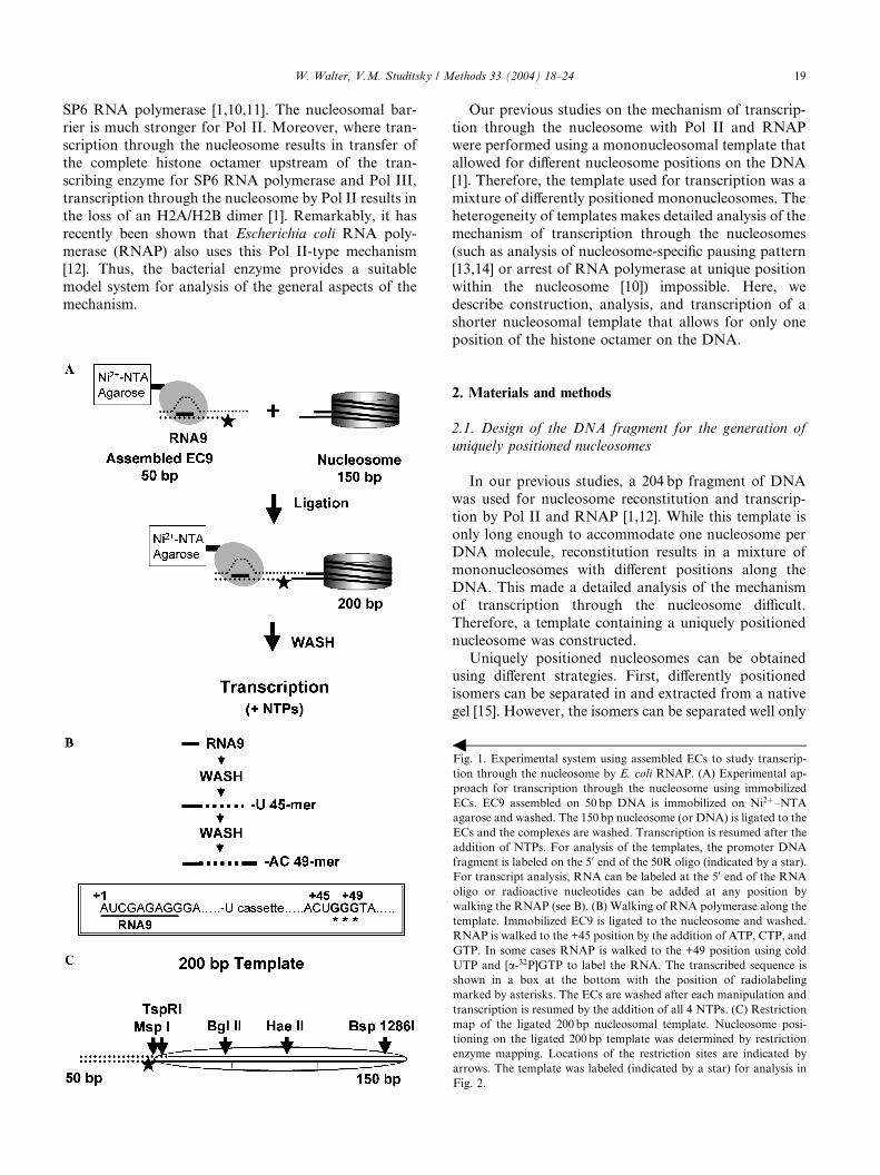
[15]). See Fig. 2 for an example of restriction enzyme
mapping on the ligated, 200 bp template. The promoter
fragment was 50 end-labeled with [c-32P]ATP (7000Ci/
mmol, ICN Biomedicals, Irvine, CA) and T4 polynu-
cleotide kinase. A map of the nucleosomes on the tem-
plate is illustrated in Fig. 1C. The majority of the ligated
Fig. 2. Restriction enzyme mapping of the 200 bp nucleosomal tem-
plate. As much as 200 bp nucleosomes were obtained by ligating 150 bp
nucleosomes to the 50 bp promoter fragment (50 end-labeled 50R oligo
annealed to 59D oligo), and the templates were analyzed by native
PAGE before and after restriction enzyme digestion. The mobilities of
the nucleosome, hexasome, and free DNA are indicated. Positions of
the restriction enzyme sites along the DNA are shown at the top. The
location of DNA labeling is indicated by a star.
W. Walter, V.M. Studitsky / Methods 33 (2004) 18–24 21
nucleosomes are sensitive to MspI digestion, showing
that ligation does not alter the nucleosome positioning.
1. Equimolar amounts of promoter oligos ([c-32P]ATPlabeled 50R-50 GGTGTCGCTTGGGTTGGCTTTT
CGGGCTGTCCCTCTCGATGGCTGTAAGT-30
and 59D-50-ACTTACAGCCATCGAGAGGGACA
CGGCGAAAAGCCAACCCAAGCGACACCGG
CACTGGG-30) in TB40 (see transcription buffers be-
low) were annealed by incubation at 45 �C for 10min,
followed by a 2min incubation at each step of a 2 �Cdecrease in temperature down to RT. Oligos are pur-chased from Oligos, Etc. Wilsonville, OR or Invitro-
gen, Carlsbad, CA.
2. Equimolar amounts of 50 bp annealed promoter oli-
gos and 150 bp nucleosomal templates were ligated
by incubation with 100 lM ATP, 1% PEG-8000,
0.5mg/ml Acet-BSA, and 50U T4 DNA ligase at
12 �C for 1–2 h.
3. Samples (10 ng aliquots) are supplemented with buf-fer providing 20mM Na–Hepes, pH 7.8, 5mM
MgCl2, 2mM spermidine (Sigma, St. Louis, MO),
and 0.5mg/ml BSA.
4. One sample is not digested, while appropriate restric-
tion enzymes (10U) are added to the others, and di-
gestion is performed at room temperature (RT) for
0.5–1 h.
5. Buffer is added providing a final concentration of10mM EDTA, 10% sucrose, and 50 lg/ml sheared
herring testes DNA (Intergen, Purchase, NY).
6. The templates are resolved by native gel electrophore-
sis (4.5% acrylamide (39:1), 5% glycerol, 20mM Na–
Hepes, pH 8, and 0.1mM EDTA) at 100V for 2.5–4 h
(depending on the size of the DNA fragment and the
degree of resolution desired) as described [9].
7. Quantitation is performed using a Cyclone StoragePhosphor System (Packard, Meriden, CT).
2.4. Transcription buffers
TB0 contains 20mM Tris–HCl, pH 7.9, 5mM
MgCl2, and 1–2mM b-mercaptoethanol (2-ME). TB40,
TB150, TB300, and TB1000 contain 40, 150, 300mM,
and 1M KCl, respectively. Acetylated BSA was pur-chased from Sigma (St. Louis, MO).
2.5. RNAP and Pol II EC assembly and promoter
initiation of RNAP
ECs are formed separately in solution, immobilized
on Ni2þ–NTA agarose (Qiagen, Chatsworth, CA) and
washed, and then ligated to the nucleosomal template(see Fig. 1A). If the templates were to be analyzed after
transcription, the promoter fragment was 50 end-labeledwith [c-32P]ATP (7000Ci/mmol, ICN Biomedicals, Ir-
vine, CA) and T4 polynucleotide kinase. It should be
noted that each wash step involves adding the wash
buffer to the resin, centrifuging for 10 s, and then col-
lecting the supernatant. The scheme is illustrated in
Fig. 1A. Assembly of RNAP and Pol II ECs from pu-rified components and ECs formed from promoter ini-
tiation of RNAP were the subject of other methods
papers [9] and will not be discussed in detail here. For
the sake of simplicity, only ECs assembled with RNAP
will be illustrated in the figures.
Authentic RNAP ECs are assembled from purified
components as described in [9] using the following oli-
gonucleotides (Oligos, Etc. Wilsonville, OR): TDS50 (50-GGTGTCGCTTGGGTTGGCTTTTCGGGCTGTCC
CTCTCGATGGCTGTAAGT-30), RNA9 (50 AUCGA
GAGG 30), and NDS59 (50-ACTTACAGCCATCGA
GAGGGACACGGCGAAAAGCCAACCCAAGCGA
CACCGGCACTGGG-30). The TDS50 oligo is labeled
with [c-32P]ATP (7000Ci/mmol, ICN Biomedicals, Ir-
vine, CA) using T4 polynucleotide kinase (New England
Biolabs, Beverly, MA) as described [9]. The Ni2þ–NTAagarose (Qiagen, Chatsworth, CA) is pre-treated with
0.2mg/ml acetylated BSA for 10min and washed two
times with 1ml TB40 prior to immobilization of the
RNAP. ECs assembled with �2–3 pmol RNAP (im-
mobilized on 50 ll of 50% resin suspension) should be
enough for about 10 reactions. The DNA in the ECs has
a sticky end that is complementary to the nucleosomal
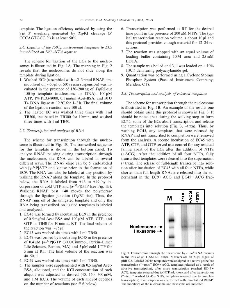
Fig. 3. Transcription through the nucleosome by E. coli RNAP results
in the loss of an H2A/H2B dimer. Markers are an MspI digest of
pBR322. Labeled 200 bp templates were analyzed in a native gel before
transcription (‘‘)trxn;’’ EC9+ACG, templates released as a result of
abortive transcription), after mock transcription (washed EC45+
ACG, templates released due to NTP addition), and after transcription
(‘‘+trxn;’’ washed EC45+NTPs, templates released due to complete
transcription). Transcription was performed with immobilized RNAP.
The mobilities of the nucleosome and hexasome are indicated.
22 W. Walter, V.M. Studitsky / Methods 33 (2004) 18–24
template. The ligation efficiency achieved by using the9 nt 30 overhang generated by TspRI cleavage (50
CCCAGTGCC 30) is at least 50%.
2.6. Ligation of the 150 bp nucleosomal templates to ECs
immobilized on Ni2þ–NTA agarose
The scheme for ligation of the ECs to the nucleo-
somes is illustrated in Fig. 1A. The mapping in Fig. 2reveals that the nucleosomes do not slide along the
template during ligation.
1. Washed EC9 (assembled with �2–3 pmol RNAP, im-
mobilized on �50 ll of 50% resin suspension) was in-
cubated in the presence of 150–200 ng of TspRI-cut
150 bp template (nucleosome or DNA), 100 lMATP, 1% PEG-8000, 0.5mg/ml Acet-BSA, and 50U
T4 DNA ligase at 12 �C for 1–2 h. The final volumeof the ligation reaction was 100 ll.
2. The ligated EC was washed three times with 1ml
TB300, incubated in TB300 for 10min, and washed
three times with 1ml TB40.
2.7. Transcription and analysis of RNA
The scheme for transcription through the nucleo-some is illustrated in Fig. 1B. The transcribed sequence
for this template is shown in the bottom panel. To
analyze RNAP pausing during transcription through
the nucleosome, the RNA can be labeled in several
different ways. The RNA9 oligo can be 50 end-labeledwith [c-32P]ATP and kinase prior to the formation of
EC9. The RNA can also be labeled at any position by
walking the RNAP along the template. In the protocolbelow, the RNA is labeled from +46 to +49 by in-
corporation of cold UTP and [a-32P]GTP (see Fig. 1B).
Walking RNAP past +40 moves the polymerase
through the ligation junction (TspRI site). Thus, the
RNAP runs off of the unligated template and only the
RNA being transcribed on ligated templates is labeled
and analyzed.
1. EC45 was formed by incubating EC9 in the presenceof 0.5mg/ml Acet-BSA and 100 lM ATP, CTP, and
GTP in TB40 for 10min at RT. The final volume of
the reaction was �75 ll.2. EC45 was washed six times with 1ml TB40.
3. EC49 was formed by incubating EC45 in the presence
of 0.4 lM [a-32P]GTP (3000Ci/mmol, Perkin–Elmer
Life Sciences, Boston, MA) and 5 lM cold UTP for
5min at RT. The final volume of the reaction was40–50 ll.
4. EC49 was washed six times with 1ml TB40.
5. The samples were supplemented with 0.5mg/ml Acet-
BSA, aliquoted, and the KCl concentration of each
aliquot was adjusted as desired (40, 150, 300mM,
and 1M KCl). The volume of each aliquot depends
on the number of reactions (see # 6 below).
6. Transcription was performed at RT for the desiredtime point in the presence of 200 lM NTPs. The typ-
ical transcription reaction volume is about 10 ll andthis protocol provides enough material for 12–24 re-
actions.
7. The reaction was stopped with an equal volume of
loading buffer containing 10M urea and 25mM
EDTA.
8. The sample was boiled and 3 ll was loaded on a 10%(19:1) denaturing polyacrylamide gel.
9. Quantitation was performed using a Cyclone Storage
Phosphor System (Packard Instrument Company,
Meriden, CT).
2.8. Transcription and analysis of released templates
The scheme for transcription through the nucleosomeis illustrated in Fig. 1B. An example of the results one
should obtain using this protocol is shown in Fig. 3. It
should be noted that during the walking step to form
EC45, some of the ECs abort transcription and release
the templates into solution (Fig. 3, )trxn). Thus, by
washing EC45, any templates that were released by
RNAP and not transcribed to completion were removed
from the analysis. A second incubation of EC45 withATP, CTP, and GTP served as a control for any residual
falling apart of the ECs after the addition of NTPs
(+ACG). After the addition of all four NTPs, fully
transcribed templates were released into the supernatant
(+trxn). The release of full-length transcript into solu-
tion after incubation of EC45 with all four NTPs, while
shorter than full-length RNAs are released into the su-
pernatant in the EC9+ACG and EC45+ACG frac-
W. Walter, V.M. Studitsky / Methods 33 (2004) 18–24 23
tions, can be confirmed in a control experiment withlabeled transcripts rather than labeled templates (see
[9]). Transcription through the nucleosome by E. coli
RNAP resulted in the appearance of a faster migrating
band (+trxn, hexasome) as compared to the mobility of
the original nucleosomes ()trxn).1. EC45 was formed by incubating EC9 (formed with 1–
1.5 pmol RNAP, immobilized on 25 ll of 50% sus-
pension) in the presence of 0.5mg/ml Acet-BSA and100 lM ATP, CTP, and GTP in TB300 for 10min
at RT. The final volume of the reaction was 35 ll.The reaction was mixed, centrifuged for 10 s, and
7 ll of supernatant was collected (-transcription con-
trol: EC9+ACG, contains templates released as a re-
sult of abortive transcription rather than fully
transcribed templates).
2. EC45 was washed three times with 0.5ml TB300, in-cubated in 0.5ml TB300 for 15min at RT, and
washed three times with 0.5ml TB300.
3. The sample was supplemented with 0.5mg/ml Acet-
BSA in a 35 ll volume. 15 ll was aliquoted into two
tubes.
4. Transcription was performed in a final volume of
15 ll for 15min at RT by the addition of NTPs.
A. Mock transcription control (EC45+ACG, con-tains templates released as a result of abortive
transcription after further incubation with
NTPs rather than fully transcribed templates):
The 15 ll reaction contained 200 lM ATP,
CTP, and GTP.
B. Transcription (EC45+NTPs, contains fully
transcribed templates): The 15 ll reaction con-
tained 200 lM of all four NTPs.5. The sample was mixed, centrifuged for 10 s, and 7 ll
of supernatant was collected from each sample.
6. Buffer is added providing a final concentration of
10mM EDTA, 10% sucrose, 50 lg/ml sheared herring
testes DNA (Intergen, Purchase, NY), and 350 ng of
carrier nucleosomes (to block non-specific interac-
tions with the wells of the gel, donor chromatin works
well).7. The templates are resolved by native gel electrophore-
sis (4.5% acrylamide (39:1), 5% glycerol, 20mM Na–
Hepes, pH 8, and 0.1mM EDTA) at 100V for 2.5–4 h
(depending on the size of the DNA fragment and the
degree of resolution desired) as described [9].
8. Quantitation is performed using a Cyclone Storage
Phosphor System (Packard, Meriden, CT).
2.9. Transcription in solution
Transcription in solution can also be performed as
described above. The reaction should be started in the
immobilized state so that excess promoter fragments,
unligated nucleosomes, and the non-transcribed tem-
plates released as a result of ECs falling apart during
walking from EC9 to EC45 can be removed from thereaction.
1. Washed EC45 (above) was eluted with 100mM imid-
azole in TB300 containing 0.5mg/ml Acet-BSA for
10min at RT. The final volume of the elution mix
was 35 ll.2. The sample was mixed, centrifuged for 10 s, and 17 ll
of supernatant was collected. 7 ll of the sample was
aliquoted into two tubes. The sample was diluted 2-fold with TB300 containing 0.5mg/ml Acet-BSA.
3. Transcription is performed in the presence of 200 lMNTPs and a mock transcription control is performed
in the presence of 200 lM ATP, CTP, and GTP for
15min at RT.
4. The reactions are stopped by the addition of tran-
scription stop/gel loading buffer providing 10mM
EDTA, 10% sucrose, 50 lg/ml sheared herring testesDNA, and �350 ng of donor chromatin.
5. Analysis of templates by native PAGE was per-
formed as described above.
2.10. Reconstitution and purification of hexasomes from
purified histones and ligation to the 50 bp promoter
fragment for use as mobility controls in native PAGE
Transcription through the nucleosome by Pol II and
E. coli RNAP results in the loss of an H2A/H2B dimer
and the formation of a subnucleosomal particle, the
hexasome [1,12]. Thus, hexasomes are reconstituted on
the 150 bp template, gel purified, and ligated to the 50 bp
promoter fragment for use as a mobility control in na-
tive gel electrophoresis. Hexasomes are reconstituted
from purified histones (Mol Cell, Walter meth). Thedetailed protocol for purification of core histones on
hydroxyapatite [20] was published and will not be dis-
cussed here. The reconstitution protocol described be-
low is a slightly modified version of the method used by
the Bradbury laboratory [21] and is described in more
detail in [9].
1. 5 lg of DNA is mixed with 1.23 lg H3/H4 and 0.67 lgH2A/H2B (ratio of H3/H4:H2A/H2B¼ 1.82).
2. Dialysis is performed at 4 �C against the same buffer
but with decreasing NaCl concentration (2, 1.5, 1,
0.75, 0.5M, and 10mM NaCl) for 1 h at each step.
3. Reconstitutes are collected in low adhesion microfuge
tubes (USA Scientific, Ocala, FL) and supplemented
with buffer providing a final concentration of 10% su-
crose.
4. The templates are resolved by native gel electrophore-sis (4.5% acrylamide (39:1), 5% glycerol, 20mM Na–
Hepes, pH 8, and 0.1mM EDTA) at 100V for 2.5–4 h
(depending on the size of the DNA fragment and the
degree of resolution desired) as described [9].
5. The appropriate band is cut out of the gel, the gel is
crushed, and the hexasomes are extracted overnight
at 4 �C in 1–2 volumes of 10mM Na–Hepes, pH
24 W. Walter, V.M. Studitsky / Methods 33 (2004) 18–24
8.0, 0.1mM EDTA, and 0.5mg/ml BSA. Note thatthe hexasome preparation always contains nucleo-
somes, so nucleosomes can also be purified at this
stage.
6. The supernatant is collected and the concentration of
the sample is determined by the specific activity of the
DNA.
7. Gel-purified 150 bp hexasomes from gel-purification
are ligated to 50 32P-labeled 50 bp promter DNA asmobility controls for the 200 bp hexasomes. Equimo-
lar amounts of the promoter fragment and the 150 bp
hexasomes are incubated in the presence of 100 lMATP, 1% PEG-8000, and 50U T4 DNA ligase
(New England Biolabs, Beverly, MA) at 12 �C for
1–2 h.
8. The samples are aliquoted and prepared for digestion
or electrophoresis as described for the mononucleo-some preparation above.
3. Concluding remarks
In summary, the protocols described above allow
formation of a uniquely positioned nucleosome that can
be ligated to any DNA sequence containing the TspRIoverhang. The yield of mononucleosomes is much
higher than after gel isolation. For additional informa-
tion and modifications of the protocol described above,
it is recommended that the reader refer to [9]. Immediate
applications of this approach include analysis of the
mechanisms of transcription through the nucleosome by
various RNA polymerases and ATP-dependent chro-
matin remodeling where mononucleosomal templatesare widely used (see [22] for review). However, similar
approaches could be used for analysis of other aspects of
chromatin function.
The protocols described above only allow formation
of end-positioned nucleosomes. More centrally posi-
tioned nucleosomes could be obtained using gel-purifi-
cation [15]. Alternatively, new stronger nucleosome
positioning sequences could be used; thus, quite strongpositioning sequences were recently selected from
chemically synthetic random DNA molecules [23]. It
remains to be established whether these synthetic se-
quences having much higher affinity to DNA than any
known natural DNA sequences [24] can properly func-
tion in the cell.
Acknowledgments
We thank M. Kashlev, N. Komissarova, and J.
Becker for providing purified E. coli core and holoen-
zyme RNAP and for helpful advice. The work was
supported by the NIH Grant GM58650 and NSF Grant
0234493 to V.M.S.
References
[1] M.L. Kireeva, W. Walter, V. Tchernajenko, V. Bondarenko, M.
Kashlev, V.M. Studitsky, Mol. Cell 9 (2002) 541–552.
[2] K. Luger, A.W. Mader, R.K. Richmond, D.F. Sargent, T.J.
Richmond, Nature 389 (1997) 251–260.
[3] D.J. Clark, in: The Nucleus, vol. 1, JAI Press, Greenwich, 1995,
pp. 207–239.
[4] G. Orphanides, D. Reinberg, Nature 407 (2000) 471–475.
[5] J.A. Knezetic, G.A. Jacob, D.S. Luse, Mol. Cell. Biol. 8 (1988)
3114–3121.
[6] E. Maldonado, R. Drapkin, D. Reinberg, Methods Enzymol. 274
(1996) 72–100.
[7] R.L. Dedrick, M.J. Chamberlin, Biochemistry 24 (1985) 2245–
2253.
[8] Y.V. Liu, D.J. Clark, V. Tchernajenko, M.E. Dahmus, V.M.
Studitsky, Biopolymers 68 (2003) 528–538.
[9] W. Walter, M.L. Kireeva, V. Tchernajenko, M. Kashlev, V.M.
Studitsky, Methods Enzymol. (2003) in press.
[10] J. Bednar, V.M. Studitsky, S.A. Grigoryev, G. Felsenfeld, C.L.
Woodcock, Mol. Cell 4 (1999) 377–386.
[11] V.M. Studitsky, G.A. Kassavetis, E.P. Geiduschek, G. Felsenfeld,
Science 278 (1997) 1960–1963.
[12] W. Walter, M.L. Kireeva, V.M. Studitsky, M. Kashlev, J. Biol.
Chem. 278 (2003) 36148–36156.
[13] V.M. Studitsky, D.J. Clark, G. Felsenfeld, Cell 83 (1995) 19–27.
[14] R.U. Protacio, K.J. Polach, J. Widom, J. Mol. Biol. 274 (1997)
708–721.
[15] V.M. Studitsky, D.J. Clark, G. Felsenfeld, Methods Enzymol. 274
(1996) 246–256.
[16] S. Pennings, G. Meersseman, E.M. Bradbury, Nucleic Acids Res.
20 (1992) 6667–6672.
[17] K. Luger, T.J. Rechsteiner, T.J. Richmond, Methods Enzymol.
304 (1999) 3–19.
[18] R.T. Utley, T.A. Owen-Hughes, L.J. Juan, J. Cote, C.C. Adams,
J.L. Workman, Methods Enzymol. 274 (1996) 276–291.
[19] G. Meersseman, S. Pennings, E.M. Bradbury, EMBO J. 11 (1992)
2951–2959.
[20] R.H. Simon, G. Felsenfeld, Nucleic Acids Res. 6 (1979) 689–696.
[21] S. Pennings, G. Meersseman, E.M. Bradbury, Proc. Natl. Acad.
Sci. USA 91 (1994) 10275–10279.
[22] G. Langst, P.B. Becker, J. Cell Sci. 114 (2001) 2561–2568.
[23] P.T. Lowary, J. Widom, J. Mol. Biol. 276 (1998) 19–42.
[24] A. Thastrom, P.T. Lowary, H.R. Widlund, H. Cao, M. Kubista,
J. Widom, J. Mol. Biol. 288 (1999) 213–229.