contents · contents foreword, barbara w. henderson, phd ... some regions to escape photodynamic...
TRANSCRIPT

�
Contents
Foreword, Barbara W. Henderson, PhD .............................................................. vii
Preface, Merrill A. Biel ........................................................................................ ix
Contributors ....................................................................................................... xi
one The Roswell Park History of PDT: 1972 to the Present: A Personal Perspective, Thomas J. Dougherty ......................................................................1
two Principles of Photodynamic Therapy-Induced Killing of Tumor Cells, Nancy L. Oleinick and David Kessel .............................................................. 19
three PDT Laser Physics and Safety, Tom Mang ....................................................... 33
four Photodynamic Therapy (PDT) in Oral Cancer, Barry L. Wenig and David Goldenberg .................................................................................... 47
five Photodynamic Therapy of Early Laryngeal Cancer, Merrill A. Biel ................ 61
six Interstitial PDT Cancer Treatment, Christian S. Betz and Colin Hopper .......... 67
seven Intraoperative Adjuvant PDT of Head and Neck Cancer, Merrill A. Biel ........ 75
eight Photodynamic Therapy (PDT) in Nasopharyngeal Cancer, I. B. Tan, H. J. Nyst, H. J. C. M. Sterenborg, R. L. P. van Veen, P. C. Levendag, D. J. Robinson, and F. A. Stewart ..................................................................... 81
nine Photodynamic Therapy for Esophageal Diseases, Wytske M. Westra and Kenneth K. Wang ..................................................................................... 93
ten The Use of Photodynamic Therapy in the Management of Lung Cancer, Eric S. Edell ................................................................................................. 107
eleven Photodynamic Therapy in Skin Cancer of the Head and Neck, Alexander Kübler and Nicolas Hunzelmann .................................................. 115
twelve Nursing Care of the Photodynamic Therapy (PDT) Head and Neck Cancer Patient, Carla Kane ............................................................... 127
thirteen Photodynamic Therapy of Recurrent Respiratory Papillomatosis, Mark J. Shikowitz, Bettie M. Steinberg, and Virginia Mullooly ...................... 133
fourteen PDT of Bacterial and Fungal Biofilms, Merrill A. Biel ................................... 153
Glossary of terms ............................................................................................ 173
Appendices:
A. Pdt Laser treatment Record .................................................................. 175
b. Formulae for Light dose ......................................................................... 177
index ............................................................................................................... 179

ix
Preface
PHOtOdyNAMiC tHERAPy (Pdt) of malignancies is another treatment modality for the management of cancer patients to be added to surgery, radiotherapy, chemo-therapy, immunotherapy and targeted mo-lecular therapies. in addition, Pdt has the potential to be employed to treat nonmalig-nant diseases including bacterial and viral infections. this book presents the history, basic science, including the molecular and cellular mechanisms of Pdt, methodology, and clinical outcomes for Pdt treatment of diseases of the head and neck. the authors, all experts and pioneers in their field, dis-cuss the indications for Pdt treatment with their advantages and pitfalls. As Pdt is an
approved therapy for treatment of head and neck cancers in many countries in the world, this text provides the clinician and basic researcher with an understanding of Pdt and how to successfully employ it for the successful treatment of head and neck cancers as well as its potential use for treat-ment of noncancerous conditions. this comprehensive book is unique in that no other scientific text has devoted itself to the presentation of Pdt treatment of head and neck and upper aerodigestive tract disease, a treatment area that has its own unique treatment issues.
Merrill A. biel

�
o n e
The Roswell Park History of PDT: 1972 to the Present
A PeRsonAl PeRsPecTive
Thomas J. Dougherty
Current StatuS
Photodynamic therapy (Pdt) develop-ment has had a long and convoluted his-tory. therefore, i begin with the current status and then explain (from my own experience) how we arrived here. As read-ers of this volume already know what Pdt is, i will not describe it, nor discuss its mechanism of action, but note that there are some really interesting new develop-ments in this area (eg, Pdt-induced im-munologic effects).1,2
Health Agency Approvals (as of 2006)Photofrin®:
• Obstructive esophageal cancer (palli-ative intent)—this first Pdt approval occurred in 1993 in Canada and 1995 in the United States.
• Early stage, microinvasive lung can-cer (curative intent)
• Endobronchial lung cancer (pallia-tive intent)
• High-grade dysplasia in barrett’s es-ophagus (curative intent), the most recently approved (2004).
the only other cancer indication of which i am aware at this time is for Fo-scan® (m-tHPC) in Europe for head and neck cancers.
However, in addition to the above, there are numerous “off-label” studies (ie, non-approved) of Photofrin-Pdt, for example, basal cell carcinoma, head and neck can-cers, prostate cancer, and as an adjunct treatment with surgery for mesothelioma, brain cancers, and head and neck cancers (etc). these are being carried out in numer-ous centers as once a drug is approved by the FdA they have little control of how it is used. However, the company selling the drug cannot advertise it or even discuss its off-label uses. this will bring on a repri-mand from the FdA to cease and desist.
ALA (protoporphyrin precursor) is ap-proved for potentially cancerous actinic keratosis.
Companies that have/had the license to Photofrin® include Oncology Research and development (1981–1985), Johnson & Johnson (1985–1987), Lederle/QLt (1987–1990), QLt (1990-2000), and Ax-can (2000-current). Some details of the change of sponsors are discussed below.

��
t w o
Principles of Photodynamic Therapy-induced Killing
of Tumor cells
Nancy L. Oleinick David Kessel
photodynamIC therapy (pdt): an IntroduCtIon
Pdt is a treatment for cancer and certain nonmalignant conditions that employs a photosensitive drug that is ‘activated’ by light in the visible range, producing a le-thal oxidative stress and cell death in the targeted tissue.1-4 this procedure differs from surgery and ionizing radiation as it can be directed with great specificity to-ward malignant tissue. but unlike chem-otherapy, Pdt requires that the precise location of a neoplastic lesion be known. it has been estimated that malignant cells can remain at surgical margins in as many as 50% of cases,5 so the use of Pdt as a surgical adjuvant might be a reasonable approach.
the FdA has approved Pdt with the photosensitizer Photofrin® for treatment of esophageal and lung cancer.6 in other countries, protocols have been approved for other indications, including the use of
Foscan® for treatment of head-and-neck cancer in Europe, and there are ongoing clinical trials in bladder, brain, skin, head-and-neck, gastrointestinal, genitourinary, and other cancers.6 Pdt has also been ap-proved for the treatment of certain non-cancerous conditions including age-related macular degeneration (using the photo-sensitizer verteporfin®), actinic keratosis (using Levulan®), and barrett’s esophagus (with Photofrin®).6
there are three components to Pdt.1-3 A photosensitizing agent is administered intravenously or topically, and after a suit-able time to permit selective drug accumu-lation, selected sites are exposed to visible light at a wavelength of light correspond-ing to an absorbance band of the sensitizer. the third component of Pdt is oxygen. in a series of steps abbreviated in Equations 1 through 4, the light energy absorbed by the photosensitizer is transferred to mo-lecular oxygen to form the highly ener-getic singlet molecular oxygen, which is

�0 • PHOTODYNAMIC THERAPY OF DISEASES OF THE HEAD AND NECK
the primary damaging species of Pdt. the ground-state photosensitizer is regenerat-ed and available to absorb more light. in order for this to occur, the tissues need to have a sufficient level of oxygenation.
Photosensitizer + Light Singlet-state Photosensitizer (1)
Singlet-state Photosensitizer Triplet-state Photosensitizer (2)
Triplet-state Photosensitizer + Oxygen Singlet Oxygen + Photosensitizer (3)
Singlet Oxygen + Cellular Target Oxidized Cellular Target (R-OOH) (4)
One of the advantages of Pdt is the high degree of specificity offered. Most photosensitizers accumulate preferential-ly in malignant or other abnormal tissue in comparison to the surrounding normal tissues, for reasons that still remain largely obscure. Moreover, light can be precisely focused onto a selected region. because of the strong oxidative stress produced, the Pdt response is unhindered by the usual modes of resistance to conventional can-cer treatments, and there is no evidence for limits on the doses that can be tolerated by patients, such as occurs with ionizing ra-diation. Pdt also can be used in combi-nation with conventional treatments. A final but very important factor is that both the photosensitizer and the wavelength of light used are inert and, therefore, harm-less, eliminating systemic toxicity.1-3
Limitations of Pdt also exist. in spite of attempts to elicit an immune or vaccine re-sponse with Pdt,7,8 at present it remains a local treatment. the photosensitizers can distribute in a tumor unevenly, allowing some regions to escape photodynamic dam-age. the photosensitizers can remain in the skin for up to several weeks, making the pa-tient sensitive to sunlight; in practice, this is a problem only for Photofrin®. the pen-
etration of photoactivating light through human tissue increases with wavelength and is most efficient at the longer wave-lengths (red and infrared light). the ener-gy of the photon decreases with increasing wavelength, so that at wavelengths above 800 to 850 nm, formation of the photosen-sitizer triplet state is inefficient. Although the longer wavelengths of light can pen-etrate deeply into tissues, local regions of high optical density can limit the exposure of certain regions to irradiation. Sensitizers in current use tend to absorb in the vicinity of 600 to 800 nm. because Photofrin® and some other porphyrins absorb only weakly in this region (the extinction coefficient of Photofrin® is approximately 5000 at 630 nm), newer photosensitizers have been de-veloped with much higher extinction coef-ficients at wavelengths greater than 650 nm that permit deep penetration of light into tissues.9-11
in tumor-bearing animals and in the clinic, Pdt can yield a complete tumor re-sponse within a few days. there are three processes that contribute to successful treatment.1-3, 9 (1) Pdt can directly damage and kill the malignant cells of the tumor, generally resulting in a 2 to 3 log reduction in viable tumor cells. (2) Pdt causes pro-found changes in the tumor vasculature, in-cluding blood flow stasis, vascular collapse, and/or vascular leakage, that can result in indirect killing of malignant cells. (3) Pdt can promote release of cytokines and other inflammatory mediators from treated cells that induce an inflammatory response and recruit additional host cells to the tumor. the contribution of each mechanism to the overall tumor response depends on the photosensitizer, the tumor, and the treat-ment parameters (eg, the dose of photosen-sitizer and the amount (fluence) of light). One critical parameter is the fluence rate, that is, the rate at which photons impinge

PRINCIPlES OF PHOTODYNAMIC THERAPY-INDuCED KIllINg OF TuMOR CEllS • ��
on the tissue. it has long been known that the delivery of oxygen in vivo to the treat-ment site can be a limiting factor in Pdt. Molecular oxygen is consumed in the pho-todynamic process in order to generate sin-glet oxygen (Equation 3), which is further fixed in oxidized substrates (Equation 4). High light fluence rates can deplete the tar-geted tissue of oxygen, limiting the impact of further photoirradiation. decreasing the rate of light delivery, or using on-off cycles, can slow oxygen consumption, permitting reoxygenation of tissues. this results in a reduced rate of vascular blockage, allowing more oxygen to be delivered via the circu-lation, producing a greater overall response (more tumor cures) at a lower total light dose. Lower fluence rate irradiation can thereby produce a higher level of tumor cell death.12
ChemICal and BIoChemICal propertIeS of the photoSenSItIzerS uSed In pdt
it was initially thought that only the com-plex mixture termed “HPd,” initially de-scribed by Schwarz, could yield a selective in vivo Pdt response, but it was quickly learned that this property is shared by many related agents.1 these include porphyrins and related structures, for example, ben-zoporphyrins, chlorins, pheophorbides, purpurins, and phthalocyanines. All of these agents have relatively hydrophobic ring systems that can bring about drug lo-calization in cellular membranes.3,4,9,10 One critical element is the ability of the agent to be a photosensitizer, that is, it must have photophysical properties that permit formation of reactive oxygen species upon irradiation.6 Some photosensitizers are not porphyrins; for example, hypericin, a pho-tosensitizer derived from St. John’s wort,
which is under investigation in Europe and Singapore.11 A large number of Pdt protocols employ the heme precursor 5-aminolevulinic acid (ALA) which is meta-bolically converted to a photosensitizer: protoporphyrin iX (PpiX), an intermedi-ate of the heme biosynthetic pathway.13,14 Upon administration of ALA (systemically, orally, or topically), PpiX is generated over the first 2 to 4 hours, reaches a maximum level, and then is lost, either by metabolic conversion to heme through the introduc-tion of iron catalyzed by the enzyme fer-rochelatase or by diffusion out of the cell. While PpiX remains at the site of its for-mation in the mitochondria, it is an effi-cient photosensitizer and can also be used for fluorescence detection of tumors. the latter are often more efficient in the gener-ation of PpiX than are normal tissues.13,14 Esters of ALA have also been employed to enhance cellular uptake of ALA.15
pdt-InduCed Cell death
Whereas high-dose Pdt can cause cell necrosis (membrane destruction with re-lease of cell contents into the environ-ment), lower doses often initiate a cell-death process termed apoptosis. this is an elegant method for eliminating cells by tak-ing advantage of an already existing process that is normally involved in programmed cell death. the literature on this topic has grown markedly since the first report of the induction of apoptosis by a photosensitizer and light.16 Pdt is an efficient inducer of apoptosis3,16 in both cultured cells and in vivo. triggering apoptosis, for example, by photodamage to anti-apoptotic proteins or by mitochondrial photodamage causing loss of cytochrome c into the cytosol, re-sults in the initiation of this intrinsic cell death process. this is a very efficient proc-

��
t h r e e
PDT laser Physics and safety
Tom Mang
IntroduCtIon
Photodynamic therapy (Pdt) is a mini-mally invasive therapy designed to treat conditions resulting from hyperproliferat-ing tissues. it utilizes a drug, the photosen-sitizer, which is also a tumor localizer, and nonthermal, low-power, visible wavelength laser for the activation of the drug to pro-duce the photodynamic effect. the only side effect of this therapy is that a patient who receives the drug will have some skin photosensitivity due to residual low levels of drug in the skin. this condition lasts ap-proximately 1 to 4 weeks, dependent on the photosensitizer and the final dose used,1 in which the patient is very sensitive to direct sunlight or extremely bright artificial lights (ie, flood lamps). Pdt has been shown to destroy various types of cancerous tumors in clinical trials. Currently, investigators are using Pdt to treat a variety of cancers including esophageal cancer, lung cancer, head and neck cancer, recurrent cutaneous breast cancer, recurrent brain tumors, Hiv-associated Kaposi’s sarcoma, squamous cell cancer, and basal cell carcinoma.
the treatment can be used at various stages of disease. Photodynamic therapy
can and has been used in conjunction with other treatments including surgery, chemo-therapy, and radiation therapy. it is a two-stage process in which the patient is given a systemic injection of the drug. the drug by itself does not have any effects, particu-larly those that are associated with chemo-therapy. the drug is inactive until triggered by light. After a waiting period of approxi-mately 40 to 50 hours (depending on the photosensitizer utilized) the laser aspect of the therapy is accomplished. this waiting period is necessary to allow the accumula-tion of the drug into the tumor and allow some clearance from normal tissues, to set up a favorable therapeutic ratio.
the drug, which is concentrated in the tissues through selective retention, is ac-tivated by the appropriate wavelength of light. the light, in all of the approved ap-plications of Pdt for oncologic use, is obtained by a laser. the activation of the drug results in selective destruction of the tumor with minimal damage to the sur-rounding tissue as a result of the tumor to normal tissue ratio and the selection of the correct light fluence and dose rate deliv-ered from the laser.2

��
f o u r
Photodynamic Therapy (PDT) in oral cancer
Barry L. WenigDavid Goldenberg
anatomy and phySIology
Most head and neck malignancies develop within the oral cavity and oropharynx. the complex anatomy of the region makes the diagnosis and treatment of these lesions particularly challenging.
Anatomythe oral cavity is bounded anteriorly by the vermilion border of the lips and poste-riorly by an imaginary perpendicular plane dropped between the soft and hard palate junction superiorly and the circumvallate papillae of the tongue inferiorly. the lateral boundaries are the buccal mucosa on each side consisting of the epithelial lining of the inner surface of the cheeks and lips. Struc-tures of significance within these bounda-ries include the lips, the upper and lower alveolar ridges, the retromolar trigone, the floor of the mouth, the anterior two-thirds of the tongue, the hard palate, the gingivae, the teeth, and the buccal mucosa.
Neoplasia of this region can be both be-nign and malignant and generally origi-nate from the mucosal lining although any of the underlying supporting tissues can result in tumor growth as well.
the major clinical concern of primary malignancies of the oral cavity rests with the propensity of these tumors to metas-tasize by lymphatic drainage. the patterns of drainage are of predictive value in the evaluation of a patient for metastases.
Physiologythe oral cavity is a complex organ com-prising muscle, glands, teeth, and special-ized sensory receptors. the orosensory and oromotor apparatus is critical for success-ful defense, reproduction, exploration and vocalization.1 Somatosensory innervation of the oral cavity is provided by the max-illary (v2) and mandibular (v3) branches of the trigeminal nerve and by the glos-sopharyngeal nerve (iX). the mandibular nerve branches to innervate the oral muco-sa of the cheek, anterior two-thirds of the tongue, mandibular dentition, gingiva, and anterior mandibular vestibule. branches of the maxillary nerve innervate the hard and soft palate, the oral mucosa of the mandib-ular vestibule, the maxillary dentition, and the gingival. Somatosensory innervation of the posterior third of the tongue is provided by the glossopharyngeal nerve.

�� • PHOTODYNAMIC THERAPY OF DISEASES OF THE HEAD AND NECK
Oral motor functions include masti-cation, swallowing, respiration, and vo-calization. Chewing, swallowing, and breathing are each produced by generators located within the brainstem and are influ-enced by descending inputs from a major regions of the neuraxis.
Motor coordination taking place on multiple levels is essential to enable the competing functions of chewing, swallow-ing, and respiration to coexist in a coor-dinated manner. Coordination must take place between motor groups and must also be found within the muscles themselves.
Gustatory or taste sensations are evoked by relatively low concentrations of chemi-cal stimuli. individual neural elements are usually sensitive to a variety of chemical stimuli. Receptor cells, afferent nerve fib-ers, and central neurons are responsive to diverse chemical stimuli that elicit differ-ent sensations in humans.
hIStory of pdt of the oral CavIty
Photodynamic therapy (Pdt) was initial-ly described as a clinical treatment over a century ago.2 in 1975 dougherty et al3 described the effect of hematoporphyrin derivative (Hpd) in combination with red light destroying tumors in mice. Clinical trials ensued in both bladder cancer and skin malignancies,4,5 resulting in the ap-proval of Photofrin® for clinical use.
the efficacy of Pdt for the treatment of malignancies is a function of the type of photosensitizer, the drug concentration and intracellular localization, the light dose, the dose rate, and the availability of oxygen. the singlet oxygen generated can directly kill tumor cells by inducing apoptosis and necrosis and by damag-ing the vasculature of the tumor and the
surrounding healthy vessels resulting in indirect tumor kill by the induction of hypoxia and starvation. the outcome is dependent on all of these mechanisms and the relative contribution of each de-pends on the treatment regimen that is selected.6 the ideal photosensitizers would be chemically pure having a pref-erential uptake in tumor, rapid clearance, and absorption at peak light wavelengths greater than 630 nm. Pdt is considered to be a local rather than systemic therapy and is accordingly thought to be suitable only for localized disease. this indication makes it an excellent choice for treatment of malignancies of the oral cavity yet, tra-ditionally, treatment has been limited to relatively small, accessible tumors. it can be used, however, in combination with debulking surgery for the palliative treat-ment of larger tumors.
Several advantages make it an excellent choice for use in oral cavity malignancies. Limited light penetration protects tissue immediately below as well as adjacent to tumor from phototoxic effects. this local-ized illumination with shielding of tissues results in tumor-specific treatment with-out resulting destruction of normal tissue. Resultant ulceration of the treated area resolves with minimal long-term seque-lae such as fibrosis being seen. by sparing tissue architecture regeneration of normal tissue is expected as noncellular support-ing elements (such as collagen and elas-tin) are preserved. Finally, retreatment is possible and repeatable without concern for excessive tissue damage.
Early stage oral cavity malignancies (t1-t2) can be treated either with surgery or radiation therapy whereas combination therapy encompassing some combination of surgery, radiation therapy, and chemo-therapeutic agents is generally reserved for late stage disease (t3-t4). Pdt appears to
PHOTODYNAMIC THERAPY (PDT) IN ORAl CANCER • ��
be equally as effective as curative surgery or radiation therapy for small, superficial tumors with reportedly high cure rates7,8 and it does have a role in the salvage and palliative treatment of large, previously treated tumors as well.9-11
biel12 reported that Pdt is effective in the treatment of carcinoma in situ (Cis) and t1 carcinomas of the oral cavity in-cluding the palate, floor of mouth, and posterior pharyngeal walls. Less success was achieved with lesions that were deep-ly infiltrating probably as a result of the inability to deliver adequate laser light to the bed. Other authors13-15 report similar observations and results.
photoSenSItIzIng agentS In oral CanCer
Early detection of oral malignancies leads to improved outcomes and survival. Al-though oral leukoplakia is a clinically de-scriptive term it is nevertheless considered to be a precancerous lesion with a preva-lence of between 1 and 4% in the general population. Malignant transformation rates are reported to be 1 to 7% for ho-mogenous thick leukoplakia, 4 to 15% for granular or verrucous leukoplakia, and 18 to 47% for erythroleukplakia.16 Photosen-sitizers selectively localize in the areas of disease and render the tissues fluorescent. these result in the following advantages: utilization of these agents as noninvasive diagnostic markers, employment of sen-sitizers as monitors following treatment, and initiation of treatment to selectively destroy targeted cells only.
Using Photofrin® in the form of a topical application Chang and Wilder-Smith17 evaluated 20 patients with oral neoplasms. differentiation was made be-tween tumors and adjacent healthy mu-
cosa with 25% displaying hyperkeratosis, 45% squamous hyperplasia, and 30% sq-uamous cell carcinomas. the authors determined that the predictive value for their method of fluorescent study was 95.65% correct for the macroscopic diag-nosis and 97.50% correct for the micro-scopic diagnosis.
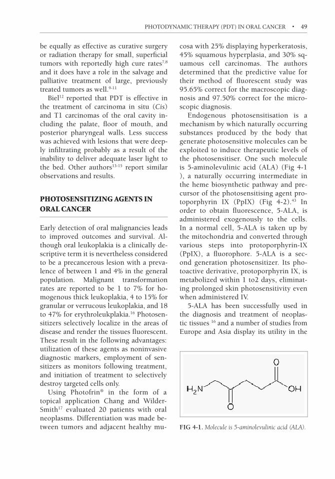
Endogenous photosensitisation is a mechanism by which naturally occurring substances produced by the body that generate photosensitive molecules can be exploited to induce therapeutic levels of the photosensitiser. One such molecule is 5-aminolevulinic acid (ALA) (Fig 4-1 ), a naturally occurring intermediate in the heme biosynthetic pathway and pre-cursor of the photosensitising agent pro-toporphyrin iX (PpiX) (Fig 4-2).43 in order to obtain fluorescence, 5-ALA, is administered exogenously to the cells. in a normal cell, 5-ALA is taken up by the mitochondria and converted through various steps into protoporphyrin-iX (PpiX), a fluorophore. 5-ALA is a sec-ond generation photosensitizer. its pho-toactive derivative, protoporphyrin iX, is metabolized within 1 to2 days, eliminat-ing prolonged skin photosensitivity even when administered iv.
5-ALA has been successfully used in the diagnosis and treatment of neoplas-tic tissues 16 and a number of studies from Europe and Asia display its utility in the
D-Aminolevulinic acid
FIG 4-1. Molecule is 5-aminolevulinic acid (ALA).

��
f i v e
Photodynamic Therapy of early laryngeal cancer
Merrill A. Biel
CARCiNOMA OF tHE LARyNX accounts for 25 to 30% of all carcinomas of the head and neck.1 Early carcinomas of the larynx (Cis, t1
, t2) and severe dysplasia
are presently treated with either radiation therapy or surgery alone. Five-year cure rates achieved with this therapy is 75 to 90%.2 Radiation therapy has the advantage of preserving the physical integrity of the larynx, thereby preserving the voice. Ra-diation therapy, however, has significant disadvantages even when small laryngeal fields of radiation are used. these disad-vantages include discomfort and mucositis during and for potential prolonged periods after therapy, permanently altered voice quality, dysphagia, chondroradionecrosis of the larynx and trachea, and the exten-sive length of therapy (6-7 weeks).3,4 Sur-gical therapy for early carcinomas, that is t1 and t2, of the larynx includes perform-ing a partial cordectomy or hemilaryngec-tomy. Although cure rates are high, surgi-cal removal of portions of the vocal cord or hemilarynx results in significant altera-tion of the quality of voice.5
Severe dysplasia and Cis may also be treated with either radiation or limited
surgery with either microsurgical tech-niques or laser excision. Le reported on 82 patients with Cis of which 15 were treated with vocal cord stripping with a 56% lo-cal control rate; 13 treated with extensive laser resection/hemilaryngectomy with a 71% local control rate; and 54 treated with radiotherapy with a 79% local control rate. Anterior commissure involvement was a significant negative prognostic factor. Subjective voice quality was good to ex-cellent in 73% of patients who underwent vocal cord stripping; 40% of those who underwent extensive resection and 68% who underwent radiation therapy.6 Zeitels reported on 7 patients with Cis undergo-ing microsurgical resection. two patients developed subsequent microinvasive can-cer requiring more aggressive treatment.7 Smith reported on 25 patients with Cis treated with surgical resection with an 88% cure rate.8 Sittel reported on laser excision of vocal cord cancers and noted a signifi-cant effect on voice with anterior commis-sure resections even when done in a staged fashion.9 Review of 10 reports of laser ex-cision treatments of Cis demonstrated a 82.5% control rate in 177 patients. Many

�� • PHOTODYNAMIC THERAPY OF DISEASES OF THE HEAD AND NECK
required multiple laser excisions.10 damm reported on 29 patients with Cis treated with laser excision. 76% (22/29) required more than one laser excision for persist-ence of disease, 9 of which were in the an-terior commissure. two-year disease-free survival was 86%. dysphonia was report-ed in all patients and none had improved voice over the pretreatment state.11 the lit-erature therefore demonstrates that surgi-cal techniques to treat Cis are best limited to those patients where the Cis does not involve the anterior commissure or the bi-lateral vocal cords.
Garcia-Serra reported on 30 patients with Cis treated with radiotherapy with an 88% local control rate. Review of litera-ture for radiotherapy of Cis demonstrated an 87.4% weighted local control rate at 5 years on 705 patients in 22 published re-ports.10
the optimal treatment for severe dys-plasia and early carcinomas of the larynx would be one that is effective, safe, repeat-able, minimally invasive, nonsurgical, and a less time-consuming therapy than radio-therapy. Photodynamic therapy is poten-tially such a treatment for severe dysplasia and early carcinomas of the larynx.
photodynamIC therapy
Photodynamic therapy (Pdt) is a mini-mally invasive treatment involving the use of a photosensitizing drug and laser light for the treatment of a variety of cancers.12 When administered, these compounds are accumulated and retained to a greater degree in malignant tissues than normal tissues. the drugs remain inactive until exposed to a specific wavelength of light. the light, usually from a laser, is transmit-ted through specially modified fiber optics and activates the drug. the resulting pho-
tochemical reaction results in the produc-tion of oxygen radicals thereby destroying diseased cells with little effect on normal tissues. to date, Pdt has been used to treat carcinomas in many organs and Photofrin-based Pdt has been approved by the Unit-ed States FdA to treat early and end stage endobronchial and esophageal squamous cell carcinomas and barrett’s dysplasia. in particular, the use of Pdt to treat early carcinomas of the head and neck has been promising.12-20
the generally accepted mechanism of action of Pdt is that there is an energy transfer process from the light activated or excited triplet state of the photosensi-tizer to oxygen producing singlet oxygen which, in turn, causes irreversible oxida-tion of some essential cellular component. it has also been shown that the vascula-ture changes within the tumor necrosis subsequent to Pdt result in ischemia that is responsible for tumor necrosis. Either or both are sufficient to explain the remarka-ble necrosis of tumors within 2–5 days fol-lowing Pdt with HPd or Photofrin.
PhotofrinR (porfimer sodium), like HPd, concentrates in malignant tissue, is activated by penetrating light (630 nm + 3 nm), produces fluorescence, and is pho-tochemically efficient. Like its predecessor HPd, PhotofrinR has produced only one major adverse reaction as a result of its use: light sensitivity. in animals, it requires about twice as much PhotofrinR as HPd to produce skin photosensitivity.
Photodynamic therapy has been dem-onstrated to be effective in the treatment of early carcinomas of the head and neck.12-20 Furthermore, preliminary studies on the treatment of benign laryngeal papilloma-tosis with Pdt have demonstrated this treatment to be safe and effective.
the advantage of Pdt treatment for early carcinomas of the larynx is the abili-

PHOTODYNAMIC THERAPY OF EARlY lARYNgEAl CANCER • ��
ty to preserve normal endolaryngeal tissue while effectively treating the carcinomas. this results in improved laryngeal func-tion and voice quality. Furthermore, Pdt requires a short duration of therapy as compared to radiation therapy, is repeat-able and carries less risk than surgical therapy, and is performed as an outpatient noninvasive treatment. importantly, the use of Pdt does not preclude the use of radiotherapy or surgery in the future for new primary or recurrent disease. if prov-en effective in a multi-institutional clinical trial, Pdt may become the first-line thera-py for treatment of early carcinomas of the larynx.
Multiple centers have reported Phase ii study data on the use of Photofrin-based Pdt to treat Cis-t2 carcinomas of the lar-ynx12-20 (table 5-1). Freche16 reported on 32 patients with t1 vocal cord carcinomas treated primarily with Pdt. twenty-five of 32 patients obtained a complete response for a complete response rate of 78%.16 Feyh treated 12 patients with Cis-t2 larynge-al carcinomas. Eleven of 12 patients ob-tained a complete response for a complete response rate of 91%.14 Gluckman treated 2 patients with t1 carcinomas of the lar-ynx both of which obtained a complete response.15 Schweitzer treated 10 patients
with Cis-t2 carcinomas of the larynx of whom 8 obtained a complete response for an 80% complete response rate.18
the largest single study of the treatment of laryngeal carcinomas with long-term follow-up has been performed by biel.12 One hundred and ten patients with recur-rent or primary CiS, t1N0, and t2N0 la-ryngeal tumors were treated with Pdt for cure with Photofrin-based Pdt at Ab-bott Northwestern Hospital (Minneapolis) from February 1990 to November 2005. three patients had recurrent CiS, 92 pa-tients had t1N0 carcinomas of the true vocal cord of which 25 were radiation fail-ures, and 15 patients had t2N0 carcino-mas of the true vocal cord of which 8 were radiation failures. All patients underwent a single microlens light treatment and most t2 tumors also underwent cylindrical dif-fuser implants into the paraglottic space. the age range was 39 to 88 years. All pa-tients were treated according to specif-ic protocols in accordance with FdA and iRb approvals. Pretreatment evaluation in-cluded a history and physical examination and endoscopic examination with tumor mapping and biopsy. Ct or MRi scanning of the larynx was used for staging prior to treatment of tumors greater than t1 or ra-diation failures. Photofrin (Axcan Pharma-
Table 5-1. Summary of Published Results with Photofrin PDT of Early Squamous Cell Cancer of the Larynx
Study Patients Lesion and Site Drug, Dose, mg/kg Response, n
Complete Partial None
Feyh et al. (14) 12 T1 and T2, larynx Photosan III 11 1 0Freche et al (16) 32 T1, larynx HPD, 3 25 7 0
Photofrin, 2Schweitzer (18) 10 T1, larynx 8 2 0Gluckman (15) 2 T1, larynx 2 0 2Biel (12,19) 110 Cis, T1, and T2,
larynxPhotofrin, 2 110 10 0

��
s i x
Interstitial PDT Cancer Treatment
Christian S. BetzColin Hopper
lImItatIonS of SurfaCe IllumInatIon pdt
the effectiveness of surface illumination Pdt is limited by the depth of penetration of light within the tissue. Longer wave-lengths have a greater depth of penetra-tion although against this is the feature of lower quantum yields with increasing wavelength. Oxygen requires 94 kJ/mol to raise it from the triplet ground state to the excited singlet state. in photodynamic therapy this energy is acquired from the photosensitiser which has itself been raised to a high energy state. Only light-absorbing compounds which can emit en-ergy greater than 94kJ/mol are therefore capable of activating ground state oxygen. this corresponds to an absorption wave-length of around 850 nm: photosensitis-ers are therefore only suitable for photo-dynamic therapy if their activation wave-length is below this figure. this equates to a depth limit of approximately 1.5 cm for surface illumination.1 Most of these problems can be overcome if light can be delivered directly into tissue using inter-stitial techniques.
Safety ConSIderatIonS— tISSue toleranCe
in order to effectively treat interstitially, it is necessary to be confident about Pdt effects on normal tissue. this is of par-ticular relevance in the head and neck with its abundance of vital structures. A number of safety studies have been car-ried out on different photosensitisers2-4 and have demonstrated the ability of nor-mal tissue to recover from the Pdt insult. bone treated with Pdt does show some slightly impaired healing. it is not en-tirely clear what mechanism is involved, but microvascular damage with reduced blood supply to the healing site is pos-sible. Fortunately, no long-term prob-lems were seen with bone healing and this is obviously of great importance for any treatment in the oral cavity. Of par-ticular relevance was the study by Kübler, who used m-tHPC on rabbit carotid and femoral vessels as well as the vagus and femoral nerves. in a dose escalation study he found that at high drug doses (0.3 mg/kg) and a short drug light interval (24 hours) with a light dose of 20 J/cm2 there

�� • PHOTODYNAMIC THERAPY OF DISEASES OF THE HEAD AND NECK
was edema, some thrombosis, and dis-ruption of the endothelial layer. He also reported up to 75% demyelination, but importantly, neither caused any clinical distress and no vessel rupture was seen.5 it is clear from these studies that Pdt is safe in close proximity with blood vessels in the normal setting. it would be quite reasonable to speculate that in a clini-cal setting, where tumor was eroding the vessel wall, Pdt might precipitate acute hemorrhage. However, as long as tumor is close but not eroding the arterial wall, treatment can be carried out safely. these studies have been performed on normal tissue models, so caution should be used when extrapolating from these animal studies to head and neck cancer where patients may have already been heavily pretreated with surgery radiotherapy and chemotherapy.
interstitial Pdt (Fig 6-1) has been suc-cessfully used in the treatment of internal organs such as the pancreas, prostate, and brain, so there is a reasonable amount of data to suggest iPdt is safe in the head and neck.
drugS In ipdt uSe
Any Pdt treatment can be carried out in-terstitially just as long as the activating wavelength can be delivered through an optical cable. Most clinical studies in the head and neck have used either Photofrin, phthalocyanines, or Foscan, although sev-eral other drugs have been investigated in other pathologies—for example, the bac-teriochlorin m-tHPbC has been used in the treatment of liver metastases from co-lon cancer6 and Pd-bacteriopheophorbide (tookad) and metallotexaphyrin (Lu-tex) are being investigated for the treatment of prostate cancer.7-9
doSImetry
there are two main methods of delivering light—point sources and diffuser fibers (Plate 10). the tissue effects of these when used interstitially can be simulated using mathematical modelling; this can be quite useful in the calculation of light distribu-tion and extent of treatment effects (Plate 11). there are limitations with these tech-niques which are related to the exact na-ture of tissue and the extent to which light can pass through the tissue. there is really no substitute for real-time monitoring of light distribution which can be done in a number of ways. isotropic detector fibers can be used to monitor light within tissues during treatment.10 Recently a more in-novative approach has been described by Gross which uses bOLd MR as a marker of photodynamic activity.11 None of these methods, however, is so far able to accu-rately predict the actual effect on the tis-sue but histologic confirmation, although such evidence is hard to come by in hu-
FIG 6-1. Foscan PDT treatment in the posterior orbit using interstitial light delivery through titanium needles. Eyesight was preserved in this patient.

INTERSTITIAl PDT CANCER TREATMENT • ��
man tissue and would certainly not be available in all cases.
Planningthe aims of planning are to iden-tify the total volume of disease and then ensure that light is delivered at sufficient energy to trigger the Pdt effect to this tumor volume. the accurate evaluation of tumor extent is usually carried out using MRi, Ct, ultrasound, and PEt Ct and a similar approach is taken to radiotherapy planning where the target volume includes a rim of normal tissue. the advantage of Pdt over radio-therapy is, of course, the absence of cumu-lative toxicity with repeat treatment.
Fiber PositioningPositioning of the fibers can be performed freehand, or in more sensitive areas under image guidance with MR, Ct and ultra-sound. One of the techniques for doing this has been described by Jäger et al where titanium needles are placed in the target tissue under MR guidance in accordance with the London rules for iPdt.12 these are loosely based on the Paris system for radiotherapy and state that light delivery systems should be parallel, equidistant and deliver consistent light along their length.
An alternative approach is with an after-loading system that is very much adapted from brachytherapy techniques. diffuser fibers can be fed through a series of plastic conduits and complex volumes of tumor can be treated (Fig 6-2).
anaeSthetIC ConSIderatIonS
there are a few important points regarding patients undergoing iPdt under general anaesthesia. Overall, the patient popula-
tion may have already had extensive sur-gery and radiotherapy. this often makes airway management problematic and an experienced airway anaesthetist is essen-tial for these cases. there are the added complications of slightly subdued light-ing and if the treatment takes place in an MR or Ct scanner, the surroundings may not be that familiar. in addition, the use of protective glasses makes the reading of color-coded drugs and monitors difficult. Care must also be taken with the use of a pulse oxymeter as this uses a red HeNe laser that can cause blistering of the finger if left in place too long.
lImItatIonS on effeCtS
Proximity to major vessels where vessels may be invaded by tumor is a great cause for concern and treatment in this situation is likely to result in catastrophic bleeding. Accurate preoperative assessment of vas-cular invasion is essential for safe treat-ment. When vascular invasion is present in a major vessel, we would advocate the prophylactic placement of a covered endo-luminal stent (Fig 6-3).
the optical properties of tissue are also of obvious importance. Light transmission
FIG 6-2. Use of transparent conduits to facilitate diffuser fiber placement in the treatment of a recurrent tongue tumor.

�0 • PHOTODYNAMIC THERAPY OF DISEASES OF THE HEAD AND NECK
through a vascular tumor would be very different to scarred or fibrotic tissue. this makes real-time dosimetry even more de-sirable.
ClInICal StudIeS
based on the theoretical and preclinical ad-vantages addressed above, several groups have endeavored to bring interstitial Pho-todynamic therapy into clinical reality (table 6-1). As early as 1988, there had been a single, sporadic report on the use of interstitial Pdt with hematoporphy-rin derivative as a photosensitizer for the
treatment of primary oral squamous cell carcinomas as well as their regional lymph node metastatases.13 the authors state that all lesions treated except for those with bony involvements showed a good re-sponse to treatment, whereas the covering skin, connective tissue, and nearby organs seemed spared from major injury.
Only since the beginning of the 21st century, scientific publications concerning the use of interstitial Pdt for the treat-ment of head and neck tumors have come up more frequently. in 2001, two groups have reported on the successful use of in-terstitial Pdt with hematoporphyrin de-rivatives and phthalocyanines in a limited number of patients with head and neck tumors of various stages.14,15
At the department of Oral and Maxil-lofacial Surgery at the University College London Hospital, interstitial Pdt using m-tHPC as a photosensitizer was established in 1997 and has since then primarily been applied for palliative treatment of advanced head and neck tumors. in an original pub-lication from 2004 by Lou et al,16 the group presented the retrospective results of a 5-year experience in this field. in total, 45 pa-tients with persistent or recurrent head and neck cancer unsuitable for further treat-ment with surgery, radiotherapy or chemo-therapy were recruited and subsequently
FIG 6-3. Placement of a covered endoluminal stent to protect carotid artery from a blowout during PDT.
Table 6-1. Medline-Listed Publications on Clinical Studies Using iPDT for the Treatment of Head and Neck Disease
Author Year Tumor Site Photosensitizer N Main Results Ref
Wang et al 1988 Oral cavity Hematoporphyrin derivative 10 “Good response” of all tumors except for those with bony involvement
13
Stranadko et al 2001 Oropharynx/ Larynx
Hematoporphyrin derivative/Al-Phthalocyanine
Fraction of 61
CR 57.4%; PR 37.7% 14
Tanaka et al 2001 Tongue Porfimer Sodium 3 CR 66.7%; PR 33.3% 15
Lou et al 2004 Oral cavity/ Oropharynx
m-THPC 45 CR 20.0%; PR 53.3% (only recurrent SCCs treated)
16
��
s e v e n
intraoperative Adjuvant PDT of Head and neck cancer
Merrill A. Biel
HEAd ANd NECK squamous cell carci-nomas with extensive soft tissue invasion are known to have high rates of local and regional recurrence. Recurrence rates of cervical nodes with extracapsular spread range from 21 to 58%.1-4 Primary squa-mous cell carcinomas of the tongue base and hypopharynx also have local and re-gional recurrence rates of 43 to 71% de-spite aggressive combined surgical and chemoradiotherapy.5-7
When patients with squamous cell carcinomas develop large invasive soft tissue recurrences of their carcinomas fol-lowing previous surgery, radiation, and chemotherapy, the likelihood of further recurrence following extensive surgical resection of the recurrent carcinoma is extremely high. in addition, these recur-rences tend to occur within the first 6 to 12 months of surgical resection.6-7
Photodynamic therapy (Pdt) has been successfully used to treat patients with early squamous cell carcinmomas of the head and neck.8-9 this is due to the abil-ity of the activating laser light to penetrate up to one centimeter into tissue resulting
in destruction of microscopic tumor with relative sparing of normal tissue when the correct drug and light dose combinations are used. Employing this principle, Pdt may be used as an adjuvant intraopera-tive therapy following resection of tumor in patients with recurrent and large infil-trating carcinomas of the head and neck, to potentially increase the rate of cure in this dismal disease by destroying micro-scopic residual disease and to provide for a greater likelihood of tumor-free margins of resection. this method of treatment may be clinically applied in two different ways: (1) Pdt for curative intent follow-ing gross tumor debulking. the goal of this treatment is to achieve complete tu-mor eradication with preservation of nor-mal vital structures such as the larynx and tongue; and (2) Pdt of the surgical resec-tion bed following complete resection of t3 and t4 tumors. the goal of this treat-ment is to increase local/regional disease control by increasing tumor-free resection margins and destroy microscopic skip le-sion disease while preserving uninvolved normal structures.

�� • PHOTODYNAMIC THERAPY OF DISEASES OF THE HEAD AND NECK
IntraoperatIve pdt preClInICal StudIeS
intraoperative Pdt in the head and neck results in the exposure of vital blood ves-sels and nerves to the Pdt treatment. in particular, the carotid artery, internal jugular vein and cranial nerves X to Xii would be commonly exposed during Pdt treatment. A partial or complete necrosis of these structures due to Pdt treatment could result in catastrophic life-threatent-ing complications including arterial rup-ture or permanent cranial nerve damage.
Several preclinical studies have been performed to evaluate the effect of Pdt on blood vessels and nerves. Grant et al used a rabbit carotid artery model to investigate the effect of disulphonated phthalocyca-nine and 5-aminolevulinic acid mediated Pdt on these vessels. three days follow-ing Pdt all treated vessels demonstrated a complete loss of endothelium with death of the media smooth muscle cells. there was no vascular occlusion, hemorrhage, or thrombosis present. Re-endotheliali-zation occurred in all vessels by 2 weeks. Furthermore, intraluminal hydrostatic distension tests performed on the vessels demonstrated no reduction in the pressure required to burst the vessels treated with Pdt versus the nontreated control carotid arteries. they concluded that despite the full thickness vessel wall cell death, Pdt treated arteries are not at risk for throm-botic occlusion, rupture or hemorrhage.10
Ris et al performed intraoperative Pdt to the blood vessels and nerves in the tho-racic cavity of mini-pigs using mtHPC as photosensitizer (0.1 mg/kg, 20 J/cm2, drug-light interval of 12 hours to 6 days). tissue damage was strongest at a drug-light interval of 12 hours and gradually became less at longer drug-light intervals and was absent by 3 days after drug injec-
tion. At a drug-light interval of 12 hours, there was severe damage of the aorta with thrombosis and necrosis of the tunica me-dia and desquamation of the endothelium; however, there was no damage to nerves. At 24 hours, only minor changes were ob-served in the aorta and the vena cava re-sponded with swelling of the endothelium and thrombosis of the vasa vasorum but did not shown any necrosis of the vessel wall or thrombosis.11
Kübler et al studied the effect of m-tHPC Pdt on large blood vessels and nerves in a rabbit model. the most severe reactions occurred at a drug dosage of 0.3 mg/kg, light dose of 20 J/cm2 and 24 hour light interval. blood vessels demonstrat-ed severe edema, media hyperplasia with loosening of the endothelium and various degrees of local thrombosis. there was no breakdown of the vessel wall or any vessel ruptures. Nerves were altered by a 75% de-myalinization but with no clinical symp-toms.12
biel studied the effect of Photofrin Pdt on the carotid artery, internal jugular vein, and vagus nerve in dogs. this study dem-onstrated that at a drug dose of 2 mg/kg and a light dose of 50J/cm2 at 150 mw/cm2, there was no histologic effect on either the blood vessels or the vagus nerve. At high-er light doses, greater than 75 J/cm2, there was loss of the arterial media and endothe-lial sloughing.13
Kübler et al demonstrated in a rat skin flap model that the addition of Photof-rin Pdt does not affect wound healing.14 However, belmont et al in a rat fasciocuta-neous flap model demonstrated that Pho-tofrin Pdt reduced the critical primary ischemic time of the rat fasciocutaneous flap, whereas white light illumination in the presence of Photofrin had no effect on the critical primary ischemic time.15
importantly, preclinical studies have

INTRAOPERATIvE ADjuvANT OF HEAD AND NECK CANCER • ��
demonstrated that intraoperative adju-vant Pdt at the time of surgery reduces the incidence of local recurrence. dilkes in a squamous cell carcinoma model and davis in a mouse neuroblastoma model demonstrated that intraoperative Pdt to the tumor bed prior to closing the wound resulted in a 50% reduction in the local re-currence rate.16-17 based on these preclini-cal studies demonstrating the potential for adjuvant intraoperative Pdt to improve cure rates, several centers have performed human clinical studies to demonstrate the efficacy of adjuvant intraoperative Pdt delivered to the tumor resection bed at the time of surgical resection.
IntraoperatIve pdt human ClInICal StudIeS
biel performed intraoperative adjuvant Pdt using Photofrin in 35 patients.8 these patients were divided into two treatment groups: (1) Pdt for curative intent fol-lowing gross tumor debulking. the goal of this treatment was to achieve complete tu-mor eradication with preservation of nor-mal vital structures such as the larynx and tongue; and (2) Pdt of the surgical resec-tion bed following complete resection of t3 and t4 tumors. the goal of this treat-ment was to increase local regional disease control by increasing tumor-free resection margins and destroy microscopic skip le-sion disease while preserving uninvolved normal structures.
in the first treatment group, Pdt for curative intent following gross tumor de-bulking, 17 patients were treated, 11 laryn-geal and 6 oral cavity. Of the 11 laryngeal, 8 were supraglottic and 3 were glottic. the oral cavity lesions were tongue and floor of mouth. the treatment consisted of the patient receiving Photofrin 2 mg/kg pre-
operatively and 2 days after the injection the patient underwent general anesthe-sia and gross but incomplete resection of the tumor mass. Residual microscopic dis-ease was confirmed with frozen section biopsies intraoperatively. Pdt was then performed to the resection site using a mi-crolens fiber at 75 to 80 J/cm2 at 150 mW/cm2. Cylindrical diffuser implantation 0.5 cm in length was placed wherever the lo-cation was safe to do so and illumination performed at 100 J/cm fiber length at 400 mW/cm fiber length. Most of the treat-ments were performed on an outpatient basis. For the laryngeal tumors treated, with follow-up to 69 months, there have been no recurrences (Plates 13 and 14). For the 6 oral cavity tumors, with follow-up to 58 months there was one recurrence that went on to conventional surgical re-section and remains free of disease.
in the second treatment group, intraop-erative adjuvant Pdt of the surgical resec-tion bed following complete resection of t3 and t4 tumors, 18 patients with recur-rent infiltrating squamous cell carcinoma of the head and neck were treated. Each patient had undergone previous treatment of the primary lesion of the head and neck with surgical resection, radiotherapy, and chemotherapy. the initial primary car-cinomas were in the larynx; tongue and floor of mouth; branchial cleft cyst; me-dial canthal skin and ethmoid sinus; and tonsil. the sites of recurrence included the pharyngoesophagus and anterior neck skin; mandible and neck; medial orbit, eth-moid and anterior skull base; neck skin, parotid, and lateral skull base; tongue and floor of mouth; and neck. in all cases, ex-tensive skin involvement with tumor was present with deep infiltration into the soft tissues as determined by Ct, MRi, and an-giographic scanning. All lesions were de-termined to be surgically resectable.

��
e i g h t
Photodynamic Therapy (PDT) in nasopharyngeal cancer
I. B. TanH. J. Nyst
H. J. C. M. SterenborgR. L. P. van Veen
P. C. LevendagD. J. Robinson
F. A. Stewart
demographICS
Nasopharyngeal carcinoma (NPC) occurs sporadically in the western world, but is endemic in certain parts of Asia, such as southern China. the worldwide incidence of NPC is 45,976, with 19,616 new cases each year in China and 4,848 new cases in indonesia.1 in western countries the inci-dence is much lower with 49 and 538 new cases each year in the Netherlands and Germany, respectively, and an intermedi-ate position for the Mediterranean basin.
Nasopharyngeal carcinomas are epithe-lial neoplasm’s, classified in three different histopathologic types (WHO classifica-tion, 1993). type i tumors are squamous cell carcinoma (SCC) with varying degree of differentiation. type ii are nonkeratiniz-ing carcinomas and type iii are undifferen-
tiated carcinomas, collectively considered as undifferentiated nasopharyngeal carci-nomas (UCNt).
treatment optIonS
Nasopharyngeal carcinomas are responsive to radiotherapy and chemotherapy and the first-line treatment for primary NPC is ra-diation.2 As the nasopharynx is located in the midline and is surrounded by critical structures, efforts should be made not to overdose these important areas. the vol-ume to be irradiated should include the nasopharynx, adjacent parapharyngeal, tissues and the cervical lymph nodes (lev-el ii-v). After about 40 to 60 Gy, the spinal cord should be shielded as a conservative estimate of the tolerance of the spinal cord

�� • PHOTODYNAMIC THERAPY OF DISEASES OF THE HEAD AND NECK
is about 50 Gy in 2-Gy daily fractions. An additional dose of 22 to 30 Gy is delivered to the nasopharynx proper. in general, 66 to 70 Gy for t1 and t2 lesions and 70 to 75 Gy for t3 and t4 tumors is required. because of the likelihood of developing cervical metastases, all of the cervical lym-phatics should be irradiated with 46 to 50 Gy to both sides of the neck in N0 patients or, a cumulative dose of 70 Gy applied to the nodes in case of positive lymph nodes at presentation (80% of the cases).3
if a “booster dose” is required for the nasopharynx after external irradiation, this can be delivered by stereotactic ra-diosurgery, intracavitary therapy, or three dimensional (3d) conformal or intensi-ty-modulated radiation therapy (iMRt) techniques.4-8 brachytherapy has also been used to deliver a higher dose to a limited volume of the nasopharynx.9-12
the best treatment for early t-stage na-sopharyngeal carcinoma is external beam radiotherapy in combination with brach-ytherapy or stereotactic radiotherapy. For advanced disease the combination of radi-otherapy and neoadjuvant or concomitant chemotherapy is standard.13,14 the combi-nation of chemotherapy and radiotherapy is an attractive therapeutic option because of a possible synergy and the potential re-duction of distant metastasis.15-16
reSultS of treatment
despite the radio-responsiveness of na-sopharyngeal tumors, good long-term survival is only achieved for patients who have early primary tumors with minimal neck disease 67 to 71% 10 years’ disease-free survival for t1, t2, and N0-1. Surviv-al is poor for patients who have extended tumors and/or extended neck nodes 29 to
54% 10 years’ disease-free survival for t3, t4, and N2, N3.17-21
Poor survival in the t4,N0-1 category is chiefly the result of the high local re-currence rate (63.8%), whereas for the t1-2,N2-3 category, it is the result of the high distant metastases rate (approximately 50%).22
retreatment of reCurrent naSopharyngeal CarCInoma
Usual treatment options for early stage re-current or persistent NPC are surgery in combination with external radiotherapy, brachytherapy alone,23-25 or in combination with external radiation,26-29 or stereotactic radiosurgery.30 the standard treatment for advanced stages of recurrent or persistent disease is chemotherapy followed by reir-radiation, or concomitant chemo-reirradi-ation.31 Pryzant et al32 reported on 53 pa-tients with locally persistent or recurrent nasopharyngeal carcinoma treated with reirradiation. Local recurrence was con-fined to the nasopharynx in 27 patients, and persistent tumor in 26 patients. the 5-year actuarial local tumor control rate was 35%, 5-year disease-free survival was 18%, and overall survival was 21%. Eight patients developed severe complications from retreatment, two involving the brain, one the spinal cord, and two the cranial nerves, all of which were fatal. the 5-year actuarial incidence of severe complications was 17%. the incidence of severe compli-cations was related to the total cumulative dose of external irradiation.
Lee et al33 described the incidence of late complications after reirradiation in 891 pa-tients with local recurrence after definitive radiation therapy for nasopharyngeal carci-noma. After external reirradiation, brachy-therapy, or a combination of both, a wide

��
n i n e
Photodynamic Therapy for esophageal Diseases
Wytske M. WestraKenneth K. Wang
ratIonale for photodynamIC therapy
Photodynamic therapy is ideally suited for the esophagus as it is so readily applied us-ing the endoscope. Photodynamic therapy has always been used in areas where pho-toradiation can be easily delivered such as the skin where it was first applied by the ancient Egyptians and Chinese to treat skin diseases, such as psoriasis and vitil-igo. this application also took advantage of the fact that phototherapy can be used to treat relatively focused areas of skin in a single application. the esophagus is easy for gastroenterologists to access via endos-copy and these endoscopes have the ap-propriate working channels that can pass the optical fibers needed for photoradia-tion. it also has the advantage of treating large areas of circumferential mucosa with a single application of light and to this day represents the technically simplest thera-py available, for treatment of large areas of esophageal mucosa.
methodS of photodynamIC therapy In the eSophaguS
Photodynamic therapy (Pdt) involves the local or systemic administration of a chemi-cal (photosensitizer) that has a known pro-pensity to photoexcitation when exposed to light of the appropriate wavelength. the absorption of light causes the photosensi-tizer to transfer from its ground state into an excited singlet state which can then ei-ther decay directly back to the ground state with fluorescence emission, or undergo fur-ther transformation into an excited triplet state. the latter can react with surround-ing oxygen to form singlet oxygen, a highly cytotoxic molecule. in the esophagus, Pdt has a few specialized requirements. Since the photosensitizer needs to react with sur-rounding oxygen to form singlet oxygen, an adequate level of molecular oxygen is needed to achieve maximal tissue damage. there is usually a requirement for the deliv-ery of supplemental oxygen as the airway can be slightly compromised by the place-ment of an endoscope into the esophagus.

�0�
t e n
The Use of Photodynamic Therapy in the Management of lung cancer
Eric S. Edell
IntroduCtIon
Lung cancer continues to be the most com-mon cause of cancer death in the United States accounting for more deaths than prostate cancer, breast cancer, and color-ectal cancer combined. Of those patients diagnosed with lung cancer, fewer than 15% of them will survive this devastating disease. the opportunity to achieve long-term survival depends on resecting lung cancer at its earliest stage which, unfortu-nately, is a rarity in this disease.
the NiH sponsored studies at the Johns Hopkins University, Mayo Clinic, and Me-morial Sloan-Kettering evaluating the ef-fectiveness of screening chest x-ray and sputum cytology on lung cancer mortal-ity. Unfortunately, there was no mortality improvement with the screening strategy; however, sputum cytology did detect more cancers in the experimental group than in the control group. it was also noted dur-ing the studies that, on occasion, patients with abnormal sputum cytology had no obvious cancer identified on routine bron-choscopic inspection. this dilemma led to the development of fluorescent detection devices that relied on photochemicals to
facilitate the localization of these bron-choscopic occult cancers. Photochemicals were also known to cause cell death and thus are effective in treating cancers of the airway. the purpose of this chapter is to review the use of both photodynamic di-agnosis and photodynamic therapy in the management of lung cancer.
photodynamIC dIagnoSIS
Photodynamic diagnosis is a term that re-fers to the use of devices which discrimi-nate normal from abnormal mucosa by dif-ferences in the wavelength of fluorescence for each. Compounds such as dihemat-oporphyrin ether (Photofrin) and hemat-oporphyrin-derivative (HPd) are photo-chemicals that were originally used for this purpose.1–4 these compounds accumulate in abnormal tissue at higher concentrations than normal tissue enabling localization of the abnormal tissue by detecting the fluo-rescence of the compounds when exposed to the appropriate wavelength of light. the amount of photochemical that accumulates in small superficial cancers of the airway is

���
e l e v e n
Photodynamic Therapy in skin cancer of the Head and neck
Alexander Kübler Nicolas Hunzelmann
IntroduCtIon
More than one-third of all cancers in the United States are nonmelanomatous skin cancers.1 Exposure to sunlight is the prin-cipal cause for this kind of tumor. For Cau-casians, their incidence is strongly associ-ated with age and cumulative ultraviolet b radiation.2,3 However, exposure to chemical carcinogens, ionizing radiation, chronic ulceration, immunsuppression, or genetic defects also account for these tumors. due to their genesis, most nonmelanomatous skin tumors are located in sun-exposed skin parts. therefore, 80% of the squamous cell cancers and basal cell carcinomas of the skin are found on the hands, arms or the head and neck.2 Even after a curative therapy the risk of subsequent skin tumors is high.4 About 50% of the patients with a history of nonmelanomatous skin cancer will develop a new skin cancer at another site within the first 5 years independent of the primary therapy.5 in patients with a genetic defect, immunsuppression (eg, after organ transplantation) or chemical-induced tumors, the risk of subsequent tumors can be even higher. these patients may suffer simultaneously from more than
one and up to several dozen skin tumors or premalignant skin lesions at various sites, not only limited to the arms, head, or neck. On account of the link between skin tumors and ultraviolet b light exposure and the changing leisure activities in western societies, nonmelanomatous skin tumors are an emerging problem in dermatology and the most common form of cancer worldwide. An increase would also be expected with the aging of the population but there are also data indicat-ing that the incidence is increasing in the younger population as well.6,7
Squamous cell carcinoma rates have in-creased 3% to over 10%.7 in New Hamp-shire (US) the incidence of squamous cell carcinoma increased by 235% in men and 350% in women and incidence rates of ba-sal cell carcinoma increased by more then 80% in men and women between 1979 to 1980 and 1993 to 1994.8 More then 1 mil-lion cases of basal cell or squamous cell carcinomas occur annually in the Unit-ed States.9 basal cell carcinoma incidence rates have increased 3% to 6% annually according to most studies throughout the industrialized world. there are marked

��� • PHOTODYNAMIC THERAPY OF DISEASES OF THE HEAD AND NECK
geographic differences in the incidence of basal cell carcinoma. these incidences are probably underestimated as nonmelano-ma skin cancers tend to be underreport-ed in cancer registries. the overall age and sex standardized annual incidence in Min-nesota, USA was 146 per 100,000 and in Australia was 726 per 100,000.10 in Wales (UK), the incidence of nonmelanoma skin cancer rose from 173 to 265 per 100,000 population between 1988 and 1998.11 Since 2001, nonmelanoma skin cancer oc-cupies second place in Russia; in 2004, 54.284 new cases were diagnosed, and the incidence of skin cancer was 38 per 100 000 population.12
despite good accessibility of this superfi-cial malignancy to diagnosis and treatment, there are many still unsolved problems. Skin tumors can be treated by surgical and nonsurgical methods. dependent on tu-mor location, size, type (primary or recur-rence), histology, patient morbidity, and preference different treatment methods like surgical excision, Mohs´ micrographic surgery, cryosurgery, curettage, laser abla-tion, or radiation therapy can be applied.6 Up to now surgery has been the mainstay of therapy for nonmelanomatous skin tu-mors. in patients with large or multifocal tumors, a good cosmetic outcome after surgery can be difficult to obtain. this is especially true for lesions on the face or in patients with multilocalized lesions, when primary wound closure after surgery can be difficult to achieve, requiring recon-struction by plastic surgery, for example, local skin flaps, skin graft, or healing by second intention.
Photodynamic therapy (Pdt) has be-come an important alternative therapeu-tic option. As most tumors of the head and neck are easy accessible for direct la-ser light illumination, Pdt has been used in the head and neck since the beginning
of the clinical application of Pdt. Cutane-ous lesions, premalignant lesions of the oral mucosa, and solid tumors have been treated so far. Pdt is also successfully em-ployed for the treatment of a number of nonmalignant diseases. Pdt is effective and has advantages over traditional treat-ment modalities.5 tumor destruction with preservation of surrounding normal tissues provides excellent cosmetic effects which is especially important in skin tumors.13-15 Pdt is convenient and well tolerated by patients. the most frequent adverse effect is photosensitivity which can be control-led by restriction of light exposure for the period of time that it is present. For medi-cal personnel Pdt is also relatively simple procedure.16
For the treatment of skin tumors, intra-venous applied, systemic photosensitiz-ers like Photofrin or Foscan can be used as well as the topical applicable photosen-sitizer aminolevulinic acid (ALA). due to the limited penetration depth of a topical applied photosensitizer and due to its hy-drophilicity, ALA is mainly used for very superficial skin tumors not thicker than 2 mm. Compared to that, systemic pho-tosensitizers are mainly used for tumors thicker than 2 mm.
SyStemIC photoSenSItIzerS
in using intravenously applied photosen-sitizers, drug accumulation is not limited to the superficial areas of the tumor as it is for ALA.13-17 instead the therapy depth depends only on the light penetration ca-pability into human tissue, which is domi-nated by the activation wavelength and op-tical properties of the illuminated tissue.18 For several studies on intravenous photo-sensitizers for Pdt of skin tumors, hemat-oporphyrin derivatives (HPd, dHE, Pho-

PHOTODYNAMIC THERAPY IN SKIN CANCER OF THE HEAD AND NECK • ���
tofrin), photosensitizers of the first gen-eration, were applied. Hematoporphyrin derivatives are activated at 630 nm which causes a maximum light penetration depth into the skin respectively a therapy depth of maximum 5 to 7 mm. various drug and light dosage combinations have been used ranging from 0.5 to 5 mg/kg and 25 to 288 J/cm2. the clinical results of these studies are difficult to compare due to the various histologic diagnosis as the distin-guish treatment parameters.19-36 there are only very few reports about the Pdt of sq-uamous cell cancer (SCC). Most of them have shown good clinical results with a complete response rate of up to 100%. Only the results of Pennington et al, who have used extreme low doses of light, were disappointing.26 therefore a wide range of tumor response rates between 0 and 100% is notified which varies by tumor histolo-gy and tumor location. but there is a clear tendency that by reducing the drug dosage and increasing the light energy a better se-lective response and better cure rate can be obtained.32 So most investigators re-ported about satisfying response rates and good cosmetical results for superficial and nodular basal cell cancer (bCC) by using hematoporphyrin derivatives. but Photof-rin-mediated Pdt seems not to be suitable for the treatment of morphoeic bCC.37
the reason for the very reserved clinical application of intravenous administered photosensitzers in skin Pdt might be the long-lasting systemic photosensitivity of the patients. due to the intravenous ap-plication of the drug a systemic photosen-sitivity of the patient occurs which forces the patient to stay indoors, out of bright daylight for up to several weeks, depend-ing on the photosensitizer and the drug dosage. For Photofrin this period can last up to 4 weeks, why this photosensitizer is not a real treatment option for skin tumors
so far. New second-generation photosen-sitizers have been introduced for clini-cal application. these drugs are activated at a longer wavelength which results in a deeper penetration in biological tissues. they also have much faster systemic elim-ination which shortens the photosensitiv-ity period.
benzoporphyrin derivate (bPd) a pho-tosensitizer of the second generation, has also be used for the treatment of skin tu-mors so far.38 by treating 27 patients with 107 primary nonmelanomatous skin can-cers and skin metastasis at various drug and light doses, a complete response rate of 57% and a partial response rate of 22% with a good cosmetic outcome could be obtained.
Compared to Photofrin the period of photosensitivity when using Focan can be shorter than 2 weeks. this is the reason why Foscan has been used in several clini-cal trials for the treatment of skin tumors and other tumors so far.
Foscan is an approved second-genera-tion photosensitizer for photodynamic therapy of recurrent head and neck can-cer.39,40 the active pharmaceutical ingre-dient of Foscan, temoporfin, (m-tHPC) is a lipophilic chlorin derivative with ab-sorption peaks in green and red spectral bands (514, 532, and 654 nm). this com-pound is characterized by a high quan-tum yield of singlet oxygen formation and high photodynamic activity. the unique photochemical properties of Foscan al-low administration of a relatively low drug and light dose to achieve good therapeu-tic results.41 there is a favorable tumor-to-normal tissue retention ratio. the longer excitation wavelength allows adequate de-struction of deep tumors of up to 1 cm when using superficial illumination tech-nique. Clinical trials of Foscan-mediated Pdt demonstrated very good results in

���
t w e l v e
Nursing Care of the Photodynamic Therapy (PDT)
Head and Neck Patient
Carla Kane
IntroduCtIon
Photodynamic therapy (Pdt) is a nonsur-gical, tissue sparing, minimally invasive technique used to treat certain types of cancer. Pdt has been FdA approved in the United States for esophageal and tracheo-bronchial cancers. Pdt has been used in investigative study treatments including: cancer of the breast, colon, bladder, brain, cervix, and skin. Pdt has been recognized in the medical field for treatment in the following conditions: age-related macular degeneration, dermatology, and barrett’s esophagus. Pdt has been used in the treat-ment of early oropharyngeal primary and recurrent cancers, and palliative treatment of refractory head and neck cancers. Pdt is also effective in the early recurrent car-cinomas (Cis, t1, or t2) of the oral cavity, larynx, pharynx, and nasopharynx.
the primary advantage of Pdt is that it causes minimal damage to healthy tissue surrounding the cancerous tumor. the side effects of Pdt are minimal, easily managed, and are not permanent. Pdt does not affect the white blood count, leaving the patient’s immune system intact, allowing the patient a better chance to fight disease.
On the other hand, chemotherapy and radiation therapy can cause significant side effects including nausea and vomit-ing, fatigue, headache, internal bleeding, diarrhea, hair loss, and blood abnormali-ties. Some patients suffer from xerostomia (severe dry mouth) after radiation therapy, resulting in significant difficulty eating and even speaking for the rest of their life.
Pdt can be used repeatedly to achieve the desired results. Pdt does not exclude concurrent treatment such as surgery, chemotherapy, or radiation therapy. be-tween 2 to 4 weeks of time should be allot-ted before commencing radiation therapy. this waiting period will allow for the in-flammatory response from the Pdt to sub-side.
Pdt is often conducted on an outpatient basis, allowing the patient to go home the same day as their procedure, reducing the overall health services consumption.
the downside of Pdt is that for about 4 to 8 weeks, the patient could develop an extreme photosensitivity reaction if the proper light precautions are not adhered to. Pdt is contraindicated in patients with porphyria or a known allergy to porphy-
���
t h i r t e e n
Photodynamic Therapy of Recurrent Respiratory Papillomatosis
Mark J. ShikowitzBettie M. Steinberg
Virginia Mullooly
IntroduCtIon
the beneficial health properties of sunlight have long been established. Romans often had a special room or “solarium” for sun-bathing. this represented an early form of heliotherapy. Hippocrates in the 4th cen-tury bc advocated the use of sunbaths for building up wasted muscles. He even in-corporated a protective head cover, some-thing we do during modern therapies.
there is evidence that a form of phy-tochemotherapy was practiced in some ancient civilizations. this involves the ad-dition of a drug to the light therapy. in in-dia, extracts of Psoralea corylifolia, now known to contain furocoumarins, was ad-ministered orally, followed by exposure to sunlight to treat vitiligo.1
the role of phototherapy as a useful medical treatment seemed to founder for a period of time. Niels Ryberg Finsen (1860–1904) was born in the Faroe islands, but studied and worked in Copenhagen. He wrote a book, which was translated into English.2 in it, he reported that sunlight or light from a carbon arc with a heat fil-
ter could be used to treat lupus vulgaris, a tuberculin skin condition. A Medical Light institute in Copenhagen was named after him. Finsen received the Nobel Prize for his work in 1903.
Queen Alexandra brought the idea of light therapy to London. She was the pres-ident of the London Hospital in White Chapel and introduced the technique in early 1900. the light source was a carbon arc and filtered through water to dissipate the heat. the light department was still in operation into the 1920s.
Again, despite success in specific dis-eases, phototherapy did not advance as hoped. there were isolated packets of dis-ease treatments, but there was no cohesive treatment theory. this included the ben-eficial role of light in treating rickets3 and psoriasis4 is well documented. the form of light treatment known to most of us is the therapy of jaundice in the newborn. Cre-mer et al (1958) made observations of the effect of sunlight and then artificial light on infants in the Rochford General Hos-pital in Essex. Up to this time, exchange transfusion was the only method for re-

��� • PHOTODYNAMIC THERAPY OF DISEASES OF THE HEAD AND NECK
ducing the effects of unconjugated bi-lirubin, which acts as a neural toxin.5
the use of photodynamic therapy (Pdt), which requires three components; visible light, photosensitizer, and oxygen to kill tumor cells is not new. this technique of Pdt was first used by vontappeiner and Jesionek in 1903 with their treatment of tumors treated with eosin topically, and then exposed to visible light.6,7 in 1905, Jesionek and vontappeiner reported the extension of this work to include two light sources (sunlight and arc lamp) with vari-ous photosensitizers. As before, the ap-plication of the photosensitizer was local at or near the surface of the lesion. the results which were mainly on basal cell carcinomas appeared promising.8 despite this apparent success, no further reports of Pdt immediately followed.
Recurrent respiratory papillomatosis (RRP) is a potentially life-threatening dis-ease with a clinical course marked by mul-tiple recurrences. Human papillomavirus (HPv) types 6 and 11, which cause RRP, exist as latent infections in morphologi-cally normal tissue of the airway and are believed to be the source of recurrent dis-ease.9-12 to achieve cure, therapy must ei-ther eliminate latent infection or prevent its activation. the HPv is a commensal or-ganism in humans, with widespread latent infection usually suppressed by the host immune system. Results of recent stud-ies suggest that the immune response to HPv proteins is altered in patients with RRP.13,14
Numerous treatment modalities have been tried for RRP, with limited success in preventing recurrent disease.15,16 these have included cryosurgery, hormones, steroids, antibiotics, autogenous vaccines, chemotherapeutic agents, interferon, and CO
2 laser excisions.17,24 Each of these ther-
apies has met with limited success. initial
report with interferon showed promise, but studies have shown continued therapy was required for even partial control.19-21 direct microlaryngoscopy with laryngeal forceps, CO
2 laser surgery and now the use
of mechanical microdebriders are the most common methods of treatment today.22,23 Our studies with Pdt show it to be effec-tive, but not ideal due to side effects.18
Current experimental treatments in-clude adjuvant indole-3-carbinol, intrale-sional mumps vaccine25,26 and intralesional cidofovir injections.27 these therapies seem to be effective in a subset of patients but not all patients respond and the mech-anism for the intralesional therapies are not known. in this chapter, we describe a possible mechanism for therapy with Pdt or intralesional injections.
photodynamIC aCtIon
the ability of visible light damage or de-struction of a living tissue with the addi-tion of a photosensitizer was first observed by Oscar Raab in 1900. At that time, he was a medical student and spent time in the pharmacological laboratory of Profes-sor Herman vontappeiner working on the behavior of paramecia in the presence of small concentrations of acridine.1,28 His in-itial findings were highly variable until he noticed that daylight seemed to be affect-ing the results. He then set up controlled experiments and showed that the acridine killed the paramecia in the presence of light but not in the dark. He also showed that light alone did not kill the paramecia in the absence of acridine.
vontappeiner as well as others went on to develop this work. it was eventually rec-ognized that the presence of oxygen was essential for the desired effects. vontap-peiner and Jodlbauer (1904) introduced

���
f o u r t e e n
PDT of Bacterial and Fungal Biofilms
Merrill A. Biel
A biOFiLM is a complex aggregation of microorganisms marked by the excre-tion of a protective and adhesive matrix. biofilms are often characterized by sur-face attachment, structural heterogeneity, genetic diversity, complex community in-teractions, and an extracellular matrix of polymeric substances.
Single-celled organisms generally ex-hibit two distinct modes of behavior. the first is the free floating, or planktonic, form in which single cells float or swim in-dependently in some liquid medium. the second is an attached state in which cells are closely packed and firmly attached to each other and usually a solid surface. the change in behavior is triggered by many factors, including quorum sensing, as well as other mechanisms that vary between species. When a cell switches modes, it undergoes a phenotypic shift in behavior in which large suites of genes are up- and down-regulated.
Formation of a biofilm begins with the attachment of free-floating microorgan-isms to a surface. these first colonists ad-here to the surface initially through weak, reversible van der Waals forces. if the col-
onists are not immediately separated from the surface, they can anchor themselves more permanently using cell adhesion molecules such as pili.1
the first colonists facilitate the arrival of other cells by providing diverse adhe-sion sites and they begin to build the ma-trix that holds the biofilm together. Some species are not able to attach to a surface on their own but are often able to anchor themselves to the matrix or directly to earlier colonists. Once colonization has begun, the biofilm grows through a com-bination of cell division and recruitment.
biofilms are usually found on solid sub-strates submerged in or exposed to some aqueous solution, although they can form as floating mats on liquid surfaces. Given sufficient resources for growth, a biofilm will quickly grow to be macroscopic. bio-films can contain many different types of microorganism, for example, bacteria, ar-chaea, protozoa, and algae; each group per-forming specialized metabolic functions. However, some organisms will form mono-species films under certain conditions.
the biofilm is held together and pro-tected by a matrix of excreted polymeric

��� • PHOTODYNAMIC THERAPY OF DISEASES OF THE HEAD AND NECK
compounds called extracellular polymer-ic substance or exopolysaccharide (EPS). this matrix protects the cells within it and facilitates communication among them through biochemical signals. Some biofilms have been found to contain wa-ter channels that help distribute nutrients and signaling molecules.
bacteria living in a biofilm usually have significantly different properties from free-floating bacteria of the same species, as the dense and protected environment of the film allows them to cooperate and in-teract in various ways. One benefit of this environment is increased resistance to de-tergents and antibiotics, as the dense ex-tracellular matrix and the outer layer of cells protect the interior of the commu-nity. in some cases antibiotic resistance can be increased 1000 fold.2 Kim Lewis of Northeastern University has discov-ered that a small fraction of cells within E. coli biofilms are dormant within the bio-film and almost immune to the effects of antibotics because of their very low level of metabolic activity. Once antibiotic lev-els drop, these dormant or “persister cells” become active and repopulate the biofilm. Persisters are not mutants, but phenotypic variants of the wild type.3 the biofilm bac-teria excrete toxins that reversibly block important processes such as translation, protecting the cell from bactericidal anti-biotics that are ineffective against inactive targets. these toxins promote the creation of the persister cells.1
biofilms have been found to be involved in a wide variety of microbial infections in the body, by one estimate 80% of all in-fections.3 infectious processes in which biofilms have been implicated include common problems such as urinary tract infections, catheter infections, middle-ear infections, sinusitis, formation of dental plaque, gingivitis, coating contact lenses,
endocarditis, infections in cystic fibrosis, and infections of permanent indwelling devices such as joint prostheses and heart valves.4
in the head and neck area biofilms are a major etiologic factor in periodontitis, wound infections, oral candidiasis, and si-nus and ear infections. biofilms have been demonstrated to be present on the re-moved tissue of patients undergoing sur-gery for chronic sinusitis.5-8 Patients with sinus biofilms were shown to have sinus mucosa that was denuded of cilia and gob-let cells whereas normal controls without biofilms had normal cila and goblet cell morphology.5 importantly, the species of bacteria from interoperative cultures did not correspond to the bacteria species in the biofilm on the respective patient’s tis-sue.6 thus, the biofilm, though a major cause of chronic sinusitis, was not present on routine culture and therefore was not treated. due to the prevalence of biofilms as a cause of disease in the head and neck region and the significant bacterial resist-ance to conventional antibiotic therapies, new modalities of treatment are necessary to address this severe medical problem.
perIodontItIS
Periodontitis is a common infectious dis-ease found in the majority of the adult population.9 Periodontitis is an inflamma-tory condition of the periodontal tissues caused by bacterial infection that results in the progressive destruction of the peri-odontal connective tissue and resorption of alveolar bone. Conventional mechani-cal plaque removal (ie, tooth root scaling and planing) is clinically effective in most cases; however, antimicrobial treatment may also be required to achieve desired results.10 Effectiveness of plaque, biofilm, and bacteria removal by mechanical means

PDT OF bACTERIAl AND FuNgAl bIOFIlMS • ���
can be improved on by the adjunctive use of antimicrobial agents. Numerous sys-temic and local antimicrobial agents have been evaluated for the treatment of perio-dontitis with various degrees of success.11-
15 Although the combination of mechani-cal and antimicrobial treatment has been shown to be more effective than either therapy alone, refractory periodontitis in many cases still remains. Periodontal bio-film is reportedly difficult to treat by either mechanical means or antimicrobial agents and is known to be contributory to chronic periodontitis.16-20 diminished clinical effec-tiveness of antibiotic therapy is reportedly due to the development of drug-resistant bacterial strains.21-26 to address the clinical needs presented by ineffective mechanical treatment and the emergence of bacterial resistance to antibiotics, a number of in-vestigators have examined the use of pho-todynamic therapy (Pdt) as an alternative to antimicrobial treatments.
For the past several decades, photo-dynamic treatment has been reported in the literature to be effective in eradicating various microorganisms using different photosensitizers, different wavelengths of light, and different light sources.27-38 Pdt has been further studied to demon-strate its effectiveness for the eradication of both gram-negative and gram-positive antibiotic resistant bacteria.38 Recent stud-ies have demonstrated the effectiveness of Pdt to eradicate bacteria associated with periodontitis including the anaero-bic, gram-negative species Porphyromonas gingivalis, Prevotella denticola, Fusobacteri-um nucleatum, and Capnocytophaga gingi-valis; the microaerophillic, gram-negative species Actinobacillus actinomycetemcom-itans and the gram-negative, facultative anaerobic species Eikenella corrodens.39-41 in addition, Pdt treatment has also been demonstrated to be effective in controlling
key virulence factors associated with peri-odontal bacteria such as endotoxins (eg, lipopolysaccharides) and enzymes (eg, proteases) which are primary contributors to the progressive tissue and bone destruc-tion associated with periodontitis.42 Most importantly, the development of bacterial resistance to Pdt would appear to be un-likely as its bactericidal activity is due to conversion of cellular oxygen into singlet oxygen and other reactive species, such as hydroxyl radicals, which disrupt a number of normal cellular functions that result in cell death.43-45
biel, teichert, Sievert et al performed an in vitro experiment to determine the effi-cacy of Pdt using a methylene blue (Mb) based photosensitizer solution at various white light doses to destroy periodontal bacteria biofilms consisting of Actinoba-cillus actinomycetemcomitans.46 this study demonstrated that white light doses of 10 and 20 J/cm2 with Mb concentrations of 200 and 300 μg/mL resulted in complete eradication of the Actinobacillus actino-mycetemcomitans biofilm (p < 0.05) (Figs 14-1 and 14-2).
Metcalf et al performed an in vitro ex-periment to evaluate erythrosine mediat-ed Pdt of Streptococcus mutans biofilms using white light.47-48 they demonstrated that one light treatment resulted in a 2 log reduction of the biofilm but that a frac-tionated light dosing scheme resulted in a 3.7 log reduction on the Streptococcus mu-tans biofilm.
Zanin, Lobo, et al performed an in vit-ro study of the effect of toluidine blue O-mediated Pdt on Streptococcus mutans, S. sobrinus, and S. sanguinis biofilms. they demonstrated a 95% reduction of S. mu-tans and S. sobrinus and a 99% reduction of S. sanguinis biofilms using toluidine blue O at 0.1mg/ml at a light dose of 85.7 J/cm2.49