contents : iop measurement convention & population means. types of tonometers. applanation...
TRANSCRIPT

Contents :
• IOP Measurement convention & Population Means .• Types of Tonometers.• Applanation Tonometers.
• GOLGMANN TONOMETER :• Parts of the instrument.• Optical principle.• Calibration of tonometer.• Performing Goldman applanation tonometer.• Factors that affect on the accuracy of IOP readings.• Disinfecting the applanation tip.• References.

• Tonometry: is the measurement of intraocular pressure (IOP), it’s performed as a part of a thorough ocular examination to help detect ocular hypertension and glaucoma and to diagnose ocular hypotony (Low IOP) in condition such as iritis, retinal detachement, postoperative wound leaks & occult perforations of the glob .


IOP Measurement conventions and Population Means :
By convention, IOP is measured in millimeters of mercury (mm Hg).
IOP, like many biologic parameters, varies in the population as a whole. In large epidemiologic studies, means IOP is 16 mm Hg, with standerd deviation of 3 mm Hg. Variables such as the time of day, age & genetic factors influence IOP. Although there is no strict cutoff between normal & abnormal IOP, most people have IOPs between 10 and 21 mm Hg .
Types of tonometers :
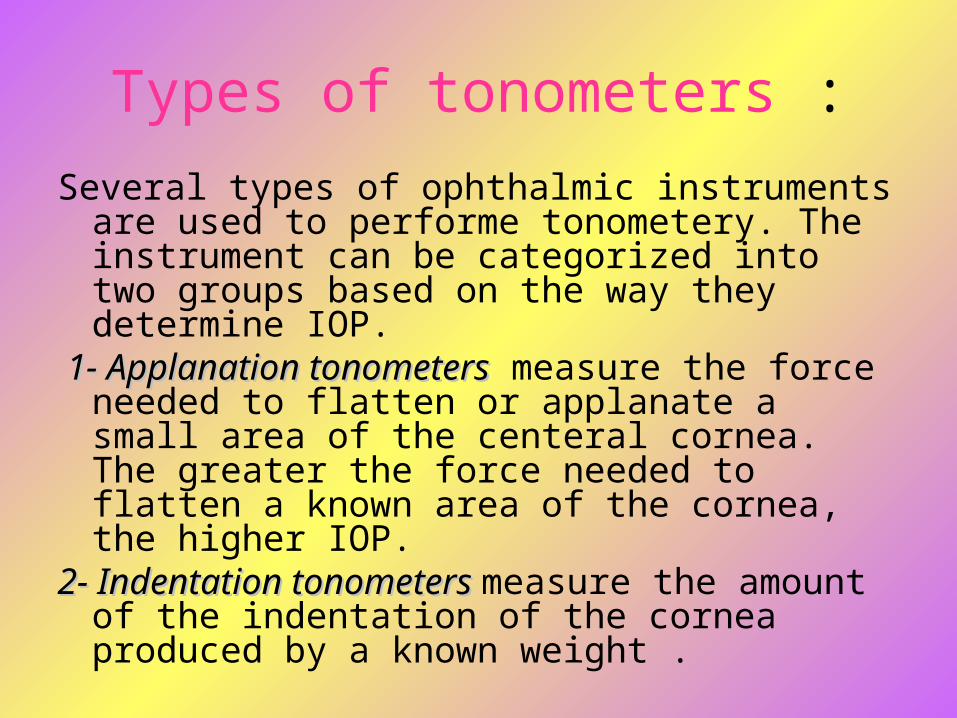
Several types of ophthalmic instruments are used to performe tonometery. The instrument can be categorized into two groups based on the way they determine IOP.
1- Applanation tonometers1- Applanation tonometers measure the force needed to flatten or applanate a small area of the centeral cornea. The greater the force needed to flatten a known area of the cornea, the higher IOP.
2- Indentation tonometers 2- Indentation tonometers measure the amount of the indentation of the cornea produced by a known weight .


Applanation tonometerApplanation tonometer

Indentation tonometerIndentation tonometer

Applanation tonometers :
• Some of the most common types of applanation tonometers and there charactaristics are listed below :
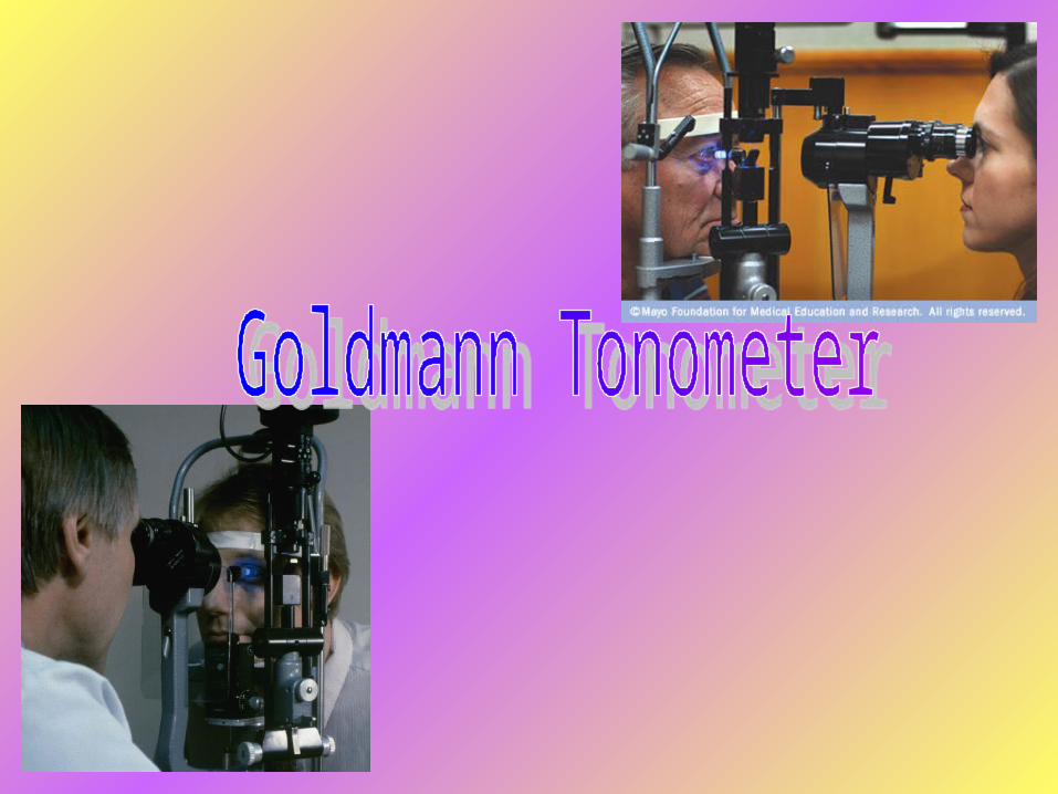
* The Goldmann TonometerThe Goldmann Tonometer is the most common tonometer. Usually mounted on the standard slit-lamp biomicroscope. It’s easy to use and measure the IOP of a seated patient with high accuracy in most clinical situations. Measurements are less precise for edematous and scarred corneas.

The Goldmann TonometerGoldmann Tonometer

* The Perkins tonometerThe Perkins tonometer is a handheld, portable applanation device. The technique for use, mechanism of action, and relative accuracy are similar to those of the slit-lamp mounted Goldmann tonometer, and it can be used with either a seated or supine patient. It’s portability makes this device useful at the bedside or in the opening room. Because it is not mounted to a stable device.However, the steadiness of both the patient and the examiner are harder to control. Nevertheless, with some practice, the perkins tonometer is a useful instrument.

The Perkins tonometerThe Perkins tonometer


* The Peneumatic tonometerThe Peneumatic tonometer is an electronic is an electronic pressure- sensing device that consist of a Gas-pressure- sensing device that consist of a Gas-filled chamber covered with a Silastic filled chamber covered with a Silastic diaphragm. The gas in the chamber escapes diaphragm. The gas in the chamber escapes through an exhaust vent. As the diaphragm through an exhaust vent. As the diaphragm touches the cornea, the gas vent is redused in touches the cornea, the gas vent is redused in size and the pressure in the chamber rises. The size and the pressure in the chamber rises. The instrument supplies a measurement reading instrument supplies a measurement reading directly in (mm Hg). The Pneumatic tonometer is directly in (mm Hg). The Pneumatic tonometer is portable, can be used with a seated or supine portable, can be used with a seated or supine patient and is specially useful in the presence of patient and is specially useful in the presence of corneal scar or corneal odema.corneal scar or corneal odema.

The Peneumatic tonometerThe Peneumatic tonometer


* The Tonopen The Tonopen like many simlar portable electronic applaning devices, contains a strain gauge and produces an electrical signal as the tip of the instrument applanates the cornea. These devices use disposable ssterile latex covers for the applanating tip, can be used with a seated or supine patient and are useful in the presence of corneal scars or edema. Some studies have found that these instruments underestimate IOP in the higher ranges.

The TonopenThe Tonopen


* The noncontact (Air-puff) tonometerThe noncontact (Air-puff) tonometer determines IOP by measuring the time necessary for a given force of air to flatten a given area of the cornea . Because the instrument does not contact the patient’s cornea, no anesthetic drops are needed. Reading obtained with these instruments correlate well with those obtained by Goldmann applanation tonometery except at high and low extremes of intraocular pressure.

The noncontact (Air-puff) tonometerThe noncontact (Air-puff) tonometer

GOLDMANN GOLDMANN TONOMETERTONOMETER

Parts of the instrument :
The Goldmann applanation tonometer consist of four principle operative parts :
I.I. The Tonometer Tip:The Tonometer Tip: The part of the instrument that contacts the patient’s cornea, contains biprism (two beam-splitting prisms) that converts a circular area of conact between the tonometer tip and the patient’s cornea into two semicircles. By properly aligning the semicircles, the examiner can determine the area of corneal applanation and measure the intraocular preasure with great accuracy.


II.II. A metal rod :A metal rod : it connects the tonometer tip to the instrument housing.
III.III. The tonometer housingThe tonometer housing : contains a mechanism that can deliver a measured force controlled by the force adjustment knob on the housing to the tonometer tip.
IV.IV. The force adjustment knobThe force adjustment knob : :on the housing is used to vary the amount of force needed to applanate the cornea. The scale reading on the knob is multiplied by 10 to express IOP in mm Hg.

Parts of the instrument:


Optical principle:
• The Goldmann Applanation tonometer relies on an interesting physical principle :
“For an ideal, dry, thin-walled sphere, the pressure inside a sphere is proportional to the force applied to it’s surface.” Unlike an ideal sphere, however, the human eye is not thin-walled and it is not dry. This produces two confounding forces : (1) a force produced by the eye’s scleral rigidity (because the eye is not a thin-walled),wich is directed away from the glob,

and (2) a force produced by the surface tension of the tear film (because the eye is not dry), wich is directed toward the glob.
Goldmann determined that when a flat surface is applied to the cornea with enough force to produce a circular area of flattening 3.06 mm in diameter, the force caused by scleral rigidity exactly cancels out the force caused by surface tension. Therfore, the applanating force required to flatten a circular area of cornea exactly 3.06 mm in diameter is directly proportional to the intraocular pressure.

Calibration of Goldmann tonometer• It is possible to check the calibration of the
tonometer; this should be done every six months. Calibration is done at dial positions 0, 2, and 6 (equivalent to 0, 20, and 60 mmHg).
• Insert the prism in the holder and place the tonometer on the slit lamp.• At dial position 0, the feeler arm should be in
free movement. If the dial is turned backwards a small way (to the equivalent of position -0.05),
the arm should fall towards the examiner.

• If the dial is turned forwards a small way (to the equivalent of position +0.05) the arm should fall towards the patient.
• If the arm doesn’t respond in the above way, the tonometer is inaccurate at dial position 1.• To check dial positions 2 and 6, the check
weight is used (this is normally found in the case with the tonometer prisms or in the drawer of the slit lamp). There are five markings engraved on the bar. These represent 0 centrally,
then 2 on either side, and 6 towards the edges.

• Line up the adjustable holder with index mark 2 on the weight. With the longer end of the bar
facing you, put it into the slot on the side of the tonometer and push it all the way in.
• Repeat the above steps (for dial position 0), with the dial now at position 2. This time, turn the dial
backwards to the equivalent of 1.95 and forwards to the equivalent of 2.05.
• To check dial position 6, move the weight bar to the end position. Repeat the steps at dial position 6, turning the dial backwards to the
equivalent of 5.9 and forwards to the equivalent of 6.1 .

• If the tonometer is inaccurate at any of these dial positions, it should be returned to the manufacturer for recalibration.


Performing Goldman applanation tonometer.
1) Insert clean tonometer tip in the biprism holder. The 180 degree marking on the tonometer tip should be aligned with the white line on the biprism holder.
2) Instill a topical anesthetic drop and fluorescein dye into each of the patient’s eyes. Many clinics use a single solution containing both the anesthesia and dye fluorescein for this test.
3) Seat the patient at the slit lamp with the patient’s forehead firmly against the headrest and chin comfortably on the chin rest.

Wetting the fluorescein with anesthesia


The patient’s eye should be aligned with the black band on the headrest column. Instruct the patient to look straight ahead and to open the eyelids wide. The examiner should be seated facing the patient, behind the slit- lamp oculars.
4) Position the cobalt filter in front of the slit-lamp illumination device. The cobalt-blue light causes the fluorascien dye on the patient’s eye to fluoresce a bright yellow-green.
5) Set the magnification of the slit lamp at low power, with the light beam at high intensity and shinig on the tonometer up at a wide angle (about 60 degree)
6) Looking from the side, use the slit-lamp control handle to align the tonometer tip with the patient’s right cornea. Adjust the numbers on the tonometer force adjustment knob to read anywhere between 1 and 2 (10 and 20 mm Hg).
7) Instruct the patient to focus on your right ear, blink once (to spread the fluorescein dye), and then try to avoid blinking. If it is necessary to hold the patient’s lids open, secure them against the bony orbit ; do not apply pressure to the globe.

Cobalt filter & gently touch of the cornea

8) If the patient wears soft lenses you should flush the eyes out very well, cleaning the patient up to remove all the fluorescein, then advise them not to wear their lenses for at least one (1) hour. Fluorescein will be absorbed into the soft lens turning it yellow. Patients who wear hard lenses should be advised not to wear their lenses as long as they would normally. Topical anesthetics soften the cornea’s epithelium making an abrasion more likely. All patients should have has much of the fluorescein dye cleaned off as possible.

Soft contact lenses

Hard contact lenses

Soft & Hard lenses

You do not have to pull the probe off the patients eye to make fine adjustments for alignment; this can be done with small movements while on the cornea. Removing the probe from the patients eye then putting it back on until you have proper alignment will result in drying and staining of the cornea.

9) Using the slit-lamp control handle, gently move the biprism forward until it just touches the cornea. Looking through the slit-lamp oculars, confirm that the biprism has just touched the cornea: the spot of fluorescein will break into two semicircles, one above and one below a horizontal line. Raise and lower the slit-lamp biomicroscope with the control handle until the semicircles are equal in size. The semicircles can be viewed monocularly through only one of the slit-lamp oculars; in most slit lamps the semicircles are viewed through the left ocular.

A. If the patient has a large amount of corneal astigmatism, the semicircles seen by the examiner through the instrument ocular will look elliptical rather than circular. An error will be introduced into the pressure determination. In this situation, rotate the tonometer tip so that the dividing line between the semicircles is 45 degree to the major axis of the ellipse.

10) Slowly and gently turn the force adjustment knob in the direction required to move the semicircles until their inner edges just touch and do not overlap.
A. If the semicircles are separated, the pressure reading will be too low if the semicircles overlap, the pressure reading will be too high.
B. If there is too fluorescein or if the examiner is applying pressure to the glob while holding the patient’s eye open, the semicircles will appear thick and an inaccurate pressure reading will result. A small pulsatile motion of the semicircles may be apparent, synchronous with the patient’s pulse.


Normal pressure need some correction

Too high pressure

Too low pressure (image “a”)

Too Fluorescein or the Probe is too far forward

11) With the slit-lamp control handle, pull the tonometer biprism away from the patient’s eye. Note the reading on the numbered dial of the force adjustment knob. Multiply the number by 10 to obtain the intraocular pressure in mm Hg and record the pressure in the patient’s medical record.
12)Repeat the procedure for the left eye.

Factors that affect on the accuracy of IOP readings:
• The accuracy of applanation tonometry is reduced in certain situations. Corneal edema predisposes to inaccurately low readings, whereas pressure measurements taken over a corneal scar will be falsely high. Tonometry performed over a soft contact lens gives falsely low values. Alterations in scleral rigidity may compromise the accuracy of measurements; for example, applanation readings that follow scleral buckling procedures may be inaccurately low.

Disinfecting the Applanation Tip:
1. Remove the tonometer up from the biprism after each use.
2. Disinfect by one of the following methods: Swab the tip with a cotton-tipped applicator
that has been soaked in isopropyl alcohol. Soak the tonometer tip in a 10% solution of
sodium hypochlorite (household bleach) or 3% hydrogen peroxide or isopropyl alcohol for 5 minutes.
Isopropyl alcohol
3. Rinse the tonometer tip with water and dry with tissue or gauze to remove residual disinfecting solution, which could damage the corneal epithelium.

References:BooksBooks:• Optics, Refraction & Contact lenses (American
Academy of ophthalmology).• Practical ophthalmology (American Academy of ophthalmology).• Clinical ophthalmoptics ….By Mohamed El-Rifi
(Cairo – Egypt).WebsitesWebsites:• American Academy of optometry.• www.webmd.com/eye-health/tonometry .• medical-dictionary.thefreedictionary.com/
applanation+tonometer