contents of male and female perineal pouches copy
TRANSCRIPT
Contents of male and female perineal pouches
By
Dr. Abdul Waheed Ansari
(Prof. & chairperson of Anatomy, RAKCOMS)
12/18/2014 1

Perineum boundaries are
12/18/2014 2
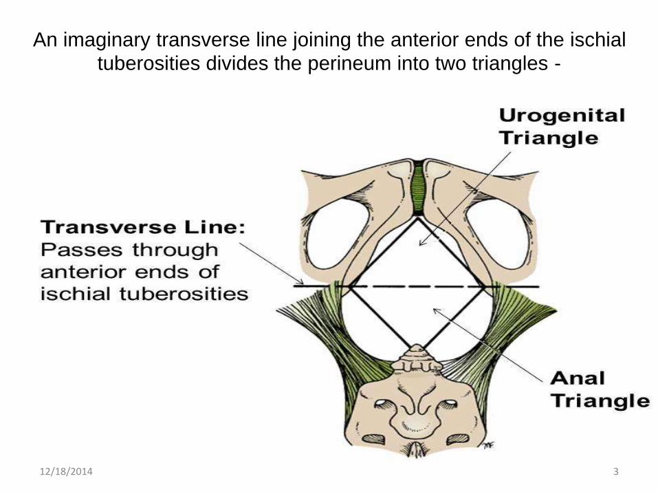
An imaginary transverse line joining the anterior ends of the ischial
tuberosities divides the perineum into two triangles -
12/18/2014 3

The perineal body
Structurally perineal body is an irregular fibro muscular mass, containing both collagenous and elastic fibers and both skeletal and smooth muscles.
It is the site of convergence of several muscles:
1. Bulbospongiosus,
2. Superficial transverse perinea
3. Superficial part of external anal sphincter
4. Sphincter urethrae and
5. Deep transverse perinea,
6. Deep part of external anal sphincter and
7. Levator ani
12/18/2014 4

12/18/2014 5
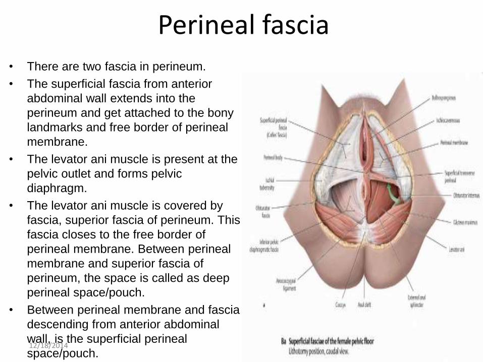
Perineal fascia• There are two fascia in perineum.
• The superficial fascia from anterior
abdominal wall extends into the
perineum and get attached to the bony
landmarks and free border of perineal
membrane.
• The levator ani muscle is present at the
pelvic outlet and forms pelvic
diaphragm.
• The levator ani muscle is covered by
fascia, superior fascia of perineum. This
fascia closes to the free border of
perineal membrane. Between perineal
membrane and superior fascia of
perineum, the space is called as deep
perineal space/pouch.
• Between perineal membrane and fascia
descending from anterior abdominal
wall, is the superficial perineal
space/pouch.12/18/2014 6

Male external genital organs The penis:
a. root of the penis: the fixed dorsal segment within the urogenital perineum, consists of two crura
and the bulb
b. penile body: the pendulous, mobile ventral segment suspended from the pubis
• 1. consists of two corpora cavernosa and one corpus spongiosum
• 2. The two corpora cavernosa fuse lying side by side, and insert into the glans
• penis to form the superior (dorsal) surface.
• 3. The thinner corpus spongiosum, containing the urethra, lies below the fused
• corpora cavernosa and forms the inferior (ventral) surface.
c. Over lining these structures are the related muscles: the ishiocavernosus & bulbospongiosus
d. Vessels and nerves of the penis
• 1. superficial dorsal vein empties into the superficial external pudendal veins
• 2. deep dorsal veins drain into the prostatic venous plexus and also make connection
• with the vertebral venous flow
• 3. two dorsal penile arteries from the internal pudendal artery
• 4. paired dorsal nerves arise from the pudendal nerve
e. Suspensory Ligament
12/18/2014 7

Contents of superficial
perineal pouch in male
1. Bulbospongiosus2. Ischiocavernosus3. Anal sphincter4. Deep Transverse Perineal5. Superficial Transverse Perineal6. Pelvic diaphragm / Levator Ani muscles7. Obturator Internus8. Perineal body9. Ano-coccygeal body 10. Sacrum and coccyx 11. Ischial tuberosities12. Sacrotuberous ligament
12/18/2014 8

External genitalia in both sexes
12/18/2014 9

Lymphatic drainage of male external genitalia
• The lymph vessels from the scrotum drain into the superficial inguinal lymph nodes.
• From most of the penis, lymph drains into the superficial inguinal lymph nodes.
• Vessels from the glans penis drain into the deep inguinal lymph nodes.
• Enlarged nodes are either due to disease in drainage areas or systemic disease.
• Drainage areas
Horizontal group:-
• External genitalia (Cancer Penis, Vulva)
• Anal canal (cancer Rectum)
• Gluteal region
• Lower third of vagina
Vertical group:
• Leg
• Systemic disease
• Lymphoma
• Tuberculosis
12/18/2014 10

Contents of superficial perineal pouch in female
1. Root (crura) of the clitoris and muscle associated with it (ischiocavenous)2. Bulbs of the vestibule and surrounding muscle (bulbospongiosus)3. Superficial transverse perineal muscles4. Branches of the internal pudendalartery5. Posterior labial branches of the perieneal branch6. Transverse perineal branches of the perieneal branch7. Posterior labial branch of Perineal branch of the pudendal nerves8. Perineal branch of posterior cutaneous nerve of thigh9. Greater vestibular glands
12/18/2014 11
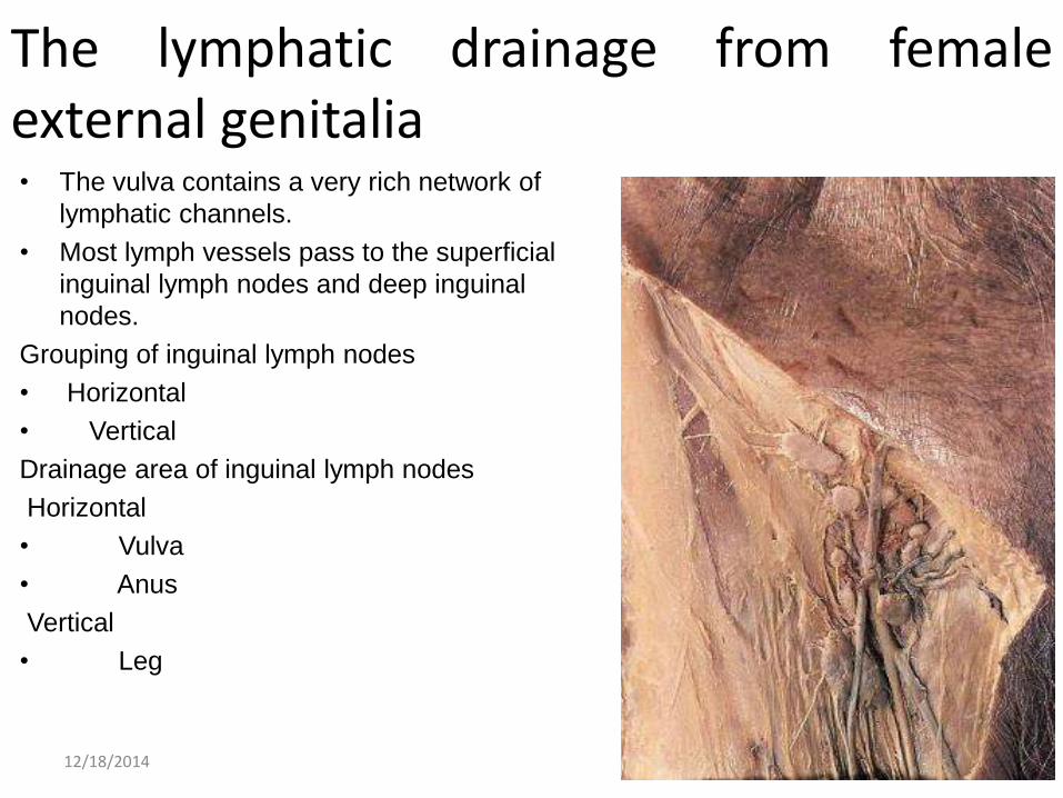
The lymphatic drainage from femaleexternal genitalia• The vulva contains a very rich network of
lymphatic channels.
• Most lymph vessels pass to the superficial
inguinal lymph nodes and deep inguinal
nodes.
Grouping of inguinal lymph nodes
• Horizontal
• Vertical
Drainage area of inguinal lymph nodes
Horizontal
• Vulva
• Anus
Vertical
• Leg
12/18/2014 12
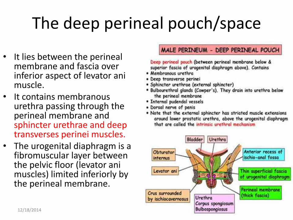
The deep perineal pouch/space
• It lies between the perineal membrane and fascia over inferior aspect of levator animuscle.
• It contains membranous urethra passing through the perineal membrane and sphincter urethrae and deep transverses perinei muscles.
• The urogenital diaphragm is a fibromuscular layer between the pelvic floor (levator animuscles) limited inferiorly by the perineal membrane.
12/18/2014 13
Contents of deep perineal pouch in female
1. Proximal part of urethra
2. External urethral sphincter muscle
3. Deep transverse perineal muscles
4. Related vessels and nerves
5. Deep artery of clitoris
6. Dorsal artery of clitoris
7. Artery to bulb of vestibule
8. Urethral artery
9. Dorsal nerve of clitoris
12/18/2014 14
Pelvic diaphragm
• The pelvic diaphragm is a muscular partition formed by the levatores ani and coccygei, with which may be included the parietal pelvic fascia on their upper and lower aspects.
• It separates the pelvic cavity above from the perineal region below. • The right and left levatores ani lie almost horizontally in the floor of
the pelvis, separated by a narrow gap that transmits the urethra, vagina, and anal canal.
• The levator ani is usually considered in three parts: 1. pubococcygeus, 2. puborectalis, and 3. iliococcygeus. • The pubococcygeus, runs backward from the body of the pubis
toward the coccyx and may be damaged during parturition.
12/18/2014 15

12/18/2014 16

The Puborectalis• The right and left
Puborectalis unite behind the anorectal junction to form a muscular sling.
• The Puborectalis fibers interdigitates with the sphincter ani externus.
12/18/2014 17
The coccygeus
• The iliococcygeus, the most posterior part of the levator ani.
• The coccygeus extends from the ischial spine to the lateral margin of the sacrum and coccyx.
• The pelvic diaphragm is supplied chiefly by the ventral rami of S3,S4.
• The diaphragm helps to support the pelvic viscera, resists increases in intra-abdominal pressure, and aids in micturition.
12/18/2014 18

References
• http://teachinganatomy.blogspot.ae/2013/06/perineum25_268.html
• http://anatomytopics.wordpress.com/2009/01/08/38-the-perineum-the-formation-of-the-placenta-the-structure-of-the-matured-placenta/
• http://download.videohelp.com/vitualis/med/male_ext_genitalia.htm
• http://www.meddean.luc.edu/lumen/meded/medicine/pulmonar/pd/pstep43.htm
• http://web.uni-plovdiv.bg/stu1104541018/docs/res/skandalakis'%20surgical%20anatomy%20-%202004/Chapter%2028_%20Pelvis%20and%20Perineum.htm
• http://unanipathy.com/videos/pelvic-floor-part-1-the-pelvic-diaphragm-3d-anatomy-tutorial/
12/18/2014 19