continuous asl perfusion fmri investigation of higher cognition: quantification of tonic cbf changes...
TRANSCRIPT

www.elsevier.com/locate/ynimg
NeuroImage 31 (2006) 376 – 385
Continuous ASL perfusion fMRI investigation of higher cognition:
Quantification of tonic CBF changes during sustained attention and
working memory tasks
Junghoon Kim,a,b,* John Whyte,a,c Jiongjiong Wang,b,d,e Hengyi Rao,b
Kathy Z. Tang,b and John A. Detreb,d,e
aMoss Rehabilitation Research Institute, Albert Einstein Healthcare Network, 1200 W. Tabor Road, Philadelphia, PA 19141, USAbCenter for Functional Neuroimaging, University of Pennsylvania, Philadelphia, PA 19104, USAcDepartment of Rehabilitation Medicine, Thomas Jefferson University, Philadelphia, PA 19107, USAdDepartment of Neurology, University of Pennsylvania, Philadelphia, PA 19104, USAeDepartment of Radiology, University of Pennsylvania, Philadelphia, PA 19104, USA
Received 25 August 2005; revised 11 November 2005; accepted 18 November 2005
Available online 19 January 2006
Arterial spin labeling (ASL) perfusion fMRI is an emerging method in
clinical neuroimaging. Its non-invasiveness, absence of low frequency noise,
and ability to quantify the absolute level of cerebral blood flow (CBF) make
the method ideal for longitudinal designs or low frequency paradigms.
Despite the usefulness in the study of cognitive dysfunctions in clinical
populations, perfusion activation studies to date have been conducted for
simple sensorimotor paradigms or with single-slice acquisition, mainly due
to technical challenges. Using our recently developed amplitude-modulated
continuous ASL (CASL) perfusion fMRI protocol, we assessed the
feasibility of a higher level cognitive activation study in twelve healthy
subjects. Taking advantage of the ASL noise properties, we were able to
study tonic CBF changes during uninterrupted 6-min continuous perfor-
mance of working memory and sustained attention tasks. For the visual
sustained attention task, regional CBF increases (6–12 ml/100 g/min) were
detected in the right middle frontal gyrus, the bilateral occipital gyri, and
the anterior cingulate/medial frontal gyri. During the 2-back working
memory task, significantly increased activations (7–11 ml/100 g/min) were
found in the left inferior frontal/precentral gyri, the left inferior parietal
lobule, the anterior cingulate/medial frontal gyri, and the left occipital
gyrus. Locations of activated and deactivated areas largely concur with
previous PET and BOLD fMRI studies utilizing similar paradigms. These
results demonstrate that CASL perfusion fMRI can be successfully utilized
for the investigation of the tonic CBF changes associated with high level
cognitive operations. Increased applications of the method to the investi-
gation of cognitively impaired populations are expected to follow.
D 2005 Elsevier Inc. All rights reserved.
Keywords: Arterial spin labeling; CASL; Cerebral blood flow; 2-back task;
Clinical neuroimaging
1053-8119/$ - see front matter D 2005 Elsevier Inc. All rights reserved.
doi:10.1016/j.neuroimage.2005.11.035
* Corresponding author. Moss Rehabilitation Research Institute, 1200 W.
Tabor Road, Philadelphia, PA 19141, USA. Fax: +1 215 456 5926.
E-mail address: [email protected] (J. Kim).
Available online on ScienceDirect (www.sciencedirect.com).
Introduction
During the past decade, functional magnetic resonance imaging
(fMRI) has become a standard tool for visualizing resting and task-
related brain activations. In comparison to positron emission
tomography (PET) or single photon emission computed tomogra-
phy (SPECT), fMRI has higher spatial and temporal resolution,
does not involve exposure to ionizing radiation, and is widely
available at medical centers. These merits have expanded the
applications of fMRI to many clinical areas including presurgical
mapping, psychopharmacology, and pediatric neuroimaging (Detre
and Floyd, 2001; Hennig et al., 2003; Honey and Bullmore, 2004;
Matthews and Jezzard, 2004; Wilke et al., 2003). Blood-
oxygenation-level-dependent (BOLD) fMRI has been the method
of choice in most occasions, due to its high sensitivity to task-
related effects and relative ease of implementation. However,
BOLD contrast is known to be a complex interaction of multiple
physiological parameters including blood flow, blood volume, and
hemoglobin oxygenation (Kwong et al., 1992; Mandeville et al.,
1999; Ogawa et al., 1993). As a result, it confounds changes from
neuronal activation with vascular effects, making the interpretation
of data difficult when circulatory changes are expected due to
cerebrovascular disease or direct vascular effects of pharmacolog-
ical intervention. Lack of an absolute measure of cerebral blood
flow also limits the application of BOLD fMRI in situations where
multiple scanning sessions need to be compared since condition
differences can arise due to either (or both) an increase in
activation or a decrease in deactivation (e.g., Poldrack, 2000). In
addition, the presence of low frequency noise in the BOLD signal
(Friston et al., 2000; Zarahn et al., 1997) precludes using a long
task block (e.g., >2 min), making it difficult to investigate slow
processes such as learning, emotion, and sustained attention in
healthy and clinical populations.
J. Kim et al. / NeuroImage 31 (2006) 376–385 377
Arterial spin labeling (ASL) fMRI is an emerging method-
ology that uses magnetically labeled arterial blood water as an
endogenous tracer to provide quantitative cerebral blood flow
(CBF) measurements (Detre et al., 1992; Williams et al., 1992).
This non-invasive method provides highly reliable measures of
CBF, making it particularly suitable for longitudinal studies of
treatment (e.g., drug or training) effects or functional recovery
processes that require assessment of baseline function and
repeated measurements across sessions (Detre and Alsop,
1999; Detre and Wang, 2002). ASL contrast, due to the
pairwise subtraction of temporally adjacent images, is also free
from the slow signal drifts present in BOLD fMRI contrast
(Aguirre et al., 2002). As a result, it is well suited for
investigating low frequency brain events such as changes related
to practice, mood, and mental set (Wang et al., 2003a,b). In
addition, recent evidence suggests that ASL fMRI may provide
contrasts with smaller intersubject variability (Aguirre et al.,
2002; Kemeny et al., 2005), reduced susceptibility artifacts in
regions of high static inhomogeneity (Tjandra et al., 2005;
Wang et al., 2004), and more specific functional localization
than BOLD fMRI (Duong et al., 2001; Luh et al., 2000).
Due to the advantageous characteristics mentioned above,
ASL perfusion fMRI has been increasingly adopted for clinical
studies of cerebral perfusion during resting states (e.g., Ances et
al., 2004; Johnson et al., 2005; Oguz et al., 2003; Rashid et al.,
2004). However, few studies have attempted to validate this
technique with cognitive activation paradigms. Previous activa-
tion studies have mainly used passive visual stimulation
(Aguirre et al., 2002; Talagala and Noll, 1998) or simple
psychomotor tasks such as finger tapping (Garraux et al., 2005;
Mildner et al., 2003; Wang et al., 2003a,b). Only two studies to
date have used cognitive paradigms such as the 2-back and verb
generation tasks (Ye et al., 1998; Yee et al., 2000). However,
these studies acquired only single slices, limiting their use to
hypothesis testing regarding a priori regions-of-interest (ROIs).
Thus, a whole-brain multi-slice study with conventional voxel-
wise group analyses is needed to validate ASL pefusion fMRI
for the investigation of the widely distributed neural networks
of high-level cognitive processes such as attention and working
memory.
It is known from previous PET studies that there are
significantly smaller CBF changes during cognitive activation
tasks compared to those during simple sensory–motor tasks
(Colebatch et al., 1991; Jonides et al., 1997; Paulesu et al.,
1993; Ramsey et al., 1996). Considering the relatively low
signal-to-noise ratio (SNR) of ASL fMRI (cf. Calamante et al.,
1999), detecting these subtle changes could be a major
challenge for studies of higher cognitive processes using this
method. The combination of high magnetic field strength and
continuous ASL (CASL) method offers an appealing approach
to improve the SNR and image coverage of ASL fMRI (Wang
et al., 2002). One way of implementing a high-field ASL fMRI
is to use a separate small RF coil for labeling (Mildner et al.,
2003; Zaharchuk et al., 1999). However, this dual-coil approach
requires special hardware and relies heavily on the labeling
geometry, which may vary from subject to subject. The added
distance for arterial transit from the carotid tagging region and
the relatively poor labeling of the vertebral arteries also limit
the practical use of this approach. Recently, we demonstrated
that whole-brain multi-slice CASL fMRI could be successfully
implemented with a single transmit– receive coil at 3.0 T by
reducing RF pulses and gradient strength appropriately (Wang et
al., 2005). Our goal in the present study was to demonstrate
that higher cognitive processes could be studied utilizing the
same imaging protocol, with appropriate sensitivity and local-
ization power.
Two cognitive tasks were selected to assess CASL fMRI’s
sensitivity to CBF changes related to high-level cognitive
processes. Both tasks are known to be Ffrontal_ tasks– that is,
they involve prefrontal areas for successful performance–by
previous PET and BOLD fMRI investigations. The first task
was a visual sustained attention task (Whyte et al., 1995). Since
sustained attention is also implicated in various clinical
disorders including attention deficit hyperactivity disorder,
traumatic brain injury, and Alzheimer’s disease, it is important
to understand the neural correlates of this cognitive function.
Previous neuroimaging studies of sustained attention in the
visual modality have consistently identified a right hemisphere
dominant fronto-parietal network (Coull et al., 1996, 1998;
Lawrence et al., 2003; Pardo et al., 1991). However, these
studies used rather short data acquisition blocks (40–90 s) that
might not be optimal for detecting aspects of sustained attention
associated with prolonged task performance. Taking advantage
of ASL fMRI’s noise characteristics, the current study was able
to employ a long (6 min) block of uninterrupted performance of
visual target detection. It was hypothesized that we would find
tonic changes of CBF in the previously identified areas of the
visual sustained attention network, including the right middle
frontal and right parietal cortices.
The second task was a working memory (2-back) task
(Cohen et al., 1997; Jonides et al., 1997). It is known from
previous neuroimaging studies that various versions of this task
invoke activations in a large-scale network including prefrontal,
premotor, supplementary motor, and parietal cortices (Cabeza
and Nyberg, 2000; D’Esposito et al., 1998; Smith and Jonides,
1998, 1999). Since this task is one of the most frequently used
high-level cognitive tasks in various clinical populations (e.g.,
Callicott et al., 2003; Kwon et al., 2001; Scheibel et al., 2003;
Sweet et al., 2004; Valera et al., 2005), implementing it with a
whole-brain multi-slice ASL approach would provide useful
comparison CBF data for future clinical neuroimaging studies. It
was predicted that we would replicate the results from prior
normative studies using the 2-back task (for review, see Owen
et al., 2005).
Materials and methods
Participants
Seventeen healthy volunteers participated in this study. Five
subjects were excluded from subsequent data analysis due to large
head movements during scanning (see Imaging data analysis for
the criteria). The remaining subjects included 9 men and 3 women
aged between 21 and 46 years (mean age = 34.4 years, SD = 9.5)
with a mean education of 13.3 years (SD = 2.0). Eleven of them
were right-handed (Edinburgh Handedness Inventory, Oldfield,
1971). Subjects had no previous history of neurological or major
psychiatric disorder and had normal or corrected-to-normal visual
acuity. After complete description of the study, subjects provided
written informed consent to the University of Pennsylvania
Institutional Review Board-approved protocol.

J. Kim et al. / NeuroImage 31 (2006) 376–385378
Cognitive tasks
Visual sustained attention task
A simple go/no-go visual reaction time task was used to
examine the neural network involved in maintaining visual
sustained attention (Whyte et al., 1995, 2004). Stimuli consisted
of pairs of vertical lines presented for a brief period in the
center of the screen. The central area of the screen was covered
by a random pattern mask with a fixation cross except when a
stimulus was presented. The mask subtended approximately 1-and 4- of horizontal and vertical visual angle, respectively.
Subjects were taught that a pair of identical lines constituted a
target, whereas a pair of grossly unequal lines constituted a foil
(one line was the same length as the target and the other was
50% shorter), and to press the button with their dominant hand
as quickly and accurately as possible in response to targets only.
They were also explicitly told that only 20% of the stimuli were
targets. A total of 60 stimuli were presented during an
uninterrupted 6-min task block with an average interstimulus
interval of 6 s (range: 4 to 8 s).
Two-back task
A letter version of 2-back task (Awh et al., 1996; Cohen et
al., 1997) was employed to examine the neural network involved
in continuous performance of a working memory task. In this
task, subjects were presented with a series of letters in the center
of the screen. The letters subtended approximately 1.5- � 1.5-of visual angle. Subjects were required to press the button
whenever each letter presented was identical to the one
presented two letters previously in the sequence. A total of
180 letters were presented with an exposure duration of 1 s and
an interstimulus interval (ISI) of 2 s. The target rate for this task
was 12%.
Experimental design and procedure
Prior to the scanning sessions, subjects were trained on the two
tasks outside of the scanner. For the visual sustained attention task,
stimulus exposure durations were individualized to avoid ceiling or
floor performance level. Average stimulus exposure duration was
62.2 ms (SD = 21.2). Details of the calibration procedure are
available elsewhere (Whyte et al., 1995). The order of task blocks
was always resting first, the sustained attention task second, and
the 2-back task last. Each task block was approximately 6 min, and
the intervals between task blocks were approximately 30 s. During
the resting condition, which was used as the baseline control,
subjects were instructed to close their eyes but stay awake. For
both tasks, responses and reaction times (RTs) were recorded for
further analysis.
Imaging data acquisition
The functional imaging was conducted on a Siemens 3.0 T
Trio whole-body scanner (Siemens AG, Erlangen, Germany),
using a standard transmit– receive head coil. An amplitude-
modulated CASL technique was implemented for perfusion
fMRI scans (Wang et al., 2005). Interleaved images with and
without labeling were acquired using a gradient echo echo-
planar imaging sequence with the following acquisition param-
eters: FOV = 22 cm, matrix = 64 � 64, TR = 4 s, TE = 17
ms, flip angle = 90-. Fourteen slices (6 mm thickness with 1.5
mm gap) were acquired from inferior to superior in a sequential
order to cover the whole brain. A delay time of 1 s was
inserted between the end of labeling pulses and image
acquisition to reduce transit-related effects. Each subject
performed three CASL scans each with 92 acquisitions
(approximately 6 min). Before the functional scans, high
resolution T1-weighted anatomic images were obtained using
3D MPRAGE: TR = 1620 ms, TI = 950 ms, TE = 3 ms, flip
angle = 15-, 160 contiguous slices of 1.0 mm thickness, FOV =
192 � 256 mm2, matrix = 192 � 256, 1NEX with a scan time
of 6 min.
Behavioral data analysis
Performance of the subjects was characterized with respect to
three dimensions: discrimination, response bias, and speed.
Discrimination was measured with dV. Response bias was charac-
terized by yes rate (total proportion of button presses without
regard to accuracy). Speed was operationalized as median RT on
hits (correct button presses to targets).
Imaging data analysis
Functional image pre-processing and individual-level analysis
were carried out using VoxBo software (Center for Functional
Neuroimaging, Philadelphia, PA, http://www.voxbo.org). The
group-level analysis was performed with Statistical Parametric
Mapping software (SPM99, Wellcome Department of Cognitive
Neurology, London, UK, http://www.fil.ion.ucl.ac.uk/spm). For
each subject, functional images were realigned to correct the
head motion using a 6-parameter rigid-body least squares
realignment routine (Friston et al., 1995). Subjects who showed
excessive head motion, defined as any translational movement
larger than 2 voxels along the x or y axis or 1 voxel size along
the z axis, were excluded from further analysis (n = 5).
Realigned images were smoothed in space with a three-
dimensional Gaussian kernel (4 � 4 � 3 voxels at FWHM).
Perfusion-weighted image series were generated by pairwise
subtraction of the label and control images followed by
conversion to absolute CBF image series based on a single
compartment CASL perfusion model (Wang et al., 2005). The
resulting CBF data sets contained 46 images for each 6-min
task block with an effective TR of 8 s. The CBF images were
then normalized to a 3 � 3 � 3 mm3 Montreal Neurological
Institute (MNI) template using bilinear interpolation.
For each cognitive task, the following statistical analyses were
conducted. First, for each subject, voxel-wise individual GLMs
were built comparing each task condition with the resting baseline
by using appropriately weighted linear contrasts. The global signal
covariate was included in the GLM to reduce spatially coherent
noise in the data (Aguirre et al., 1998). The perfusion MRI data are
known to be free from any substantial temporal autocorrelation
(Aguirre et al., 2002; Wang et al., 2003a,b). Therefore, no filtering,
autocorrelation modeling, or smoothing was done for the time
series. The resulting parameter estimates were then fed into a
random effects model to allow population-level inferences (Holmes
and Friston, 1998). Areas of significant activation were identified
at the cluster level (Forman et al., 1995) for the P value smaller
than 0.005 (t = 3.11) and the cluster extent size larger than 50
voxels. These criteria yielded a level of P < 0.005 for each
significant cluster after correction for multiple comparisons. The

J. Kim et al. / NeuroImage 31 (2006) 376–385 379
resultant activation clusters were used as functionally defined
regions of interest (ROIs) for the subsequent ROI analysis.
A public domain software package MRIcro (http://www.
psychology.nottingham.ac.uk/staff/cr1/mricro.html) was used to
project group activation data to the Colin-brain atlas (Van Essen
et al., 2001) in MNI space for display purposes. Activation peaks
in MNI space were converted to Talaraich coordinates (Talairach
and Trounoux, 1988) to allow better comparison with locations of
activations from previous studies. This non-linear conversion was
achieved using a MATLAB program provided with SPM
extensions (http://www.mrc-cbu.cam.ac.uk/Imaging/Common/
mnispace.shtml). Anatomical labels for these activation peaks
were determined using the Talairach Daemon (Lancaster et al.,
2000). To calculate the mean CBF increase (and % CBF change
equivalent) of activation relative to the resting baseline, the
adjusted (for the global signal) CBF time series for each voxel
from each subject was averaged across all the voxels in each ROI
and then averaged across subjects.
Results
Behavioral results
Subjects performed the visual target detection task with a dV(mean T standard deviation) of 1.41 T 1.11 and an RT of 979 T 362
ms. The yes rate was 0.29 T 0.12 (where target rate = 20%). The dV
Fig. 1. Mean CBF images in a representative subject during th
and the RT of the 2-back task were 3.03 T 1.06 and 717 T 137 ms.
The yes rate was 0.12 T 0.02 (where target rate = 12%).
Imaging results
Fig. 1 shows the quantitative mean CBF images in a
representative subject, with fourteen slices covering the whole
brain from the top of the brain to the cerebellum. Without using a
separate labeling coil, we were able to obtain clear quantitative
perfusion contrasts of the gray and white matter. The calculated
mean CBF of the group (n = 12) was 51.69 ml/100 g/min T 12.7
for the whole brain, which is comparable to previously reported
mean CBF values for the whole brain using PET and ASL methods
(e.g., Lassen, 1985; Yee et al., 2000).
Significant task-related CBF changes were observed in many
hypothesized areas for each task. Tables 1 and 2 list the clusters
showing significant CBF changes for each task. The coordinates of
the local maxima and percent CBF changes associated with each
region are also reported. Fig. 2 illustrates resampled axial images
of the activated areas for the two tasks.
For the visual sustained attention task, four activated clusters
were identified including the right middle frontal gyrus (BA 9),
bilateral occipital gyri (BA 18), and the anterior cingulate/medial
frontal gyrus (BA 32/8) (Fig. 2; Table 1). All of these areas showed
increased CBF during performance of the visual attention task
relative to the resting baseline. In addition to these activated areas,
there were Fde-activated_ regions more active in the resting
e entire experiment. Total acquisition time was 18 min.

Table 1
Clusters of significant CBF changes during the visual sustained attention task compared to the resting baselinea
Type Size (voxels) Anatomical label BA Talairach coordinates Z score D CBF T SD
(ml/100 g/min)
% CBF
change T SDx y z
Activation 113 R. middle frontal gyrus 9 45 22 29 3.64 7.6 T 4.6 13.2 T 7.2
111 R. middle occipital gyrus 18 30 �84 2 3.28 12.1 T 8.8 23.4 T 18.6
R. cuneus 18 18 �99 8 3.19
97 L. middle occipital gyrus 18 �33 �85 �1 3.81 10.8 T 8.7 22.0 T 20.1
70 R. medial frontal gyrus 8 9 29 48 4.02 5.9 T 5.3 9.2 T 9.0
L. cingulate gyrus 32 �6 22 38 3.79
Deactivation 394 L. cingulate gyrus 24 �18 �18 40 4.01 �4.9 T 2.3 �12.4 T 7.9
L. superior
temporal gyrus
41 �33 �31 13 3.93
N/A N/A �33 �25 34 3.34
125 L. superior temporal gyrus 22 �65 �5 9 3.57 �7.1 T 4.6 12.2 T 7.7
L. superior temporal gyrus 21 �53 �15 �2 3.21
L. middle temporal gyrus 21 �68 �26 �1 3.20
106 R. cingulate gyrus 24 15 �18 42 3.60 �6.0 T 6.3 �13.4 T 17.2
R. postcentral gyrus 3 27 �33 49 3.49
53 R. superior frontal gyrus 10 12 62 25 3.27 �8.8 T 6.4 �17.6 T 16.6
R. medial frontal gyrus 10 6 59 8 3.08
a Cluster sizes are in voxels. Coordinates of local maxima at least 16 mm apart are reported per each cluster (maximum 3 maxima). The anatomical labels of
the nearest gray matter within a 7 � 7 � 7 mm range were also reported. DCBF and % CBF values were calculated for each cluster. R. = Right. L. = Left. BA =
Brodmann area. SD = standard deviation.
J. Kim et al. / NeuroImage 31 (2006) 376–385380
baseline compared to the active task block. These areas included
the left superior/middle temporal gyri, the bilateral posterior
cingulate gyri, and the right superior/medial frontal gyrus. As
shown in Table 1, the magnitude of % CBF changes ranged from 9
to 23 % (6–12 ml/100 g/min) at the cluster level. Sizes of the ROIs
and standard deviations of the CBF values are also reported.
For the letter 2-back task, the task versus resting contrast
showed strong left-hemispheric lateralization with activation in the
left inferior frontal/precentral gyri (BA 9/6), the left inferior
parietal lobule (BA 40), the left middle occipital gyrus (BA 37),
and the anterior cingulate/medial frontal gyri (BA 32/6) (Fig. 2;
Table 2). Regions that were deactivated during the task block
Table 2
Clusters of significant CBF changes during the 2-back task compared to the resti
Type Size (voxels) Anatomical label BA Talaira
x
Activation 194 L. inferior parietal lobule 40 �50L. inferior parietal lobule 40 �33
186 L. inferior frontal gyrus 9 �50L. precentral gyrus 6 �39
141 L. medial frontal gyrus 6 �6R. cingulate gyrus 32 9
L. medial frontal gyrus 6 �3120 L. middle occipital gyrus 37 �48
Deactivation 432 L. medial frontal gyrus 6 �6R. cingulate gyrus 24 12
R. paracentral lobule 6 9
399 R. insular 13 36
R. insular 13 39
R. superior temporal gyrus 22 62
392 L. superior temporal gyrus 22 �53L. insular 13 �45L. middle temporal gyrus 21 �50
61 R. superior frontal gyrus 10 15
a The details of this table are the same as those for Table 1.b Based on n = 11. One subject’s resting CBF value for this region showed ex
compared to the baseline included bilateral posterior cingulate/
medial frontal gyri, bilateral superior/middle temporal gyri,
bilateral posterior insular cortices, and the left superior frontal
gyrus. As shown in the Table 2, the magnitude of % CBF changes
ranged from 10 to 26% (7–11 ml/100 g/min) at the cluster level.
Discussion
The purpose of the present study was to demonstrate the
feasibility of CASL perfusion fMRI for the investigation of higher
cognitive processes. Utilizing our amplitude-modulated CASL
ng baselinea
ch coordinates Z score D CBF T SD
(ml/100 g/min)
% CBF
change T SDy z
�33 40 3.70 8.3 T 6.1 15.7 (T13.0)�50 41 3.00
4 30 3.84 7.7 T 5.0 14.1 (T11.6)
22 27 2.84
17 43 3.86 7.3 T 4.2 11.5 (T6.6)14 38 3.60
3 55 3.28
�64 �7 3.27 11.1 T 13.8b 26.0 (T39.3)b
�20 56 4.07 �5.6 T 2.6 �12.1 (T6.3)
�13 39 3.94
�24 51 3.74
�5 20 3.65 �5.6 T 2.7 �11.5 (T6.1)�22 29 3.54
3 �5 3.25
�12 1 3.91 �6.9 T 3.4 �10.2 (T3.9)
�20 15 3.63
�1 �18 3.37
62 22 3.45 �6.5 T 4.2 �13.5 (T10.3)
treme value due to artifacts and was excluded.
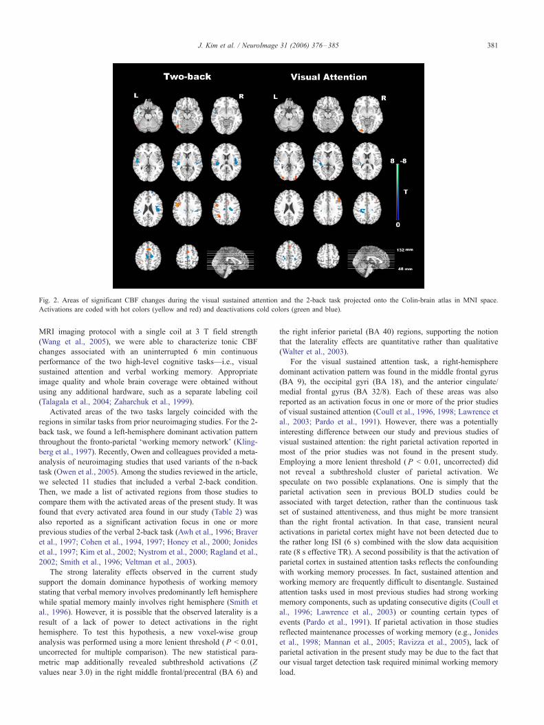
Fig. 2. Areas of significant CBF changes during the visual sustained attention and the 2-back task projected onto the Colin-brain atlas in MNI space.
Activations are coded with hot colors (yellow and red) and deactivations cold colors (green and blue).
J. Kim et al. / NeuroImage 31 (2006) 376–385 381
MRI imaging protocol with a single coil at 3 T field strength
(Wang et al., 2005), we were able to characterize tonic CBF
changes associated with an uninterrupted 6 min continuous
performance of the two high-level cognitive tasks—i.e., visual
sustained attention and verbal working memory. Appropriate
image quality and whole brain coverage were obtained without
using any additional hardware, such as a separate labeling coil
(Talagala et al., 2004; Zaharchuk et al., 1999).
Activated areas of the two tasks largely coincided with the
regions in similar tasks from prior neuroimaging studies. For the 2-
back task, we found a left-hemisphere dominant activation pattern
throughout the fronto-parietal Fworking memory network’ (Kling-
berg et al., 1997). Recently, Owen and colleagues provided a meta-
analysis of neuroimaging studies that used variants of the n-back
task (Owen et al., 2005). Among the studies reviewed in the article,
we selected 11 studies that included a verbal 2-back condition.
Then, we made a list of activated regions from those studies to
compare them with the activated areas of the present study. It was
found that every activated area found in our study (Table 2) was
also reported as a significant activation focus in one or more
previous studies of the verbal 2-back task (Awh et al., 1996; Braver
et al., 1997; Cohen et al., 1994, 1997; Honey et al., 2000; Jonides
et al., 1997; Kim et al., 2002; Nystrom et al., 2000; Ragland et al.,
2002; Smith et al., 1996; Veltman et al., 2003).
The strong laterality effects observed in the current study
support the domain dominance hypothesis of working memory
stating that verbal memory involves predominantly left hemisphere
while spatial memory mainly involves right hemisphere (Smith et
al., 1996). However, it is possible that the observed laterality is a
result of a lack of power to detect activations in the right
hemisphere. To test this hypothesis, a new voxel-wise group
analysis was performed using a more lenient threshold (P < 0.01,
uncorrected for multiple comparison). The new statistical para-
metric map additionally revealed subthreshold activations (Z
values near 3.0) in the right middle frontal/precentral (BA 6) and
the right inferior parietal (BA 40) regions, supporting the notion
that the laterality effects are quantitative rather than qualitative
(Walter et al., 2003).
For the visual sustained attention task, a right-hemisphere
dominant activation pattern was found in the middle frontal gyrus
(BA 9), the occipital gyri (BA 18), and the anterior cingulate/
medial frontal gyrus (BA 32/8). Each of these areas was also
reported as an activation focus in one or more of the prior studies
of visual sustained attention (Coull et al., 1996, 1998; Lawrence et
al., 2003; Pardo et al., 1991). However, there was a potentially
interesting difference between our study and previous studies of
visual sustained attention: the right parietal activation reported in
most of the prior studies was not found in the present study.
Employing a more lenient threshold (P < 0.01, uncorrected) did
not reveal a subthreshold cluster of parietal activation. We
speculate on two possible explanations. One is simply that the
parietal activation seen in previous BOLD studies could be
associated with target detection, rather than the continuous task
set of sustained attentiveness, and thus might be more transient
than the right frontal activation. In that case, transient neural
activations in parietal cortex might have not been detected due to
the rather long ISI (6 s) combined with the slow data acquisition
rate (8 s effective TR). A second possibility is that the activation of
parietal cortex in sustained attention tasks reflects the confounding
with working memory processes. In fact, sustained attention and
working memory are frequently difficult to disentangle. Sustained
attention tasks used in most previous studies had strong working
memory components, such as updating consecutive digits (Coull et
al., 1996; Lawrence et al., 2003) or counting certain types of
events (Pardo et al., 1991). If parietal activation in those studies
reflected maintenance processes of working memory (e.g., Jonides
et al., 1998; Mannan et al., 2005; Ravizza et al., 2005), lack of
parietal activation in the present study may be due to the fact that
our visual target detection task required minimal working memory
load.

J. Kim et al. / NeuroImage 31 (2006) 376–385382
The areas of deactivation largely concur with previous studies
(Binder et al., 1999; Mazoyer et al., 2001; Shulman et al., 1997).
Deactivated areas included the superior/middle temporal gyrus
(BA 21/22), the posterior cingulate (BA 24), and the superior
frontal gyrus (BA 10). Different from the activated areas,
deactivated foci showed a large overlap between two tasks,
supporting the existence of a common Fdefault_ network (Gusnard
and Raichle, 2001). The only difference is the fact that the 2-back
task showed larger and more bilateral areas of deactivation
compared to the visual sustained attention task.
Percent CBF changes during the visual sustained attention task
ranged from 9 to 23% (6–12 ml/100 g/min), based on the
functionally defined ROIs in normalized space. Previous PET
studies of visual sustained attention (Coull et al., 1996, 1998;
Pardo et al., 1991) typically did not report mean CBF increases or
% CBF changes for activated regions due to their global
normalization procedure (e.g., artificially setting the global signal
to 50 ml/100 g/min), precluding comparison with the current
findings. For the 2-back task, we found % CBF changes ranging
from 10 to 26% (7–11 ml/100 g/min). Some PET studies reported
2–7% CBF changes for the 2-back condition compared to the 0-
back control (e.g., Jonides et al., 1997; Kim et al., 2003). However,
% CBF changes are likely to be influenced by many factors such as
the nature of the control condition, the sizes of smoothing kernels
and ROIs and whether normalization was done or not. Thus,
cautious efforts to exactly replicate prior studies in terms of data
acquisition and imaging analysis parameters are needed in the
future to make precise comparisons of % CBF change values
between studies.
Several limitations of the present study should be recognized.
First, the temporal resolution of the current CASL method was
rather low compared to that of a BOLD fMRI experiment, which
has typically a TR of 2–4 s. However, for some research questions,
this limitation is outweighed by the CASL technique’s ability to
measure activation associated with prolonged mental activity and
tonic task sets, as well as its ability to study longitudinal change in
performance during learning or neurologic recovery, and its ability
to distinguish the direct cardiovascular effects of psychoactive
drugs from secondary effects related to changes in cognitive
processing. In addition, novel labeling paradigms have been
proposed recently to improve the temporal resolution of ASL
methods, even allowing event-related fMRI designs (Wong et al.,
2000; Yang et al., 2000).
Another potential limitation of the present study is related to the
nature of the resting condition used as the baseline. We used a
resting condition with closed eyes because this condition could
yield a physiological baseline (Gusnard and Raichle, 2001), and it
was used as a control condition for a majority of previous PET
studies of sustained attention (Coull et al., 1996; Kinomura et al.,
1996; Pardo et al., 1991). In fact, one might argue that the resting
baseline is most suitable for a sustained attention task since most
control tasks would also induce a sustained attention load.
However, utilizing a more specific control condition such as 0-
back, 1-back, or variants of visual fixation will eventually be more
helpful in isolating specific cognitive processes. It still remains as
an empirical question whether CASL perfusion fMRI can detect
subtle condition differences to isolate more specific subcompo-
nents of higher cognition.
Lastly, one can be concerned about the fixed order of the task
blocks in the current study since time-dependent physiologic noise
such as fatigue or adaptation effects might have affected the results.
However, these effects, if any, did not prevent us from finding
distinct activation patterns of the two tasks. Good agreement on
activation sites with previous studies of each task also indicates
that observed activation differences between the two tasks are not
merely due to the time-related effects.
ASL perfusion fMRI is a completely non-invasive technique that
shows the capability to quantify absolute CBF and stable noise
characteristics over the entire spectrum. CBF measurements with
ASL perfusion MRI have recently been shown to agree with results
from 15O-PET (Ye et al., 2000) and dynamic susceptibility contrast
agent approach (Siewert et al., 1997; Wolf et al., 2003). ASL
perfusion measurements both at rest and during task activation have
also been demonstrated to be highly reproducible across intervals
varying from a few minutes to a few days (Floyd et al., 2003; Wang
et al., 2003a,b). In addition, as reviewed in Introduction, this method
may provide (1) reduced motion and susceptibility artifacts in
regions of high static inhomogeneity, (2) smaller intersubject
variance, and (3) potentially greater spatial resolution. On the other
hand, the current state of the method has several technical limitations
including (1) fewer number of slices, (2) low temporal resolution,
and (3) relatively low SNR. Because of its superior sensitivity,
BOLD fMRI will be the method of choice when a maximum
detection power is needed and when the subject of interest is the
processing of specific events. However, due to the merits mentioned
above and continuing technical improvements to come, ASL
perfusion fMRI will be increasingly used for basic and clinical
neuroimaging applications, particularly when longitudinal stability
(e.g., studies of drug treatment or training effects) and slow changes
in mental state (e.g., task set, learning, emotion, and sustained
attention) are of interest. The present study has for the first time
demonstrated that the CASL perfusion fMRI methodology can be
successfully utilized for the study of higher cognition such as
sustained attention and working memory. An extensive number of
ASL studies of higher cognitive processes in healthy and clinical
populations are expected to be seen in the near future.
Acknowledgments
The authors wish to thank Daniel Kimberg, Ph.D. and Geoffrey
K. Aguirre, MD, Ph.D. for their helpful comments on data analysis
and Monica Vaccaro, M.A. for her help in subject screening. The
assistance of MRI technician Doris Cain is also gratefully
acknowledged. This study is supported by grant R24HD39621
from the NCMRR, NICHD, NIH, and P30NS045839 from the
NINDS, NIH. This project is also funded, in part, under a grant
with the Pennsylvania Department of Health. The Department
specifically disclaims responsibility for any analyses, interpreta-
tions, or conclusions.
References
Aguirre, G.K., Zarahn, E., D’Esposito, M., 1998. The inferential impact of
global signal covariates in functional neuroimaging analyses. Neuro-
Image 8 (3), 302–306.
Aguirre, G.K., Detre, J.A., Zarahn, E., Alsop, D.C., 2002. Experimental
design and the relative sensitivity of BOLD and perfusion fMRI.
NeuroImage 15 (3), 488–500.
Ances, B.M., McGarvey, M.L., Abrahams, J.M., Maldjian, J.A., Alsop,
D.C., Zager, E.L., et al., 2004. Continuous arterial spin labeled

J. Kim et al. / NeuroImage 31 (2006) 376–385 383
perfusion magnetic resonance imaging in patients before and after
carotid endarterectomy. J. Neuroimaging 14 (2), 133–138.
Awh, E., Jonides, J., Smith, E.E., Schumacher, E.H., Koeppe, R.A., Katz,
S., 1996. Dissociation of storage and rehearsal in verbal working
memory: evidence from positron emission tomography. Psychol. Sci. 7
(1), 25–31.
Binder, J.R., Frost, J.A., Hammeke, T.A., Bellgowan, P.S., Rao, S.M., Cox,
R.W., 1999. Conceptual processing during the conscious resting state. A
functional MRI study. J. Cogn. Neurosci. 11 (1), 80–95.
Braver, T.S., Cohen, J.D., Nystrom, L.E., Jonides, J., Smith, E.E., Noll,
D.C., 1997. A parametric study of prefrontal cortex involvement in
human working memory. NeuroImage 5 (1), 49–62.
Cabeza, R., Nyberg, L., 2000. Imaging cognition: II. An empirical review
of 275 PET and fMRI studies. J. Cogn. Neurosci. 12 (1), 1–47.
Calamante, F., Thomas, D.L., Pell, G.S., Wiersma, J., Turner, R., 1999.
Measuring cerebral blood flow using magnetic resonance imaging
techniques. J. Cereb. Blood Flow Metab. 19 (7), 701–735.
Callicott, J.H., Mattay, V.S., Verchinski, B.A., Marenco, S., Egan, M.F.,
Weinberger, D.R., 2003. Complexity of prefrontal cortical dysfunction
in schizophrenia: more than up or down. Am. J. Psychiatry 160 (12),
2209–2215.
Cohen, J.D., Forman, S.D., Braver, T.S., Casey, B.J., Servan-Schreiber, D.,
Noll, D.C., 1994. Activation of the prefrontal cortex in a nonspatial
working memory task with functional MRI. Hum. Brain Mapp. 1 (4),
293–304.
Cohen, J.D., Perlstein, W.M., Braver, T.S., Nystrom, L.E., Noll, D.C.,
Jonides, J., et al., 1997. Temporal dynamics of brain activation during a
working memory task. Nature 386 (6625), 604–608.
Colebatch, J.G., Deiber, M.P., Passingham, R.E., Friston, K.J., Frackowiak,
R.S., 1991. Regional cerebral blood flow during voluntary arm and hand
movements in human subjects. J. Neurophysiol. 65 (6), 1392–1401.
Coull, J.T., Frith, C.D., Frackowiak, R.S., Grasby, P.M., 1996. A fronto-
parietal network for rapid visual information processing: a PET study of
sustained attention and working memory. Neuropsychologia 34 (11),
1085–1095.
Coull, J.T., Frackowiak, R.S., Frith, C.D., 1998. Monitoring for target
objects: activation of right frontal and parietal cortices with increasing
time on task. Neuropsychologia 36 (12), 1325–1334.
D’Esposito, M., Aguirre, G.K., Zarahn, E., Ballard, D., Shin, R.K., Lease,
J., 1998. Functional MRI studies of spatial and nonspatial working
memory. Brain Res. Cogn. Brain Res. 7 (1), 1–13.
Detre, J.A., Alsop, D.C., 1999. Perfusion magnetic resonance imaging with
continuous arterial spin labeling: methods and clinical applications in
the central nervous system. Eur. J. Radiol. 30 (2), 115–124.
Detre, J.A., Floyd, T.F., 2001. Functional MRI and its applications to the
clinical neurosciences. Neuroscientist 7 (1), 64–79.
Detre, J.A., Wang, J., 2002. Technical aspects and utility of fMRI using
BOLD and ASL. Clin. Neurophysiol. 113 (5), 621–634.
Detre, J.A., Leigh, J.S., Williams, D.S., Koretsky, A.P., 1992. Perfusion
imaging. Magn. Reson. Med. 23 (1), 37–45.
Duong, T.Q., Kim, D.S., Ugurbil, K., Kim, S.G., 2001. Localized cerebral
blood flow response at submillimeter columnar resolution. Proc. Natl.
Acad. Sci. U. S. A. 98 (19), 10904–10909.
Floyd, T.F., Ratcliffe, S.J., Wang, J., Resch, B., Detre, J.A., 2003.
Precision of the CASL-pefusion MRI technique: global and regional
cerebral blood flow within vascular territories at one hour and one
week. J. Magn. Reson. Imaging 18, 649–655.
Forman, S.D., Cohen, J.D., Fitzgerald, M., Eddy, W.F., Mintun, M.A., Noll,
D.C., 1995. Improved assessment of significant activation in functional
magnetic resonance imaging (fMRI): use of a cluster-size threshold.
Magn. Reson. Med. 33 (5), 636–647.
Friston, K.J., Ashburner, J., Frith, C.D., Poline, J.-B., Heather, J.D., 1995.
Spatial normalization and registration of images. Hum. Brain Mapp. 3,
165–189.
Friston, K.J., Josephs, O., Zarahn, E., Holmes, A.P., Rouquette, S., Poline,
J., 2000. To smooth or not to smooth? Bias and efficiency in fMRI time-
series analysis. NeuroImage 12 (2), 196–208.
Garraux, G., Hallett, M., Talagala, S.L., 2005. CASL fMRI of subcortico-
cortical perfusion changes during memory-guided finger sequences.
NeuroImage 25 (1), 122–132.
Gusnard, D.A., Raichle, M.E., 2001. Searching for a baseline:
functional imaging and the resting human brain. Nat. Rev., Neurosci.
2 (10), 685–694.
Hennig, J., Speck, O., Koch, M.A., Weiller, C., 2003. Functional magnetic
resonance imaging: a review of methodological aspects and clinical
applications. J. Magn. Reson. Imaging 18 (1), 1–15.
Holmes, A., Friston, K.J., 1998. Generalizability, random effects, and
population inference. NeuroImage 7, S754.
Honey, G.D., Bullmore, E., 2004. Human pharmacological MRI. Trends
Pharmacol. Sci. 25 (7), 366–374.
Honey, G.D., Bullmore, E.T., Sharma, T., 2000. Prolonged reaction time to
a verbal working memory task predicts increased power of posterior
parietal cortical activation. NeuroImage 12 (5), 495–503.
Johnson, N.A., Jahng, G.H., Weiner, M.W., Miller, B.L., Chui, H.C., Jagust,
W.J., et al., 2005. Pattern of cerebral hypoperfusion in Alzheimer
disease and mild cognitive impairment measured with arterial spin-
labeling MR imaging: initial experience. Radiology 234 (3), 851–859.
Jonides, J., Schumacher, E.H., Smith, E.E., Lauber, E.J., et al., 1997. Verbal
working memory load affects regional brain activation as measured by
PET. J. Cogn. Neurosci. 9 (4), 462–475.
Jonides, J., Schumacher, E.H., Smith, E.E., Koeppe, R.A., Awh, E.,
Reuter-Lorenz, P.A., et al., 1998. The role of parietal cortex in verbal
working memory. J. Neurosci. 18 (13), 5026–5034.
Kemeny, S., Ye, F.Q., Birn, R., Braun, A.R., 2005. Comparison of
continuous overt speech fMRI using BOLD and arterial spin labeling.
Hum. Brain Mapp. 24 (3), 173–183.
Kim, J.J., Kim, M.S., Lee, J.S., Lee, D.S., Lee, M.C., Kwon, J.S., 2002.
Dissociation of working memory processing associated with native and
second languages: PET investigation. NeuroImage 15 (4), 879–891.
Kim, J.J., Kwon, J.S., Park, H.J., Youn, T., Kang do, H., Kim, M.S., et al.,
2003. Functional disconnection between the prefrontal and parietal
cortices during working memory processing in schizophrenia:
a[15(O)]H2O PET study. Am. J. Psychiatry 160 (5), 919–923.
Kinomura, S., Larsson, J., Gulyas, B., Roland, P.E., 1996. Activation by
attention of the human reticular formation and thalamic intralaminar
nuclei. Science 271 (5248), 512–515.
Klingberg, T., O’Sullivan, B.T., Roland, P.E., 1997. Bilateral activation of
fronto-parietal networks by incrementing demand in a working memory
task. Cereb. Cortex 7 (5), 465–471.
Kwon, H., Menon, V., Eliez, S., Warsofsky, I.S., White, C.D., Dyer-
Friedman, J., et al., 2001. Functional neuroanatomy of visuospatial
working memory in fragile X syndrome: relation to behavioral and
molecular measures. Am. J. Psychiatry 158 (7), 1040–1051.
Kwong, K.K., Belliveau, J.W., Chesler, D.A., Goldberg, I.E., Weisskoff,
R.M., Poncelet, B.P., et al., 1992. Dynamic magnetic resonance imaging
of human brain activity during primary sensory stimulation. Proc. Natl.
Acad. Sci. U. S. A. 89 (12), 5675–5679.
Lancaster, J.L., Woldorff, M.G., Parsons, L.M., Liotti, M., Freitas, C.S.,
Rainey, L., et al., 2000. Automated Talairach atlas labels for functional
brain mapping. Hum. Brain Mapp. 10 (3), 120–131.
Lassen, N.A., 1985. Normal average value of cerebral blood flow in
younger adults is 50 ml/100 g/min. J. Cereb. Blood Flow Metab. 5 (3),
347–349.
Lawrence, N.S., Ross, T.J., Hoffmann, R., Garavan, H., Stein, E.A., 2003.
Multiple neuronal networks mediate sustained attention. J. Cogn.
Neurosci. 15 (7), 1028–1038.
Luh, W.M., Wong, E.C., Bandettini, P.A., Ward, B.D., Hyde, J.S., 2000.
Comparison of simultaneously measured perfusion and BOLD signal
increases during brain activation with T(1)-based tissue identification.
Magn. Reson. Med. 44 (1), 137–143.
Mandeville, J.B., Marota, J.J., Ayata, C., Moskowitz, M.A., Weisskoff,
R.M., Rosen, B.R., 1999. MRI measurement of the temporal evolution
of relative CMRO (2) during rat forepaw stimulation. Magn. Reson.
Med. 42 (5), 944–951.

J. Kim et al. / NeuroImage 31 (2006) 376–385384
Mannan, S.K., Mort, D.J., Hodgson, T.L., Driver, J., Kennard, C., Husain,
M., 2005. Revisiting previously searched locations in visual neglect:
role of right parietal and frontal lesions in misjudging old locations as
new. J. Cogn. Neurosci. 17 (2), 340–354.
Matthews, P.M., Jezzard, P., 2004. Functional magnetic resonance imaging.
J. Neurol., Neurosurg. Psychiatry 75 (1), 6–12.
Mazoyer, B., Zago, L., Mellet, E., Bricogne, S., Etard, O., Houde, O.,
et al., 2001. Cortical networks for working memory and executive
functions sustain the conscious resting state in man. Brain Res. Bull.
54 (3), 287–298.
Mildner, T., Trampel, R., Moller, H.E., Schafer, A., Wiggins, C.J., Norris,
D.G., 2003. Functional perfusion imaging using continuous arterial spin
labeling with separate labeling and imaging coils at 3 T. Magn. Reson.
Med. 49 (5), 791–795.
Nystrom, L.E., Braver, T.S., Sabb, F.W., Delgado, M.R., Noll, D.C., Cohen,
J.D., 2000. Working memory for letters, shapes, and locations: fMRI
evidence against stimulus-based regional organization in human
prefrontal cortex. NeuroImage 11 (5 Pt. 1), 424–446.
Ogawa, S., Menon, R.S., Tank, D.W., Kim, S.G., Merkle, H., Ellermann,
J.M., et al., 1993. Functional brain mapping by blood oxygenation level-
dependent contrast magnetic resonance imaging. A comparison of signal
characteristics with a biophysical model. Biophys. J. 64 (3), 803–812.
Oguz, K.K., Golay, X., Pizzini, F.B., Freer, C.A., Winrow, N., Ichord, R.,
et al., 2003. Sickle cell disease: continuous arterial spin-labeling
perfusion MR imaging in children. Radiology 227 (2), 567–574.
Oldfield, R.C., 1971. The assessment and analysis of handedness: the
Edinburgh inventory. Neuropsychologia 9 (1), 97–113.
Owen, A.M., McMillan, K.M., Laird, A.R., Bullmore, E., 2005. N-back
working memory paradigm: a meta-analysis of normative functional
neuroimaging studies. Hum. Brain Mapp. 25 (1), 46–59.
Pardo, J.V., Fox, P.T., Raichle, M.E., 1991. Localization of a human system
for sustained attention by positron emission tomography. Nature 349
(6304), 61–64.
Paulesu, E., Frith, C.D., Frackowiak, R.S., 1993. The neural correlates of the
verbal component of working memory. Nature 362 (6418), 342–345.
Poldrack, R.A., 2000. Imaging brain plasticity: conceptual and methodo-
logical issues—A theoretical review. NeuroImage 12 (1), 1–13.
Ragland, J.D., Turetsky, B.I., Gur, R.C., Gunning-Dixon, F., Turner, T.,
Schroeder, L., et al., 2002. Working memory for complex figures: an
fMRI comparison of letter and fractal n-back tasks. Neuropsychology
16 (3), 370–379.
Ramsey, N.F., Kirkby, B.S., Van Gelderen, P., Berman, K.F., Duyn, J.H.,
Frank, J.A., et al., 1996. Functional mapping of human sensorimotor
cortex with 3D BOLD fMRI correlates highly with H2(15)O PET rCBF.
J. Cereb. Blood Flow Metab. 16 (5), 755–764.
Rashid, W., Parkes, L.M., Ingle, G.T., Chard, D.T., Toosy, A.T., Altmann,
D.R., et al., 2004. Abnormalities of cerebral perfusion in multiple
sclerosis. J. Neurol., Neurosurg. Psychiatry 75 (9), 1288–1293.
Ravizza, S.M., Behrmann, M., Fiez, J.A., 2005. Right parietal contributions
to verbal working memory: Spatial or executive? Neuropsychologia 43
(14), 2057–2067.
Scheibel, R.S., Pearson, D.A., Faria, L.P., Kotrla, K.J., Aylward, E.,
Bachevalier, J., et al., 2003. An fMRI study of executive functioning
after severe diffuse TBI. Brain Inj. 17 (11), 919–930.
Shulman, G.L., Corbetta, M., Buckner, R.L., Fiez, J.A., Miezin, F.M.,
Raichle, M.E., et al., 1997. Common blood flow changes across visual
tasks: I. Increases in subcortical structures and cerebellum but not in
nonvisual cortex. J. Cogn. Neurosci. 9 (5), 624–647.
Siewert, B., Schlaug, G., Edelman, R.R., Warach, S., 1997. Comparison of
EPISTAR and T2*-weighted gadolinium-enhanced perfusion imaging
in patients with acute cerebral ischemia. Neurology 48, 673–679.
Smith, E.E., Jonides, J., 1998. Neuroimaging analyses of human working
memory. Proc. Natl. Acad. Sci. U. S. A. 95 (20), 12061–12068.
Smith, E.E., Jonides, J., 1999. Storage and executive processes in the
frontal lobes. Science 283 (5408), 1657–1661.
Smith, E.E., Jonides, J., Koeppe, R.A., 1996. Dissociating verbal and
spatial working memory using PET. Cereb. Cortex 6 (1), 11–20.
Sweet, L.H., Rao, S.M., Primeau, M., Mayer, A.R., Cohen, R.A., 2004.
Functional magnetic resonance imaging of working memory among
multiple sclerosis patients. J. Neuroimaging 14 (2), 150–157.
Talagala, S.L., Noll, D.C., 1998. Functional MRI using steady-state arterial
water labeling. Magn. Reson. Med. 39 (2), 179–183.
Talagala, S.L., Ye, F.Q., Ledden, P.J., Chesnick, S., 2004. Whole-brain 3D
perfusion MRI at 3.0 T using CASL with a separate labeling coil. Magn.
Reson. Med. 52 (1), 131–140.
Talairach, J., Trounoux, P., 1988. Co-Planar Stereotaxic Atlas of the Human
Brain. Thieme Medical Publishers, New York.
Tjandra, T., Brooks, J.C., Figueiredo, P., Wise, R., Matthews, P.M., Tracey,
I., 2005. Quantitative assessment of the reproducibility of functional
activation measured with BOLD and MR perfusion imaging: implica-
tions for clinical trial design. NeuroImage 27 (2), 393–401.
Valera, E.M., Faraone, S.V., Biederman, J., Poldrack, R.A., Seidman,
L.J., 2005. Functional neuroanatomy of working memory in adults
with attention-deficit/hyperactivity disorder. Biol. Psychiatry 57 (5),
439–447.
Van Essen, D.C., Lewis, J.W., Drury, H.A., Hadjikhani, N., Tootell,
R.B., Bakircioglu, M., et al., 2001. Mapping visual cortex in
monkeys and humans using surface-based atlases. Vision Res. 41
(10–11), 1359–1378.
Veltman, D.J., Rombouts, S.A., Dolan, R.J., 2003. Maintenance versus
manipulation in verbal working memory revisited: an fMRI study.
NeuroImage 18 (2), 247–256.
Walter, H., Bretschneider, V., Gron, G., Zurowski, B., Wunderlich, A.P.,
Tomczak, R., et al., 2003. Evidence for quantitative domain dominance
for verbal and spatial working memory in frontal and parietal cortex.
Cortex 39 (4–5), 897–911.
Wang, J., Alsop, D.C., Li, L., Listerud, J., Gonzalez-At, J.B., Schnall, M.D.,
et al., 2002. Comparison of quantitative perfusion imaging using arterial
spin labeling at 1.5 and 4.0 Tesla. Magn. Reson. Med. 48 (2), 242–254.
Wang, J., Aguirre, G.K., Kimberg, D.Y., Detre, J.A., 2003a. Empirical
analyses of null-hypothesis perfusion FMRI data at 1.5 and 4 T.
NeuroImage 19 (4), 1449–1462.
Wang, J., Aguirre, G.K., Kimberg, D.Y., Roc, A.C., Li, L., Detre, J.A.,
2003b. Arterial spin labeling perfusion fMRI with very low task
frequency. Magn. Reson. Med. 49 (5), 796–802.
Wang, J., Li, L., Roc, A.C., Alsop, D.C., Tang, K., Butler, N.S., et al.,
2004. Reduced susceptibility effects in perfusion fMRI with single-
shot spin-echo EPI acquisitions at 1.5 Tesla. Magn. Reson. Imaging
22 (1), 1–7.
Wang, J., Zhang, Y., Wolf, R.L., Roc, A.C., Alsop, D.C., Detre, J.A., 2005.
Amplitude-modulated continuous arterial spin-labeling 3.0-T perfusion
MR imaging with a single coil: feasibility study. Radiology 235 (1),
218–228.
Whyte, J., Hart, T., Vaccaro, M., Grieb-Neff, P., Risser, A., Polansky, M.,
et al., 2004. Effects of methylphenidate on attention deficits after
traumatic brain injury: a multidimensional, randomized, controlled
trial. Am. J. Phys. Med. Rehabil. 83 (6), 401–420.
Whyte, J., Polansky, M., Fleming, M., Coslett, H.B., Cavallucci, C., 1995.
Sustained arousal and attention after traumatic brain injury. Neuro-
psychologia 33 (7), 797–813.
Wilke, M., Holland, S.K., Myseros, J.S., Schmithorst, V.J., Ball Jr., W.S.,
2003. Functional magnetic resonance imaging in pediatrics. Neuro-
pediatrics 34 (5), 225–233.
Williams, D.S., Detre, J.A., Leigh, J.S., Koretsky, A.P., 1992. Magnetic
resonance imaging of perfusion using spin inversion of arterial water.
Proc. Natl. Acad. Sci. U. S. A. 89 (1), 212–216.
Wolf, R.L., Alsop, D.C., McGarvey, M.L., Maldjian, J.A., Wang, J., Detre,
J.A., 2003. Susceptibility contrast and arterial spin labeled perfusion
MRI in cerebrovascular disease. J. Neuroimaging 13 (1), 17–27.
Wong, E.C., Luh, W.M., Liu, T.T., 2000. Turbo ASL: arterial spin labeling
with higher SNR and temporal resolution. Magn. Reson. Med. 44 (4),
511–515.
Yang, Y., Engelien, W., Pan, H., Xu, S., Silbersweig, D.A., Stern, E., 2000.
A CBF-based event-related brain activation paradigm: characterization

J. Kim et al. / NeuroImage 31 (2006) 376–385 385
of impulse-response function and comparison to BOLD. NeuroImage
12 (3), 287–297.
Ye, F.Q., Berman, K.F., Ellmore, T., Esposito, G., van Horn, J.D., Yang, Y.,
et al., 2000. H(2)(15)O PET validation of steady-state arterial spin
tagging cerebral blood flow measurements in humans. Magn. Reson.
Med. 44 (3), 450–456.
Ye, F.Q., Smith, A.M., Mattay, V.S., Ruttimann, U.E., Frank, J.A.,
Weinberger, D.R., et al., 1998. Quantitation of regional cerebral blood
flow increases in prefrontal cortex during a working memory task: a
steady-state arterial spin-tagging study. NeuroImage 8 (1), 44–49.
Yee, S.H., Liu, H.L., Hou, J., Pu, Y., Fox, P.T., Gao, J.H., 2000. Detection
of the brain response during a cognitive task using perfusion-based
event-related functional MRI. NeuroReport 11 (11), 2533–2536.
Zaharchuk, G., Ledden, P.J., Kwong, K.K., Reese, T.G., Rosen, B.R., Wald,
L.L., 1999. Multislice perfusion and perfusion territory imaging in
humans with separate label and image coils. Magn. Reson. Med. 41 (6),
1093–1098.
Zarahn, E., Aguirre, G.K., D’Esposito, M., 1997. Empirical analyses of
BOLD fMRI statistics. I. Spatially unsmoothed data collected under
null-hypothesis conditions. NeuroImage 5 (3), 179–197.